Delbrück, Reimers and Schönborn: Enzyme activity pattern in Dupuytren's contracture tissue 931 J. Clin. Chem. Clin. Biochem.
Vol. 19, 1981, pp. 931-941
A Comparative Study of the Activity of Lysosomal and Main Metabolic Pathway Enzymes in Tissue Biopsies and Cultured Fibroblasts from Dupuytren's Disease and Palmar Fascia On the pathobiochemistry of connective tissue proliferation, I.
By A. Delbrück, E. Reimers
1) and /. Schönborn *)
(Received July 2,1980/January 19, 1981)Respectfully dedicated to Prof. Dr. Fritz Hartmann, Hannover, on the occasion of his 60th birthday
Summary: Activities of ten main metabolic pathway enzymes and seven lysosomal enzymes were determined in speci- mens from human normal palmar fascia and Dupuytren's contracture. The activities of the enzymes tested are 2-3 times higher in fresh specimens of Dupuytren's contracture. There are no differences in the activity distribution patterns of both these specimens, or in the absolute activities calculated in relation of the glyceraldehyde-3-phosphate dehydrogenase activities. With the exception of adenylate kinase and pyruvate kinase, the activities of main metabolic pathway enzymes based on the DNA content showed significantly lower activities in Dupuytren 's contracture tissue than in palmar fascia. Lysosomal enzymes exhibit no significant differences of activity in the respective specimens.
However, the lysosomal enzyme activities of cultured fibroblasts are lower than the corresponding activities from tissue specimens. The enzyme activities per DNA content in cultured fibroblasts are 10-50 times higher than in tissue specimens. The enzyme activities in cultured fibroblasts decrease with age or density of the cells in culture. The in- creased metabolic activity of the diseased tissues in Dupuytren's contracture is due to the higher cell content of the afflicted portions of the tissue, but individual enzymes show no qualitative changes in activity and there are no in- creases of enzyme activity per cell (DNA).
Eine vergleichende Untersuchung über die Aktivitäten lysosomaler und Hauptketten-Enzyme in Biopsiematerial undFibroblasten in der Gewebekultur von Palmarfascie undDupuytren'schem Kontrakturgewebe
Zur Pathobiochemie der Bindegewebsproliferation, 1. Mitteilung
Zusammenfassung: Die Aktivitäten von 10 Hauptkettenenzymen und sieben ly sosomalen Enzymen wurden in Ge- websproben von normalen menschlichen Palmaraponeurosen und vonDupuytren'scher Kontraktur bestimmt. Die ermittelten Enzymaktivitäten lagen in den frischen Proben der Dupuytren'sehen Kontraktur 2-3 mal höher als in Gewebe Von Pailmarfaszie. Differenzen in der relativen Enzymverteilung zwischen den Proben von Dupuytren'scher Kontraktur und Pälmarfaszie konnten nicht festgestellt werden. Ebenfalls fanden sich keine signifikanten Unterschiede beim Bezug der Enzymaktivitäten auf die Glycerinaldehydphosphatdehydrogenase als Bezugssystem. Die Aktivitäten von Hauptkettenenzymen im Bezug auf den DNA-Gehalt der Gewebsproben waren im Dupuytren 'sehen Kontraktur- gewebe signifikant niedriger als in dem Vergleichsmaterial der Palmaraponeurose mit der Ausnahme von Adenylatkin- ase und Pyruvatkinase. Lysosoinale Enzyme zeigten keine signifikanten Unterschiede in ihrer Aktivität in den entspre- chenden Geweben. In Fibroblasten, welche aus den entsprechenden Gewebsproben gezüchtet wurden, waren die Ak- tivitäten lysosomaler Enzyme vergleichsweise niedriger als in den zugehörigen Gewebsproben. Die Aktivitäten in den Fibroblasten im Bezug auf den DNA-Gehalt fänden sich 10-50 mal höher als in den entsprechenden Gewebsproben.
Die Enzymaktivitäten in Fibroblasten nehmen mit zunehmender Dichte und Alter der Kultur ab. Die erhöhte meta- bolische Aktivität der betroffenen Gewebspartien bei der Dupuytren 'sehen Kontraktur muß auf den erhöhten Zell- gehalt der Gewebe zurückgeführt werden und geht nicht mit qualitativen Veränderungen der Aktivitäten einzelner Enzyme oder einer Zunahme der Eiizymaktivitäten in den Zellen einher.
Introduction other cases are characterized by uncontrolled tissue
proliferation leading to severe destruction and malfunc- Malfunction of tissue repair plays a major role when ^
rf connective tissue structures. The progressive con- considering pathological states of connective tissues. In ^
addition to those connective tissue diseases which are i^ j^ publication contains data from the Doctoral Thesis of characterized by lack of defence or repair reactions, E. Reimers and /. Schönborn.
0340-076X/81 /OO19-0931 $02.00
© by Walter de Gruyter & Co. · Berlin · New York
932 Delbr ck, Reimers and Sch nborn: Enzyme activity pattern in Dupuytren's contracture tissue
tracture of palmar and plantar fascia first described in 1614 by E. Plater (1) and investigated in 1832 by Dupuytren (2) is morphologically characterized by heavy cell proliferation and destruction as well as rebuilding of fibre proteins (3). In contrast to the large number of publications on the morphological changes of the tissue structure in Dupuytren disease, only a few papers have been published on the pathdbiochemistry of this disorder. They indicate an increased collagen and glycosaminoglycan content and metabolism of the afflicted tissue portions (4-8).
Data from an earlier study performed in our laboratory on connective tissue demonstrated an increase in the activity of main metabolic pathway enzymes m Dupuy- tren tissue (11). The enzyme activity pattern showed a similar distribution to that found in other connective tissues, with relatively high activities of phosphate trans^
ferases. During the course of our investigation, Hoopes et al. published data on enzymes of glucose metabolism in Dupuytren contracture and the adjacent skin showing elevated activities in both these tissues (12). The goal of the present study was the confirmation of our earlier results on a larger group of patients with Dupuytren contracture. Furthermore a comparative study of enzyme activities in cultured fibroblasts grown from nor- mal palmar aponeurosis and Dupuytren contracture should reveal basic data for the interpretation of in-vitro experiments on the pathobiochemistry of connective tissue proliferation.
The enzymes2) chosen for activity determination were lactate dehydrogenase representing glycolysis, glyceral- dehyde-3-phosphate dehydrogenase for the Embden- Meyerhof pathway, adenylate kinase, pyruvate kinase
and 3-phosphoglycerate kinase as phosphate transfer enzymes, malate dehydrogenase and isocitrate dehydro- genase for the Krebs cycle, glucose-6-phosphate dehydro- genase for the hexose monophosphate shunt, glutamate- oxalacetate transaminase, glutamate-pyruvate trans- aminase and glutamate dehydrogenase for amino acid metabolism, collagen peptidase and cathepsin B l representing collagen breakdown and 0-glucuronidase, a-N-acetyl-glucosaminidase, á-L-fucosidase, arylsulfatase for carbohydrate decomposition, and acid phosphatase.
Material and Methods Tissue specimens
Tissue,, specimens were taken during surgery from patients aged 40-70 years suffering from Dupuytren's contracture of the hand.
Normal aponeurosis specimens are obtained by autopsy within 24 h post mortem. The specimens were freed from adjacent tissue and blood, cut into small pieces and homogenized with an ultra- turrax-homogenizer (Janke and Kunkel, Stauffen, Germany) at 4 °C in 0.15 mol/1 KC1,1 ml/1 Trition X 100 ten times for 15 minutes, followed by 30 minutes intermission to avoid an increase in temperature. The homogenate was extracted for 60 minutes by stirring at + 4 °C followed by a second homo- genisation as above. The extract was cleared by centrifugation at 100000s for 30 minutes (Superspeed 50, MSB Ltd., London
SW 1, England). The pellet was extracted by acetone for dry weight estimation and DNA assay.
The biopsy specimens were macro scop ically examined and the nodular portions selected for the extraction and enzyme activity determinations (in the following referred to zsDupuytren's contracture).
One part of these specimens underwent microscopic examination and could be characterized as cell-rich proliferating tissue (pro- liferating stage). From suitable specimens thin unsuspected portions of biopsies were collected. They showed microscopic- ally no or only slight proliferation (thin portion of Dupuytren's contracture).
The reference group (palmar fascia) exhibited a normal micro- scopical tissue structure.
Enzyme activity determinations
Enzyme activity determination was performed in triethanol- amine buffer 50 mmol/1 and ethylenediamine tetraacetate (di- sodium salt) 3 mmol/1, pH 7.5 at 25 °C and measured at the appropriate wavelength in the Photometer Eppendorf (Eppen- dorf Ger tebau GmbH, Hamburg, Germany) or the Beckman 25 Spectrophotometer (Beckman Instruments GmbH, Munich, Germany) if not otherwise stated.
The substrate concentrations in the tests were:
Lactate dehydrogenase
NADH 0.1 mmol/1 j MgCl2 5 mmol/1, pyruvate 1.25 mmol/1 Glyceraldehyde&phosphate dehydrogenase
NADH 0.1 mmol/1, glutathione (GSH) 2.5 mmol/1, MgS04
10 mmol/1, 3-phosphqglycerate 3 mmol/1, phosphoglycerate kinase 4000 U/l, ATP 5 mmol/1
2) Enzymes
Adenylate kinase (ATP: AMP phosphotransferase, EC 2.7.4.3) Acid phosphatase (Orthophosphoric-monoesterphospho- hydrolase, EC 3.1.3.2)
Arylsulfatase (EC 3.1.6.1) Cathepsin Bl (EC 3.4.22.2) Collagen peptidase (EC 3.4.4.-) á-Æ,-Fucosidase (EC 3.2.1.51)
Glyceraldehyde-3-phosphate dehydrogenase
(D-Glyceraldehyde-3-phosphate: NAD oxidoreductase phosphorylating, EC L2.1.12)
á-N-Acetyl-glucosaminidase
Glutamate dehydrogenase CL-Glutamate: NAD (P)oxido- reductase (deaminating), EC 1.4.1.3)
0-GIucuronidase (j3-jD-Glucuronide-glucuronohydrolase, EC 3.2.1.31)
Glutamate-oxalaeetate transaminase (L-Aspartate:
2-oxoglutarate aminotransferase, EC 2.6.1.1) Glutamate-pyruvate transaminase (Z^Alanine:
2-oxoglutarate aminotransferase, EC 2.6.1.2)
Glucose-6-:phosphate dehydrogenase (D-Glucose-6-phosphate:
NADP oxidoreductase EC 1.1.1.49)
Isocitrate dehydiogenase (threo-D-Isoci tr te: NADP oxidoreductase (decarboxylating) EC 1.1.1.42)
Lactate dehydrogenase (É,-Lactate: NAD oxidoreductase, EC 1.1.1.27)
Malate dehydrogenase (i-Malate: NAD oxidoreductase, EC 1.1.1.37)
Pyruvate kinase (ATP: pyruvate phosphotransferase, EC 2.7.1.40)
3-Phosphoglycerate kinase (ATP: 3-phosphcwD-glycerate l^phosphotransferase EC 2.7.2.3)
J, Clin. Chem. Clin. Biochem. / Vol. 19,1981 / No. 9
Deibr ck, Reimers and Sch nborn: Enzyme activity pattern in Dupuytren*s contracture tissue 933 Pyruvate kinase
NADH 0.25 mmoi/1, phosphoenolpyruvate 2.5 mmol/1, MgSO4 10 mmol/1, KCl 100 mmol/l, lactate dehydrogenase 7200 U/l ADP 1.25 mmol/1
Adenylate kinase
NADH 0.1 mmol/1, MgSO4 50 mmol/1, KCl 50 mmol/1, phos- phoenolpyruvate 1.25 mmol/1, lactate dehydrogenase 18000 U/l, pyruvate kinase 6000 U/l, ATP l mmol/1, AMP l mmol/1 Malaie dehydrogenase
NADH 0.2 mmol/1, oxalacetate 0.2 mmol/1 Glucose-6-phosphate dehydrogenase
NADH 0.5 mmol/1, glucose-6-phosphate 2.4 mmol/1 Isocitrate dehydrogenase
NADP 0.3 mmol/1, MnCl2 5 mmol/1, isocitrate 1.9 mmol/1 Glutamate-oxalacetate transaminase
NADH 0.25 mmol/1, 2-oxoglutarate 9 mmol/1, KCl 30 mmol/1, malate dehydrogenase 22000 U/l, Æ,-aspartate O.I mol/1, phos- phate buffer 50 mmol/1, pH 7.5
Glutamate-pyruv ate transaminase
NADH 0.3 mmol/1, 2-oxoglutarate 11.25 mmol/1, lactate dehydrogenase 9000 U/l, L-alanine 0.3 mol/1
Glutamate dehydrogenase
ADP 1.0 mmol/1, NADH 0.15 mmol/1, ammonium sulphate 80 mmol/1, 2-oxoglutarate 8 mmol/1
3-Phosphoglycerate kinase
NADH 0.1 mmol/1, EDTA 2.6 mmol/1, glycerate-3-phosphate 6.0 mmol/1, MgSO4 7.5 mmol/1, glutathione (GSH) 2.5 mmol/1, glyceraldehyde-3-phosphate dehydrogenase 7000 U/l, ATP 2.5 mmol/l
The lysosomal enzyme activity determinations were carried out as follows:
Arylsulfatase
Sodium acetate buffer 72 mmol/1, pH 6.2; 4-nitrophenyl-hydro- gensulphate potassium salt 10 mmol/1, incubation for 60 min at 37 °C. Reaction stopped by addition of NaOH final concentra- tion 1.33 mmpl/1. Photometer Eppendorf 405 nm ((13), modified)
-Ghicuronidase
Sodium acetate buffer 96 mmol/1, nitrpphenyl-^-/>-glucuronide 10 mmol/1, pH 4.8, incubation for 60 min at 37 °C Reaction stopped by addition of gly cine carbonate final concentration 133 mmol/l, pH 10. Photometer Eppendorf 405 nm ct-N-Acetyl-glucosaminidase
Sodium citrate buffer 39 mmol/1, pH 4.2, p-nitrophenyl-2- acetamido-2-deoxy-a^glucopyranoside 7.98 mmol/1, incuba- tion for 60 min at 37 °C. Reaction stopped by addition of gly cine NaOH buffer, final concentration 320 mmol/l, pH 10.4.
Photometer Eppendorf 405 nm (15) QL-L-Fucosidase
Qtrate phosphate buffer 53 mmol/1, pH 6.1, jMiitrophenyl-D- fucoside 0.5 mmol/1, incubation for 60 min at 37 °C. Reaction stopped by addition of hydrogen carbonate buffer final con- centration 100 mmol/1, pH 10.6. Photometer Eppendorf 405 nm (16).
Collagen peptidase
Tris-HCl buffer 120 mmol/1, pH 7.2, 4-phenylazobenzyl-oxy- carbonyl-£-prplyl-£-leucyl-glycyl-L-prolyl-/>-arginine-dihydrate
0.41 Mmol/l, incubation 60 min at 37..-C. Reaction stopped by addition of citric acid, final concentration 30 mmol/1, extraction of the reaction product with b€nzene. Photometer Eppendorf 334 nm (17).
Cathepsin B l
Hydrogenc rbonate/carbonate buffer 190 mmol/1, pH 6.0, Ethy- lenediaminetetraacetate disodium salt 0.95 mmol/1, cysteine hydrochloride 1.9 mmol/l, a-benzoyl-/),Z,-arginine-2-naphthyl- amide 2.19 mmol/1 in dime thy Isu If oxide, incubation for 10 min at 25 °C. Photometer Eppendorf 340 nm (18,19)
Acid phosphatase
Citrate buffer 40 mmol/l, pH 4.8; Na-p-nitrophenylphosphate 4.6 mmol/l, 37 °C. Photometer Eppendorf 405 nm (20).
Enzyme activities are expressed in U (ìðéïß substrate turnover per minute under the given conditions).
Protein assay: was performed according to Weichselbaum (21) in extracts of biopsy specimens; and by the method of Lowry et al. (22) for autopsy and fibroblast extracts.
Blood contamination: was calculated by haemoglobin determina- tion according to Crosby & Furth (23).
DNA determination: modified according to Burton (24). Fol- lowing enzymatic proteolysis by papain (25), precipitation of DNA by HC1O4 (final concentration 1 mol/l) and centrifuga- tion at 2400 £ 15 minutes. Hydrolysis of the pellet in 0.5 mol/1 HC1 at 70 °C for 20 minutes, photometry with diphenyl- amine reagent at 600 nm Beckman Spectrophotometer 25.
Hydroxyproline assay: according to Stegemann (26).
Cell culture conditions: minimum essential medium with Earl's salt and 10% fetal calf serum at 37 °C; 95% air/5% CO2 (25).
Cell extract: cells are freed from culture flasks by pronase, washed with isotonic sodium chloride and resuspended in 0.15 mol/1 KCl containing 0.1 ml/1 Triton X 100 (27) and 2.5 mmol/1 ethylenediaminetetraacetate disodium salt. An aliquot of the suspension was taken for cell counting in the TOA cell counter (Colora Me technik Lorch, Germany). The remaining suspension was treated for 10 minutes by ultrasonic irradiation, centrifuged for 20 minutes at 100000£. The super- natant was used for enzyme activity determination and protein assay.
Results
Enzyme activity determinations in strong tissues such as palmar fascia depend as much on the method used to disrupt the tissue structure and to extract the homo- genates as on the test conditions chosen for the activity assays. The methods applied in this study include the addition of triton X 100 to the extraction medium which resulted in extraction rates five times higher for lyso- somal enzymes, while no effect in the yield of main metabolic enzymes could be observed. Triton X 100 did not interfere with the activity estimation of these enzymes. Repeated breakdown and extraction of the tissue specimens leads to a gain of less than 10% of the enzyme activities present in the first extract; only the first extract was taken for enzyme determinations in this study. The extraction of cultured fibroblasts is practic- ally complete; after extraction, cell residues show no measurable activity of main metabolic pathway enzymes or lysosomal enzymes.
For interpretation of the data on the enzyme activities in the different specimens, wet weight, dry weight, DNA
J. Ciin. Chem. Clin. Biochem. /Vol. 19,1981 / No. 9
<: ul
934
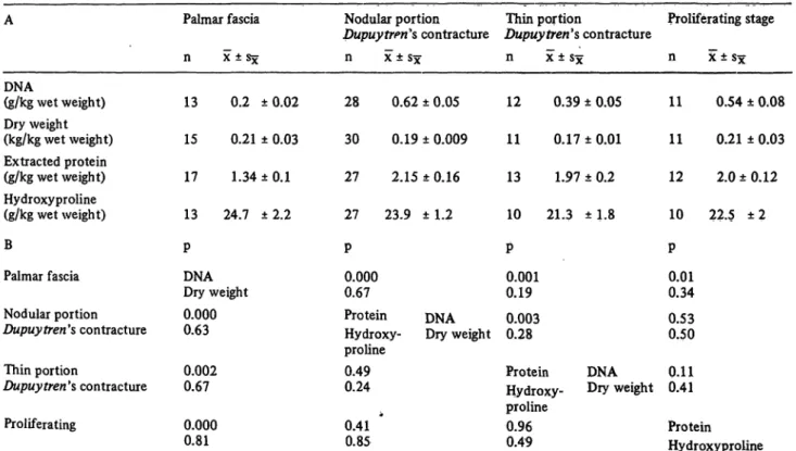
Delbr ck, Reimers and Sch nborn: Enzyme activity pattern in Dupuytren's, contracture tissueand hydroxyproline content and the amount of extract- able protein were determined (tab. 1A). Dry weight and hydroxyproline content do not differ between Dupuy·
trends contracture and palmar fascia. The extracted pro- tein values of Dupuy tren's contractures are found to be twice as high as those of palmar fascia and the DNA con- tent about four times higher in Dupuy trerfs contracture than in palmar fascia. The respective data for the thin portion ofDupuytren tissue are between those for Dupuytren's contracture and palmar fascia values. The
statistical significance of these findings is listed in table IB. Enzyme activity represented by the activity of glyceraldehyde-3-phosphate dehydrogenase follows
the DNA content of the specimens (fig. 1) in the palmar fascia andDupuytren's contracture. The mactoscopically classified nodular portions (Dupuytrerfs contracture, fig. 1) and the tissue specimens which were histplogic- ally classified as proliferating state of Dupuy tren's con- tracture showed no significant differences in dry weight, extractable protein, hydroxyproline or DNA content.
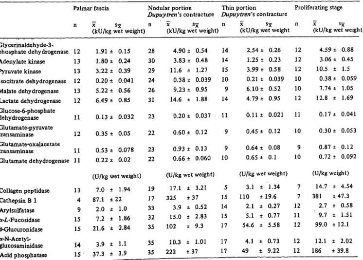
The results of the enzyme activity determinations are summarized in figure 2 and tables 2A and 2B. The activity distribution patterns are characterized by high activities oflactate dehydrogenase, m late dehydrogen- ase and phosphotransferases while the activities of the transaminases, glutamate dehydrogenase and glucose-6-
Tab. 1. Deoxyribonucleic acid content, dry weight, extracted protein and hydroxyproline content of palmar fascia and Dupuy tren's contracture (subgroups). A: content per kg wet weight. B: significance level of differences between the specimen groups.
A
DNA(g/kg wet weight) Dry weight (kg/kg wet weight) Extracted protein (g/kg wet weight) Hydroxyproline (g/kg wet weight) B
Palmar fascia Nodular portion Dupuy tren's contracture Thin portion
Dupuy tren's contracture Proliferating
Palmar fascia ç ÷ ± 5 ÷ 13 0.2 ±0.02 15 0.21 ± 0.03 17 1.34 ±0.1 13 24.7 ±2.2 Ñ
DNADry weight 0.000 0.63 0.002 0.67 0.000 0.81
Nodular portion Dupuy trends contracture
ç ÷ ± 5 ÷ 28 0.62 ± 0.05 30 0.19 ± 0.009 27 2.15 ± 0.16 27 23.9 ±1.2 Ñ
0.000 0.67
Protein DNA Hydroxy- Dry weight proline
0.490.24
0.41 * 0.85
Thin portion
Dupuy tren's contracture ç ÷ ± 8÷
12 0.39 ± 0.05 11 0.17 ±0.01 13 1.97 ± 0.2 10 21.3 ±1.8 Ñ
0.001 0.19 0.003 0.28
Protein DNA Hydroxy- D*y weight proline
0.96 0.49
Proliferating stage ç ÷"±8÷
11 0.54 11 0.21 12 2.0 ± 10 22.5 Ñ
0.010.34 0.530.50
±0.08
±0.03 0.12
± 2
0.110.41
Protein Hydroxyproline
-0.6
•å É ÏË
10.2
>%
°0.1
-| I 0.05
^"2,
o,f 0.04h^« o>
I I 0.03
É l°· 02
HID Sl-o
6 S - g
å _
*f
•3 0.6
O)
*0)
J£0)
·<.
O)
0,1
D
Polmorfoscio àõÑ^Ãâç Proliferation Dupuytren s thin portion stage macroscopically
nodular portion
Fig. 1. Comparison of dry weight, extracted protein, hydroxyproline, deoxyribonucleic acid contents and glyceraldehyde-3-phqsphate dehydrogenase activity in fresh specimens from palmar fascia and Dupuytreh's disease.
L Clin, Chem: Clin. Biochem. / Vol. 19,1981 / No. 9
Delbr ck, Reimers and Sch nborn: Enzyme activity pattern in Dupuytren^ contracture tissue 935
10 ·
ö*
óé
É
Ë
É é · ß 5
·££>*
"- 10"1 ·
"C óï
"•Ñ
"ï
S 10*·
ßï-
3Dupuytren's contracture
¢
MDH ~
GAPDH ADK
GLDHGUI CAT- Â
G-6-PDH £tiQ GLRD
satr FUCD
GLAMD ARY-S
Palmar fascia
MDH ' PK
ADK GAPDH
GOT GPT IDH GLDH
GG-PDH CAT'S
A-PHO GLRD COL-P FUCD
GLAMD ARY-S
Tendon
MDH
GOT
GLDH
IDH G-6-PDH
Articular cartilage
LDH PK
GAPDH MDH
GOT IDH
GLDH G-6-PDH
Fig. 2. Enzyme activity distribution patterns fromDupuytren contracture and human palmar fascia in comparison to human tendon and articular cartilage (11). Logarithmic scale. Activity in kU/kg wet weight.
Abbreviations: LDH: Lactate dehydrogenase MDH: Malate dehydrogenase GAPDH: Glyceraldehyde-3-phosphate dehydro- genase PK: Pyruvate kinase IDH: Isocitrate dehydrogenase ADK: Adenylate kinase ARS: Arylsulfatase CAT-B: Cathep- sin B 1 A-PHO: Acid phosphatase GLAMD: á-N-Acetyl-glucosaminidase COL-P: Collagen peptidase FUCD: á-Æ,-Fucosidase GOT: Glutamate-oxalacetate transaminase GPT: Glutamate-pyruvate transaminase GLRD: 0-Giucuronidase G-6-PDH: Glu- cose-6-phosphate dehydrogenase PGK: 3-Phosphoglycerate kinase GLDH: Glutamate dehydrogenase
The line under each abbreviation refers to the enzyme activity (ordinate).
Tab. 2 A. Enzyme activities in U/kg wet weight of palmar fascia and of Dupuytren's contracture and subgroups.
Palmar fascia Nodular portion Thin portion
Dupuytren's contracture Dupuytren's contracture ç ÷ 8÷ ç ÷ 8÷ ç ÷ 8÷
(kU/kg wet weight) (kU/kg wet weight) (kU/kg wet weight Glycerinaldehyde-3-
phosphate dehydrogenase Adenylate kinase Pyruvate kinase
Isocitrate dehydrogenase Malate dehydrogenase Lactate dehydrogenase Glucose-6-phosphate dehydrogenase Glutamate-pyruvate transaminase
Glutamate-oxalacetate transaminase
Glutamate dehydrogenase 12 13 13 12 13 12 11 12 11 11
1.91 ± 1.80 ± 3.22 ± 0.20 ± 5.22 ± 6.49 ± 0.13 ± 0.35 ± 0.53 ± 0.22 ±
0.15 0.24 0.39 0.041 0.56 0.85 0.032 0.05 0.078 0.02
28 30 29 24 26 31 23 22 23 22 (U/kg wet weight) Collagen peptidase 13 7.0 ± 1.94 19
4.90 ± 3.83 ± 11.6 ± 0.38 ± 9.23 ± 14.6 ± 0.20 ± 0.60 ± 0.93 ± 0.66 ± (U/kg wet
17.1 ± 0.54 0.48 1.27 0.039 0.95 1.88 0.037 0.12 0.13 0.060 weight)
3.21 14 14 15 10 9 14 11 9 9 10
5
2.54 ± 0.26 1.25 ± 0.23 3.99 ± 0.58 0.21 ± 0.039 6.10 ± 0.52 4.79 ± 0.95 0.1 1 ± 0.021 0.45 ± 0.12 0.64 ± 0.08 0.65 ± 0.1 (U/kg wet weight)
3.1 ± 1.34
Proliferating stage ç ÷ ä÷
(kU/kg wet weigh 12
12 12 10 10 12 11 10 9 10
4.59 ± 3.06 é 10.5 ± 0.38 ± 7.74 ± 12.8 ± 0.17 é 0.30 ± 0.87 ± 0.72 ±
0.88 0.45 1.5 0.059 1.05 1.69 0.041 0.053 0.12 0.092 (U/kg weight) 7 14.7 t 4.54 Collagen peptidase
Cathepsin B 1 Aiylsulfatase á-Æ,-Fucosidase
-Glucuronidase á-N-Acetyl- glucosaminidase Acid phosphatase
13 4 9 15 15 14 15
7.0 87.1 2.0 7.2 21.6
3.9 37.3
±
±
±
±
±
* 1.94 22
1.0 1.86 2.84 1.1 3.9
19 17 33 32 35 35 35
17.1 325
3.9 15.0 102
10.3 222
± 3.21
±37
± 0.52
± 2.83
± 9.3
± 1.01
±37
5 15 14 15 17 17 17
3.1 110
2.1 5.1 54.6
4.1 49
±
±
±
±
±
±
± 1.34 19.6
0.27 0.77 5.58 0.73 9.22
7 7 12 11 12 12 12
14.7 381
2.7 ' 9.7 99.0 12.1 186
± 4.54
±47.3
± 0.58
± 1.51
± 12.1
± 2.02
±39.8
936
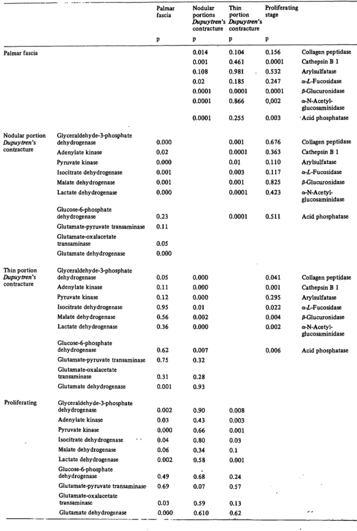
Delbr ck, Reimers and Sch nborn: Enzyme activity pattern in Dupuytren's contiacture tissueTab. 2B. Levels of significance of enzyme activities per kg wet weight between palmar fascia, Dupuy trends contracture and subgroups.
Palmar fascia
Nodular portion Dupuy trends contracture
Thin portion Dupuytren's contracture
Proliferating
Glyceraldehyde-3-phosphate dehydrogenase
Adenylate kinase Pyruvate kinase Isocitrate dehydrogenase Malate dehydrogenase Lactate dehydrogenase Glucose-6-phosphate dehydrogenase
Glutamate-pyruvate transaminase Glutamate-oxalacetate
transaminase
Glutamate dehydrogenase Glyceraldehyde-3-phosphate dehydrogenase
Adenylate kinase Pyruvate kinase Isocitrate dehydrogenase Malate dehydrogenase Lactate dehydrogenase Glucose-6-phosphate dehydrogenase
Glutamate-pyruvate transaminase Glutamate-oxalacetate
transaminase
Glutamate dehydrogenase Glyceraldehyde-3-phosphate dehydrogenase
Adenylate kinase Pyruvate kinase Isocitrate dehydrogenase Malate dehydrogenase Lactate dehydrogenase Glucose-6-phosphate dehydrogenase
Glutamate-pyruvate transaminase Glutamate-oxalacetate
transaminase
Glutamate dehydrogenase
Palmar fascia
P
0.000 0.02 0.000 0.001 0.001 0.000
0.23 0.11 0.05 0.000
0.05 0.11 0.12 0.95 0.56 0.36
0.62 0.75 0.31 0.001
0.002 0.03 0.000 0.04 0.06 0.002 0.49 0.69 0.03 0.000
Nodular Thin Proliferating portions portion stage Dupuy trends Dupuy tren's
contracture contracture Ñ
0.014 0.001 0.108 0.02
ï.üüüé
0.0001 0.0001
0.000 0.000 0.000 0.01 0.002 0.000
0.007 0.32 0.28 0.93
0.90 0.43 0.66 0.80 0.34 0.58 0.68 0.07 0.59 0.610
Ñ 0.104 0.461 0.981 0.185 0.0001 0.866 0.255
0.001 0.0001 0.01 0.003 0.001 0.0001
0.0001
0.008 0.003 0.001 0.03 0.1 0.001 0.24 0.57 0,13 0.62
Ñ 0.156 0.0001 0.532 0.247 0.0001 0,002 0.003
0.676 0.363 0.110 0.117 0.825 0.423
0.511
0.041 0.001 0.295 0.022 0.004 0.002
0.006
Collagen peptidase Cathepsin  1 Arylsulfatase a-£-Fucosidase /3-Glucuronidase a-N-Acetyt glucosaminidase
•Acid phosphatase
Collagen peptidase Cathepsin  1 Arylsulfatase a-jL-Fucosidase 0-Glucuronidase onN-Acetyl- glucosaminidase Acid phosphatase
Collagen peptidase Cathepsin  1 Arylsulfatase á-Æ,-Fucosidase 0-Glucuronidase a-N-Acetyl- glucosaminidase Acid phosphatase
•x *
J. Clin. Chem. Clin. Bioehefh. / Vol. 19,1981 / No. 9
Delbr ck, Reimers and Sch nborn: Enzyme activity pattern in Dupuytren^ contracturc tissue 937
phosphate dehydrogenase are much lower and are in the
range of the lower limit of analytical accuracy. The enzyme patterns resemble those of other connective tissues such as tendon or cartilage (fig. 2; (29), (30)).
The activities of lysosomal enzymes are two to three orders of magnitude lower than the activities of main metabolic pathway enzymes. In palmar fascia and Dupuytren's contracture the activities of cathepsin B l
and acid phosphatase exceed those of other lysosomal enzymes. The absolute activities calculated on the wet weight basis of the specimens are two to three times higher in Dupuy trends contracture than in palmar fascia.
The thin portion of Dupuy tren^ contracture is signifi- cantly different from Dupuytren's contracture and from palmar fascia with respect to the enzyme activities, whereas the proliferating stage does not differ from the total Dupuy trends contracture group but differs from the palmar fascia group. Calculated on the basis of DNA, the absolute activities for both lysosomal and main metabolic pathway enzymes are somewhat higher in palmar fascia than in Dupuy tren's contracture (tab. 3).
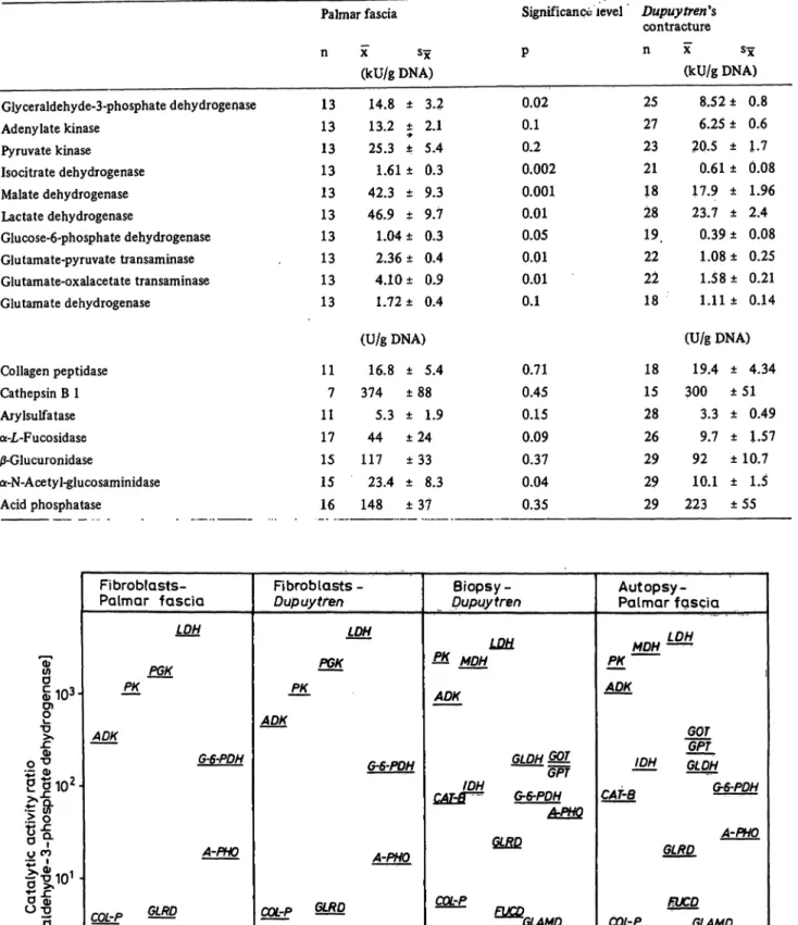
Enzyme activities based on the glyceraldehyde-3-phos- phate dehydrogenase activity show no statistically signif- icant differences in specimens from Dupuy tren's con- tracture or palmar fascia (tab. 4, fig. 3).
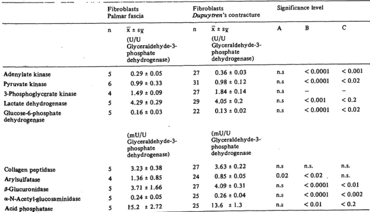
The absolute activities as well as the enzyme activity distribution patterns of fibroblasts from palmar fascia
and Dupuy tren's contracture are identical. In particular, the activities of lysosomal enzymes do not differ signif- icantly between both strains of cultured fibroblasts. The activity distribution patterns (based on the activity of glyceraldehyde-3-phosphate dehydrogenase) of main metabolic pathway enzymes from fibroblasts and the respective tissue specimens from palmar fascia and Dupuy fren's contracture (fig. 3) differ only slightly.
While adenylate-kinase and pyruvate-kinase are lower, lactate dehydrogenase and glucose-6-phosphate dehydro- genase show a higher activity in fibroblasts than the corresponding tissue specimens (statistical significance see table 3). Due to the small number of samples of palmar fascia cell strains tested, the significance of these differences are less than those for Dupuy trends contracture tissue specimens and Dupuytren's contrac- ture cell cultures. However, lysosomal enzyme activity patterns are different for tissue specimens and cultured fibroblasts. In general, the activities of these enzymes are higher in relation to the main metabolic pathway enzymes in tissue specimens than in the respective cultured fibroblasts. This is especially true for j3-glucuron- idase and á-N-acetyl-glucosaminidase (fig. 3).
A great difference in overall activities could be shown by comparing tissue enzymes with cultured fibroblast enzymes on the basis of the DNA content. Enzyme activities per g DNA are 10 to 50 times higher in fibro-
Tab. 3. Enzyme activities in cultured fibroblasts from Dupuytren's contracture and palmar fascia. U/U glyceraldehyde-3-phosphate dehydrogenase. Significance level of differences between both fibroblast groups (A); between fibroblasts from Dupuy tren's contracture and Dupuytren's contracture tissue (B); and fibroblasts from palmar fascia and palmar fascia tissue (C) (see table 2A).
Fibroblasts Palmar fascia
Adenylate kinase Pyruvate kinase
3-Phosphoglycerate kinase Lactate dehydrogenase Gluco se-6-pho spha te dehydrogenase
Collagen peptidase Arylsulfatase 0-Glucuronidase
a-N-Acetyl-giucosaminidase Acid phosphatase
n
5 6 4 5 5
5 4 5 5 5
× ± 5 ÷
(U/UGly ceraldehy de^3- phosphate dehydrogenase)
0.29 ± 0.05 0.99 ± 0.33 1.49 ±0.09 4.29 ± 0.29 0.16 ± 0.03 (mU/U
Glyceraldehyde-3- pihosphate dehydrogenase)
3.23 ± 0.38 1.36 ± 0.85 3.71 ± 1.66 0.24 ± 0.05 15.2 ±2.72
Fibroblasts Significance level Dupuy tren 's contracture
n
27 31 27 29 22
27 24 27 25 25
÷ ± 5 ÷ A (U/UGlyceraldehyde-3- phosphate dehydrogenase)
0.36 ± 0.03 n.s 0.98 ±0.1 2 n.s 1.84 ±0.14 n.s 4.05 ± 0.2 n.s 0.1 3 ±0.02 n.s (mU/U
Glyceraldehyde-3- phosphate dehydrogenase
3.63 ± 0.22 n.s 0.85 ± 0.05 0.02 4.09 ±0.31 n.s 0.26 ± 0.04 n.s 13.6 ± 1.3 n.s
B
< 0.0001
< 0.0001
—
< 0.001
< 0.0001
n.s.
<0.02 .
< 0.0001
< 0.0001
<0.01 C
< 0.001
<0.02
—
<0.2
<0.02
n.s.
n.s.
<0.01
< 0.002
<0.2
J. Clin. Chem. din·. Biochem. / Vol. 19, 1981 / No. 9
938
Delbr ck, Reimers and Sch nborn: Enzyme activity pattern in Dupuytren'* contracture tissue Tab. 4. Comparison of enzyme activities calculated on the DMA basis in specimens from Dupuy trends contracture and normal palmarfascia.
Palmar fascia ç ÷ Sx
(kU/g DNA) Glyceraldehyde-3-phosphate dehydrogenase
Adenylate kinase Pyruvate kinase Isocitrate dehydrogenase Malate dehydrogenase Lactate dehydrogenase
Glucose-6-phosphate dehydrogenase Glutamate-pyruvate transaminase Glutamate-oxalacetate transaminase Glutamate dehydrogenase
Collagen peptidase Cathepsin B 1 Arylsulfatase a-L-Fucosidase 0-Glucuronidase
a-N-Acetyl-glucosaminidase Acid phosphatase
13 13 13 13 13 13 13 13 13 13
11 7 11 17 15 15 16
14.8 ± 3.2 13.2 ± 2.1·*
25.3 ± 5.4 1.61 ± 0.3 42.3 ± 9.3 46.9 ± 9.7 1.04 ± 0.3 2.36 ± 0.4 4.10 ± 0.9 1.72 ± 0.4 (U/gDNA)
16.8 ± 5.4 374 ± 88
5.3 ± 1.9 44 ± 2 4 117 ±33 23.4 ± 8.3 148 ± 37
Significance level Dupuy trends contracture
Ñ n * sx
(kU/g DNA) 0.02
0.1 0.2 0.002 0.001 0.01 0.05 0.01 0.01 0.1
0.71 0.45 0.15 0.09 0.37 0.04 0.35
25 27 23 21 18 28 19.
22 22 18
18 15 28 26 29 29 29
8.52 ± 0.8 6.25 ± 0.6 _20.5 ± 1.7 0.61 ± 0.08 17.9 ± 1.96 23.7 ± 2.4
0.39 ± 0.08 1.08 ± 0.25 1.58 ± 0.21 1.11 ± 0.14 (U/g DNA)
19.4 ± 4.34 300 ± 51
3.3 ± 0.49 9.7 ± 1.57 92 ± 10.7 10.1 ± 1.5 223 ± 55
§ |éï»·
ï
•D
of
Ï ð 1Ð ·
Ç
.> 05! II»
D ö1
Ï Ö
óé 1
5
Åéç-é
Fibroblasts- Palmar fascia
LOH PK PGK
ADK
G-6-PDH
A-PHO
CQlrP SL Q ARY-S
GLAMD
Rbroblasts - Dupuytren
LDH PGK PK ADK
G-6-PDH
A-PHO
rrv-p GLRD
7*^* ~
ARY-S
GLAMD
Biopsy - Dupuytren
£& MDH ADK
GLDHSSL IDH GPT
CAT-ff^ G-6-PDH
A.PHQ
GLRD
CO£rP
GLAMD
ARV-S
•
Autopsy - Palmar fascia
LDH PK
ADK
~GPfGOT IDH GLDH
f* C DfMJ f* A T O \TQ~r Ufi
\*f\i-Q
A-PHO GLRD
f-UCD COL-P GLAMD
ARY-S
Fig. 3. Comparison of enzyme activity distribution patterns from cultured fibroblasts (Dupuytren** contracture, palmar fascia) with the respective patterns of tissue specimens on the basis of glyceraldehyde-3-phosphate dehydrogenase activity, Logarithmic scale. Explanations see fig. 2.
J. Clin. Chem. Clin. Biochem. / Vol. 19,1981 / No. 9
Delbr ck, Reimers and Sch nborn: Enzyme activity pattern in Dupuytren's contracture tissue 939
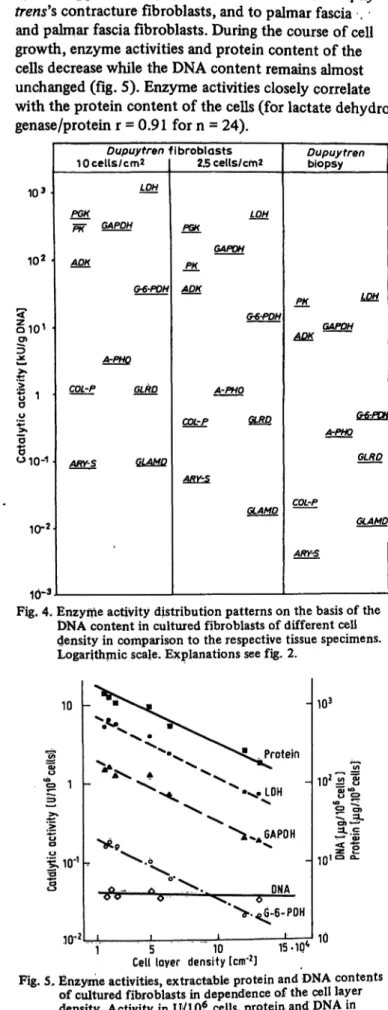
blasts depending on the duration of the growth period
and/or the density of fibroblasts in the cell layer (fig. 4, 5). This applies to Dupuytren's contracture and Dupuy- trens's contracture fibroblasts, and to palmar fascia . and palmar fascia fibroblasts. During the course of cell growth, enzyme activities and protein content of the cells decrease while the DNA content remains almost unchanged (fig. 5). Enzyme activities closely correlate with the protein content of the cells (for lactate dehydro- genase/protein r = 0.91 for n = 24).
103·
102
010'
B
ÁÎ
º é
0
"B
5 ßï--"
ºÏ-2
10-3
Dupuytren 1 10 cells/ cm2
LOH PGK
PR" GAPDH
ADK
G-6-POH
A-PHO COL-P GLRD
ARY-S G1AVQ
ibroblasts 2.5 ce(ls/cm2
LQH PGK
GAPDH PK_
ADK
G*'PDH
A-PHO COL-P GLJRD
ARY-S
CLAMP
Dupuytren biopsy
PK_
^ezsa
&6-PDH A-PHO
GLRD
COL-P
GLAMD ARY-S
Fig. 4. Enzyme activity distribution patterns on the basis of the DNA content in cultured fibroblasts of different cell density in comparison to the respective tissue specimens.
Logarithmic scale. Explanations see fig. 2.
10
t;
ï'jL 10"1
3¼ ONA
5 10 15-1Q*
Cell loyer density (cm'2]
103
8
10
Fig. 5, Enzyme activities, extractable protein and DNA contents of cultured fibroblasts in dependence of the cell layer density. Activity in U/10* cells, protein and DNA in Mg/ÉÏ* cells. Logarithmic scale. Abbreviations see fig. 2.
Discussion
Since the tissue samples were homogeneous and the blood impurities negligible (0.01%), the possibility of falsification of the enzyme patterns by enzymes from other sources was excluded. However, due to the diffi- culties of equally disrupting strong connective tissue portions, the scatter of the results within the individual groups is relatively high. A mean of 90% of the total enzyme activities was extracted by the method applied in this study. On the other hand, fibroblast extraction leads to more precise results with smaller deviation from the mean. The latter can be achieved only when fibro- blasts of the same cell density in culture are taken for extraction. The methodological scatter of the results partly explains the poor significance of differences be- tween the groups for those enzymes with low activities in the tissues. For interpretation of the data on enzyme activities, the metabolic capacity of the respective tissue as well as that of the individual cell must be taken into consideration. The basis for a comparative interpreta- tion should be first the wet weight, then the DNA content or the cell number. Extracted protein, however, did not prove to be a suitable reference in the tissues under investigation. The missing correlation between values for extracted protein and DNA content (fig. 1) shows that, in addition to the metabolically active enzyme proteins, other proteins such as neutral salt-soluble collagen are extracted under the experimental condi- tions applied in this study. Due to the constant correla- tion of enzyme activities in different tissues, extractable enzyme proteins offer a further possibility of a reference system (31). For instance, glyceraldehyde-3-phosphate dehydrogenase as a key enzyme of glycolysis (32) could be used as reference for the comparison and interpreta- tion of enzyme patterns of the specimen groups under discussion.
The data on the enzyme activities in palmar fascia and Dupuytren's contracture allow the conclusion that the higher metabolic activity of Dupuytrends contracture does not depend on changes in the enzymatic activities within the individual cell, but rather on the larger number of cells present in the diseased portions of the tissue, as seen in the morphological examination of the Dupuytren** contracture samples (3). These findings are in agreement with the higher DNA content of the Dupuy- tren's contracture samples compared with palmar fascia specimens. However, the enzyme activities based on the DNA content are lower inDupuytren's contracture than in palmar fascia. These differences might be explained by a higher DNA content per cell in proliferating cells ofDupuytren's contracture compared to the non- proliferating cells in palmar fascia. On the other hand, preliminary experiments on main metabolic pathway enzyme activities in growing fibroblasts (fig.' 5) showed a decrease in enzyme activity and protein content per cell of cultured fibroblasts with increasing age and density of cells. If cell density is the regulating factor
J. Clin. Chem. Clin. Biochem. / Vol. 19,1981 / No. 9
940
Dclbrück, Reimers and Schöuborn: Enzyme activity pattern in Dupuytren's contracture tissuefor the activity of cell enzymes, the difference in enzyme activity per DNA (or cell) could be attributed to the isolated localisation of cells in palmar fascia in contrast to the proliferating cell clusters in Dupuytren's contrac- ture (see below).
Although historically Dupuytren's contracture exhibits tissue breakdown alongside tissue regeneration, the activities of lysosomal peptidases (cathepsin B 1, collagen peptidase) involved in collagen breakdown (33, 34, 25), the carbohydrases and the acid phosphatase do not significantly differ between palmar fascia and Dupuytren's contracture on the tissue DNA basis. This means that the elevated metabolic turnover of extra- cellular compounds is provided by an increased cell number in Dupuytren's contracture.
The data oflioopes et al. (12) differ considerably from the results of this study. Based on the DNA content, the authors reported an increase in enzyme activities in specimens of Dupuytren's contracture which varied between 1 and 10 times from enzyme to enzyme. How- ever, the methods applied for enzyme activity determina- tions were different. Hoopes et al. incubated tissue slices instead of testing tissue extracts which means that enzymes or substrates have to penetrate extracellular matrix and fibres prior to enzymatic action on the sub- strates. This extracellular compartment is assumed to be a stronger barrier for both enzymes and substrates in the case of the intact palmar fascia than in the pathologi- cal structure of Dupuytren's contracture. In addition, the penetration rates of the different substrates or enzyme proteins may vary, thus causing different eleva- tion rates of individual enzymes as reported by the authors. Calculating the activities determined by Hoopes et al. on the basis of glyceraldehyde-3-phosphate de- hydrogenase, the differences in enzyme activities are in the range of 1-2 fold, depending on the activity of glyceraldehyde-3-phosphate dehydrogenase given by the assay method. Moreover, tissue slices contain more or less intact cells, subcellular microstructures, and sub- strates in various concentrations, providing metabolic reaction sequences which may interfere with the specific enzyme reactions to be determined. These methodologi- cal differences apply also to the data published by Hoopes et al. on enzyme activities in hyperytrophic
scars and keloids (36). The difficulties involved in the techniques used by the authors are evident by the fact that, in normal dermis, no measurable giyceraldehyde-3- phosphate dehydrogenase activity was detected; this seems very unlikely in view of the high activities of other enzymes of the Embden-Meyerhof pathway in the examined specimens. Although the data of Hoopes et al.
are not comparable to the results given in this paper, they do demonstrate a considerable metabolic activity of palmar fascia and Dupuytren.
Studies on the synthesis of collagen and acid glycosamino- glycans in cultured fibroblasts have shown an enhanced
[3H]hydroxyproline and [35S]sulfate incorporation in cells cultured from Dupuyfren's contracture biopsies compared to palmar fascia fibroblasts (26). Correspond- ing differences in the determined enzyme activities are missing in the respective fibroblasts (fig. 3). However, there are some distinct differences between enzyme activities in fibroblasts and the respective tissue speci- mens from which fibroblasts were derived (fig. 3, tab. 3), with respect to the overall activity and the relationship of the activities of lysosomal enzymes to those of main metabolic pathway enzymes. These differences very probably have a methodological basis. In the case of the tissue specimens, the extraction procedure includes the extracellular compartment, and this may contain varying relative quantities of individual secreted lysosomal enzyme proteins. However, the fibroblasts were isolated from the surrounding medium prior to the extraction. In this way the extracellular portion of the lysosomal enzyme activities was removed before activity determina- tions. Enzyme activity determinations in isolated cells offer the opportunity to distinguish between intracellular activity of lysosomal enzymes and the activity of the secreted enzyme proteins. When setting up cell culture experiments along with enzyme activity assays in tissue specimens, it should be possible to answer the question whether enzyme activities derive from the specific cells of the respective tissue or originate from other sources such as granulocytes or macrophages. No final explana- tion can be offered as to the differences in enzyme activity per g DNA between tissue and the respective cultured fibroblasts (fig. 4). Experimental errors in DNA determination can be excluded since the respec- tive data from tissue and cultured fibroblasts concur with those of the literature (12,37). The data on the dependence of enzyme activity upon cell density in cultured fibroblasts suggest a mechanism by which the enzyme activity is regulated by extracellular factors, such as cell to cell interaction or the state of extra- cellular matrix. Future studies must be performed to reveal more detailed information on this matter and prove the possibility of establishing similar data in in vivo systems of proliferating cells. For the present, it must be emphasized that work on the metabolism of fibroblasts, using cultured cells, should take into con- sideration the comparability of the state of cell growth and the density of the cells under investigation.
Acknowledgements
The authors are very indebted to Prof. Dr. med. Köhnlein and his colleagues from the Department to Plastic and Reconstruc- tion Surgery, Hannover Medical School, for their assistance in collecting the specimens of Dupuytren's contracture and to the colleagues from the Department of Pathology (Prof. Dr. med.
Georgii) for providing the autopsy specimens, especially to Dr. Schnaidt for preparing the slices and assistance in inter- pretation of the microscopical morphology. The authors express their gratitude to Dr. Törek, Department of Biometrics,, Han- nover Medical School for their valuable assistance in statistical evaluation of the experiments.
J.Clin.Chem.Clin.Biochem./Vol. 19,1981 /No. 9
Delbruck, Reimers and Schönborn: Enzyme activity pattern in Dupuytren's contracture tissue 941 References
J. Plater, F. (1614), Obscrvationum in homines affectibus plc- 19.
risque corpori et animo functionum laesionc, dolorc, aliavc molestia et vitio insensis libri tres, 2. Aufl. Basel (Neuaufla- 20 gen 1630,1641)
2. Dupuytren, G. (1832), Lecons orales de cliniquc chirurgicalc 21 faites a THotel Dieu de Paris, G. Balliere, Vol. l (l 832). 22.
3. Mackenzie, D. H. (1979), The differential diagnosis of fibro- blastic disorders. Blackwell scientific publications, Oxford 23.
and Edinburgh, p. 82-86. 24 4. Luck, J. V. (1959), J. Bone Jt. Surg. 41A, 635. 25.
5. Tyrkkoe, J. & Viljanto, J. (1975), Ann. Chir. Gynaegol. 26 Fcnn. 64, 288-291.
6. Tessari, L. & Paring L. (1958), Arch. Ortop. 72, 435-441. 27 7. Lagier, R. & Exer, B. (1960), Virchow's Archiv 333, 68-80.
8. Carr, T. L. (1979), Hand 1, 50-55. 28.
9. Viljanto, J., Seppaelac, P. O. & Lethonen, A. (1971), Ann.
Rheum. Dis. 30, 423-427. 29.
10. Hunter, A. A., Ogdon, C. & Morris, M. G. (1975), Brit. J. 30.
Plast. Suig. 28,10-18.
11. Delbruck, A. (1962), Klin. Wochenschr. 40, 677-684. 31.
12. Hoopes, J. E., labaley, M. E., Chi-Tsung Su, Wilgies, E. F. S.
& Im, M. J. C. (1977), J. Hand. Surg. 2, 62-65. 33.
13. Webb, E. C & Morrow, P. F. W. (1959), Biochem. J. 73,
7-15. 34.
14. Szasz, G. (1967), Clin. Chim. Acta 15, 275-282.
15. Weissmann, B., Rowin, G., Marshall, J. & Friedrici, D. 35.
(1967), Biochemistry 6, 207-214. 36.
16. Levy, G. A. & McAllan, A. (1961), Biochem. J. 80,435-
439. 37.
17. Cries, G., Buresch, H. & Strauch, L. (1970), Experientia 25,31-33.
18. Barret, A. J. (1972), Anal. Biochem. 47, 280-293.
Lee, H. J., La Rue, J. N. & Wilson, J. B. (1971), Anal. Bio- chem. 41, 397-401.
DiPietro, D. L. & Zcngerle, F. S. (1967), J. Biol. Chem. 242, 3391-3396.
Wcichselbaum, T. E. (1946), Amcr. J. Clin. Pathol. 10,40.
Lowry, D. H., Rosenbrough, N. J., Fair, A. L. & Randall, R. J. (1951), J. Biol. Chem. 193, 265-275.
Crosby, W. H. & Furth, F. W. (1956), Blood 11, 380.
Burton, K. (1956), Biochem. J. 62, 315.
Delbruck, A. & Schröder, H. unpublished results.
Stegemann, H. (1958), Hoppe-Seylcr's Z. Physiol. Chem.
311,41-45.
Watthiaux, R. & C. DC Duve (1956), Biochem. J. 63, 606- 608.Niehe, N. H., Statistical package social sciences Me. Graw- hill, New York, 2. ed. 1975.
Dclbruck, A. (1964), Enzymol. Biol. Clin. 4, 84-106.
Patzschke, E. & Delbruck, A. (1967), Enzymol. Biol. Clin.
5,421-450.
Dclbrück, A., Zebc, E. & Bücher, Th. (1959), Biochem.
Zschr.Jjy, 273-296.
Tappel, A. L. in Dingl, J. T. & Fell, B. Ed. (1969), The lyso- somes in biology and pathology, Vol. 2, pp. 207-244.
Stojan, B., Müller, W., Wurm, K. & Tariverdian, M. (1973), Schweiz. Med. Wochenschr. 103, 337-341.
Lindner, J. (1972), Archiv Dcrmatol. Forsch. 244, 104-112.
Hoopes, J. E., Su, C. T. & Im, M. J. C. (1971), Plast.
Reconstr. Surg. 47, 132-137.
Leyva, A. jr. & Kelley, W. N. (1974), Anal. Biochem. 62, 173-179.
Prof. Dr. med. A. Dclbrück Abt. IV (Klinische Chemie) Zentrum für Laboratoriumsmedizin der Mcd. Hochschule Hannover Zcntrallabor im Krankenhaus Oststadt Podbielskistraße 380
D-3000 Hannover 51
J. Clin. Chem. CÜn. Biochem. / Vol. 19,1981 / No. 9