Towards Elucidation of the Phenazine Biosynthesis Pathway of Pseudomonas with the Structural and
Functional Analysis of the enzymes PhzA, B, G and BcepA
DISSERTATION
zur Erlangung des akademischen Grades eines Doktors der Naturwissenchaften (Dr. rer. nat.) des Fachbereichs Chemie der Universität Dortmund
Angefertigt am Max-Planck-Institüt für Molekulare Physiologie In Dortmund
Eingereicht von
Ekta G. Ahuja (Niki)
Aus Ahmedabad/Indien
Dortmund, 2006
1. Gutachter : Prof. Dr. R.S.Goody
2. Gutachter : Prof. Dr. R. Winter
Pathway of Pseudomonas with the Structural and Functional Analysis of the enzymes PhzA, B, G
and BcepA
DISSERTATION
Submitted towards partial fulfillment of requirement for the degree of
DOKTOR DER NATURWISSENCHAFTEN (DR. RER. NAT.)
in the Department of Chemistry, University of Dortmund, Germany
By
Ekta G. Ahuja (Niki)
Max Planck Institute for Molecular Physiology
&
Department of Chemistry, University of Dortmund
2006
Die vorliegende Arbeit wurde in der Zeit von Januar 2003 bis September 2006 am Max- Planck-Institut für Molekulare physiologie in Dortmund unter der Anleitung von Prof. Dr.
Roger S Goody, Dr. Wulf Blankenfeldt und Prof. Dr. Roland Winter durchgeführt.
Hermit versichere ich an Eides statt, dass ich die vorliegende Arbeit selbständig und nur mit den angegeben Hilfsmitteln angefertigt habe.
Dortmund, 2006 Ekta G Ahuja (Niki)
Symbols and Abbreviations iv
1.0 INTRODUCTION
1.1 Discovery of phenazines. 2 1.2 Prevalence of phenazine producers. 2 1.3 Chemical properties of phenazines. 4 1.4 Biological significance of phenazines. 6 1.5 Mode of action of phenazines. 7 1.6 Biosynthesis of phenazines. 9 1.6.1 The Shikimate pathway. 9
1.6.2 Assembly of the tricyclic phenazine ring scaffold. 10 1.6.3 Genetic basis of phenazine biosynthesis. 10 1.6.4 Phenazine biosynthesis in other organisms. 12 1.6.5 Phenazine biosynthesis in Pseudomonas. 14 1.6.6 Enzymes of the phenazine biosynthesis operon. 15 1.7 Generation of phenazine diversity. 18 1.8 Regulation of phenazine production. 20 2.0 OBJECTIVES OF THIS WORK 21 3.0 RESULTS & DISCUSSIONS
Section I – Sequences analysis, cloning and crystallisation
3.1 Sequence analysis 22 3.1.1 PhzA, PhzB and BcepA. 22 3.1.2 Analysis of genes similar of PhzA/B. 24 3.1.3 Sequence analysis of PhzG. 25 3.2 Cloning, over-expression and purification. 26 3.3 Crystallisation of native proteins. 27 3.4 Crystallisation of protein complexes. 29
Section II – Data collection and structural analysis
3.5 Data collection, structure determination and Quality of models. 30
3.5.1 PhzA 31
3.5.2 BcepA and its complex with B75. 32 3.5.3 PhzG and its complex with PCA. 33 3.6 Structural features of PhzA from Pseudomonas fluorescens. 37 3.6.1 The monomer of PhzA. 37
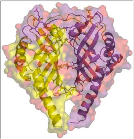
3.6.2 Dimer of PhzA and the dimer interface. 38 3.6.3 Active centre cavity and binding of MES. 40 3.6.4 Ligand docking studies. 41 3.7 Structural analysis of BcepA from Burkholderia cepacia. 44 3.7.1 Monomer of BcepA. 44
3.7.2 Dimer of BcepA and dimer interface. 45 3.7.3 The active site cavity and binding of B75. 47 3.8 Comparison of the structures of PhzA and BcepA. 50
3.9 PhzA, BcepA and homologous structures. 54 3.9.1 The C-terminal region. 56 3.9.2 The sheet-loop-sheet motif. 57
3.9.3 Function of homologous structures and implications.
for PhzA & BcepA. 58 3.10 PhzG & Complex of PhzG with PCA 60
3.10.1 Monomeric structure description. 60 3.10.2 The functional dimer of PhzG. 61 3.10.3 Active site cavity and binding of FMN. 64 3.10.4 PhzG in complex with PCA. 67 3.10.5 Comparison of PhzG and E.coli PNPOx. 69
Section III – Investigating intermediates of the phenazine biosynthesis pathway
4.0 Intermediates formed during the biosynthesis of PCA. 73 4.1 HPLC experiments. 73 4.2 Mass Spectroscopic measurements ESI-MS. 75 4.3 APCI-MS measurements. 79
4.3.1 Effects of various enzymes of DHHA. 79 4.3.2 Formation of 1,2,4-trihydro-3-oxo-anthranilic acid. 80 4.3.3 Formation of Mw 274. 81
4.3.4 Formation of Mw 228 83 4.3.5 Formation of PCA. 84 4.3.6 Implications of APCI results on phenazine biosynthesis. 85
Section IV – Interactions between the seven enzymes
5.0 Interactions between enzymes of the ‘phz’ operon. 91 5.1 Bacterial two-hybrid assays on indicator agar. 91 5.2 Liquid β-galactosidase activity assay. 93
Section V – Measuring oxygen consumption
6.0 Measurements using Clarke’s electrode. 95 7.0 CONCLUSIONS AND FUTURE DIRECTIONS 98 8.0 ZUSAMMENFASSUNG 101
9.0 MATERIALS AND METHODS
9.1 Materials 103
9.1.1 Chemicals & Enzymes 103
9.1.2 Materials 103
9.1.3 Kits 103
9.1.4 Microorganisms 104 9.1.5 Plasmid vectors 104 9.1.6 Media and antibiotics 104
9.1.6 Buffers 104
9.2 Molecular Biology Methods. 105 9.2.1 Agarose gels 105 9.2.2 Isolation of plasmid DNA 105
9.2.5 Preparation of Competent cells. 105
9.2.6 Ligation 106
9.2.7 Transformation 106 9.2.8 Glycerol Stocks 106 9.2.9 DNA sequencing 106 9.2.10 Cloning of phzA, bcepA, B and G. 106 9.3 Biochemical Methods 107 9.3.1 Growth and harvest of protein expressing bacteria. 107 9.3.2 Protein purification (Ni-NTA chromatography). 108
9.3.3 SDS-PAGE 108
9.3.4 Removal of 6xHis-tag and Size exclusion
chromatography. 108
9.3.5 Determination of protein concentration. 109 9.3.6 Final concentration and storage of protein. 109 9.3.7 Seleno-L-methionine labelled protein. 109 9.3.8 Bacterial two-hybrid system. 109 9.3.9 Liquid β-galactosidase assay. 112 9.3.10 High Pressure Liquid Chromatography (HPLC) 112 9.3.11 Mass Spectroscopy. 112 9.3.11.1 MALDI-TOF MS 112 9.3.11.2 ESI-MS 113 9.3.11.3 APCI-MS 114 9.3.12 Measurements with Clarke’s electrode. 116 9.4 Crystallographic methods 116 9.4.1 Crystallisation by Vapour-diffusion. 118 9.4.2 Crystal Soaking 119
9.3.1 PhzA 119
9.3.2 PhzB 119
9.3.3 BcepA 120
9.3.4 PhzG 120
9.3.5 Complex of PhzG with PCA 120 9.4.3 Cryocrystallography 120 9.4.4 Data collection 121 9.4.5 Data Processing 121 9.4.6 Determination of Phases. 123 9.4.7 Refinement 127 9.4.8 Validation 128 9.4.9 Representation of structures. 128
10.0 REFERENCES 129
ACKNOWLEDGEMENTS vi
SYMBOLS AND ABBREVIATIONS Å Ångstrom (0.1nm)
ADP Adenosinediphosphate
APCI Atmospheric pressure controlled ionisation ATP Adenisinetriphosphate
BSA
Bovine Serum Albuminbp base pair
cAMP cyclic adenosine 5’ phosphate CCD Charge coupled devices C-terminus Carboxyterminus
Da Dalton
DNA Desoxyribonucleic acid EDTA Ehtylendiamintetraacetate
ESRF European Synchrotron Radiation Facility
E. coli Escherichia coliESI-MS electrospray ionisation mass spectroscopy
F
calc, F
obsStructure-factor amplitudes (calc:calculated, obs: observed) FMN flavin mononucleotide
FPLC Fast Performance Liquid Chromatography g gram
h hour
HEPES 4-(2-Hydroxyethyl)-piperazin-1-ethan-sulfonsäure HPLC
High Performance Liquid ChromatographyIPTG Isopropyl-β-D-1-thiogalactopyranosid kb kilo-base pair
kDa kilo-Dalton kJ kilo-Joule
λ wavelength,
Lambda
MAD multiple wavelength anomalous dispersion MALDI matrix assisted Laser desorption/ionization MES
min minute
MIR multiple isomorphous replacement mM milimolar
MR molecular replacement
NCS non-crystallographic symmetry
nm Nanometer
NMR nuclear magnetic resonance N-terminus Aminoterminus
PCA Phenazine-1-carboxylate acid PCR
Polymerase Chain ReactionPDB Protein Data Bank
PDC Phenazine-1,6-dicarboxylate acid
PEG Polyetheleneglycol
rmsd root mean square deviations rpm revolution per minute RT room temperature s second
Se Selinium
SAD Single-wavelength anomalous dispersion
SDS-PAGE Sodiumdodecylsulphate-Polyacrylamide gel electrophoresis SeMet Seleno-L-Methionine
TEMED N,N,N,N -Tetramethyl-Ethylenediamine TRIS Tris-(hydroxymethyl)-aminomethane
U Units UV Ultraviolet V Volt µl microliter
µM micromolar
Amino Acids Abbreviations
Name 3-letter code 1-letter code
Alanine Ala A
Argenine Arg R
Asparagine Asn N
Aspartate Asp D
Cysteine Cys C
Glutamine Glu E
Glutamate Gln Q
Glycine Gly G
Histidine His H
Isoleucine Ile I
Leucine Leu L
Lysine Lys K
Phenylalanine Phe F
Methionine Met M
Proline Pro P
Serine Ser S
Threonine Thr T
Tryptophane Trp W
Tyrosine Tyr Y
Valine Val V
INTRODUCTION
Introduction
Begin at the beginning and go on till you come to the end: then stop.
- The King (Alice in wonderland, Lewis Carroll)
1.0 Introduction
Microorganisms produce an array of low molecular weight organic compounds with a wide range of biological activities. These compounds have interested chemists for more than a century and a versatile gamut has been isolated, analyzed and characterised. However, this process is by no means complete and the inventory of newly discovered compounds increases with each passing year.
The compounds discovered so far have diverse chemical structures with intricate, idiosyncratic molecular frameworks displaying a remarkable variety and subtlety (Vining, 1990). They were recognised as products of specialised metabolism and the reason for their existence though not immediately apparent, was speculated on ever since their discovery more than a century ago. ‘Secondary metabolites’
was the name given to these compounds by plant phytologists (Barger, 1907) and this term was brought into common use by J. D. Bu’Lock (Bu’Lock, 1961).
The biosynthesis of all secondary metabolites has been found to arise from intermediates or products of primary metabolism (Vining, 1990). Unlike intermediates and cofactors which participate in cell-structure syntheses, energy transduction and other cellular processes, secondary metabolites do not seem to be essential either for growth or reproductive metabolism of the organism. These compounds are usually distinctive products of a particular group of organisms, sometimes even of a single strain and require a particular set of physiological conditions for the initiation of their synthesis. Prior to the antibiotic era, relatively few microbial products were discovered based on biological activity. However, in the late nineteenth century, the biochemical basis of microbial antagonism of secondary metabolites was discovered and since then these metabolites have been intensely investigated and a plethora of these compounds has been isolated and examined.
Phenazines produced under quorum-sensing control in pseudomonads have long been categorised as ‘secondary metabolites’ (Leisinger et al., 1979; Vining 1990;
Kerr 2000). They satisfy the paradigms of secondary metabolites, namely, they
are natural products synthesised by cells after reaching the stationary phase,
display no obvious effects on cell growth and have highly sensitive requirements
I. INTRODUCTION
of media, pH and temperature for their biosynthesis. However, recently, phenazines have been discovered to play more essential roles namely, as biocontrol agents and facilitators of root colonisation in plants (Lugtenberg et al., 1999; Haas et al., 2000; Lee et al., 2000), in transcriptional modulation (Goh et al., 2002), mineral reduction and scavenging (Hernandez et al., 2004) etc, all of which have far-reaching consequences not only for the well-being but also ultimately survival of the organism. Therefore, this notion of phenazines being secondary metabolites, indeed the phenomenon of secondary metabolism, is now being questioned and revised (Price-Whelan et al., 2005).
1.1 Discovery of phenazines
As early as the 1860s, a blue colouration was observed in the pus of injured soldiers, which on microscopic examination, was found to be caused by a rod- shaped micro-organism, named Bacillus pyocyaneus (later Pseudomonas
aeruginosa) (Villavicencio et al., 1998). Again, in 1889, it was discovered that theconcentrated cell-free culture fluid of the same Pseudomonas aeruginosa was capable of killing several different kinds of bacteria. This concentrate was used as therapy for meningitis, influenza and diphtheria until the first few decades of the last century (Leisinger et al., 1979). The active compound in this culture- concentrate was isolated and this blue-coloured organic molecule named
‘pyocyanin’ (Fordos, 1859; Emmerich & Low, 1899). The discovery of antimicrobial properties of this compound aroused further interest and instigated deeper investigation into the ‘secondary metabolites’ of pseudomonads and of other micro-organisms in general (Woodruff, 1966; Aoyagi et al., 1978) . This led to the discovery of a range of compounds of bacterial origin, similar to pyocyanin, which were collectively designated as ‘phenazines’.
1.2 Prevalence of phenazine producers
As mentioned in the preceding paragraph, the earliest known phenazines were
discovered from the species Pseudomonas aeruginosa. These included, first, the
blue-coloured pyocyanin and later the green chlororaphine (Leisinger et al.,
1979). For a long time, it was thought that this organism was the sole synthesiser of phenazines in nature. However, in the second half of the 20
thcentury a range of micro-organisms capable of synthesising phenazines were discovered. It is now known that micro-organisms are the exclusive source of phenazines in nature and phenazine producers are widely distributed in the biosphere.
Phenazine producers have been identified as organisms belonging to a range of species like Methanosarcina maze (Archebacterium) (Abken et al., 1998),
Pantoea agglomerans Eh1087 (Enterobacteriaceae) (Giddens, 2002), membersof
Streptomyces genus (Actinobacteria) (Umezawa et al., 1951; Turner, 1986;Maul et al., 1999), Pseudomonads,
Burkholderia cepacia and B. phenazinium(Proteobacteria) (Arima et al., 1964; Chang et al., 1969; Byng, 1976; Brisbane et al., 1987; Thomashow et al., 1995; Chin-A-Woeng et al., 1998), Pelagobacter
variabilis and Vibrio (marine bacteria) (Laursen et al., 2004; Turner, 1986; Chin-A-Woeng et al., 2003; Giddens et al., 2002) etc.
This long, albeit incomprehensive list also serves to highlight the fact that phenazine producers are spread over both gram-positive and gram-negative proteobacteria. Overall, phenazine producing species are more abundant among gram-negative bacteria with a high G+C genomic content. There is a considerable overlap in phenazine derivatives produced by these organisms;
several different organisms are found to produce the same compound as are
individual species known which produce a variety of phenazines in a growth-
condition-dependent manner. Most intensively studied phenazine producers are
the members of the fluorescent Pseudomonas species, in which these
compounds were first identified (Thomashow et al., 1998). Fluorescent
pseudomonads include organisms like Pseudomonas aeruginosa, P. fluorescens,
P. chloraraphis, P. putida etc. which are gram negative, strictly aerobic, polarflagellated and rod-shaped.
I. INTRODUCTION
1.3 Chemical properties of phenazines
Chemically, phenazines belong to the alkaloid class of compounds which contain a basic amino group in their structure. Phenazines are water-soluble and are secreted into media at concentrations as high as grams per litre of bacterial culture (Kerr, 2000; Smirnov, 1990). Most species synthesise two or more species-specific phenazines except Pseudomonas fluorescens which, so far, is known to produce only phenazine-1-carboxylic acid (PCA) (Mavrodi et al., 1992).
The relative amount of phenazines produced by a species is directly co-related with growth-conditions (van Rij et al., 2004).
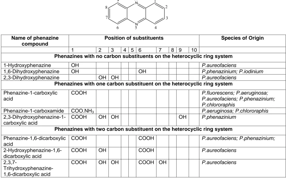
These nitrogen-containing heterocyclic compounds are substituted at different points around their rings, which alters their solubility and biological activity to suit environmental demands of phenazine producing species. (Table 1.1; overleaf).
Phenazines have a characteristic absorption spectrum which includes two intense peaks in the UV range (between 250-290 nm and 350-400 nm) and at least one more in the visible range (400-600 nm) (Britton, 1983). This peak in the visible range varies, depending on the aforementioned modifications of the core phenazine structure and gives rise to a range of brilliant colours, from bright blue of 1-hydroxy-5-methylphenazine (pyocyanin (PYO)), lemon yellow of phenazine- 1-carboxylic acid (PCA) to the bright orange of 2-hydroxy phenazine (2-OHPCA) (Figure 1.1).
a b
Figure 1.1: Streak plate of various phenazine-producing Pseudomonads.
(‘a’;
From Kerr et al 2000) and aqueous solution of phenazines. (‘b’;
From Price- Whelan et al. 2005)
N
N 1
8 2 9 10
3 4 5
6 7
Name of phenazine compound
Position of substituents Species of Origin
1 2 3 4 5 6 7 8 9 10
Phenazines with no carbon substituents on the heterocyclic ring system
1-Hydroxyphenazine OH P.aureofaciens
1,6-Dihydroxyphenazine OH OH P.phenazinium; P.iodinium 2,3-Dihydroxyphenazine OH OH P.aureofaciens
Phenazines with one carbon substituent on the heterocyclic ring system Phenazine-1-carboxylic
acid
COO H P.fluorescens; P.aeruginosa;
P.aureofaciens; P.phenazinium;
P.chlororaphis
Phenazine-1-carboxamide COO.NH3 P.aeruginosa; P.chlororaphis 2,3-Dihydroxyphenazine-1-
carboxylic acid
COOH OH OH OH P.phenazinium
Phenazines with two carbon substituent on the heterocyclic ring system Phenazine-1,6-dicarboxylic
acid
COOH COOH P.aureofaciens; P.phenazinium;
2-Hydroxyphenazine-1,6- dicarboxylic acid
COOH OH COOH P.aureofaciens
2,3,7-
Trihydroxyphenazine- 1,6-dicarboxylic acid
COOH OH OH COOH OH P.aureofaciens
Table 1.1: Some of the phenazines synthesised by various species of Pseudomonas. (From Turner & Messenger, 1986)
I. INTRODUCTION
Till date more than 50 phenazine compounds of bacterial origin have been identified (Laursen & Neilson, 2004). The Pseudomonas species synthesise simple phenazines, (Table 1.1) whereas other, more complex phenazine aldehydes, thioesters, ester and amides are made by Streptomyces and other phenazine producers. This work focuses on the phenazines associated with pseudomonads.
1.4 Biological significance of phenazines
The highlight of biological significance of phenazines is their ability to act as broad-spectrum antimicrobial, antiparasite, antimalarial agents affecting a vast range of organisms (Yang et al., 2005; Cerecetto et al., 2004; Turner et al., 1986;
Budziekiewicz, 1993; Hollstein et al., 1973; Handelsmann et al., 1996; Mavrodi et al., 2001; Kitahara et al., 1982; Baron et al., 1989).
The root-associated soilborne pseudomonads - P. fluorescens, P. chlororaphis and
P. aureofaciens produce PCA, phenazine-1-carboxamide (PCN), andhydroxyphenzaine-1-carboxylic acid (OH-PCA) respectively, which inhibit infection by soilborne phytopathogenic fungi (Chin-A-Woeng et al 2003) and bacterial root diseases (‘take-all decline’ disease), thus securing survival of the host in the rhizosphere and acting as potent biocontrol agents (Giddens et al., 2002; Thomashow et al., 1988).
The opportunistic pathogen P.aeruginosa produces an array of phenazines
including pyocyanin (PYO), PCA, 1-hydroxyphenazine and aerugenosin A and B
(Byng et al., 1979; Hassan et al., 1980, Holliman, 1969). Of these, the
phenazines pyocyanin (PYO) and PCA are implicated in a number of instances
of disease pathogenesis. PYO generates pathogenic symptoms leading to the
effective killing of the nematode Caenorhabditis elegans by P. aeruginosa
Rahme et al., 1997; Mahajan-Miklos et al., 1999). It is also found to be essential
for lung infection in mice and the pathogenicity of P. aeruginosa in ‘burned
mouse’ model (Cao et al., 2001; Lau, 2004; Ran et al., 2003). In case of cystic
fibrosis patients, where lung infection by P. aeruginosa is highly prevalent and
results in a high rate of mortality, phenazine compounds have been detected at
concentrations as high as 10
-4M (Wilson, 1988). Both PCA and PYO alter expression of immunomodulatory proteins of human airway epithelial cells (Denning et al., 2003; Lau et al., 2005), lead to neutrophil inflammation (Look et al. 2005), induce apoptosis of neutrophils (Usher et al., 2002) and thus among other factors, contribute towards the persistence of P. aeruginosa infections (Lau et al., 2004).
Phenazines are capable of mineral reduction and iron sequestering (Hernandez et al., 2004; Schroth et al., 1982), enabling accessibility of these essential minerals not only to phenazine producing species, but also other organisms present in soil, thus ultimately influencing the micro flora in a given environment.
Finally, the phenazines produced by P. agglomerans on the stigmas of apple flowers contribute towards the ability of this bacterium to suppress colonization by phytopathogenic Erwinia amylovora which causes fire-blight disease in apples (Giddens et al., 2003). These examples clearly emphasise the role of phenazines as broad, host-nonspecific pathogenicity factors which contribute towards the ecological fitness of phenazine-producing strains in their natural habitats.
1.5 Mode of action of phenazines
The biological activities of phenazines have interested chemists for a long time
and an estimated 6000 phenazine compounds have been synthesised or
modified after their discovery from biological sources. These include chemically
synthesised phenazines which play an important role in physical and
electrochemical research e.g. construction of microbial fuel cells for generation of
green and renewable energy (Rabaey et al., 2005). Additionally, phenazines
were designed to exploit their DNA intercalative properties, e.g. [5,4-ab]-
phenazine and phenazine-5,10-dioxide to investigate their antiproliferative, thus
anti-tumour and anti-cancer activities (Fernandez et al., 2001; Gamage et al.,
2002; Phillips et al., 2004; Yang et al., 2005). Although the exact mechanism or
effect of intercalation of DNA by phenazines is not well understood, it is thought
that phenazines act by hindering DNA biosynthesis, replication or processing
(Cerecetto et al., 2004);
I. INTRODUCTION
These examples clearly indicate that phenazines, rather than performing one dedicated purpose, seem to be capable of performing multiple roles. This has led to intensification in research towards understanding the mode of action of phenazines. The evident non-selective toxicity of phenazines was thought to arise from a mode of action directed against one of the more universal primary metabolic pathway reactions (Vining, 1991). In 1934, Friedheim and co-workers showed through potentiometric studies, that PYO in mixture with its reduced derivative acted as a reversible redox system. The same group also reported that PYO was capable of increasing the rate of respiration of both mammalian and bacterial cells.
Almost all the effects of phenazines mentioned in the previous paragraph (except DNA intercalation) can be attributed to this one essential feature of phenazines - their capacity for undergoing redox transformations (Hassan et al., 1980; Muller, 1995; Gardner, 1996; Denning et al., 1998). Phenazines are thought to diffuse across, or insert into membranes and readily undergo redox-cycling in the presence of molecular oxygen, resulting in the uncoupling of oxidative phosphorylation and generating reactive oxygen species like superoxide (O
2-) hydroxyl (OH
-) and hydrogen peroxide (H
2O
2) (Hassan et al., 1980; Turner, 1986). The accumulation of these radicals is toxic not only to bacterial but also fungal and eukaryotic cells (Toohey et al., 1965; Hasset et al., 1992; Mahajan et al., 1999) thus conferring host-nonspecific pathogenicity to organisms producing phenazines. The finding of Friedheim about increased rates of respiration has been reiterated by O’Malley et al., (2003) and Lau et al., (2004) who showed that the effects of PCA and PYO on both bacterial and eukaryotic host cells result from oxidative activity and the inactivation of proteins involved in oxidative stress responses. Moreover, Hassett and co-workers (Hassett et al., 1992, 1995) have found that the superoxide dismutases of pyocyanin producing P. aeruginosa possess a higher activity than other known dismutases, which would protect this organism from the harmful effects of phenazines.
Though the redox activity of phenazines is well known and the mode of action is
hypothesised, the exact mechanism of how phenazines generate these species
is not well understood. A more detailed understanding of mode of action of various phenazines, their influence on the molecular level and their metabolism is only just emerging (Hernandez et al., 2001; Rabaney et al., 2005; Look et al.
2005).
1.6 Biosynthesis of phenazines 1.6.1 The Shikimate pathway
As mentioned earlier, all secondary metabolites are known to arise from some modification of the primary metabolic pathways of cells. The discovery of the Shikimic acid pathway for the biosynthesis of aromatic amino acids - tryptophan, tyrosine and phenylalanine in the 1960s (Balinsky and Davis, 1961) and the availability of isotopically labelled compounds resulted in the investigation of some metabolic intermediates of the shikimate pathway as a possible precursor of phenazines.
Three important studies in the 1960-70 period established beyond doubt that the Shikimic acid pathway was indeed the primary metabolic pathway which branched into the phenazine biosynthesis. The first of these studies was carried out in 1963 by Millican (Millican, 1963) and showed that biosynthetically prepared [U
14C] shikimic acid was the precursor of pyocyanin synthesised by P.
aeruginosa;
with the recovery of around 16% of this radiolabelled shikimate in pyocyanin. A second set of experiments using isotope-competition technique (MacDonald, 1963; Levitch et al., 1964, 1966; Chang et al., 1968) confirmed that shikimic acid was the precursor for biosynthesis not only of pyocyanin but also phenazine-1-carboxylic acid and oxychlororaphin. Lastly, experiments by Longley et al. and Calhoun et al., (1972) used mutants unable to degrade shikimic acid and blocked at various points on the branched pathway to aromatic amino acids.
Their experiments not only validated the conclusions by the previous two sets of experiments, but went further and identified chorismic acid as the branching point of the shikimic acid pathway to phenazine biosynthesis (Figure 1.2; overleaf).
The next question in elucidating the phenazine biosynthesis pathway was the
identification of the compound that led to the formation of the tricyclic scaffold of
I. INTRODUCTION
PENTOSE PHOSPHATE
PATHWAY GLYCOLYSIS
Erythrose-4-phosphate Phosphoenol pyruvate
3-Deoxy-D-arabinoheptulosnate -7-phospho (DAHP)
Shikimate COOH
HO OH
OH
O COOH
OH
COOH O
chorismic acid
Aromatic amino acids Phenazines
N N OH
HO OH
COOH
COOH
HO OH
OH
OH
OH
Shikimic acid Iodinin
N N
N N OH
O
COOH
COOH
OH O
COOH C COOH
COOH
CH2
C COOH CH2
COOH
Chorismic acid
4,9-dihydroxyphenazine- 1,6-dicarboxylic acid
Phenazine-1-carboxylic acid
N N
COOH
COOH OH
OH
Phenazine-1,6-dicarboxylic acid B
A
Figure 1.3: The proposed intermediates of the phenazine biosynthesis pathway.
Figure 1.2: The shikimic acid pathway and the two compounds investigated as the potential branching points for the biosynthesis of phenazines.
1.6.2 Assembly of the tricyclic phenazine ring scaffold
To further understand the mechanism of phenazine ring assembly, initial studies on Iodinin were initiated, which led to the hypothesis that the phenazine moiety was formed by the diagonal, asymmetrical incorporation of two shikimic acid units. 4,9-dihydroxyphenazine-1,6-dicarboxylic acid was hence proposed as the key intermediate in the biosynthesis of all phenazines (Podojil et al., 1970).
However, studies on phenazine ring assembly of pyocyanin and phenazine-1- carboxylic acid by Herbert et al., (1972) and Hollstein et al., (1972 & 1973) led to a different hypothesis - that of diagonal, symmetric incorporation of two chorismic acid units leading to formation of the phenazine scaffold, with phenazine-1,6- dicarboxylic acid (PDC) as the common precursor of all phenazines (Herbert et al., 1976; Etherington et al., 1979) (Figure 1.3). This paradigm was accepted, despite lack of any significant incorporation of [U
14C] chorismic acid into phenazines observed by Hollstein (Hollstein et al., 1972, 1973). Meanwhile, search continued for more evidence for chorismic acid being the compound that dimerises to form the phenazine ring system, with anthranilic acid also being considered. The discovery of two sets of anthranilate synthase genes in pyocyanin producing strains of P. aeruginosa by Essar et al., (1990) gave some credence to this hypothesis; although all previous attempts to demonstrate incorporation of isotopically labelled anthranilate into phenazine were unsuccessful (Carter et al., 1961; Millican, 1962).
Thus, although the shikimic acid pathway was accepted as the primary metabolic pathway which branched off into phenazine biosynthesis, the mechanism of assembly of the tricyclic scaffold of phenazines and identity of the intermediate formed in this process remained unclear.
1.6.3 Genetic basis of phenazine biosynthesis in pseudomonads.
Further progress in understanding the phenazine biosynthesis pathway came in
1995, when Pierson and co-workers identified the biosynthetic genes responsible
for the production of phenazine in P. aureofaciens (Pierson et al., 1995). Soon, a
I. INTRODUCTION
phenazine biosynthesis locus was also identified, cloned and sequenced in P.
fluorescens (Mavrodi et al., 1998) and P. aeruginosa (Mavrodi et al., 2001). This
locus comprised of a conserved seven-gene operon phzABCDEFG which was found to be sufficient for the production of PCA from chorismic acid. (Figure 1.4).
Both
P. aureofaciens and P. fluorescens contain one, whereas P. aeruginosapossesses two copies of the phenazine biosynthesis operon. PCA is the end- product of this ‘core’ phenazine biosynthesis pathway.
8505 bp
XhoI SmaI SmaI PstI XbaI
PstI PstI KpnI
EcoRI
EcoRI EcoRI
BglII BglII BglII
0.0kb 2.0kb 4.0kb 6.0kb 8.0kb
RBS RBS RBS RBS RBS
RBS RBS
RBS RBS
phzD phzG
phzI phzR phzA phzB phzC phzE phzF
Figure 1.4: Phenazine biosynthesis operon from P.fluorescens.
The nucleotide sequences of the phenazine biosynthetic operon of these organisms were found to be very homologous, with an identity of 70-95%. This implies that the manner of phenazine biosynthesis and the functions of enzymes involved in these organisms are identical.
The discovery of this ‘core’ phenazine biosynthesis operon in Pseudomonas led to further investigation into the presence of this operon in other phenazine producing species.
1.6.4 Phenazine biosynthesis operon in other organisms
The seven-gene sequence from Pseudomonas was used as a probe to find
similar genes in other phenazine-producing organisms. However, this probe
yielded no results in case of Burkholderia cepacia, Burkholderia phenazinium
(formerly,
Pseudomonas cepacia and P. phenazinium respectively),and
Brevibacterium iodinum. This led to the conclusion that although the phenazinebiosynthesis operon is conserved in the Pseudomonal species, other phenazine
producing species might utilise a modified or different operon for phenazine
biosynthesis. To test this hypothesis, further sequence comparison between the newly sequenced genomes of various microorganisms were initiated using only five of the seven enzymes i.e. PhzA, D, E, F and G. This probe found matches in the genomes of B. cepacia, Erwinia carotovora and the first gram positive bacteria Brevibacterium lingens.
B. cepacia was previously called Pseudomonas cepacia (Ballard et al., 1970)
and was known to produce the phenazines shown in the figure below:
N N
COO.CH3
OH OH
COOCH3
4,9-dihydroxyphenazine -1,6-dicarboxylic acid
dimethyl ester
N N
COOH
COOH
phenazine-1,6-dicarboxylic acid
N N
COO.CH3
COOCH3
N N
COO.CH3
OH COOCH3 phenazine-1,6-dicarboxylic acid
dimethyl ester
4-hydroxyphenazine -1,6-dicarboxylic acid
dimethyl ester Figure 1.5: Phenazines produced by B. cepacia.
On further investigation, the phenazine cluster of B. cepacia was found to consist six instead of seven genes, with only one copy of the highly identical phzA-B genes of Pseudomonas.
Investigation into this phzA/B-like gene from Burkholderia cepacia (named BcepA
in this work) was initiated, to gain further insight into the structure and function of
phzA/B-like genes of Pseudomonas. This study is the first to undertake theinvestigation of an enzyme of the phenazine biosynthesis operon of B. cepacia
I. INTRODUCTION
and no other information is available at this point regarding the specific details of how this pathway operates, the substrates, intermediates involved etc.
This, however, is not the case for the phenazine biosynthesis operon of
Pseudomonas, for which a comprehensive study was undertaken by MacDonaldand co-workers (MacDonald et al. 2001).
1.6.5 Phenazine biosynthesis in Pseudomonas.
MacDonald et al., (2001) expressed all or a subset of seven genes of the phenazine biosynthesis operon of P. fluorescens in Escherichia coli. Feeding experiments were then carried out using exclusively, chorismic acid, anthranilic acid or 2-amino-2-deoxychorismic acid (ADIC), to re-examine the branching point of phenazine biosynthesis from shikimic acid pathway and the process of phenazine ring assembly.
Their results proved conclusively that chorismic acid was the branching point and it was converted to ADIC by the enzyme PhzE. Also, neither anthranilic acid (no conversion to PCA) nor chorismic acid (conversion to ADIC by PhzE, but no PCA formation) were utilised for the assembly of the phenazine scaffold. ADIC however, was completely converted to PCA. Moreover, incubation of ADIC with the enzyme PhzD yielded trans-2, 3-dihydro-3-hydroxyanthranilic acid (DHHA) and incubation of DHHA with PhzA-G showed complete conversion to PCA. Both these compounds were thus confirmed as intermediates of phenazine biosynthesis pathway.
PDC, the previously hypothesised intermediated, was not detected in these
experiments. To further explore the possibility of PDC formation, [11-C
13]-labelled
PDC was incubated with PhzA-G extract. Less than 1% of this labelled PDC was
found to be converted into PCA. Moreover, the incubation of radiolabelled PDC in
the presence of radiolabelled ADIC with the same set of enzymes (PhzA-G)
yielded no conversion of labelled PDC to PCA. Thus, PDC was ruled out as an
intermediated of the phenazine biosynthesis pathway of P. fluorescens. Instead,
the dimerisation of two molecules of oxidised DHHA was suggested as a
possible mode of phenazine scaffold assembly.
These observations were collated and presented in the form of a scheme for the biosynthesis of PCA from chorismic acid by MacDonald and co-workers (shown overleaf) which included the newly identified intermediates and the role of enzymes involved.
1.6.6 Enzymes of the phenazine biosynthesis operon
While studying the various enzymes involved in the phenazine biosynthesis operon of P. fluorescens, MacDonald et al. compared the sequences of these seven enzymes with proteins of known function. Their results are tabulated in table below.
Gene Size(amino acids) Similar Enzymes
PhzA
163 PhzB
PhzB
162 PhzA
PhzC
400 Plant 3-deoxy-D-arabino-heptulosonate-7- phosphate (DAHP) synthase.
PhzD
207 Bacterial Isochorismate
PhzE
637 Bacterial Anthranilate synthase
PhzF
278 Unknown
PhzG
222 Bacterial pyridoxyamine 5’-phosphate oxidase
Table 1.2: Enzymes similar to those of the phenazine biosynthesis pathway.There is more than 70% similarity between the genes phzA and B, which were found to be important for quantitative synthesis, but not essential for the biosynthesis of PCA (MacDonald et al. 2001). Both these enzymes however, are conserved in the phenazine biosynthesis operons of all Pseudomonas species.
The enzymes PhzA-B were thought to act past the formation of DHHA, but no further information, either structural or biochemical was available, since these enzymes showed no similarity to any enzymes of known function in the NCBI database.
Of the rest of the enzymes PhzC, D, E, F and G, which are essential for PCA
production, PhzC, D, E and G were found to have counterparts in the bacterial
I. INTRODUCTION
world. PhzC is a 3-deoxy-arabino-heptulosonate-7-phosphate (DAHP) synthase and acts more upstream (prior to formation of chorismic acid), ensuring adequate availability of chorismate for phenazine biosynthesis. PhzE, which acts after PhzC, shows similarity to bacterial anthranilate synthase and catalyses the conversion of chorismate to ADIC. This enzyme is followed by PhzD, an isochorismatase, which catalyses the conversion of ADIC to DHHA. This conclusion was recently confirmed by the crystal structural analysis of PhzD by Parson et al., (2003).
Recently, Blankenfeldt et al., (2004) shed more light on the mode of action of the homodimeric enzyme PhzF through structural and biochemical analysis. They showed that PhzF acts as an isomerase, converting DHHA to its highly reactive ketone form - 1,2,4-trihydro-3-oxo-anthranilic acid. Blankenfeldt and co-workers also hypothesised that PhzF is an enzyme with dual functions and also catalyses the condensation of two molecules of this ketone to form the phenazine ring scaffold. This role was formerly thought to be performed by PhzG (MacDonald et al., 2001). The modified scheme proposed by Blankenfeldt et al is depicted in the Figure 1.6, overleaf.
The last of the seven enzymes, PhzG, is similar to bacterial pyridoxyamine-5- phosphatase. It was thought to act in steps past the formation of 2,3-dihydro-3- oxo anthranilic acid, but its exact role in PCA biosynthesis could not be deduced.
The functions of the enzymes PhzC, D, E and F were thus known or could be accurately surmised. However, no such conclusion could be drawn for PhzA, B and G. The clarification of the structure and roles of these three enzymes was one of the goals of this investigation.
As mentioned previously, the end product of core phenazine biosynthesis
operon,
phzABCDEFG, is PCA, which is the only phenazine compoundsynthesised by P. fluorescens. PCA however, is modified to other phenazine
derivatives in case of Pseudomonas strains like P. aeruginosa, P. chlororaphis
etc. The genetic basis of generation of phenazine diversity and the organisation
of phenazine biosynthesis operon in other organisms is described herewith.
COOH
O
OH
COOH
COOH
O COOH NH2
COOH COOH
OH O
NH2 NH2
H H
PhzE PhzD
PhzF
N N COOH
COOH N
N COOH
N N COOH
[PhzA,B,G]
[PhzA,B,G]
COOH NH2
OH PhzF
Phenazine-1-carboxylic acid Mw224
Partially aromatised PCA Mw 228
Schiff's Base Mw 274
Chorismic Acid Trans-2,3-Dihydro- 1,2,4-trihydro-3-oxo-anthranilic acid
2,3-dihydrobenzoic acid (DHHA)
2-amino-2deoxychorismic acid (ADIC)
-CO2, -H2 -2H2
O COOH
OH
COOH O
COOH NH2
O COOH
NH2
COOH H2N
O
[O], CO2
PhzE PhzD
chorismic acid 2-amino-2-deoxychorismic acid (ADIC)
Trans-2,3-dihydro-3- hydroxyanthranilic acid (DHHA)
PhzF and PhzA,B
Phenazine-1-carboxylic acid (PCA)
COOH NH2
OH
PhzG
N N COOH Phosphoenol
pyruvate Erythrose-4- phosphate
COOH O(P)
HO OH
OH
PhzC primary shikimate
pathway enzymes
3-Deoxy-D-arabinoheptulosonic acid-7-phosphate (DAHP)
2,3-Dihydro-3-oxo-anthranilic acid O
O
COOH
PhzF 2x
a
b
Figure 1.6: (a) Scheme of Phenazine biosynthesis
proposed by MacDonald et al., (2001) & (b) modified by Blankenfeldt et al., (2004)
I. INTRODUCTION
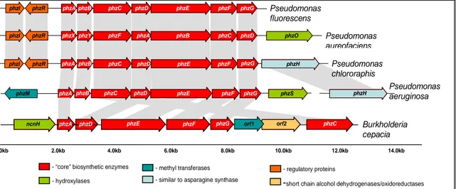
1.6 Generation of phenazine diversity.
Diversity in phenazine biosynthesis is achieved by the presence of other genes, associated with the core phenazine biosynthesis operon, which are capable of a wide range of modifications of PCA. Such genes were identified in P.
chlororaphis, P. aeruginosa and P. aureofaciens and the organisation of
phenazine biosynthesis operon in these organisms is compared to that of P.
fluorescens in the figure below:
phzR phzY phzF phzA phzB phzC phzD phzO
phzI phzX
phzI phzR phzA phzB phzC phzD phzE phzF phzG phzH
phzM phzA phzB phzE phzF phzG phzS phzH
phzR phzB phzC phzD phzE phzF phzG
phzI phzA Pseudomonas
fluorescens
Pseudomonas aureofaciens
Pseudomonas chlororaphis
Pseudomonas aeruginosa
0.0kb 2.0kb 4.0kb 6.0kb 8.0kb 10.0kb 12.0kb 14.0kb
- regulatory proteins - “core” biosynthetic enzymes
- hydroxylases
- methyl transferases
-short chain alcohol dehydrogenases/oxidoreductases - similar to asparagine synthase
phzC
ncnH phzA phzD phzE phzF phzG orf1 orf2 phzC Burkholderia
cepacia
phzD
Figure 1.7 The organisation of the phenazine biosynthetic operon from various
organisms
(From Mavrodi et. al., 2005)In case of P. chlororaphis, the biosynthetic gene phzH is found downstream of phzG. PhzH belongs to the asparagine snythase enzyme family and catalyses the conversion of PCA to PCN via a transamidase reaction. The transfer of phzH gene to the phenazine biosynthetic operon of P. fluorescens and P. aureofaciens enabled these strains to produce PCN and to act as biocontrol agents in tomato (Chin-A-Woeng et al., 2001). Thus, validating the observation that PCA is not only the end product of the enzymes PhzABCDEFG, but it is the compound that is produced in all phenazine synthesising Pseudomonas.
In
P. aureofaciens, the gene phzO is localised immediately downstream of thecore biosynthetic operon as well. Belonging to the family of two-component non-
haem, flavin-diffusible bacterial aromatic monooxygenases, phzO catalyses the
conversion of PCA to 2-OHPCA.
P. aeruginosa in addition to phzH contains two other genes – phzM and phzS
associated with this pathway. phzM is located upstream of the operon and catalyses the formation of PYO. PhzS is a protein similar to bacterial monooxygenases and catalyses the conversion of PCA to both PYO and 1- hydroxyphenazine. Figure 1.8 summarises the conversion of PCA to different derivates.
The current knowledge of the enzymes involved in phenazine biosynthesis and modification is still not complete and it is expected that further research into this field will result in the identification of more such enzymes. This also holds true about the regulation of phenazine biosynthesis, which, though not yet fully understood, is known to be regulated by the mechanism of quorum sensing in
Pseudomonas.N N
COOH N N
COOH
N N
COOH
N N
O N N
N N
CONH2
N N
OH
OH
OH
CH3
CH3
Phenazine-1-carboxylic acid (PCA)
5-methylphenazine-1- carboxylic acid betaine
Pyocyanin 2-hydroxyphenazine- carboxylic acid 2-hydroxyphenazine
Phenazine-1- carboxamide (PCA) 1-hydroxyphenazine
PhzO
PhzS PhzH
PhzM
PhzS PhzM
Figure 1.8: Derivatives of
PCA and the enzymes
involved in its modification.
I. INTRODUCTION
1.8 Regulation of phenazine production
Quorum sensing is a mechanism that enables bacteria to regulate their gene expression in a population-density-dependent manner, thus adjusting various metabolic processes according to environmental conditions. In this process, autoinducer signal molecules convey population density information from neighbouring sister cells. These autoinducer molecules are diffusible across cell membranes and belong to N-acyl-
L-homoserine lactone (N-AHLs) class of molecules in gram negative bacteria like pseudomonas.
In
P. fluorescens, P. aureofaciens and P. chlororaphis, two genes, phzI andphzR, located directly upstream to the phenazine biosynthesis operon function as regulators of N-acyl-homoserine lactone mediated gene expression of the ‘phz’
operon. Thus, these two genes link quorum sensing to phenazine biosynthesis and act as regulating sub-circuit in these Pseudomonas strains. However, this is not the only mode of regulation. In P. aeruginosa, the genes regulating phenazine biosynthesis are not located upstream of the core biosynthetic pathway, but elsewhere in the genome. In this organism, the expression of phenazine biosynthesis is thought to be regulated in a more complex manner, by not one, but two sets of hierarchically organised quorum sensing cascades ‘las’
and ‘rhl’. Thus, the regulation of phenazine biosynthesis is varied and the current knowledge still incomplete. However, a detailed description of regulation of phenazine biosynthesis is outside the scope of this work.
This work concerns itself chiefly with the investigation of the enzymes PhzA, PhzB, PhzG (P. fluorescens) and BcepA (B. cepacia); and the elucidation of the
‘core’ phenazine biosynthesis pathway, with respect to the intermediates
generated during the formation of PCA.
Objectives of this work
‘Now, I give you fair warning,' shouted the Queen.
Alice in wonderland, Lewis Carroll
II. OBJECTIVES OF THIS WORK
2.0 Objectives of this work
As discussed in the introduction, phenazines are versatile and multi-functional compounds; but relatively little information is available about the biosynthesis of these molecules in nature. Chemically modified or synthesised phenazines are being explored for various applications, however, no efficient method of chemical synthesis of phenazines has yet been found. Tapping into the natural methods of phenazine production, as employed by microorganisms like pseudomonads, could potentially provide means to generate novel phenazines for various application from pH indicators (exploiting their unique spectral properties, which vary with change in pH), and antibiotics, to DNA intercalators (as anti-tumour agents). It could also provide insights into the development of inhibitors of phenazine biosynthesis and other pharmaceutical applications and help tackle the problem of fatal infections by phenazine-producing micro-organisms in immuno-compromised individuals.
To this effect, this project intends to deepen the understanding of the functioning
of the ‘core’ phenazine biosynthesis pathway involving enzymes PhzA-G and
leading to the formation of PCA. The specific aims of this work encompasses
firstly, the over-expression, crystallisation and elucidation of the three-
dimensional structures of the enzymes PhzG, PhzA and similar enzymes (PhzB
from
P. fluorescens and BcepA from Burkholderia cepacia) to discover the roleplayed by these enzymes in phenazine biosynthesis. Secondly, this work intends
to investigate the exact mechanism of phenazine biosynthesis to establish the
identity of the substrates and products of the various enzymes as well as the
intermediates formed during the production of PCA by using various biochemical
and mass spectroscopic methods.
Results & Discussions
‘What is the use of repeating all that stuff, if you don't explain it as you go on? It's by far
the most confusing thing I ever heard!’
- The Mock Turtle (Alice in wonderland, Lewis Carroll)
III. RESULTS AND DISCUSSION
Section I
This section describes the details of cloning, over-expression and crystallisation of PhzA, PhzB, BcepA and its complex, PhzG and PhzG complexed with PCA.
The results of the bioinformatics analysis undertaken for each of the proteins are
also discussed here.
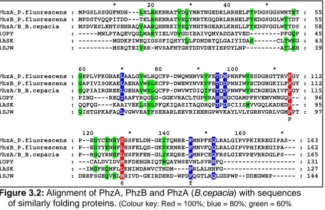
3.1 Sequence Analysis 3.1.1 PhzA, PhzB and BcepA
The sequences of proteins PhzA, B and A_Bcep were found to be 70%
identical to each other and upto 60% identical to sequences of similar proteins from other Pseudomonads (Figure 3.1). The sequences were compared using the program BLAST (BLASTp, NCBI. Altschul et al., 1997).
Figure 3.1: Multiple alignment of proteins similar to PhzA and B.
PhzA_P.fluorescens : PhzB_P.fluorescens : PhzA_P.chlororaphis : PhzY_P.chlororaphis : PhzX_P.chlororaphis : PhzA_P.aeruginosa : PhzA_Pan.agglomerans : PhzA/B_B.cepacia :
* 20 * 40 * MPGSLSSGGF-NDHLEL--RRKNRATVDQYMR-TNGEDRLRRHELFTPDGSGGSWNT MPDSTVQQPITDDT-EL--RRKNRATVEQYMR-TKGQDRLRRHELFTEDGSGGLWTT MPASLSPSSF-NDHLEL--RQKNRATVEQYMR-TNGKDRLRRHELFTQDGSGGSWNT MSNSAAQQLTANDTTEL--RRKNRATVEQYMR-TKGQDRLRRHELFTEDGTGGLWTT MPASLSPSGF-NDHLEL--RQKNRATVEQYMR-TNGKDRLRRHELFTQDGSGGSWNT MREYQRLKGF-TDNLEL--RRRNRATVEHYMR-MKGAERLQRHSLFVEDGCAGNWTT ---MYLTDEDAIRI--REINRQVVSQYLSSTRGIARLKRHELFAEDGEGGLWTT MSDVESLENTSENRAQVAARQHNRKIVEQYMH-TRGEARLKRHLLFTEDGVGGLWTT m 1 6 R NRatVeqY6r t G RL RHeLFt DG gG W T
: 53 : 53 : 53 : 54 : 53 : 53 : 49 : 56
PhzA_P.fluorescens : PhzB_P.fluorescens : PhzA_P.chlororaphis : PhzY_P.chlororaphis : PhzX_P.chlororaphis : PhzA_P.aeruginosa : PhzA_Pan.agglomerans : PhzA/B_B.cepacia :
60 * 80 * 100 * ETGEPLVFKGHAKLAALGVWLHQCFPDWQWHNVRVFETDNPNHFWVESDGRGTTRVP DTGAPIVISGKAKLAEHAVWSLKCFPDWEWYNVKVFETDDPNHIWVECDGHGKILFP ETGEPLVFKGHSKLAALGEWLEKCFPDWQWHNVRVFETDNPNHFWVESDGRGKTLVP DTGAPIVISGKAKLAEHAVWSLKCFPDWEWYNVKVFETDDPNHIWVECDGHGKILFP ETGKPLVFKGHTKLAALGVWLEKCFPDWQWHNVRVFETDNPNHFWVESERRGKTLVP ESGEPLVFRGHESLRRLAEWLERCFPDWEWHNVRIFETEDPNHFWVECDGRGKALVP ETGEPIIIRGIENLEKHAKWSLECFPDWEWYNIKIFTTDNPDHVWVECDGRGLIRFP DSGQPIAIRGREKLGEHAVWSLQCFPDWVWTDIQIFETQDPNWFWVECRGEGAIVFP 3G P6 G kL W CFPDW W 16 6FeTd1P1h WVE dg G P
: 110 : 110 : 110 : 111 : 110 : 110 : 106 : 113
PhzA_P.fluorescens : PhzB_P.fluorescens : PhzA_P.chlororaphis : PhzY_P.chlororaphis : PhzX_P.chlororaphis : PhzA_P.aeruginosa : PhzA_Pan.agglomerans : PhzA/B_B.cepacia :
120 * 140 * 160 * GYPEGYCENHYIHSFELDNGKITQNREFMNPFEQLRALGIPVPKIKREGIPAS---- GYPEGYYENHFLHSFELQDGKVKRNREFMNVFQQLRALGIPVPHIKREGIPA--- GYPEGYCENHYIHSFELDDGKITQNREFMNPFQQLRALGIPVPKIKREGIPAS---- GYPEGYYENHFLHSFELEDGKVKRNREFMNVFQQLRALGIPVPQIKREGIPT--- GYPEGYCENHYIHSFELDDGKITQSREFMNPFEQLRALGIPVPRIKREGIPAS---- GYPQGYCENHYIHSFELENGRIKRNREFMNPMQKLRALGIAVPQIKRDGIPT--- GYPESYYENHFIHSFELRNGLIVRNREFMNPVTQLKCLGIDVPRIKREGIPS--- GYPRGQYRNHFLHSFRFENGLIKEQREFMNPCEQFRSLGIEVPEVRRDGLPS--- GYP gy eNH56HSFel 1G 6 nREFMNp ql4aLGI VP 64ReG6P
: 163 : 162 : 163 : 163 : 163 : 162 : 158 : 165
(Colour key: Red = 100%; blue = 80%; green = 60% similarity)
The potential folds of these proteins were identified using the structure
prediction program 3DPSSM (Kelley et al., 2000), which predicts protein fold
based on the sequences of proteins. 3DPSSM predicted these three proteins
to be similar in fold to the members of the nuclear transport factor-2 (NTF2-
like) superfamily. This superfamily includes members of the NTF2 family, Δ
5-
3-ketosteroid isomerases, scytalone dehydratases, and the beta subunit of
ring-hydroxylating dioxygenases. This family represents a classic example of
divergent evolution where the proteins have a common fold, but diverge
greatly in their sequence and function. For example, nuclear transport factor 2
(NTF2) mediates the nuclear import of RanGDP and binds to both RanGDP
III. RESULTS AND DISCUSSIONS
and ‘FxFG’ repeat-containing nucleoporins while ketosteroid isomerases catalyze the isomerization of Δ
5-3-ketosteroid to Δ
4-3-ketosteroid, by intramolecular transfer of the C4-beta proton to the C6-beta position. While the function of the beta subunit of the ring-hydroxylating dioxygenases is not known, scytalone dehydratase catalyzes two reactions in the biosynthetic pathway producing fungal melanin. Members of the NTF2-like superfamily are widely distributed among bacteria, archaea and eukaryotes. In the sequence alignment shown in Figure 3.2, sequences of one representative each of ketosteroid isomerase (1OPY), nuclear transport factor-2 (1ASK) and scytalone dehydrates (1SJW) have been aligned to the sequences of PhzA, PhzB and BcepA. No meaningful similarity can be observed between these sequences.
Thus, although tertiary structure of the PhzA group of enzymes (PhzA, B and BcepA) could be predicted with some confidence, no conclusion as to the potential function could be drawn from the sequence alone.
Figure 3.2:
Alignment of PhzA, PhzB and PhzA (B.cepacia) with sequences of similarly folding proteins.
(Colour key: Red = 100%; blue = 80%; green = 60%similarity)
PhzA_P.fluorescens : PhzB_P.fluorescens : PhzA/B_B.cepacia : 1OPY : 1ASK : 1SJW :
* 20 * 40 * MPGSLSSGGFNDH---LELRRKNRATVDQYMRTNGEDRLRRHELFTPDGSGGSWNTET MPDSTVQQPITDD---TELRRKNRATVEQYMRTKGQDRLRRHELFTEDGSGGLWTTDT MSDVESLENTSENRAQVAARQHNRKIVEQYMHTRGEARLKRHLLFTEDGVGGLWTTDS ---MNLPTAQEVQGLMARYIELVDVGDIEAIVQMYADDATVED---PFGQP ---MGDKPIWEQIGSSFIQHYYQLFDNDRTQLGAIYIDAS---CLTWEG ---MSRQTEIVRR-MVSAFNTGRTDDVDEYIHPDYLNP---ATLEH
: 55 : 55 : 58 : 45 : 43 : 39
PhzA_P.fluorescens : PhzB_P.fluorescens : PhzA/B_B.cepacia : 1OPY : 1ASK : 1SJW :
60 * 80 * 100 * GEPLVFKGHAKLAALGVWLHQCFP-DWQWHNVRVFETDNPNHFWVESDGRGTTRVPGY GAPIVISGKAKLAEHAVWSLKCFP-DWEWYNVKVFETDDPNHIWVECDGHGKILFPGY GQPIAIRGREKLGEHAVWSLQCFP-DWVWTDIQIFETQDPNWFWVECRGEGAIVFPGY PIHG----REQIAAFYRQGLG-GGKVRACLTGPVRASHNGCGAMPFRVEMVWNGQP-- QQFQG----KAAIVEKLSSLPFQKIQASITAQDHQPTPDSCIISMVVGQLKADEDP-- GIHTGPKAFAQLVGWVRATFSEEARLEEVRIEERGPWVKAYLVLYGRHVGRLVGMPPT P
: 112 : 112 : 115 : 96 : 95 : 97
PhzA_P.fluorescens : PhzB_P.fluorescens : PhzA/B_B.cepacia : 1OPY : 1ASK : 1SJW :
120 * 140 * 160 * P--EGYCENHYIHSFELDN-GKITQNRE-FMNPFEQLRALGIPVPKIKREGIPAS-- P--EGYYENHFLHSFELQD-GKVKRNRE-FMNVFQQLRALGIPVPHIKREGIPA--- P--RGQYRNHFLHSFRFEN-GLIKEQRE-FMNPCEQFRSLGIEVPEVRRDGLPS--- ---CALDVIDVMRFDEHGRIQTMQAYWSEVNLSVREPQ--- --IMGFHQMFLLKNINDAWVCTNDM----FRLALHNFG--- DRRFSGEQVHLMRIVD-GKIRDHRD-WPDFQGTLRQLGDPWP--DDEGWRP---
6 f
: 163 : 162 : 165 : 131 : 127 : 144
3.1.2 Analysis of genes similar to PhzA/B.
An analysis of the evolution of various genes similar to PhzA/B, including those from B. cepacia, Erwinia carotovora and P. agglomerans (Figure 3.3) indicated the possibility that the pseudomonal phzA/B genes had arisen later in the evolutionary timeline and that these genes were orthologs of BcepA and
Erwinia carotovora-A (ErwA). BcepA and ErwA are thus the predecessors ofgenes of the PhzAB family in Pseudomonas. This leads to the conclusion that an event of gene duplication might have occurred in the genomes of
Pseudomonas, which led to the formation of two copies of PhzA-like genes inthis species. Thus the genes phzA and phzB are infact paralogs. This explains the high level of sequence similarity between phzA/B-like genes among
Pseudomonas.Figure 3.3: Evolutionary tree of the sequences similar of PhzA/B.