• Keine Ergebnisse gefunden

Ionic currents and intrinsic properties of key interneurons and their influence on network activity in a chain of coupled oscillators

N/A
N/A
Protected

Academic year: 2021

Aktie "Ionic currents and intrinsic properties of key interneurons and their influence on network activity in a chain of coupled oscillators"

Copied!
201
0
0

Wird geladen.... (Jetzt Volltext ansehen)

Volltext

(1)

Ionic currents and intrinsic properties of key interneurons and their influence on network activity

in a chain of coupled oscillators

Inaugural – Dissertation

zur Erlangung des Doktorgrades

der Mathematisch-Naturwissenschaftlichen Fakultät der Universität zu Köln

vorgelegt von Laura Schläger aus Starnberg

Köln, 2018

(2)

Berichterstatter/in: Dr. Carmen Wellmann Prof. Dr. Ansgar Büschges

Tag der mündlichen Prüfung: 03.09.2018

(3)

Zusammenfassung………….……….………..

Abstract………..………..

1. Introduction………..………

1.1 Central mechanisms for rhythm generation and coordination…….…….………

1.2 The swimmeret system of the crayfish………..………

1.3 Cellular activity pattern of swimmeret neurons……….………..……….

1.4 Cellular properties of rhythmic neuronal networks…………...…….………

1.5 Ion channels mediating inward currents……….………..

1.5.1 Sodium channels………..………...

1.5.2 Calcium channels……….………...

1.5.3 HCN channels……….………….

1.6 Ion channels mediating outward currents……….………….

1.7 Computational model of the swimmeret system………

1.8 Objectives………

2. Material and Methods………...

2.1 Dissection………

2.2 Electrophysiology………...

2.2.1 Extracellular recordings………...

2.2.2 Intracellular recordings………...

2.3 Morphology………..

2.4 Experimental Procedure………

2.5 Statistics………...

2.6 Computational modeling of a half center CPG model………...

3. Results………

3.1 Unveiling the PIR phenotype in an intact swimmeret network…....…………..

3.1.1 Breaking down network activity to elucidate cellular properties………..

3.2 Cellular properties in neurons of the swimmeret system – PIR……….

3.2.1 PIR in four types of spiking neurons in the swimmeret system…..…….

3.2.2 PIR in two types of nonspiking neurons of the swimmeret system….….

3.3 Ionic bases for PIR phenotype………

I III

1

1

3

6

7

8

9

10

11

12

13

14

17

17

17

17

19

19

20

25

26

28

28

30

33

33

51

57

(4)

3.3.1.1 I

CaL

and I

H

contribute to PIR responses in PSE…………...58

3.3.1.2 I

CaL

and I

H

contribute to PIR responses in RSEs………....60

3.3.2 Coordinating Neurons ………63

3.3.2.1 Different currents contribute to PIR responses in ASC…...63

3.3.2.2 I

CaL

, I

H

and I

NaP

contribute to PIR responses in DSC……...67

3.3.3 CPG neuron – I

H

contributes to PIR responses in IPS……...70

3.3.4 I

NaP

contributes to PIR responses in ComInt1……….…72

3.3.5 Other cellular properties affected by ion channel blocker application – effects on membrane input resistances……….…….76

3.4 Functional relevance of identified ionic conductances during fictive locomotion……….…..80

3.4.1 Contribution of I

CaL

during rhythmic motor activity………..80

3.4.2 Contribution of I

H

during rhythmic motor activity……….85

3.4.3 Contribution of I

NaP

during rhythmic motor activity………..89

3.5 Short excurse into roles of outward currents during fictive locomotion - I

A

…...91

3.5.1 Contribution of I

A

during rhythmic motor activity...………..91

3.6 Different composition of ionic conductances mediate similar CPG activities – A computational model approach………...…...99

4. Discussion……….…...101

4.1 Membrane properties in key interneurons of the swimmeret system – PIR…..………101

4.1.1 PIR responses in four groups of spiking neurons………..…..102

4.1.1.1 Lacking voltage dependence of PIR responses………...102

4.1.1.2 Variances of PIR amplitudes within groups of spiking swimmeret neurons………..……….103

4.1.1.3 Ionic bases for PIR responses in four classes of spiking neurons….104 4.1.2 PIR responses in the nonspiking CPG neuron – IPS……….….109

4.1.3 ‘PIR-like’ responses in ComInt1……….….113

4.2 Functional relevance of identified ionic currents during fictive locomotion….………..………..113

58 60 63 63 67 70 72

76

80 80 85 89 91 91

99 101

101 102 102

103 104 109 113

113

(5)

4.2.2 Functional relevance of I

H

during rhythmic motor activity………….…….

4.2.3 Functional relevance of I

NaP

during rhythmic motor activity……….…….

4.2.4 Functional relevance of I

A

during rhythmic motor activity……….

4.3 Different composition of ionic conductances mediate similar

CPG activities ……….….………

4.4 Comments on synaptic isolation………

4.5 Summary - Ionic currents in a chain of coupled oscillators………..…

4.6 Outlook……….

5. References………

6. Appendix……….…..

116 117 118

120

122

123

124

125

178

(6)

I

Zusammenfassung

Der Post-Inhibitorische Rebound (PIR) stellt einen essentiellen Mechanismus in der Generierung rhythmischer Aktivitäten von neuronalen Netzwerken dar. Erzeugt durch eine Phase hemmender Eingänge, kann dieser Mechanismus neue neuronale Erregung erzeugen und fördert daher, zellautonom, rhythmische Aktivität. In dieser Arbeit habe ich den Mechanismus des PIRs im Zusammenhang mit rhythmischer Bewegung der vier Schwimmbeinpaare am Flusskrebs untersucht. Das neuronale Netzwerk welches die sehr gleichmäßige und koordinierte Schwimmbeinbewegung erzeugt ist weitestgehend identifiziert. Des Weiteren ist bekannt, dass alle Neurone die bei der Generierung dieser Bewegung involviert sind, ein ähnliches Aktivitätsmuster zeigen. Das Membranpotential aller dieser Neurone oszilliert in der gleichen Frequenz wie die Beinbewegung. Es ist daher davon auszugehen, dass die Generierung dieser Oszillationen ein wichtiges Phänomen für den Erhalt der rhythmischen Schwimmbeinbewegung darstellt. Es ist jedoch nicht bekannt ob und in wie fern zellulären Eigenschaften, wie z. B. ein PIR, an der Generierung beteiligt sind. Um dies zu untersuchen habe ich das abdominale Nervensystem isoliert und intrazelluläre Ableitungen an den Neuronen, welche bewiesenermaßen die Schwimmbeinbewegung generieren und koordinieren, durchgeführt. Um einen PIR zu erzeugen wurden die Neurone durch eine negative Strominjektion für 3 Sekunden hyperpolarisiert. Dies induzierte in fast allen Neuronen, die hemmende synaptische Eingänge erhalten, ein „sag-Potential“ während der Stimulation und anschließend einen PIR. Dies waren sowohl zwei exzitatorische Gruppen von Motoneuronen, Power Stroke Erreger (PSE) und Return Stroke Erreger (RSE), als auch die koordinierenden Neurone, das Aszendierende- (ASC

E

) und das Deszendierende Koordinierende Neuron (DSC). Im Gegensatz dazu wurde nicht in allen der untersuchten Neurone des Zentralen Mustergenerators (CPG), die Inhibitoren des Power Stroke (IPS), ein PIR erzeugt. In jenen IPS jedoch die einen PIR erzeugten, konnte ein Einfluss des Halte-Membran Potentials auf die Größe des PIR erkannt werden. In einer weiteren Neuron Gruppe, dem Kommissuralen Interneuron 1 (ComInt1), welches erregende synaptische Eingänge bekommt, konnte unerwarteter Weise auch ein PIR erzeugt werden.

Im weiteren Verlauf der Experimente erforschte ich welche Ionenströme der

Generierung der beobachteten PIRs zugrunde liegen. Dabei untersuchte ich

(7)

II klassische Ionenströme welche in anderen Nervensystemen an der Generierung von PIRs beteiligt sind. Dies sind der L-typ Calciumstrom (I

CaL

), der hyperpolarisiert- aktivierte Kationenstrom (I

H

) als auch der persistierende Natriumstrom (I

NaP

).

Bei einer Hemmung von I

CaL

durch Applikation des bekannten Antagonisten Nifedipin, konnte eine Zunahme des PIR in 13 von 21 aktionspotential-generierenden Neuronen beobachtet werden. Der PIR in den übrigen 8 Neuronen wurde durch die Hemmung von I

CaL

reduziert. Der PIR des CPG Neurons, IPS, wurde nicht durch Nifedipin beeinflusst. Bei einer anschließenden Zugabe eines bekannten Antagonisten von I

H

, Zd7288, reduzierte sich der PIR in allen Motorneuronen, IPS wie auch in 4 ASC

E

s. In den übrigen ASC

E

s und DSCs konnte die Hemmung von I

H

den induzierten PIR nicht reduzieren. Dies wurde jedoch erreicht als I

NaP

durch die Hinzugabe von Riluzole, geblockt wurde.

Um die Bedeutung der identifizierten Ionenströme für die Generierung dieser Aktivitätsmuster zu verifizieren, wurden die oben beschriebenen Ionenkanalblocker einzeln, während rhythmischer Motoraktivität, appliziert. Dabei wurden sowohl die Aktivitäten einzelner Neurone, die Koordination zwischen den Segmenten als auch die Rhythmus-Eigenschaften des gesamten Netzwerkes analysiert. Hemmung von allen vorherigen Ionenströmen (I

CaL

, I

H

, I

NaP

), sowie eines transienten Auswärtsstroms I

A

durch 4-AP, hatte weitreichende Effekte auf die Rhythmusgenerierung. Ein Effekt auf die intersegmentale Koordination konnte bei einer Hemmung von I

H

, I

NaP

und I

A

beobachtet werden. Während die Blockierung von I

CaL

, I

H

, und I

A

ebenfalls eine Reduzierung der Aktionspotential Weiterleitungsgeschwindigkeit verursachte.

Die beschriebenen Ergebnisse zeigen, dass der PIR einen wichtigen Mechanismus

der Neurone im Schwimmbeinsystem des Flusskrebses darstellt. Hervorgerufen

durch unterschiedliche Ionenströme, induziert der PIR neue neuronale Erregung

welche wahrscheinlich essentiell für rhythmische Aktivitätsmuster der Neurone ist.

(8)

III

Abstract

Post-inhibitory rebound (PIR) plays an important role in producing rhythmic network activity. By inducing new excitation, following a phase of inhibition it promotes generation of rhythmic cellular activity. I investigated this property in identified neurons of the swimmeret system. These neurons are characterized by membrane potential oscillations and account for the well-coordinated generation of Power- and Return-Stroke movements of four pairs of pleopods (swimmerets) at the abdomen of crayfish. I isolated the abdominal nervous system and performed current clamp recordings with sharp electrodes at the dendritic aborizations of the neurons. To induce a PIR, hyperpolarizing current pulses at different holding potentials and with different amplitudes were applied. All spiking neurons that are known to receive inhibitory synaptic input possessed the ability to produce a PIR, which was accompanied by a small sag potential during the hyperpolarization. These are the Coordinating Neurons, Ascending Coordinating Neuron (ASC

E)

and Descending Coordinating Neuron (DSC) and the motor neurons Power Stroke Exciter (PSE) and Return Stroke Exciter (RSE). In contrast, not all neurons of the central pattern generator (CPG), Inhibitor of Power Stroke (IPS), generated rebound responses when being released from hyperpolarizing current injections. PIR rebounds elicited in IPS, depended on the holding potentials. This was contrary to the spiking neurons, which were not affected by changes in holding potentials.

One neuron in the swimmeret system, the Commissural Interneuron1 (ComInt1), which is characterized solely by excitatory synaptic input, and was therefore not expected to have a PIR, also showed the ability to produce one.

Different ionic currents are shown in various studies to account for the generation of a PIR. Those are the L-type calcium current (I

CaL

), the hyperpolarization activated cation current (I

H

), and in some systems the persistent sodium current (I

NaP

). In the following experiments, I investigated the ionic basis of the PIR in the above described neuron groups of the swimmeret system.

Blocking I

CaL

, by applying the channel antagonist Nifedipine, increased PIR

responses in 13 of 21 spiking neurons, while there was no effect on PIR in IPS. In the

remaining spiking neurons blocking I

CaL

reduced PIR responses. In all MNs, IPS, as

well as in 4 ASC

E

s the subsequent block of I

H

completely suppressed all PIR

(9)

IV responses. Blocking I

H

failed to abolish PIRs in two ASC

E

s and two DSCs. The remaining PIR responses in those Coordinating Neurons were either abolished or reduced when blocking I

NaP

by additional treatment with Riluzole.

To test the significance of the identified ionic conductances for generation of rhythmic motor output, I blocked the respective ionic currents individually while monitoring fictive locomotion of the system on the network-, as well as on the single cell level.

During this series of experiments also the contribution of the transient potassium current, I

A

, was investigated by application of 4-AP. Confirming the importance of the above identified currents (I

CaL

, I

H

and I

NaP

), all ion channel blockers altered the ability of the entire system to produce a steady motor rhythm. Similar strong effects were observed when blocking I

A

.

These results of this study demonstrate that PIR is an essential mechanism for the

neurons in the swimmeret system to induce new excitability after a phase of

inhibition. This suggests that PIR plays a crucial role in establishing rhythmic cellular

activity. Furthermore, I could show that PIR responses were mediated by different

ionic current depending on the neuron group.

(10)

1

1. Introduction

Escaping predators-, or rivals, seeking new food sources or mating partners, the ability to locomote is essential for every organism to survive. During all modes of locomotion the muscle synergies, controlled by the central nervous system, perform rhythmically reoccurring cycles of activities. To ensure functional motor output the rhythmic activity patterns controlling individual limb movements need to be precisely controlled, but, additionally, coordinated with other limbs, similarly taking part during the same process of locomotion. Thereby, biophysical properties of the neurons involved in the induction, coordinating, and execution of rhythmic motor commands are of tremendous importance. Such properties like ionic conductances, as principal creators of single neuronal activities need to be precisely regulated. Therefore, when trying to unveil how the nervous system generates behaviors, like locomotion, it is essential to identify the underlying ionic conductances.

1.1 Central mechanisms for rhythm generation and coordination

Great progress has been made to discover the neuronal networks behind the generation of motor commands. Much of this progress has been achieved by work on lower vertebrates and invertebrate nervous systems. With their characteristics small number of participating neurons essential elements for the generation of functional motor output were identified (Johnston and Levine, 1996; Skinner and Mulloney, 1998; Stein, 2008; Wallén and Williams, 1984). In particular, small neuronal networks were detected that tightly control rhythmically occurring movements without the need of sensory information or central drive from higher brain regions (Bucher, 2009;

Büschges et al., 1995). In the swimmeret system, for example, interneurons were identified that generate rhythmic activity of antagonistic muscles during swimming.

More precisely, it was shown that two groups of interneurons in each hemi segment

mutual inhibit each other to induce alternating activity of the corresponding motor

neurons (MNs) (Heitler and Pearson, 1980). Such micro circuits, generating

organized and repetitive motor patterns are called central pattern generators (CPGs)

(Marder et al., 2005). Some of the best understood rhythm generating neuronal

networks for locomotion are those, driving crayfish swimmeret beating (Mulloney and

Smarandache-Wellmann, 2012), leech- (Kristan and Calabrese, 1976), lamprey-

(Cohen and Wallén, 1980; Grillner, 2003) and tritonia swimming (Getting et al., 1980;

(11)

2 Sakurai and Katz, 2009), stick insect walking (Büschges, 2012), and locust flight (Ramirez and Pearson, 1993).

Critical factors for the generation of rhythmic neuronal network activities are synaptic transmissions that promote the expression of intrinsic electrical properties in postsynaptic neurons. Such intrinsic electrical properties are able to shape the final form of neuronal output. For example, spike frequency adaptation intrinsically reduces the activity of a neuron by reducing the instantaneous spike frequency during prolonged excitation (el Manira et al., 1994). Thereby, this process can ultimately result in the termination of a burst of action potentials (APs). On the other hand different membrane properties can increase the excitation of a neuron. In some neurons, brief depolarizing stimuli elicit the generation of plateau potentials. In crayfish, plateau potentials of abdominal stretch receptor neurons have the characteristic to outlast the duration of the stimulus, which in turn result in stronger transmitter release to the postsynaptic neuron (Barrio et al., 1991). Another membrane property, that is particularly important for the activity pattern of CPGs based on mutual inhibition, is the post-inhibitory rebound (PIR). This property evokes a depolarization of the membrane potential, elicited by prior hyperpolarizing stimuli (Friesen, 1994). Thereby, it serves as a mechanism to escape from inhibitory synaptic input. The PIR has been shown to be of particular importance for rhythmically active neurons (Angstadt et al., 2005; Ascoli et al., 2010; Bertrand and Cazalets, 1998; Wang et al., 2011).

All neurons that are part of a rhythmically active neuronal network express one or several of these intrinsic properties. Thereby, in addition to CPG neurons, MNs, transmitting information about motor commands to the muscles, but also neurons responsible for the correct coordination of different CPGs rely on such properties.

The latter are of particular importance to establish a functional motor output. Moving

body segments or limbs can be only properly coordinated to achieve different kinds of

synchronicity if rhythm generating networks “talk to one another”. The mechanism for

the coordination of CPGs has been thoroughly investigated in terrestrial as well as

aquatic living systems. However, the cellular basis for coordination is fairly

understood in most systems. In terrestrial living animals, like the stick insect for

example, intersegmental coordination primarily relies on sensory information, while

central coupling between CPG networks are rather week (Borgmann et al., 2007,

(12)

3 2012; Büschges and Gruhn, 2007; Büschges et al., 1995; Mantziaris et al., 2017).

Similar high importance of sensory feedback for intersegmental coordination has been found in the locust (Macmillan and Kien, 1983) and cat (Andersson et al., 1978;

Conway et al., 1987).

In aquatically living systems, the analysis of the central network architecture is facilitated, given the homogeneous environment and thereby the resulting minor role of sensory information. In the leech, intersegmental projection neurons, spanning asymmetrically through several body segments, have been identified to be essential for the proper induction of antiphasic dorsal and ventral muscle contractions during swimming (Brodfuehrer et al., 1995; Friesen, 1989). Forward movement of the lamprey on the other hand, is produced by alternating muscle contractions between the two body sides (Wallén and Williams, 1984). Intersegmental coupling, through ascending and descending fibers, is believed to produces the caudally propagating undulatory wave that propels the animal forward (Cohen et al., 1992; Dale, 1986;

Guan et al., 2001). This is supported by a gradient of excitability from caudal to rostral, which is reversed, when the animal swims backwards (Matsushima and Grillner, 1992). However, the exact neuronal network establishing rhythm generation and coordination of segments has not been identified.

Although, plenty of research has been done to unveil the mechanisms of rhythm generation and coordination in many systems, the sheer complexity of most nervous systems obstructs its understanding to its full capacity. Only if all neurons responsible for rhythm generation, coordination, and execution of motor commands can be identified, and their different tasks properly assigned, the important biophysical properties that are crucial for neuronal activity, can be unveiled. Only hereby a full understanding can be gained of how the nervous system generates behaviors.

1.2 The swimmeret system of the crayfish

The abdominal nervous system of the crayfish contains neuronal networks, in which

all cellular components mediating rhythm generation and intersegmental coordination

for locomotion are identified. This network generates rhythmic beating of four pairs of

swimmerets that are used for forward thrust of the animal through the water. The

activity happens in a strictly coordinated manner. All pairs of swimmerets move

(13)

4 synchronously in a metachronal wave from posterior to anterior during each cycle of activity.

Swimmerets are pleopods at the abdomen of the crayfish that are organized into the protopod at the base and more distal segment, comprised of the endopod and the exopod (Storch and Welsch, 2014). The swimmerets move in two phases, the Power Stroke (PS, retraction) and the Return Stroke (RS, protraction) when the animal swims (Mulloney et al., 2003; Paul and Mulloney, 1985a, 1985b; Smarandache- Wellmann et al., 2013). During the PS, the swimmerets extend from the flexed resting position and produce a posterior fluid thrust. During the RS, the swimmerets move back to their original starting position.

The PS and RS muscles are innervated by 70 MNs, 32 excitatory Power Stroke Exciter (PSE) and 33 excitatory Return Stroke Exciter (RSE) (Figure 1) as well as three inhibitory Power Stroke Inhibitor (PSI) and two inhibitory Return Stroke Inhibitor (RSI) (Mulloney et al., 2003; Smarandache-Wellmann et al., 2013). The axons of the MNs innervating the PS muscles project through the posterior branch of Nerve1. The axons of the MN innervating the RS muscles project through the anterior branch (Mulloney and Hall, 2000; Mulloney et al., 2003).

The alternating PS and RS activity of each swimmeret is driven by its own local, central pattern generating circuit (CPG), which is located in each segments’

hemiganglion (Heitler, 1978; Mulloney et al., 2003; Paul and Mulloney, 1985a, 1985b;

Smarandache-Wellmann et al., 2013). The CPGs consists of five identified unilateral

non-spiking interneurons: Three Inhibitors of Power stroke (IPS: IPS

Wedge

, IPS

Tangent

,

IPS

Orthogonal

) and two Inhibitors of Return Stroke (IRS: IRS

Hook

, IRS

NoHook

)

(Smarandache-Wellmann et al., 2013). These neurons form reciprocal inhibitory

synaptic connections, thereby creating a half center oscillator (Figure1). At this point

the functional relevance of three homolog copies of IPS and two of IRS has not been

unveiled. Despite slight differences in their morphology, all neurons have a similar

influence on the motor output (Smarandache-Wellmann et al., 2013). However,

IRS

Hook

has been shown to be of specific importance for the intersegmental

coordination (Smarandache-Wellmann et al., 2014) (see below).

(14)

5 Figure 1 Ipsilateral connectivity between each microcircuit. IPS and IRS inhibit each other, forming the pattern generating kernel. IPS inhibits PSE and ASC

E

, IRS inhibits RSE and DSC. ASC

E

and DSC send excitatory long range projections to ComInt1 in all other ganglia.

Dashed boxes show the location of the Lateral Neuropil. IPS: Inhibitor of Power Stroke, IRS: Inhibitor of Return Stroke, PSE: Power Stroke Exciter, RSE: Return Stroke Exciter, ASC

E

: Ascending Coordinating Neuron, DSC: Descending Coordinating Neuron, ComInt1:

Commissural Interneuron 1, PS: Power Stroke, RS: Return Stroke, A1 – A6:

abdominal ganglia 1 – 6.

The coordination between the segments is established by the neurons of the coordinating network, composed of the Coordinating Neurons, Ascending Coordinating Neuron (ASC

E

) and Descending Coordinating Neuron (DSC), and the nonspiking Commissural Interneuron 1 (ComInt1) (Figure1) (Namba and Mulloney, 1999; Tschuluun et al., 2001). Both Coordinating Neurons receive inhibitory synaptic input from the CPG; ASC

E

from IPS, and DSC from IRS (Smarandache-Wellmann and Grätsch, 2014). Hence, both neurons carry efference copies of their home ganglions motor output and transmit this information about timing, duration, and strength of the motor output to their target ganglia. This information is then decoded by excitatory postsynaptic potentials (EPSPs) in ComInt1 (Mulloney et al., 2006, 2006; Namba and Mulloney, 1999; Smarandache et al., 200). The intersegmental coordination between each ipsilateral swimmeret group is achieved by the long range projections of ASC

E

and DSC (Mulloney et al., 2003; Namba and Mulloney, 1999).

Each ASC

E

and DSC projects to all anterior (ASC

E

) or posterior (DSC) abdominal

(15)

6 ganglia (Figure1). Therefore, each ComInt1 receives the information from Coordinating Neurons from three different ganglia (Smarandache et al., 2009). A crucial factor for the proper coordinated activity is the gradient of synaptic strength between different Coordinating Neurons in ComInt1 (Smarandache et al., 2009). The size of EPSPs, elicited in ComInt1, differs depending on the distance and position of the home module of the respective Coordinating Neuron. EPSPs with the largest amplitudes are always elicited by the nearest posterior ASC

E

. Smaller EPSPs are elicited from Coordinating Neurons originating from more remote segments. The coordinating information received by ComInt1 is then transmitted to the local CPG via an electrical synapse to IRS

Hook

(Mulloney and Hall, 2003; Smarandache et al., 2009;

Smarandache-Wellmann et al., 2014). The coordinating network of ASC

E

, DSC, and ComInt1 is necessary and sufficient to establish precisely coordinated swimmeret movement that maintains its phase delay of 0.23 ± 0.07 independent of the frequency (Blumenthal and Smarandache-Wellmann, in preparation, Hughes and Wiersma, 1960; Ikeda and Wiersma, 1964).

1.3 Cellular activity pattern of swimmeret neurons

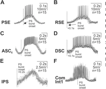
The swimmeret system generates rhythmic and coordinated motor output even deprived of sensory and central input. All neurons that take part in rhythm generation, coordination, or execution of motor commands, have oscillating membrane potentials around voltages between approximately -55 mV and -45 mV (Mulloney et al., 2006;

Paul and Mulloney, 1985; Sherff and Mulloney, 1997; Smarandache-Wellmann et al., 2013, 2014). At a closer look, similarities as well as disparities between membrane potential oscillation shapes in different neuron groups become apparent. Oscillations generated by PSE, RSE, and IPS are characterized by similar shape and shallow depolarizing slope (Figure 2 A, B, E), while oscillations in ASC

E

s are mostly characterized by steeper slopes (Figure 2 C) (Schneider, 2017; Sherff and Mulloney, 1997). However, some ASC

E

s are characterized by the same steepness as observed in PSE and RSE (Schneider, 2017; Sherff and Mulloney, 1997). Oscillations in DSC have a broader depolarized phase compared to the other neuron types (Figure 2 D) (personal communication C. Smarandache-Wellmann).

Oscillations in ComInt1 are accompanied by different sized EPSPs (Smarandache-

Wellmann et al., 2014). However, ComInt1 recordings vary with regard to proportion

(16)

7 of oscillation amplitude and EPSP amplitudes (Figure 2 F) (personal communication F. Blumenthal).

All spiking neurons produce bursts of action potentials (APs) during the most depolarized phase of the oscillation. ASC

E

is characterized by a higher amount of generated APs than the remaining spiking neurons, PSE, RSE, and DSC (personal communication F. Blumenthal), while PSE and RSE produce the least amount of APs. The elicited spike train in each spiking neuron is characterized by spike frequency adaptation and reduced first spike threshold in ASC

E

, PSE and RSE, but not in DSC (Schneider, 2017).

Figure 2: Neurons in the swimmeret system oscillate. A, B, C, D, E, F Overdraws of four representative spiking neurons (PSE, RSE, ASC

E

, and DSC) and two nonspiking neurons (IPS and ComInt1) triggered to PS burst onset (each n = 15).

PSE: Power Stroke Exciter, RSE: Return Stroke Exciter, ASC

E

: Ascending Coordinating Neuron, DSC: Descending Coordinating Neuron, IPS: Inhibitor of Power Stroke, ComInt1: Commissural Interneuron 1, PS: Power Stroke.

Experimental depolarizing current injections differentially affect membrane potential oscillations in the different neurons groups, especially when comparing IPS to the other neuron groups. Depolarizing current injections in IPS is shown to decrease membrane potential oscillations strongly, while the amplitudes remain similar or even tend to increase in in the other neurons of the swimmeret system (IRS, PSE, RSE, ASC

E

, DSC, and ComInt1) (Mulloney, 2003; Schneider, 2017; Smarandache- Wellmann et al., 2013; personal communication F. Blumenthal).

1.4 Cellular properties of rhythmic neuronal networks

Despite our good understanding of the cellular components and synaptic contacts in

each microcircuit of the crayfish abdominal nervous system, it remains unknown how

single cell activity is established. Although some neuron groups have been shown to

(17)

8 receive the same synaptic input from the CPG, their activity patterns differ. Therefore it seems likely that the discrepancies in neuronal activity pattern arise from discrete ion channel compositions in the respective neuron groups.

Ion channels are specialized proteins in the membrane of neurons that are able to respond to very small stimuli. Once activated, they can open within milliseconds to mediate fast diffusion of ions across the membrane leading to rapid change in potential. Ions will either enter the cell or diffuse out of the cell according to their electro-chemical gradient between the cytoplasmic side and the extracellular space.

In the case of sodium for example the ionic concentration is much higher in the extracellular space than inside the neuron. When channels open, sodium will diffuse into the cell and thereby cause an accumulation of positively charged ions inside the cell, leading to a depolarization of the membrane (Alberts et al., 2002).

Ion channels are highly specific with regard to their permeability for ions. Depending on their pore lining structure, some channels are selectively permeable to one ion type, while others are less specific and will allow more than one ion type to diffuse through the pore of the channel. Essential for proper signaling, mediated by ions channels are the gating mechanisms of the channel. Depending on the type of ion channel, they will open or close caused by chemical or electrical signals. Ligand- gated ion channels open when a ligand binds to specific binding site on the channel itself, or to a binding site in close proximity to the channel. Those ligands can either be transmitters in the extracellular space or intracellular mediators such as ions or nucleotides (Alberts et al., 2002).

Voltage-gated ion channels opens upon voltage changes of the membrane potential.

If membrane potentials de- or hyperpolarize, e.g. due to excitatory or inhibitory

synaptic input, the likelihood of intrinsic ion channels to open changes. Some ion

channels open at more depolarized potentials, while other channels are sensitive to

hyperpolarized potentials. Likewise, closing of ion channel can be similarly

depending on the membrane potential. Other ion channels are constantly open and

allow continuing diffusion of ions across the membrane (Alberts et al., 2002).

(18)

9 1.5 Ion channels mediating inward currents

1.5.1 Sodium channels

Sodium channels play a crucial role during membrane potential depolarization of a neuron, thereby, enabling the neuron to transmit electrical signals over long distances (Marban et al., 1998). Properties of sodium channels were first characterized by Hodgkin and Huxley, when discovering voltage-gated ion channels in the squid axon (Hodgkin and Huxley, 1952). Mammalian sodium channels consist of the α, and the smaller auxiliary β1 and β2 subunits, whereas the principle α subunit is necessary for functionality of the channel (Catterall, 1992; Marban et al., 1998). The α subunit is composed of four homologous domains, each consisting of six transmembrane domains. Four of these transmembrane domains form the central pore that ensures the selectivity and conductance properties of the channel by their pore lining domain P between the fifth and the sixth transmembrane domain (Marban et al., 1998). Experiments in Xenopus oocytes expressing sodium channels revealed that the fourth transmembrane domain with its positively charged arginin and lysine residues serves as a voltage sensor (Stühmer et al., 1989).

Experiments treating the intracellular surface of the squid neuronal membranes with pronase, or site-directed antibodies against the intracellular loop between domains III and IV, blocked inactivation of sodium channels (Armstrong et al., 1973; Vassilev et al., 1988). Rescue experiments adding a synthetic peptide consisting of three amino acids (Ile-Phe-Met) that resembled the same sequence of the hydrophobic loop between domains III and IV, could reestablish inactivation (Eaholtz et al., 1994).

These experiments led to the conclusion that the inactivation site lies between those two domains.

However, not all sodium channels are characterized by fast opening and closing

kinetics. In experiments on vertebrate guinea pig cerebellar Purkinje neurons (Kay et

al., 1998), dorsal root ganglion neuron of the rat (Elliott and Elliott, 1993; Roy and

Narahashi, 1992), as well as different mollusk neurons (Gilly et al., 1997; Kiss, 2003)

and jellyfish MNs (Anderson, 1987), sodium channels were detected with slow

activating kinetics and were either noninactivating or slowly inactivating. Furthermore,

in cat layer V pyramidal neurons, persistent sodium channels were shown to activate

at more negative membrane voltages than the transient sodium channels (Crill,

1996). Persistent sodium channels were shown to have important functions in setting

(19)

10 the resting membrane potential in cerebellar Purkinje neurons (Kay et al., 1998).

Furthermore, they were found to be involved in rhythmically active neurons during maintenance of pacemaker activities in neurons of the preBötzinger Complex (Del Negro et al., 2002; Peña et al., 2004), promotion of subthreshold oscillation in the leech heart interneurons (Opdyke and Calabrese, 1994), generation of plateau potentials in crayfish abdominal stretch receptor neurons (Barrio et al., 1991), and PIR responses of leech MNs (Angstadt and Simone, 2014).

The transient sodium current, as well as one type of I

NaP

were shown to be sensitive to Tetrodotoxin (TTX) application (Gilly et al., 1997; Kiss, 2003). In a modeling study the target site of TTX at the sodium channel could be identified as the extracellular loop between S5 and S6 (Lipkind and Fozzard, 1994). Another subgroup of persistent sodium channels was not TTX-sensitive but could be blocked by low concentrations of Riluzole (Harvey et al., 2006; Miles et al., 2005; Tsuruyama et al., 2013; Urbani and Belluzzi, 2000). Because the effect of Riluzole was predominantly seen after sodium channels were activated and entered the inactivation state, it was concluded that Riluzole binds at the inactivation loop of the channel (Benoit and Escande, 1991).

1.5.2 Calcium channels

Five classes of voltage activated calcium channels give rise to a depolarizing inward current carried by calcium ions. These channels are the L-type, P/Q type, N- type, R- type, and the T-type channels (Simms and Zamponi, 2014). Calcium channels are composed of different proteins that determine their ion selectivity, as well as their opening and closing kinetics. The α

1

subunit, a homologue to the α subunit of sodium channels, is comprised of four domains, each with six transmembrane domains, including the voltage sensor in S4 and the pore lining P region. The auxiliary subunits α

2

, β, γ, and δ modify the specific channel properties of the different channel types (Catterall, 2011).

The voltage sensitivity varies between the different channels types. P/Q-type, N-type,

and R-type calcium channels open at depolarized membrane voltages. They are

therefore often referred to as high-voltage activated calcium channels. Depending on

the system, the L-type calcium channel has been shown to be either high- (Snutch et

al., 2013) or low voltage activated (Wang et al., 2011; Xu and Lipscombe, 2001).

(20)

11 Together with the T-type calcium channel, which also opens at hyperpolarized voltages (Armstrong and Matteson, 1985), they were both shown to be involved in PIR responses in lamprey commissural interneurons and MNs (Wang et al., 2011) and rat caudal hypothalamic neurons (Fan et al., 2000). The L-type calcium current (I

CaL

), without interference of the T-type calcium current (I

CaT

), was furthermore seen to induce PIR responses in cerebellar nuclei neurons (Zheng and Raman, 2011).

Furthermore, the L-type calcium channel was shown to be involved the generation of plateau potentials in rat deep dorsal horn neurons (Morisset and Nagy, 1999), and pacemaker activity in dopaminergic Substantia Nigra neurons (Branch et al., 2014), generating membrane potential oscillations in striatal neurons (Song et al., 2016) and unidentified neurons of the swimmeret system of the crayfish (Chrachri, 1995). L-type calcium channels have been shown to be blocked by different dihydropyridines, Nitrendipine, Nifedipine, and Nimodipine by binding to a specific region close to the pore and the hypothesized activation gate at the α1- subunit (Snutch et al., 2013;

Tikhonov and Zhorov, 2009; Wang et al., 2011).

1.5.3 HCN channels

Hyperpolarization-activated cyclic-nucleotid-gated (HCN) channels activate slowly at hyperpolarized membrane voltages and give rise to slow inward currents depolarizing the neurons (I

H

) (Angelo and Margrie, 2011). Therefore, they contribute to several processes in non-pacing-, as well as rhythmically active neurons in the brain. HCN channels contribute to long term potentiation (Mellor et al., 2002) and modulation of synaptic transmission (Beaumont and Zucker, 2000). In rhythmically active neurons, they were shown to modulate pacemaker activity in preBötzinger complex neurons (Thoby-Brisson et al., 2000), thalamocortical relay neurons during sleep (McCormick and Pape, 1990), and CA1 hippocampal stratum oriens-alveus interneurons. They also contribute to membrane resonance in inferior olive neurons (Matsumoto- Makidono et al., 2016). Furthermore, this current often mediates PIR responses in pyloric neurons of the stomatogastric nervous system (Harris-Warrick et al., 1995), leech heart interneurons (Angstadt and Calabrese, 1989), CA1 pyramidal neurons (Surges et al., 2006), and neonatal rat MNs (Bertrand and Cazalets, 1998).

HCN channels are not selective for one specific ion type but allow both sodium and

potassium to diffuse through the membrane. HCN channels are comprised of six

transmembrane domains (S1 – S6), forming a tetrameric complex (Biel, 2010). The

(21)

12 channels are characterized by a pore lining loop between S5, S6, and a cyclic nucleotide binding domain at the C- terminal region. The positively charged fourth transmembrane domain functions as the voltage sensor and shares close homology with the voltage sensor in voltage-gated potassium channels (Kaupp and Seifert, 2001). HCN channels typically open at hyperpolarization of the membrane.

Depending on the neuron type, the half-maximal activation voltages vary between -65 mV and -95 mV (Pape, 1996) and inactivate at -43 mV in thalamocortical relay neurons (McCormick and Pape, 1990), -50 mV in neurons of preBötzinger complex (Thoby-Brisson et al., 2000), or -35 mV in dorsal gastric MNs of the stomatogastric nervous system (Kiehn and Harris-Warrick, 1992). When open, HCN channel conduct sodium and potassium in a ratio of 1:4 to 1:6, which is blocked by millimolar concentrations of cesium (Edman et al., 1987; Gauss et al., 1998;

Ludwig et al., 1998) or Zd7288 by binding at the residues Ala425 and Ile432 located at the S6 transmembrane domain at the inner pore of the channel (Cheng et al., 2007).

1.6 Ion channels mediating outward currents

Potassium-carried outward currents are of equal importance for the generation of rhythmic neuronal activity. Leading to outward diffusion of positively charged ions, these channels are key mediators to hyperpolarize the membrane.

Voltage-gated potassium channels (K

(V)

-channels) are comprised of four α subunits, each consisting of six transmembrane domains (S1 – S6) and a diverse number auxiliary β subunits that are arranged barrel-like and embedded into the membrane of the neuron to form a pore (Biggin et al., 2000). Despite the variety of different potassium channels, all show the same structural attributes that ensure the selectivity of these channels for potassium. This main structure is the pore loop between the fifth and sixth transmembrane domain (Biggin et al., 2000). The S4 transmembrane domain is the voltage sensor of the channel. Furthermore, the β subunit influences the conductance through the channel by serving as an inactivation gate that blocks the channel after activation or by influencing the cell surface expression of the α subunit (Biggin et al., 2000).

The first voltage-gated potassium channel was found by the detection of a mutated

shaker gene in Drosophila melanogaster (Ferrus et al., 1990). After the

(22)

13 characterization of the shaker gene, three other related potassium channel genes were cloned, shab, shaw, and shal (Salkoff et al., 1992; Wicher et al., 2001; Xu et al., 1995). All four are highly conserved between flies, mice, and humans (Salkoff et al., 1992). The genes shaker and shal encode for transient potassium currents (I

A

) with fast activation and inactivation kinetics. The genes shab and shaw encode for moderate activation and slow inactivation kinetics of potassium currents (Hille, 2001).

Depending on the cell, different compositions of these genes are expressed.

An important feature of transient potassium channels is their sensitivity to hyperpolarized membrane voltages. At subthreshold membrane potentials transient potassium channels open rapidly, reducing the amount of positively charged ions inside the cell (Kim et al., 2005). Thereby, the activity of this channel is important for shaping APs, spike frequency, delay of rebound firing, and rhythmic bursting (Ascoli et al., 2010; Harris-Warrick et al., 1995; Hartline and Gassie, 1979; Hille, 1992; Kim et al., 2005; Serrano and Getting, 1989; Tierney and Harris-Warrick, 1992). A selective antagonist of I

A

is 4-Aminopyridine (4-AP), which has been shown to block the transient potassium channel from the cytoplasmatic site when the channels are open (Kiss et al., 2002; Yao and Tseng, 1994).

Another group of potassium channels open due to accumulation of intracellular Ca

2+

ions. The outward current counteracts depolarizations mediated by calcium influx.

Calcium-gated potassium channels play an important role in inducing spike afterhyperpolarizations, which directly affects spike frequency adaptation and finally termination of AP bursts (el Manira et al., 1994). The family of calcium gated potassium channels can be divided into three groups, the big conductance (BK) intermediate (IK), and the small conductance (SK) channels (Vergara et al., 1998). IK and SK channels open solely to changes in intracellular calcium concentrations while the BK channel is also sensitive to changes in the membrane voltage. The structure of calcium- gated potassium channels is similar to those of voltage-gated potassium channels, besides a calcium binding site at the C-terminal region (Schreiber and Salkoff, 1997).

1.7 Computational model of the swimmeret system

The fundamental aim of working on the neuronal network controlling swimmeret

movements is to achieve a better understanding of how the nervous system

(23)

14 generates behavior. Regarding the extensive investigations in this system and accumulation of new insight about the network architecture, we are getting closer to understanding basic principles for rhythmogenesis, and coordination. Thereby a lot of progress has been made about possible intersegmental connections, mediating the 0.25 phase difference between the segments, by the used of simplified mathematical models (Skinner and Mulloney, 1998; Zhang et al., 2014).

In the stomatogastric nervous system extensive theoretical work has been performed identifying possible ionic currents that take part in evolving neuronal activity pattern.

Thereby it was revealed how the same firing pattern can be generated by variable ionic current compositions (Prinz et al., 2004; Soofi et al., 2012), but also, how homeostatic plasticity is able to adapt ionic conductances to maintain phase constancy between neurons (Marder and Prinz, 2002).

At this point, little is known about intrinsic neuronal properties that contribute to the generation of rhythmic and coordinated motor output in the swimmeret system.

Regarding that most of the identified synaptic connections between individual network components are shown to be inhibitory (Mulloney, 2003; Murchison et al., 1993; Skinner and Mulloney, 1998; Smarandache-Wellmann and Grätsch, 2014), suggest that either escape- or a release mechanism could play a role during rhythmic neuronal activities of the participating neurons. Especially the activity pattern of the half center oscillator formed by IPS and IRS neurons is likely to depend on either of these two processes. A study by Daun and colleagues simulated the activity of half center oscillators based on different combinations of ionic currents (Daun et al., 2009). Thereby it could be learned that particularly the half center oscillator based on I

NaP

and I

L

was able to perform the greatest range of oscillations periods.

1.8 Objectives

Ionic currents and synaptic input cooperatively establish the activity of individual

neurons within a network. This is achieved, foremost, by a precise interplay of

intrinsic ionic conductances, which fine-tune single neuronal activities to meet the

correct phasing and magnitude needed for a functional network activity. At this point

little is known about any ionic currents that contribute to generating neuronal

activities in the swimmeret system. However, in order to fully comprehend, how the

neuronal network of the swimmeret system operates, the intrinsic properties

(24)

15 underlying the activity pattern of each neuron need to be identified. Thereby, the expression of specific membrane responses, upon de- or hyperpolarizing stimuli, can facilitate the analysis of the contributing ionic conductances.

In other systems, like clione (Pirtle and Satterlie, 2004), leech (Angstadt et al., 2005), lamprey (Matsushima et al., 1993), or xenopus tadpole (Li and Moult, 2012), key neurons for rhythmogenesis are shown to receive inhibitory synaptic input, and additionally, have the ability to escape from this inhibition. Thereby, induced by negative potential changes, hyperpolarization-activated ion channels open, and allow influx of positively charged ions, which ultimately depolarizes the neuron and thus promotes the next oscillation. Possible ion channels with low voltage sensitivities are the T-type calcium channel, the L-type calcium channel, and/or the HCN channel.

Because most of the neurons in the swimmeret system are shown to receive inhibitory synaptic input (Mulloney, 2003; Murchison et al., 1993; Skinner and Mulloney, 1998; Smarandache-Wellmann and Grätsch, 2014), the question arises, whether these neuron possess the ability to escape from inhibition, as well. This could represent a possible mechanism to escape the hyperpolarized membrane potential during the oscillation and promote the depolarization. Furthermore, by a correct interplay of underlying ionic conductances the specific phasing of the oscillation could be established.

Analyzing the expression of PIR responses represents a means to gain a better understanding of escape mechanisms and the underlying ionic conductances. In a variety of neuronal networks, the presence PIR responses, mediated by one or several different ionic currents have been described. One ionic current, I

H

, has been shown to underlie or contribute to PIR generation in several vertebrate (Ascoli et al., 2010; Johnson and Getting, 1991; Matsushima et al., 1993) and invertebrate species (Calabrese et al., 1995; Golowasch et al., 1992; Harris-Warrick et al., 1995).

However, in other species or even in other neurons types of the same species, PIR responses have been shown to be mediated by calcium or sodium carried currents (Angstadt and Simone, 2014; Angstadt et al., 2005; Fan et al., 2000; Wang et al., 2011; Zheng and Raman, 2011).

In the first part of this thesis I explored the presence PIR responses in key neurons of

the swimmeret system for rhythm generation, intersegmental coordination and

(25)

16 execution of motor commands. I monitored the voltage responses of synaptically isolated neurons to hyperpolarizing current injections in discontinuous current clamp mode.

In the second part of this thesis, I performed occlusion experiments to unveil the ionic bases of the evoked PIR responses, and in addition, tested the significance of identified ionic currents for the generation of fictive locomotion in the third part of this thesis.

In a reduced mathematical model, I furthermore tested the capability of swimmeret

CPGs to perform similar network activities with disparate underlying ionic

conductances.

(26)

17

2. Material and Methods

The experiments were carried out on adult crayfish, Pacifastacus leniusculus. The animals were caught in nearby rivers and ponds and kept in fresh water tanks at temperatures of 14 °C and a 12 hour day / night cycle.

2.1 Dissection

All experiments were performed on isolated abdominal nerve cord preparations. To prepare the dissection, the animals were anesthetized by keeping them on ice for 20 – 30 min. Afterwards the claws were cut at the base near the thorax and the left and right uropodes were removed (Seichter et al., 2014). To exsanguinate the animal 50 ml cold ringer solution containing (in mM): 195.0 NaCl, 5.4 KCl, 13.5 CaCl

2

, 2.6 MgCl

2

(ph: 7.4) was rinsed though the claw opening. In a next step the animal was decapitated and the walking legs were cut near the base. A following transverse cut through the thorax at the level of the second walking leg separated the abdomen with the last three thoracic segments from the remaining cephalothorax. By separating the ventral and dorsal abdomen from another the ventral nerve cord could be exposed and removed from the abdominal plate. The abdominal nerve cord (including abdominal ganglia 1 to 6 as well as the last two thoracic ganglia) was then pinned down with the dorsal side facing up in a Sylgard lined Petri dish filled with cold ringer solution. The anterior and posterior branches of N1 were separated to record the RS (anterior branch) and PS (posterior branch) activities. In order to facilitate intracellular recordings at the region of the Lateral Neuropil and to allow diffusion of superfused drugs into the tissue, the ganglia sheaths from ganglia A2 - A5 were removed with fine scissors.

2.2 Electrophysiology

2.2.1 Extracellular recordings

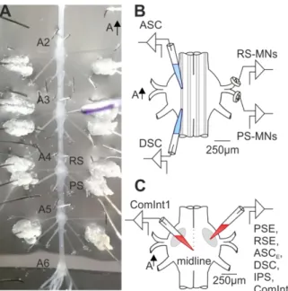
I performed up to fourteen simultaneous extracellular recordings (Figure 3 A, B). I

used two differential stainless steel pin electrodes to record the activity pattern of all

motor neurons (MNs) innervating the Power Stroke (PS) and Return Stroke (RS)

muscles. Thereby, I placed the electrodes either at the anterior branch of Nerve1

(27)

18 (N1) for RS recordings, or at the posterior branch of N1 for PS recordings (Figure 3 A, B). The nerve endings were wrapped around each of the pin electrodes and insulated from surrounding saline with petroleum jelly. RS activities were only recorded from ganglia A3 and A4. The signals from the pin electrodes were amplified 1000 fold (Model MA 102, Electronics laboratory, University of Cologne) and band pass filtered from 300 Hz to 2 kHz.

Additionally to MN recordings I performed extracellular recordings of the axons of the Coordinating Neurons, Ascending Coordinating Neuron (ASC

E

) and Descending Coordinating Neuron (DSC). Therefore I placed two differential suction electrodes (MWE-F15B, Warner Instruments, Hamden, CT, USA, M-3333, Narishige, Tokio, Japan) on the dorsal surface of the ganglia (Figure 3 A, B). Suction electrodes were pulled from a P-87 micropipette puller (Sutter Instruments, Novato, CA, USA) from borosilicate capillaries (outer diameter: 1.5 mm, inner diameter: 0.86 mm, Sutter) and broken until the tip diameter was slightly larger than the Lateral Giant Axon. In order to record from ASC

E

I placed the electrode dorsally to the Lateral Giant Axon on the anterior Minuscule Tract, during DSC recordings on the posterior Minuscule Tract (Figure 3 A, B). To increase the recording quality I applied negative air pressure to the electrode which improved the contact of the electrode to the tissue. The recorded signal was 50 - fold preamplified (MA103, Electronics laboratory, University of Cologne) and transmitted to the extracellular amplifier.

Figure 3. Experimental setup and recording sites. A: Isolated abdominal nervous system (A2 to A6) with intracellular electrode and extracellular pin electrodes at anterior and posterior branches of N1. B:

Schematic illustration of ganglion shows the placement of ASC & DSC suction electrode as well as differential pin electrodes for RS- and PS MNs recordings. C: Sharp electrode placement for intracellular recordings from PSE, RSE, ASC

E

, DSC, IPS and ComInt1. A2 to A6: abdominal ganglion 2 to abdominal segment 6, N1: Nerve 1, RS: Return Stroke, PS: Power Stroke, MN: motor neurons, PSE:

Power Stroke Exciter, RSE: Return Stroke

Exciter, ASC: Ascending Coordinating

Neuron, DSC: Descending Coordinating

Neuron, IPS: Inhibitor of Power Stroke,

ComInt1: Commissural Interneuron 1.

(28)

19 Signals of suction electrode and pin electrode recordings were digitized in a sampling frequency of 10 kHz with an analogue/digital (A/D) converter (Digidata 1440A, Molecular Devices, Sunnyvale, CA, USA) and recorded on a personal computer with Clampex (Molecular Devices).

2.2.2 Intracellular recordings

To analyze the cellular properties of the different neurons in the swimmeret system I performed intracellular recordings at the dendritic aborizations in the Lateral Neuropil or at the midline of the ganglion (Figure 3 C). The intracellular electrodes were pulled from borosilicate glass capillaries with filaments (outer diameter: 1 mm; inner diameter: 0.5 mm). The electrodes were filled with intracellular electrode solution containing 1 M KAc and 0.1 M KCl and 1% Dextran Texas Red with resistances between 30 – 60 MΩ. Electrode liquid junction potential and capacitance were compensated prior neuron penetration. During intracellular recordings electrode capacitance was compensated in regular intervals. The intracellular signal was amplified 1000 fold (SEC-05X, npi electronic, Tamm Germany), digitized (10 kHz) and recorded with Clampex (Molecular Devices). The recordings were performed in discontinuous single electrode current clamp with switching frequencies of 35 kHz.

Based on different electrophysiological properties of the recorded neurons (effect of de- or hyperpolarizing current injections on the PS / RS motor output of the home ganglion (PSE, RSE, and ComInt1) or the target ganglion (ASC

E

and DSC)) and specific phase relationships to the PS MNs bursts they could be classified during the experiment to the different neuron classes. The classifications during the experiments were later verified by their morphology through single neuron labeling by intracellular dye injection (PSE, RSE (Sherff and Mulloney, 1997), ASC

E

, DSC (Namba and Mulloney, 1999), IPS (Smarandache-Wellmann et al., 2013), ComInt1 (Smarandache-Wellmann et al., 2014)).

2.3 Morphology

To verify the identity of the intracellularly recorded neurons by their morphology I

stained the neurons iontophoretically with 1 % Dextran Texas Red, by delivering

depolarizing current pulses (1 nA; pulse durations: 250 ms, frequency: 2 Hz).

(29)

20 Ganglia in which neurons were labeled were subsequently fixed in 4 % paraformaldehyde at least 2 h at room temperature, washed 3 times in PBS for 15 min each, and dehydrated in an ascending ethanol series of 30 %, 50 %, 70 %, 90 % and 100 %. Additionally the ganglia were cleared with methyl salicylate to improve the visibility of the stained neurons. Images were taken in 10 fold to 20 fold magnification either on a fluorescence microscope (BX61, Olympus, Hamburg, Germany) or confocal laser scanning microscope (LSM 500 Meta, Zeiss, Oberkochen, Germany), installed on an inverted Zeiss Axiovert 100M. Z-stacks were converted of 20 to 25 optical slices by the image processing software Image J (Version 1.45s).

2.4 Experimental Procedure

After a settling period of up to 15min following membrane penetration with the intracellular electrode the superfusion of the preparation with different bathing solutions was initialized with a peristaltic pump (Alitea, Sweden). When searching for neurons intracellularly, the volumetric flow rate was set to 0.5 ml / min and increased to maximum flow rates of 2 ml / min during the experiment. To achieve comparable effects of applied blockers, all preparations were superfused with 2 µM carbachol diluted in ringer solution.

During the experiments ringer solution containing different drugs were applied to the abdominal nervous system while analyzing cellular and system properties.

Drugs

A mixture of different synaptic blockers with concentrations of (in µM): 10 Picrotoxin

(PTX), 10 6-cyano-7-nitroquinoxaline-2,3-dione (CNQX), 10 6,7-dinitroquinoxaline-

2,3-dione (DNQX), 10 D-2-amino-5-phosphonopentanoic acid (D-AP5), and 10

Tubocurarin was applied to the abdominal nervous system to achieve synaptic

isolation. The synaptic blockers were mixed just prior to application from defrosted

stock solutions. To investigate the contribution of specific ion channels during

characteristic membrane responses Nifedipine, 4-(N-ethyl-N-phenylamino)-1,2-

dimethyl-6-(methylamino) pyrimidinium chloride (Zd7288) and Riluzole were added to

the synaptic blocker solution and subsequently applied to the preparation.

(30)

21 The channel blockers were prepared as 40 mM (Nifedipine), 50 mM (Zd7288), and 70 mM (Riluzole) stock solutions, dissolved either in DMSO or ringer solution.

Defrosted drugs were diluted to the synaptic blocker solution immediately before application and kept in light shielded containers (40 µM Nifedipine, 200 µM Zd7288, and 70 µM Riluzole).

During system analysis the drugs mentioned above, as well as 4 mM 4- Aminopytidine (4-AP) (diluted from 4 M stock solution in ringer) were added to ringer solution without synaptic blockers and applied to the preparation individually during each experimental approach.

Protocols

While superfusing the abdominal nervous with synaptic blockers and/or ion channel blockers the cellular input resistance (R

IN

) was measured by applying brief hyperpolarizing current pulses of -1 nA for 200 ms at an interval of 0.2 Hz (Cornerstone, S-900 Stimulator, Dragan Corporation, USA). The membrane potential was always set to similar values, allowing the neurons to oscillate around -50 mV to increase comparability between cells and experimental conditions.

Cellular properties

Post-inhibitory rebound (PIR) responses were induced, in order to characterize membrane properties of the neurons in the swimmeret system. Each protocol consisted of 15 sweeps at three different holding potentials (V

H

)(V

H

= -40 mV, V

H

= -50 mV, and V

H

= -60 mV). Negative currents were injected hyperpolarizing the neurons for 3 s to three different test potentials (V

T

) (V

T

= -80 mV, V

T

= -90 mV, and V

T

= -100 mV) to elicit PIR responses (Figure 4). Protocols were executed with the build in stimulation tool in Clampex. The injected current had to be adjusted from cell to cell to reach the aimed test potentials. Due to large differences in currents needed to reach V

T

values between different neurons and neuron groups the emphasis during analysis was put on membrane potentials reached, not currents amplitudes injected.

The above mentioned channel blockers (Nifedipine, Zd7288, and Riluzole) were

applied to the preparation while PIR responses were evoked in order to evaluate the

contribution of different ionic currents for PIR generation.

(31)

22 Figure 4. PIR response of a PSE.

Intracellular recording of a PSE during hyperpolarizing current injection leading to a PIR response. The holding potential of this neuron was set to V

H

= -50 mV. During the protocol the neuron was step wise hyperpolarized to V

T

= -80 mV, V

T

= -90 mV, and V

T

= -100 mV for 3 s. Dashed boxes (i, ii) indicate the analyzed parameters during the PIR stimulations. i: PIR amplitude and spike frequency, ii: sag potential amplitude, PSE: Power Stroke Exciter, V

H

: holding potential, V

T

: test potential.

Analysis

In all cells the following parameters of the evoked PIR were analyzed in matlab (version R2014b, MathWorks, Natick, MA, USA). Figures were processed with Corel Draw X6 (Corel Corporation, Ottawa, Canada). Voltage traces of all 15 sweeps were taken for calculation of the median and interquartile ranges (IQR).

Holding Potential (V

H

)

V

H

values were identified by calculating the mean voltage potentials for each 15 sweeps in a time range between 1.5 s before onset of the stimulation, and onset of the stimulation. In order to reduce false depolarized V

H

values due to strong depolarizations during action potentials (APs), APs were removed from the voltage traces by smoothing single voltage traces with a sliding window calculating the mean voltage data points between each neighboring data point in a factor of 900.

Test Potential (V

T

)

V

T

was detected by calculating the mean maximum hyperpolarized membrane potential during the current injection. Single voltage traces were smoothed to keep consistency for PIR- and sag amplitude calculations (below).

PIR amplitude

PIR depolarization amplitudes were measured between V

H

and the maximum

depolarized potential (between termination of current injection and 2.5 s post

(32)

23 termination of stimulation) (Figure 4 i). This time period was taken for analysis to be certain that all PIR responses had already ended during the measurement.

Sag amplitude

Mean voltage values between beginning of current injection and 1 s after beginning current injection, were subtracted from mean voltage values between 1 s before current injection termination and termination of current injection, in order to calculate sag potential amplitudes (Figure 4 ii).

Spiking characteristics

Spiking cells (PSE, RSE, ASC

E

and DSC) elicited rebound spikes when released from hyperpolarizing current injections. One parameter that was analyzed was the latency until first rebound spikes were elicited. This value was measured as the time period of release from hyperpolarizing current pulse until the first APs occurred. The other parameter analyzed was the mean instantaneous APs frequency before and after PIR stimulation (Figure 4 i). In control condition, the maximum instantaneous spiking frequency was compared between the three different V

T

at V

H

= -40 mV to ensure all neurons reached spike threshold. During experiments identifying ionic conductances mediating PIR responses, maximum instantaneous frequencies were compared between experimental conditions elicited from V

H

= -50 mV to V

T

= -100 mV. This more hyperpolarized V

H

was used for comparison because channel blocker application tented to increase instantaneous spike frequencies in many cells and some neurons seemed to enter sodium block at V

H

= -40 mV.

System properties

During ion channel blocker application the following parameters were analyzed to

assess the contribution of different ion channels for the generation of rhythmic motor

output. The synchronicity between two PS recordings (in neighboring segments), as

well as the relative time delay between two segments, were compared in order to

analyze intersegmental coordination. Changes in rhythm generation were monitored

by calculating, in single segments, period, burst duration and duty cycles. To explore

which ionic currents where contributing to rhythmogenesis and coordination,

Nifedipine (40 µM), Zd7288 (200 µM), Riluzole (70 µM) and 4-AP (4 mM) were

applied individually to the abdominal nervous system.

Abbildung

Figure  2:  Neurons  in  the  swimmeret  system  oscillate.  A,  B,  C,  D,  E,  F  Overdraws  of  four  representative  spiking  neurons (PSE, RSE, ASC E , and DSC) and  two nonspiking neurons (IPS and ComInt1)  triggered to PS burst onset  (each n =  15)
Figure  3.  Experimental  setup  and  recording  sites.  A:  Isolated  abdominal  nervous  system  (A2  to  A6)  with  intracellular  electrode  and  extracellular  pin  electrodes  at  anterior  and  posterior  branches  of  N1
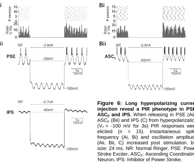
Figure  6 :  Long  hyperpolarizing  current  injection  reveal  a  PIR  phenotype  in  PSE,  ASC E   and  IPS
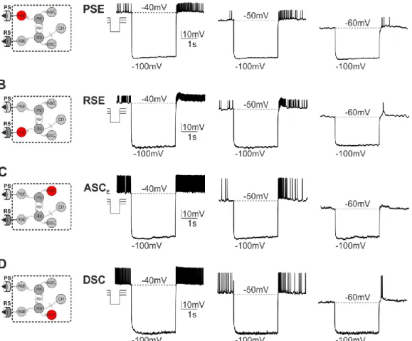
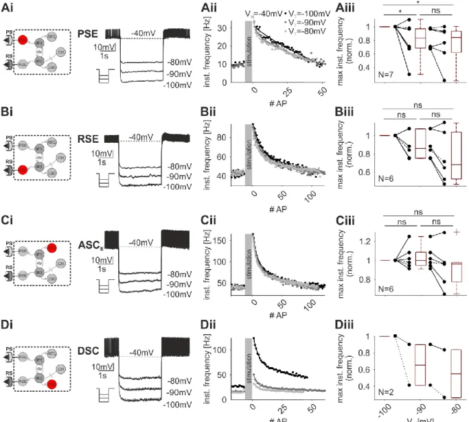
Figure  8:  PIR  responses  in  four  groups  of  spiking  neurons.  Schematic  of  recorded  neuron  group
+7

Referenzen

ÄHNLICHE DOKUMENTE

Finally, it is our conjecture that Classic Actors and Processes are mostly used in a fine-grained concurrency setting and lead to a style of programming where the state of an

11 Based on the discussion above, it is obvious that, a higher rewetting temperature means an earlier initiation of the rewetting which will shorten the whole quenching

In Figure 4.12, there is also a noticeable influence displayed due to a change in film thickness, especially in the region, where the transition from a transparent to an opaque

-I- Zone II is characterized by high substrate temperatures (0.3 < T/T m < 0.5) at which both surface and grain boundary diffusion occur at significant levels. The grain

While only considering those oscillating mitochondria that belong to the largest frequency cluster, the findings reveal significant correlations between

Moreover, expression of constitutively active Notch1 in hematopoietic progenitors resulted in a severely altered lymphoid development in the bone marrow, namely a block in

Temperature As the water vapor variation with the 27-day solar forcing is not consistent with the results from the analysis of NLC properties, especially concerning the phase lag of

In this respect Peripheral Bursters are similar to the cardiac ganglion oiPortunus (Benson, 1980), the PD-unit in the stomatogastric ganglion of the lobster (Ayers &