Involvement of CTGF in eye
development and the pathology of glaucoma
Dissertation
ZUR ERLANGUNG DES DOKTORGRADES DER NATURWISSENSCHAFTEN
(DR. RER. NAT.) DER FAKULTÄT FÜR BIOLOGIE UND VORKLINISCHE MEDIZIN
DER UNIVERSITÄT REGENSBURG
Durchgeführt am Lehrstuhl für Humananatomie und Embryologie der Universität Regensburg
vorgelegt von Andrea E. Dillinger aus Landau a. d. Isar
im Jahr 2018
Promotionsgesuch wurde eingereicht am:
28.03.2018
Die Arbeit wurde angeleitet von:
Prof. Dr. Rudolf Fuchshofer
Unterschrift
Meinen Eltern
1. Introduction ... 3
1.1. Mouse eye development ... 3
1.1.1. Embryonic mouse eye development ... 3
1.1.2. Postnatal mouse eye development ... 5
1.1.2.1. Postnatal development of the anterior eye structures ... 5
1.1.2.2. Postnatal development of the retina ... 7
1.1.3. Signaling pathways in eye development ... 8
1.2. The pathology of POAG ...11
1.2.1. The trabecular meshwork and aqueous humor outflow ...11
1.2.1.1. Structure and function of the trabecular meshwork ...11
1.2.1.2. Pathological changes in the trabecular outflow pathway ...12
1.2.2. The optic nerve head and the Lamina cribrosa ...13
1.2.2.1. Structure of the optic nerve head and the Lamina cribrosa ...13
1.2.2.2. The structure of the optic nerve head in mice ...14
1.2.2.3. Pathological changes in the optic nerve head and the Lamina cribrosa15 1.2.2.4. Pathological changes in the peripapillary sclera ...16
2. Aim of the study ...18
3. Material and methods ...20
3.1. Materials ...20
3.1.1. Reagents ...20
3.1.2. Enzymes and Reagent-Kits ...23
3.1.3. Oligonucleotide primers and Taqman probes ...23
3.1.4. Antibodies and molecular weight standards ...25
3.1.5. Chemical composition of gels, solvents and buffers ...27
3.1.6. Laboratory Equipment ...29
3.1.7. Consumables ...30
3.2. Cell linies ...32
3.3. Animal models ...32
3.3.1. Animals and animal husbandry ...32
3.3.2. βB1- CTGF mice ...32
3.3.3. CTGFLacZ/+ mice ...33
3.3.4. CD1 wildtype mice ...33
3.3.5. CTGFCoin/Coin;CAGGCre-ER mice ...33
3.4. Biomolecular Techniques ...33
3.4.2.1. Genotyping of βB1-CTGF1 mice ...34
3.4.2.1. Genotyping of CTGFLacZ/+ mice...35
3.4.2.3. Genotyping of CTGFCoin/Coin;CAGGCre-ER mice ...36
3.4.3. Agarose gel-electrophorese ...38
3.5. Expression analysis ...38
3.5.1. RNA isolation...38
3.5.2. RNA quantification ...39
3.5.3. cDNA synthesis ...39
3.5.4. Real-time RT-PCR ...40
3.5.5. TaqMan® Gene Expression Assay ...40
3.6. Biochemical techniques ...41
3.6.1. Protein isolation via peqGold TriFast™ Trizol method ...41
3.6.2. Bicinchoninic Assay (BCA assay) ...42
3.6.3. SDS polyacrylamide gel-electrophorese ...42
3.6.4. Semidry-Blotting ...43
3.6.5. Detection of specific proteins ...43
3.6.6. Coomassie staining ...44
3.7. Cell culture ...44
3.7.1. Cell lines and culture conditions ...44
3.7.2. General working conditions ...44
3.7.2.1. Passaging of cells ...45
3.7.3. Isolation of murine optic nerve astrocytes ...45
3.7.4. In vitro experiments ...45
3.7.4.1. Treatment with CTGF and TGF-β2 ...45
3.7.4.2. Transfection of siRNA coated nanoparticles (NP) ...46
3.7.4.3. Cultivation of murine ON astrocytes on increasing substratum stiffness ...46
3.8. In vivo experiments ...46
3.8.1. Preparation of anterior eye segments, retinae and corneal-scleral rims ...46
3.8.2. Preparation of optic nerves and optic nerve heads ...46
3.8.3. Preparation of retinal flat mounts ...47
3.8.4. Transcardial perfusion ...47
3.8.5. Perfusion of porcine and human eyes ...47
3.9. Histological techniques ...48
3.9.1. Cryo embedding and preparation of sections ...48
3.9.4. Immunohistochemical staining of cryo sections ...49
3.9.5. Immunohistochemical staining of paraffin sections ...52
3.9.6. Immunohistochemical staining of retinal flat mounts ...54
3.9.7. Immunocytochemical staining ...54
3.9.8. Phalloidin labeling ...55
3.9.9. β-galactosidase activity staining ...55
3.9.10. β-galactosidase activity staining of retinal flat mounts...56
3.10. Preparation of Polydimethylsiloxane Cell Substrata ...56
3.11. Image Analysis ...58
3.12. Light and Fluorescence Microscopy ...58
3.13. Statistical Analysis ...58
4. Results ...59
4.1. CTGF expression in the mouse eye ...59
4.1.1. Verification of heterozygous CTGF knockout ...60
4.1.2. CTGF distribution during embryonic eye development ...60
4.1.3. CTGF expression during postnatal development and in the adult eye ...61
4.1.3.1. CTGF expression in the anterior eye segment during postnatal development and in the adult eye ...63
4.1.3.2. CTGF expression in the posterior eye during development and in the adult eye ...66
4.1.3.3. Immunohistochemical localization of CTGF expression in the anterior eye segment ...69
4.1.3.4. CTGF expressing cell types in the developing and adult retina ...71
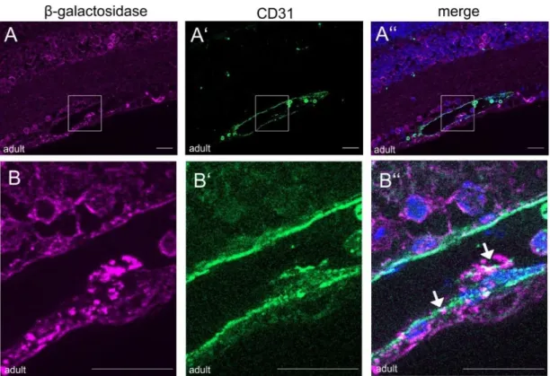
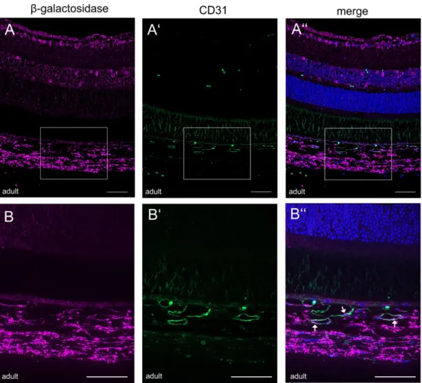
4.1.3.4.1. CTGF expression in the retinal and choroidal vasculature ...73
4.1.3.4.2. CTGF expression in retinal glial cells ...76
4.2.3.4.3. CTGF expression in retinal interneurons ...80
4.1.3.5. CTGF expressing cell types in the developing and adult ON ...82
4.2. Effect of mechanical stress and increasing substratum stiffness on astrocyte reactivity ...88
4.2.1. GFAP and CTGF alterations in the ONH of 2-month-old βB1-CTGF1 mice 88 4.2.3. Establishment of murine optic nerve astrocytes ...93
4.2.3. Increasing substratum stiffness cause reactive changes in murine optic nerve astrocytes ...94
4.2.4. Potential mechanisms of astrocyte reactivity ...96
4.2.4.1. Trp-channels expression related to increasing substratum stiffness ...96 4.2.4.2. Effect of increasing substratum stiffness on Piezo channel expression 98
4.2.4.3.1. Caveolin1 expression murine ON astrocytes ...99
4.2.4.3.2. Effect of increasing substratum stiffness on Caveolin1 and 2 expression ... 101
4.2.4.3.3. Effect of TGF-β2 and CTGF on Caveolin1 in vitro ... 102
4.2.4.3.4. Caveolin1 in the ONH of the murine glaucoma model ... 103
4.3. Intracameral delivery of layer-by-layer coated siRNA nanoparticles ... 104
4.3.1. CD44 expression in HTM cells in vitro ... 105
4.3.2. CD44 expression in βB1-CTGF1 mice in vivo ... 106
4.3.3. Localization and expression of CD44 in the chamber angle of POAG patients ... 107
4.3.4. Perfusion of porcine eyes with nanoparticles ... 108
4.3.5. Perfusion of human eyes with nanoparticles ... 111
4.3.6. CTGF silencing by nanoparticles ... 113
4.4. Effect of conditional CTGF knockdown on mouse eye development ... 114
4.4.1. Verification of conditional CTGF knockdown ... 115
4.4.2. Morphological analysis of CTGF knockdown ... 116
4.4.3. Effect of conditional CTGF knockout on astrocyte network in the retina ... 119
4.4.4. Effect of conditional CTGF knockdown on astrocyte structure in the ON .. 120
5. Discussion ... 123
5.1. Summary ... 123
5.2. CTGF expression in ocular tissues and cell types during development and in the adult eye ... 124
5.2.1. Involvement of CTGF in the development of the anterior eye segment ... 124
5.2.2. Involvement of CTGF in the development of posterior eye structures ... 127
5.2.2.1. Involvement of CTGF in development of the retina ... 127
5.2.2.2. Involvement of CTGF in the development of the ONH ... 129
5.3. Astrocyte reactivity in the ONH of a murine glaucoma model ... 130
5.3.1. Mechanosensing of murine ON astrocytes ... 131
5.4. Intracameral delivery of layer-by-layer coated siRNA nanoparticles ... 133
5.5. Outlook ... 135
6. Conclusion ... 137
7. References ... 138
8. Supplement ... 172
8.1. List of Abbreviations ... 172
8.2. Congress contributions ... 174
Abstract
Primary open angle glaucoma, a neurodegenerative disease of the optic nerve (ON), is one of the leading cause of blindness worldwide (Quigley 1996). In progression of the disease the optic nerve head (ONH) undergoes marked structural extracellular matrix changes (ECM), which lead to its permanent deformation and can contribute to the degeneration of ON axons. The changes in ECM in the ONH correlate with an increased reactivity of astrocytes. An intraocular pressure (IOP), which is too high for the health of the ON axons, was identified as the major risk factor for the development for the POAG (Collaborative-Normal-Tension-Glaucoma-Study-Group, 1998; The AGIS Investigators2000; Gordon et al. 2002; Lichter et al., 2001; Johnson et al., 2002; Kass et al. 2002; Leske et al., 2003). In POAG the homeostatic balance of different growth factors, affecting the resistance of the outflow pathway is altered (Inatani et al. 2001, Min et al. 2006, Ochiai & Ochiai 2002, Ozcan et al. 2004, Picht et al. 2001, Tripathi et al.
1994, Trivedi et al. 2011). In the last years, the Connective Tissue Growth Factor (CTGF) came into focus as a lens-specific overexpression of CTGF in the mice results in an elevated IOP and a significant progressive loss of retinal ganglion cell (RGC) axons over time (Junglas et al., 2012). CTGF is a member of matricellular regulatory proteins and downstream mediator of Transforming Growth Factor β2, and is involved in many different functions, like migration, adhesion, differentiation and ECM production. By its modular organization CTGF can arrange various interactions with different growth factors, integrins and ECM proteins. In this study we investigated the involvement of CTGF during eye development and the pathology of POAG. The effect of alterations in substratum stiffness on an increased astrocyte reactivity and the implication of possible mechanosensors was analyzed. The final aspect of the study was to develop siRNA coated NP for specific delivery to the AH outflow pathway tissue and cells.
During eye development CTGF is expressed in various eye structures. In early embryonic development an expression in the outer and inner layer of the optic cup as well as the lens vesicle was observed. In the following embryonic and postnatal developmental stages, CTGF expression was detected in tissues of the anterior eye segment like corneal endothelium, epithelium and stroma, in the TM and ciliary body. In the posterior eye segment an expression could be found in the retina, choroidea, sclera, ONH and dura mater. During the development of the eye CTGF expression showed tissue specific changes in the cornea, the ciliary body, the retina, the ONH and the dura mater, whereas the expression the TM remained constantly high. The characterization of the CTGF expressing cells types revealed that the trabecular meshwork cells, Müller cells, amacrine cells, ON astrocytes and endothelial cells of the SC and of the retinal and choroidal vasculature, were the source of the CTGF signal. An important role for CTGF
in the correct formation of astrocytic morphology in the retina and the ONH could be shown in CTGF conditional knockdown mice.
Beside the involvement of CTGF in developmental processes a correlation between CTGF expression and pathological changes in the murine glaucoma model was observed. An increased astrocyte reactivity was associated with an enhanced CTGF synthesis in the ONH related to the chronic elevated IOP, whereas none of those changes were observed in the adjacent parts of the ON. Interestingly, cultured murine ON astrocytes could sense changes in stiffness of the surrounding ECM causing the same changes like in the in vivo glaucoma model. The analysis of mechanosensitive ion channels showed a broad spectrum of expression changes dependent on the substratum stiffness, but the most promising changes were seen in Caveolin expression. An enhanced Caveolin synthesis in ON astrocytes was detected on increasing substratum stiffness and after treatment with CTGF and TGF-β2 in vitro and in the ONH of the murine glaucoma model.
Layer-by-layer coated NPs were designed and successfully delivered by anterior chamber perfusion to the outflow pathway tissues of porcine and human eyes. CD44 was identified as an ideal target for HA coated NPs, as CD44 is permanently present in cultured HTM cells, in the anterior chamber angle of murine and human donor eyes. The implication of CD44 in POAG could be shown by the enhanced expression in glaucomatous SC cells and the anterior chamber angle of human glaucomatous donor eyes. Finally, siRNA coated NP’s could successfully reduce CTGF synthesis in HTM cells in vitro.
We observed that CTGF is highly expressed in various ocular structures and we conclude that CTGF plays an essential role in the formation of astrocyte structures during development. Further we state that astrocyte reactivity induced by increasing stiffness of their surrounding matrix lead to increased levels of CTGF, which in turn contribute to the changes in ECM observed in POAG. Furthermore, the results of this study indicate that siRNA delivery to the AH outflow pathway tissue and cells based on HA-coated NPs could have a great potential in treatment of POAG.
1. Introduction
Glaucoma, a neurodegenerative disease of the optic nerve (ON), is one of the leading causes of blindness worldwide (Quigley 1996). Within the heterogenous group of disease the Primary Open Angle Glaucoma (POAG) is the most common form (Tham et al. 2014).
An intraocular pressure (IOP), which is too high for the health of the ON axons, was identified as the major risk factor for the development of POAG (1998, 2000, Gordon et al. 2002, Johnson et al. 2002, Kass et al. 2002, Leske et al. 2003, Lichter et al. 2001) In recent years, the Connective Tissue Growth Factor (CTGF) came into focus as a lens- specific overexpression of CTGF in mice results in an elevated IOP and a significant progressive loss of Retinal Ganglion Cell (RGC) axons over time (Junglas et al. 2012)).
These investigations point out that CTGF is a crucial player in the pathogenesis of POAG and can function as point of action to develop therapeutic approaches for treatment to reduce IOP in POAG. CTGF (also known as CCN2) is a member of matricellular regula- tory proteins, which also includes Cyr61, NOV, WISP1, WISP2 and WISP3 (Bork 1993, Dockrell et al. 2009, Moussad & Brigstock 2000, Perbal 2004) The several proteins of this family have a 40-60% sequence homology and are characterized by four highly con- served domains. Module 1 is an insulin-like growth factor (IGF) – binding domain, module 2 is a von Willebrand type C domain (Bork, 1993), module 3 is a thrombospondin – 1 domain and module 4 is a C-terminal domain containing a putative cysteine knot (Groten- dorst et al. 1996) . The CCN proteins are associated with the ECM and are involved in many biological processes, like adhesion, proliferation, differentiation, apoptosis, pro- duction of the ECM, migration and angiogenesis (Brigstock 2003) . CTGF acts primarily on the expression of ECM proteins and the organization of the actin cytoskeleton (Heusinger-Ribeiro et al. 2001) and can arrange various interactions with growth factors, integrins and ECM components (Abreu et al. 2002, Bork 1993, Bornstein 2001, Lau &
Lam 1999) . By its modular organization CTGF can arrange various interactions with different growth factors, integrins and ECM proteins (Abreu et al. 2002, Dudley et al.
1995, Furuta & Hogan 1998, Jena et al. 1997, Luo et al. 1995, Sanford et al. 1997, Wawersik et al. 1999). Since CTGF has a broad range of functions within the cellular arrangement, it is of huge interest to identify it defined role in development and mainte- nance of ocular structures in order, to exclude side effects of potential CTGF reducing POAG treatments.
1.1. Mouse eye development
1.1.1. Embryonic mouse eye development
For an ideal function of the eye, particularly of the retina, which transduces the light into electrical signals, the individual tissues in the eye follow a defined morphogenesis and development to ensure an optimal function. The development of the mouse eye starts
shortly after gastrulation, identifiable by first visible signs of the eye field (Adelmann HB 1929, Li et al. 1997, Wilson & Houart 2004).The eye primordium is defined in the medial anterior neural plate, comprising all progenitors of neural-derived eye structures (Li et al.
1997, Wilson & Houart 2004, Zaghloul et al. 2005).As a first step of eye morphogenesis, the walls of the diencephalon evert at E 8.5-9.0, to form the optic vesicles resulting in a close contact with the surface ectoderm, to generate the lens placode (Spemann H.H.
1901) (Figure 1-1 A, B). The optic vesicle forms the lens vesicle and the bilayered optic cup (Pei & Rhodin 1970) (Figure 1-1 C). The presumptive corneal epithelium, remaining a 1-2 cell layer until birth, is formed by the overlying surface ectoderm (Figure 1-2 A).
The distal optic vesicle becomes the inner layer of the optic cup, the future neural retina and the proximal optic vesicle becomes the retinal pigmented epithelium (RPE). The layers of the optic cup are connected to the diencephalon by the optic stalk (Figure 1-1 C).
Figure 1-1: Schematic representation of vertebrate eye development. (A) Forming of the optic vesicle. (B) Generation of presumptive RPE, neural retina, optic stalk and lens placode. (C) Forming of the optic cup and lens vesicle. (D-E) Maturation of the optic cup. Lens vesicle becomes a robust structure. Optic stalk gives rise to the optic nerve and surface ectoderm gives rise to the cornea epithelium.: C: cornea; L: lens; LP: lens placode; LV: lens vesicle; MS: mesenchyme;
NR: neural retina; ON: optic nerve; OS: optic stalk; OV: optic vesicle; RPE: retinal pigment epithelium; S: sclera; SE:
surface ectoderm. (Adapted and modified from Adler and Canto-Soler, Molecular mechanisms of optic nerve vesicle development: Complexities, ambiguities and controversies)
Forming the presumptive corneal stroma and endothelium, the neural crest-derived mesenchymal cells start to migrate between the surface ectoderm and the lens vesicle (Figure 1-2 B). Mesenchymal cells differentiate into corneal stroma keratocytes, which synthesize specialized extracellular matrix (ECM), like collagen and a variety of proteoglycans resulting in the transparence of the cornea (Figure 1-2 C). The formation
of the iris and the ciliary body starts with growing of the edge of the optic cup into the cavity between corneal endothelium and lens along the anterior lens surface.
Figure 1-2: Initial steps of corneal development. (A) Forming of presumptive epithelium by the overlying surface ectoderm. (B) Mesenchymal cells migrate between the surface ectoderm and lens vesicle. (C) Mesenchymal cells differentiate to form the corneal endothelium and stroma. (Adapted from Zavala et al., 2013).
The origins of iris and ciliary body are mesenchymal cells migrating into the anterior eye.
The anterior and posterior chamber are constituted by the initial formation of the iris, which separates the cavity between lens and cornea. At the final stages of embryonic development, a distinct demarcation between developing iris and cornea and the formation of the ciliary body gives rise to the first appearance of the iridocorneal angle, between the iris root and the cornea (Figure 1-3 A, small angle recess depicted with a).
Within the iridocorneal angle, the trabecular meshwork (TM) and the Schlemm’s canal (SC) develop in the postnatal period of eye development. The future location of the TM is already present at the later stages of embryonic development (Figure 1-3 A, arrow).
1.1.2. Postnatal mouse eye development
1.1.2.1. Postnatal development of the anterior eye structures
In contrast to the human eye, the mouse eye is not completely developed at birth. The different eye structures are already existing, but lids are still closed, while iris, ciliary body, cornea, retina and TM are still immature (GYLLENSTEN & HELLSTROM 1954, Smith et al. 2001).Therefore, the maturation of the individual tissues take place during postnatal development.(Baulmann et al. 2002, 2002, Smith et al. 2001). The cornea is the outermost tissue of the eye. At birth, the three layers of the cornea are well defined.
The corneal epithelium, providing the barrier to the external environment and protecting the eye from infectious agents, is 1-2 cell layers thick and proliferates rapidly within the first two weeks to form a 5-6 cell layered epithelium. The corneal stroma, composing 90% of corneal thickness, exhibits the key component for corneal transparency with its collagenous lamellae orientated parallel to the corneal surface. It decreases in cell density during postnatal development. The corneal endothelium, a monolayer of
endothelial cells, forms the surface to the anterior chamber and is essential for hydration and nutrient supply from the aqueous humor (AH) for the corneal stroma. The specialized basement membrane of the corneal endothelium, the Descement’s membrane is first evident within the first days after birth.
In comparison to the cornea, the other structures of the anterior eye segments undergo their main morphogenesis during the postnatal development. At birth the first appearance of the iridocorneal angle with densely packed mesenchyme and elongated and less rounded cells is determined. Some of the cells start to form the iris stroma and synthesizes pigment, which makes them distinguishable from the future TM (Figure 1-3 B; depicted with arrow). By formation of the ciliary body, the iris and the ciliary body are separated and can be well defined within the first four days after birth (Figure 1-3 B, ciliary processes depicted with arrow; Figure 1-3 C; Smith et al., 2001). From postnatal day 2 (P2) to P4, the mesenchyme of the future TM is densely packed (Figure 1-3 C) (Smith et al. 2001). Within the dense mass of mesenchymal cells, the cells elongate in the region of the developing TM and small spaces are filled with extracellular fibers, which are orientated into lamellae and beams.
Figure 1-3: Development of iridocorneal angel in mice. (A) At E16.5, a small angle recess is present (a) and the location of the future TM is visible (arrow). (B) At P0, the mesenchyme of iris and TM is distinguishable (arrow) and the ciliary body starts to form (arrowhead). (C) At P4, a long angle recess can be recognized, and iris and ciliary body are well formed. TM is depicted with arrows. (D) At P8, in the region of the TM the cells are more elongated and small spaces are filled with extracellular fibers (TM depicted with arrow). (Adapted from (Smith et al. 2001)).
For proper maturation of the TM the intertrabecular spaces must be opened up, involving morphogenesis of the mesenchyme mass, without cell death and atrophy (Figure 1-4 A;
TM depicted with arrow; (Smith et al. 2001)). For generation of the SC the scleral blood vessels next to the iridocorneal angle combine to form the SC, which is in close contact to the TM. By P14, the SC (Figure 1-4 A; SC depicted with arrow) appears as a small lumen lined with endothelial cells (Figure 1-4 A; vascular structures depicted with arrowhead), followed by the presence of abundant giant vacuoles at P18. Finally, by P21 the iridocorneal angle is fully developed. To increase the intertrabecular spaces the remodeling of ECM in the TM continues in the following weeks and is completed around seven weeks after birth. (Figure 1-4) (Smith et al. 2001).
Figure 1-4: Postnatal development and remodeling of iridocorneal angel in mice. (A) At P14, the SC (arrow) is evident and contains vascular structures (arrowhead). The developing ciliary muscle is present (open arrow) and a deep angle recess can be observed (a). (B) At P21, the development of structures of the iridocorneal angle is completed. The SC extends from the posterior ciliary body to the end of Descement’s membrane (arrow). (Adapted from Smith et al., 2001).
1.1.2.2. Postnatal development of the retina
The mouse retina exhibits an immature retinal vasculature and a persistent hyaloid vessel at birth (GYLLENSTEN & HELLSTROM 1954). Angiogenesis in the mouse retina occurs within the first three weeks after birth, starting with the radial outgrowth of vessels of the ON at P1 to form the superficial plexus. In this initial outgrowth, the retinal vessels follow the astrocyte network, which also spread from the ON (Jiang et al. 1995, Ling et al. 1989, Stone & Dreher 1987). The vertical sprouting of superficial capillaries generates the deep plexus from P7 to P12, located in the outer plexiform layer (OPL) and the intermediate plexus from P12 to P15, resident in the inner plexiform layer (IPL). The mature retina is a neuronal network with a stratified organization, composed of specialized sensory and projective neurons (Figure 1-5 A). In the process of retinal neurogenesis retinal progenitor cells run through symmetric and asymmetric cell division.
RGC, horizontal interneurons, cone photoreceptors and amacrine cells are generated
during the early neuronal differentiation, followed by a second differentiation wave, producing rod photoreceptors, bipolar interneurons and Müller glia cells. The stratified composition of the retina is well organized, as the sensory neurons, like cone and rod photoreceptors are arranged in the outer nuclear layer (ONL) and the horizontal, bipolar and amacrine interneurons in the inner nuclear layer (INL) (Figure 1-5 B). RGC, functioning as projective neurons, are located in the ganglion cell layer (GCL) (Figure 1- 5 B). In the OPL and IPL the synaptic connections are formed (Figure 1-5 B). Additionally, the specialized radial glia, the Müller glia cells structure and control the retinal environment (Figure 1-5 B). They span the entire neural retina, and together with astrocytes form the inner limiting membrane (ILM), the innermost boundary of the retina, and the outer limiting membrane (OLM), by creating junctional complexes with photoreceptors. The Müller cells provide support for the retinal neurons, by removing metabolites, and maintaining the inner retinal-blood barrier and regulating the blood flow (Choi & Kim 2008, Metea & Newman 2006, Tout et al. 1993, Tsacopoulos & Magistretti 1996).
Figure 1-5: Stratified organization of the Retina. (A) The retina can be divided into different layers. ILM: inner limiting membrane; NFL: nerve fiber layer; GCL: ganglion cell layer; IPL: inner plexiform layer; INL: inner nuclear layer; OPL: outer plexiform layer; ONL: outer nuclear layer; OLM: outer limiting membrane; PL: photoreceptor layer; RPE: retinal pigmented layer (Adapted and modified from (Willermain et al. 2014)). (B) Schema of retinal cell types. (Adapted and modified from (Gramage et al. 2014)).
1.1.3. Signaling pathways in eye development
The accurate morphogenesis of the eye is based on many different, intricately engaging processes. For this precise progression, a predefined interaction of transcription factors
and signaling pathways is obvious. A breakdown of this interaction results in false development of different eye tissues, which could lead to various developmental disorders and reduced visual function or even to blindness. A multitude of studies could identify several transcription factors expressed during eye development and essential for its morphogenesis. The signaling pathways of retinoic acid (RA), fibroblast growth factors (FGFs), sonic hedgehog (Shh), WNTs and the, transforming growth factor β (TGF-β) superfamily are known to be involved in the interaction between the neuroepithelium, the surface ectoderm and the extraocular mesenchyme (Ashery-Padan & Gruss 2001, Chow
& Lang 2001, Dudley et al. 1995, Furuta & Hogan 1998, Gehring & Ikeo 1999, Jena et al. 1997, Luo et al. 1995, Lupo et al. 2005, Martinez-Morales et al. 2005, Sasagawa et al. 2002, van Raay & Vetter 2004, Wawersik et al. 1999). One of the key regulators of the eye development is Pax6, a member of the paired box family of transcription factors, which is necessary for eye formation (Ashery-Padan & Gruss 2001, Chow & Lang 2001, Gehring & Ikeo 1999). Pax6 is essential for the establishment of lens progenitor cells and the multipotency of retinal progenitor cells during the early stages of development (Cvekl & Duncan 2007, Marquardt et al. 2001). In later stages, Pax6 regulates the development of the anterior chamber, the lacrimal gland and the neuroretina (Cvekl &
Tamm 2004, Makarenkova et al. 2000, Marquardt & Gruss 2002). Different matricellular proteins and growth factors are co-expressed with Pax6, the co-localization and interaction with TGF-β leads to the hypothesis that the activity of Pax6 is controlled by TGF-β signaling (Shubham & Mishra 2012). TGF-β itself is directly involved in the morphogenesis of the eye, since homozygous TGF-β knockout mice show a reduced corneal stromal layer and a hypercellularity of the posterior chamber (Saika et al. 2001, Sanford et al. 1997). Similarity in the developmental defects of both homozygous TGF- β and heterozygous Pax6 knockout mice confirms the hypothesis that Pax6 activity during eye formation can be controlled by TGF-β signaling (Baulmann et al. 2002, Collinson et al. 2001, Saika et al. 2001, Sanford et al. 1997). Another group of proteins involved in the morphogenesis of the eye are the bone morphogenic proteins (BMPs), belonging to the TGF-β superfamily. Mice with a BMP-4 or BMP-7 deficiency show impaired lens induction and formation (Dudley et al. 1995, Furuta & Hogan 1998, Jena et al. 1997, Luo et al. 1995, Wawersik et al. 1999).
The important role of CTGF during skeletal development was demonstrated in CTGF deficient mice, which show multiple skeletal defects, like expanded hypertrophic zones of long bones (Ivkovic et al. 2003). CTGF can arrange interactions with different growth factors, which are essential for the development of the eye (Abreu et al. 2002, Dudley et
al. 1995, Furuta & Hogan 1998, Jena et al. 1997, Luo et al. 1995, Sanford et al. 1997, Wawersik et al. 1999).
Figure 1-6: Protein structure of CTGF and interaction partners. The CTGF protein is characterized by four highly conserved domains and can arrange various interactions with different proteins. IGBP: insulin-like growth factor binding domain; VWC: von Willebrand factor-C repeat domain; TSP1: thrombospondin type 1 repeat module; CT: C-terminal cystine knot; VEGF: vascular endothelial growth factor; TGFβ: transforming growth factor β; BMP4: bone morphogenic protein 4; DCN: decorin.
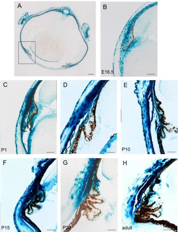
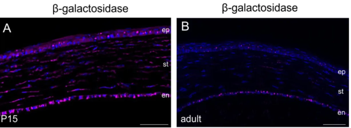
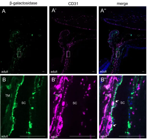
The binding of CTGF to BMPs or TGFβ, causes the inhibition of BMP and enhancement of TGF-β signaling (Abreu et al. 2002). Additional to the interaction with growth factors implicated in differentiation and morphogenesis, CTGF can coalesce interactions with vascular endothelial growth factor (VEGF), which is essential for angiogenesis during development (Ferrara et al. 1996). CTGF forms VEGF-CTGF complexes, leading to an inhibition of VEGF induced angiogenesis and in contrast VEGF can induce CTGF (He et al. 2003, Inoki et al. 2002, Jang et al. 2004, Kuiper et al. 2007, Suzuma et al. 2000). The interaction of CTGF with different factors essential for eye formation leads to the hypothesis that CTGF may play a role during the development of the mouse eye. Little is known about the expression pattern of CTGF during the development of the eye and its role in the early and later stages of the morphogenesis. The expression of CTGF was already shown in pericytes, endothelial cells and tip cells of the superficial plexus in CTGFp-GFP mice, which express GFP under the CTGF promotor (Pi et al. 2011) during development. The role of CTGF during the development of the superficial plexus, was shown by blocking CTGF with a specific antibody, which results in a reduced development (Pi et al. 2011). The exact distribution pattern and function of CTGF during embryonic and postnatal mouse eye development is still unclear, therefore it was of huge interest to analyze the CTGF promotor activity during embryonic and postnatal
development and in the adult eye. CTGFLacZ+/-mice, which express β-galactosidase under the control of the CTGF promotor were used for histological and immunohistochemical studies. Since the embryonic lethality of a CTGF knockout, CTGFCoin/Coin;CAGGCre-ER mice,which allow a conditional deletion of CTGF via the Cre-loxP system were used to study the effect of CTGF deficiency on eye development.
1.2. The pathology of POAG
1.2.1. The trabecular meshwork and aqueous humor outflow 1.2.1.1. Structure and function of the trabecular meshwork
Under normal conditions the IOP is in a steady state, as the outflow rate across the TM is equal to the production of aqueous humor (AH) by the ciliary body epithelium, lining the ciliary processes (Tamm 2009). The AH is a clear fluid, which fills the anterior and posterior chamber and provides nutrition for avascular structures, such as cornea and lens. Additionally, the AH is necessary for removing excretory products from metabolism, transporting neurotransmitter, stabilizing the ocular structure and contributes to the regulation of the homeostasis. The main drainage system is the conventional or trabecular outflow pathway, including the TM (corneo-scleral meshwork and the juxtacanalicular connective tissue (JCT)), the endothelial lining of SC, the collector channels and the aqueous veins (Figure 1-7 B).
Figure 1-7: Schematic representation of TM components and AH drainage. (A) Three layers of TM (shown in cutaway views): uveal, corneoscleral, and juxtacanalicular layer. (B) Schematic representation of different AH outflow pathways.
(Adapted from: (A) Shield and Kriegelstein, 1993 (B (Crawley et al. 2012))
The TM is enclosed in the internal scleral sulcus, a slight groove in the region of the corneo-scleral limbus (Figure 1-7 A). The TM is assembled from connective tissue
beams and lamellae, of densely packed collagen and elastic fibers, covered with TM cells (Marshall et al. 1991a, 1991b). The inner layer of the TM, the uveal meshwork consists of three layers of connective tissue beams and TM cells, with irregular intertrabecular fenestrations (Lütjen-Drecoll et al. 1981, Lütjen-Drecoll 1999, Lutjen- Drecoll E 1994, 2001, Tamm 2009) The corneo-scleral TM is similarly constructed, composed of 8-15 layers of perforated sheets of fibers, also covered by TM cells, with intertrabecular spaces becoming smaller closer to the SC. The lamellae and beams in the uveal and corneo-scleral TM are built up by densely packed collagen, mostly collagen type I and III and elastic fibers (Marshall et al. 1991a). The cells in the inner portions of the TM function mostly as phagocytic pre-filters, removing cellular debris. The outermost portion, the JCT represents almost all resistance to aqueous humor outflow, therefore the intertrabecular spaces become smaller (Figure 1-7 A) (Lütjen-Drecoll et al. 1981, Lütjen-Drecoll 1999, Lutjen-Drecoll E 1994, 2001, Tamm 2009)). In the JCT the discontinuous distribution of several layers of JCT cells are embedded in loose connective tissue. The ECM in the outflow pathway is constituted of several different components like laminin, type IV collagen, perlecan and fibronectin (Fuchshofer et al.
2006, Tamm 2009, Tawara et al. 1989, Ueda et al. 2002). The arrangement of the JCT cells and the ECM fibrils presents an irregular network. The ground substance in this network is constituted of proteoglycans and hyaluron (Gong et al. 1992, Lütjen-Drecoll et al. 1990, Tawara et al. 1989). As the SC is the proximate structure in the AH drainage system, the JCT cells are in contact to the endothelial cells lining the inner wall of SC (Grierson et al. 1979, Grierson & Lee 1974, 1978, Lütjen-Drecoll et al. 1981, Lütjen- Drecoll 1999). The AH is transported though the inner wall of SC by giant vacuoles and transendothelial pores (Braakman et al. 2015, Johnson 2006). Finally, the AH is drained to several collector channels, which are connected to the lumen of the SC. While the structure of the human TM and SC is similar to that in mice, the porcine outflow pathway differs by formation of an angular aqueous plexus (McMenamin & Steptoe 1991).
1.2.1.2. Pathological changes in the trabecular outflow pathway
In the pathogenesis of POAG no macroscopic changes are observed in the TM and SC confine it from other forms of glaucoma. Indeed, in POAG structural changes are observed in the TM, especially in the JCT, affecting the normal outflow pathway of AH, and creating an increased resistance and finally an elevation in IOP (Acott & Kelley 2008, Gordon et al. 2002, Johnson 2006, Leske et al. 2003). A characteristic sign of POAG is the increased fibrillar ECM in the JCT (Tektas & Lütjen-Drecoll 2009). The nature of this so called sheath-derived (SD) material has not been identified, but there is evidence that collagen type VI and FN are included (Hann et al. 2001, Lütjen-Drecoll et al. 1989, Lütjen- Drecoll 1999). It is thought that this accumulation of ECM contributes to the increased
outflow resistance in glaucoma. In addition to the alterations in ECM organization and structure, the outflow resistance is influenced by the actomyosin contractility of TM cells (Tian et al. 2000, Wiederholt et al. 2000).
TGF-β2, the predominant isoform of transforming growth factor β in the eye seems to be involved in the pathogenesis of POAG. TGF-β2, present in the normal AH, was shown by many studies to be elevated in the AH of POAG patients (Inatani et al. 2001, Min et al. 2006, Ochiai & Ochiai 2002, Ozcan et al. 2004, Picht et al. 2001, Tripathi et al. 1994, Trivedi et al. 2011). TGF-β2 causes ECM remodeling and organization, by an increased synthesis and a reduced degradation of ECM components. For instance, the synthesis of plasminogen activator inhibitor-1 (PAI-1) is induced by TGF-β2, leading to a reduced activity of the matrix metalloproteinase 2 (MMP2) (Fuchshofer et al. 2003). In recent years, the downstream mediator of TGF-β2, CTGF was implicated in the pathological changes occurring in the TM and SC in POAG. Analysis of CTGF levels in the AH of glaucoma patients, revealed a slight increase in POAG (Browne et al. 2011).
Interestingly, another study detected highly increased levels of CTGF in the AH of POAG patients (Fahmy IA). Furthermore, enhanced levels were detected in SC cells of glaucomatous donors (Overby et al. 2014). The implication of CTGF in the ECM reorganization and actin cytoskeleton arrangement supports the assumption that it is involved in the processes observed in the TM and SC in POAG. This assumption can be confirmed as TM cells cultured on stiffer substratum manifest higher levels of CTGF, furthermore CTGF modulate the actin cytoskeleton of TM cells and the effect of TGFβ2 on the ECM is mediate by CTGF (Junglas et al. 2009, Junglas et al. 2012, Raghunathan et al. 2013). Intriguingly, the lens specific overexpression of CTGF in mice results in an elevated IOP and a significant progressive loss of RGC axons over time (Junglas et al.
2012).
Therefore, CTGF can function as the ideal point of action for therapeutic treatment strategies, as a reduction would cause a distinct effect on the dysregulation in the tissue of the outflow pathway tissue and thus may regulate the IOP.
1.2.2. The optic nerve head and the Lamina cribrosa
1.2.2.1. Structure of the optic nerve head and the Lamina cribrosa
The optic nerve head (ONH) is a light site in the sclera in the posterior eye (Figure 1-8 A) (Bellezza et al. 2000), where the axons of the RGCs leave the eye. Besides the axons of the RGCs, the ON contains astrocytes, mircoglia, oligodendrocytes, blood vessels and depending on the species Lamina cribrosa (LC) cells. In humans, a prominent structure within the ONH is the Lamina cribrosa (LC; Lamina cribrosa sclerae), a sieve-like plate of connective tissue and elastic fibers, which are lined by astrocytes (Figure 1-8 B)
(Hernandez et al. 2008). In addition to astrocytes, the LC cells, a GFAP negative unique cell type within the ONH, are also located in the LC (Hernandez et al. 1988).
Figure 1-8: Vertical section of human ONH and 3D-representation of LC. (A) Vertical section of the ONH divided into its different parts. (B) 3-D representation of LC, depicting different constituents of connective tissue in different regions.
SNFL: surface nerve fiber Layer; PLR: prelaminar region; LCR: lamina cribrosa region; RLR: retrolaminar region. (Adapted from: (A (Hayreh 2011); Ischemic Optic Neuropathies) (B (Sigal IA et al. 2009))
Astrocytes, lining the connective tissue plates in the LC in primates and forming the glial tubes in mice are the important common element of both structures. Type 1B astrocytes are the major cell type in the ONH and are accountable for ECM macromolecule synthesis in the LC (Hernandez et al. 1991, Pena et al. 1996, Ye et al. 1994). In general, astrocytes have a widely spread field of function. They are responsible for maintaining the structural rigidity, regulating blood flow, releasing neurotransmitter, modulating synaptic function and plasticity, keeping the extracellular environment and are involved in scarring and repair processes (Haydon 2001, Iadecola & Nedergaard 2007, Magistretti 2006, Nedergaard et al. 2003, Newman 2003, Rossi et al. 2007, Takano et al. 2006, Ullian et al. 2001)). Within the LC they provide a supporting function to the surrounding axons, by their connections to the connective tissue and the surrounding blood vessels (Hernandez et al. 2008).
1.2.2.2. The structure of the optic nerve head in mice
In comparison to the human optic nerve head, in mice, a dense meshwork built up by astrocytes missing the connective tissue plates and with only low expression of ECM components, like collagen can be found. Because of the formation of glial tubes, illustrating a honeycomb structure where the axons of the RGCs pass through, this structure is called the glial lamina (Figure 1-9) (Fujita et al. 2000, Howell et al. 2007, May
& Lütjen-Drecoll 2002, Morcos & Chan-Ling 2000, Sun et al. 2009). In addition to the mostly absence of ECM, the transition zone between unmyelinated and myelinated
region is directly behind the LC in primates, but more posteriorly in mice (Oyama et al.
2006, Sun et al. 2009).
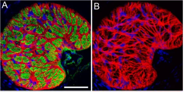
Figure 1-9: Section through a glial lamina of mouse ONH. (A) Honeycomb structure of the glial lamina constituted of astrocytes (red). Axons are counterstained with green (neurofilament). (B) Astrocytes (red) network forming glial tubes.
(Adapted from Lye-Barthel et al., 2013).
1.2.2.3. Pathological changes in the optic nerve head and the Lamina cribrosa The characteristic sign of POAG is the irreversible and progressive loss of axons of the RGCs. This significant damage is suggested to occur mostly at the level of the LC in the ONH (Hayreh 1978, Quigley et al. 1992). The distinct structural changes occurring in the LC lead to the permanent deformation, called excavation or cupping, differentiating it from other optic neuropathies (Figure 1-10) (Quigley 1983).
Figure 1-10: The ONH of healthy and glaucomatous patients. (A) The healthy, normal appearance of the ONH. (B) Typical changes in the ONH of glaucomatous patients, with tissue loss and cupping of the ONH. (Adapted from: (Quigley 2011)).
Thereby, it is hypothesized that a disruption of the retrograde and anterograde transport of neurotrophins is generated by axonal compression, which results in the death of RGC cells (Pease et al. 2000, Quigley et al. 2000). In the glaucomatous ONH a wide variety of structural changes in the ECM and in the major cell type, the astrocytes arise in response to elevated IOP (Fukuchi et al. 1992, Fukuchi et al. 1994, Gong et al. 1997, Hernandez et al. 1990, Hernandez 1992, Hernandez et al. 1994, Morrison et al. 1989, Morrison et al. 1990, Pena et al. 1996, Pena et al. 1998, Pena et al. 1999, Quigley 1983, Quigley et al. 1991). The ECM of the LC is dramatically reorganized, such as abnormal deposition of elastin, increased levels of collagen type IV and the connective tissue sheath around the capillaries in the prelaminar region are thickened (Hernandez 1992, Hernandez et al. 1994, Pena et al. 1998, Pena et al. 2001, Tektas et al. 2010). In summary, these changes include variations in both quality and quantity of ECM components (Hernandez 2000, Hernandez et al. 2002, Pena et al. 2001). Molecular signaling mechanisms contributing to or involved in these processes are not known yet, but increased amounts of TGF-β2 were detected in the ONH of glaucomatous patients (Pena et al. 1999, Zode et al. 2011). As astrocytes and LC cells assemble the LC it is likely that one or both cell types are contributing to structural changes developing in the LC (Hernandez 2000). There is evidence that reactive astrocytes are the major source of growth factors and ECM component upregulation, as it was shown that TGF-β2 is extensively localized to reactive astrocytes in the glaucomatous ONH (Pena et al. 2001).
Furthermore, previous studies of our group and others could show that TGF-β2 leads to an increase in ECM protein level in ONH astrocytes, and that this effect is dependent on its downstream mediator CTGF (Fuchshofer et al. 2005, Neumann et al. 2008, Zode et al. 2011). In the glaucomatous ONH, a change in astrocyte morphology occurs, indicated by cellular hypertrophy, increased GFAP immunoreactivity, rounded cell bodies with retracted cell processes and a migration from the core of the cribriform plates into the nerve bundles (Varela & Hernandez 1997). These changes are associated with alterations in gene expression (Liu & Neufeld 2000, Neufeld et al. 1997, Yan et al. 2000) These reactive changes, including migration, hypertrophy, upregulation of GFAP and gliosis are observed in human glaucoma and animal models of glaucoma (Hernandez 2000, Lye-Barthel et al. 2013, Morrison et al. 1990, Quigley et al. 1991, Sun et al. 2010, Sun & Jakobs 2012). Similar results could be shown for LC cells which react to the TGF- β2 treatment with increased ECM expression and synthesis (Kirwan et al. 2005, Kirwan et al. 2009, Zode et al. 2011)
1.2.2.4. Pathological changes in the peripapillary sclera
In addition, to the alteration occurring in the ONH, implications of the peripapillary sclera, the part of the sclera around the ONH, in the pathogenesis of the POAG are existing. In
general, the sclera, built up by different collagen types, elastic fibrils and proteoglycans, constitutes three quarters of human ocular circumference (Rada et al. 2006). The structure of the peripapillary sclera differs in the arrangement of collagen and elastic fibrils, as they are oriented circumferentially around the ONH. By this formation, a mechanical reinforcement against the stress in this region can be provided (Gelman et al. 2010, Girard et al. 2009b, Girard et al. 2009a, Hernandez et al. 1987, Pijanka et al.
2012, Quigley et al. 1991, Yan et al. 2000). As corneal hysteresis and myopia, both linked to the thinning of sclera are glaucoma risk factors, there is strong evidence that the composition of the sclera is a crucial component of glaucoma development (Boland &
Quigley 2007, Congdon et al. 2006). Increased scleral stiffness was shown in human glaucoma and experimental glaucoma models, in monkey and mice (Coudrillier et al.
2012, Downs et al. 2008, Hommer et al. 2008, Nguyen et al. 2013). Furthermore, scleral cross-linking is associated with greater RGC and axon loss (Kimball et al. 2014). As the astrocytes in the ONH stay in close contact with their processes to the peripapillary sclera and are sensitive to mechanical stress, it can be suggested that the mechanical behavior of the sclera influences the stress and deformation of the LC and that this mechanical stress is sensed by astrocytes, causing changes occurring in the ONH and its astrocytes (Coudrillier et al. 2012, Coudrillier et al. 2013, Girard et al. 2009b, Girard et al. 2009a, Hernandez 2000, Norman et al. 2011, Rogers et al. 2012, Sigal et al. 2005, Sigal et al.
2011).
2. Aim of the study
The aim of the study is to investigate the involvement of CTGF in eye development and the pathology of POAG. The expression of CTGF during eye development and in the adult eye will be investigated in different ocular tissues. Furthermore, the CTGF expressing cell types will be identified. It is also of interest to clarify the involvement of CTGF in the ONH pathology of POAG. Therefore, the correlation between CTGF expression changes and astrocyte reactivity in the ON and ONH will be examined related to mechanical stress in vivo and by increasing substratum stiffness in vitro. Additionally, possible mechanosensing proteins will be investigated. Furthermore, an aim of this study is to develop siRNA coated layer-by-layer nanoparticles as a new tool to reduce CTGF in the AH outflow pathway tissue and cells.
The following experiments were carried out to achieve these aims:
• Analysis of CTGF promotor activity in CTGFLacZ+/- mice during development and in the adult eye
• Identification of CTGF expressing ocular cell types in CTGFLacZ+/- mice during development and in the adult eye
• Analysis of CTGF and GFAP levels in the ON and ONH of 1-month and 2-month- old βB1-CTGF1 mice
• Analysis of the effect of increasing substratum stiffness on CTGF levels and reactivity in murine ON astrocytes in vitro
• Analysis of the effect of increasing substratum stiffness on Trp-channel and Caveolin1 expression in murine ON astrocytes
• Analysis of Caveolin1 expression in murine ON astrocytes after CTGF and TGF- β2 treatment, and in the ON and ONH of βB1-CTGF mice
• Evaluation of CD44 prevalence in HTM cells after CTGF and TGF-β2 treatment, in the outflow pathway structures of βB1-CTGF mice and glaucomatous and healthy human donor eyes
• Analysis of nanoparticle distribution after perfusion of porcine and human eyes with different nanoparticles
• Evaluation of CTGF knockdown after transfection with siRNA coated nanoparticles in HTM cells
• Generation and confirmation of a conditional, inducible CTGF knockout
• Analysis of the influence of conditional CTGF knockout on eye morphology
3. Material and methods 3.1. Materials
3.1.1. Reagents
Reagents Source of supply
2,4,6-Tri(dimethylaminomethyl) phenol (DPM-30)
Roth, Karlsruhe, Germany
0.05 % Trypsin/EDTA PAA The Cell Culture Company, Pasching, Austria
2-Mercaptoethanol Roth, Karlsruhe, Germany
Acetic acid, glacial Merck, Darmstadt, Germany
Acetone Merck, Darmstadt, Germany
Albumin Fraction V (BSA) Roth, Karlsruhe, Germany
Agarose Biozym Scientific, Oldendorf, Germany
AK10 plasticizer silicone oil Wacker Chemie, Munich, Germany Ammonium persulfate (APS), 10%
(w/v)
Roth, Karlsruhe, Germany
Aminopropyl-triethoxysilane (APTES) Sigma-Aldrich, Taufkirchen, Germany Astrocyte growth supplement (AGS) Sciencell, Carlsbad, USA
BC Assay Reagent A+B Interchim, Wörgl, Österreich BC Assay Reagent 1+2 Roth, Karlsruhe, Germany
CDP-Star Roche, Penzberg, Germany
Chloroform Roth, Karlsruhe, Germany
Coomassie®Brillant Blue R-250 Sigma-Aldrich, Taufkirchen, Germany Connective tissue growth factor Self purification
Connective tissue growth factor Prospec, Rehovot, Israel Connective tissue growth factor EMP, Ingolstadt, Germany
cross linker 210 Evonik Hanse GmbH, Gesthach, Germany
Desoxynukleosid-Triphosphate (dNTPs)
Qiagen, Hilden, Germany
Dimethylsulfoxid (DMSO) Roth, Karlsruhe, Germany
Divinyl tetramethyl disiloxane Evonik Hanse GmbH, Gesthach, Germany
DL-Dithiothreitol (DTT) Sigma-Aldrich, Taufkirchen, Germany Dulbecco’s Modified Eagle Medium
(DMEM) F12
PAA The Cell Culture Company, Pasching, Austria
Dulbecco’s Modified Eagle Medium (DMEM)
PAA The Cell Culture Company, Pasching, Austria
EDTA Roth, Karlsruhe, Germany
Ethanol 100% Roth, Karlsruhe, Germany
Ethidiumbromide Serva, Heidelberg, Germany
Fetal calve serum (FCS) Biochrom AG, Berlin, Germany Fluorescent Mounting Medium DakoCytomation, Hamburg, Germany
Formaldehyde Roth, Karlsruhe, Germany
Epon Serva, Heidelberg, Germany
Gelatine Sigma-Aldrich, Taufkirchen, Germany
Glutaraldehyde, 25% in water Serva, Heidelberg, Germany
Glycerine Roth, Karlsruhe, Germany
Glycin Merck, Darmstadt, Germany
Guanidin HCl Roth, Karlsruhe, Germany
Hydrochloric acid (37%) Merck, Darmstadt, Germany
Isoflurane Baxter, Heidelberg, Germany
Isopropanol Roth, Karlsruhe, Germany
Ketamine Wirtschaftsgenossenschaft Deutscher
Tierärzte (WDT), Garbsen, Germany Luminata Forte, Western HRP Millipore Cooperation, Billerica, USA Magnesium chloride (50 mM) Bioline, Luckenwalde, Germany
Methanol Merck, Darmstadt, Germany
methylhydrosiloxane-dimethylsiloxane AB116655 cross linker
ABCR GmbH, Karlsruhe, Germany
Milk powder (MM) Roth, Karlsruhe, Germany
N,N,N`,N`,-Tetramethylethylendiamine, (TEMED)
Roth, Karlsruhe, Germany
Paraformaldehyde (PFA) Sigma-Aldrich, Taufkirchen, Germany
PBS PAA The Cell Culture Company, Pasching, Austria
Penicilllin-Streptomycin PAA The Cell Culture Company, Pasching, Austria
peqGold TriFastTM (Trizol) PeqLab, Erlangen, Germany
Phalloidin Sigma-Aldrich, Taufkirchen, Germany
Phosphatase-Inhibitor-Mix Sigma-Aldrich, Taufkirchen, Germany Potassium chloride Roth, Karlsruhe, Germany
Potassium dihydrogen phosphate Roth, Karlsruhe, Germany
Protease-Inhibitor-Mix M Serva Electrophoresis GmbH, Heidelberg, Germany
Proteinase K Sigma-Aldrich, Taufkirchen, Germany
Pt-catalyst 510 Evonik Hanse GmbH, Gesthach, Germany
Rotiphorese® Gel 30 (30 % Acrylamid, 0.8 % Bisacrylamid; 37.5:1)
Sigma-Aldrich, Taufkirchen, Germany
Saccharose Roth, Karlsruhe, Germany
SDS (Sodium dodecylsulfat) Roth, Karlsruhe, Germany
SIH-terminated polydimethyl siloxane Evonik Hanse GmbH, Gesthach, Germany
Sodium chloride Roth, Karlsruhe, Germany
Sodium dihydrogen phosphate Merck, Darmstadt, Germany Sodium hydroxide Roth, Karlsruhe, Germany
Sodium phosphate Roth, Karlsruhe, Germany
SYBR-Green I Qiagen, Hilden
Tissue-Tek® Sakura, Zoeterwoude, Netherlands
Transforming Growth Factor 2 (TGF- β2)
R&D Systems, Minneapolis, USA
Tris HCl Roth, Karlsruhe, Germany
Triton X 100 Roth, Karlsruhe, Germany
Tween 20 Roth, Karlsruhe, Germany
Vectashield Mounting Medium for Fluorescence with DAPI
Vector Laboratories, Burlingame, USA
vinyl-functional polydimethylsiloxane Evonik Hanse GmbH, Gesthach, Germany Water Rotisolv (Rnase-free) Roth, Karlsruhe, Germany
Xylazine Serumwerk Bernburg, Bernburg, Germany
Table 3-1: Reagents
3.1.2. Enzymes and Reagent-Kits
Enzymes and Reagent-Kits Source of supply
qScript™cDNA Synthesis Kit Quanta Biosciencies, Gaithersburg, USA
Mango Taq Bioline, Luckenwalde, Germany
Proteinase K Roth, Karlsruhe, Germany
Taq DNA Polymerase Self purification
Universal SYBR Green Master (ROX) Roche, Mannheim, Germany
TaqMan®Fast Advanced Master Mix (2x) ThermoFisher, Darmstadt, Germany Table 3-2: Enzymes and Reagent Kits
3.1.3. Oligonucleotide primers and Taqman probes
Primer Species Orientation Sequence
βB1 mus
musculus
forward GGAAGTGCCAGCTCATCAGT
βB1 mus
musculus
reverse GTGCGGGACAGAAACCTG
SV40 mus
musculus
forward GTGAAGGAACCTTACTTCTGTGGTG
SV40 mus
musculus
reverse GTCCTTGGGGTCTTCTACCTTTCTC
LacZ mus
musculus
forward GCCGTCTGAATTTGACCTGA
LacZ mus
musculus
reverse TCTGCTTCAATCAGCGTGCC
CAG-Cre mus musculus
forward ATGCTTCTGTCCGTTTGCCG
CAG-Cre mus musculus
reverse CCTGTTTTGCACGTTCACCG
Coin/Coin mus musculus
forward CACTTTCTACTCTGTTGAC
Coin/Coin mus musculus
reverse CCTTACATGTTTTACTAG