The regulation of connective tissue growth factor expression influences the viability of human trabecular meshwork cells
Sabrina Kuespert
#, Benjamin Junglas
#, Barbara M. Braunger, Ernst R. Tamm, Rudolf Fuchshofer *
Institute of Human Anatomy and Embryology, University of Regensburg, Regensburg, Germany Received: June 6, 2014; Accepted: October 13, 2014
Abstract
Connective tissue growth factor (CTGF) induces extracellular matrix (ECM) synthesis and contractility in human trabecular meshwork (HTM) cells. Both processes are involved in the pathogenesis of primary open-angle glaucoma. To date, little is known about regulation and function of CTGF expression in the trabecular meshwork (TM). Therefore, we analysed the effects of different aqueous humour proteins and stressors on CTGF expression in HTM cells. HTM cells from three different donors were treated with endothelin-1, insulin-like growth factor (IGF)-1, angio- tensin-II, H
2O
2and heat shock and were analysed by immunohistochemistry, real-time RT-PCR and Western blotting. Viability after H
2O
2treat- ment was measured in CTGF silenced HTM-N cells and their controls. Latrunculin A reduced expression of CTGF by about 50% compared to untreated HTM cells, whereas endothelin-1, IGF-1, angiotensin-II, heat shock and oxidative stress led to a significant increase. Silencing of CTGF resulted in a delayed expression of
aB-crystallin and in reduced cell viability in comparison to the controls after oxidative stress. Con- versely, CTGF treatment led to a higher cell viability rate after H
2O
2treatment. CTGF expression is induced by factors that have been linked to glaucoma. An increased level of CTGF appears to protect TM cells against damage induced by stress. The beneficial effect of CTGF for viability of TM cells is likely associated with the effects on increased ECM synthesis and higher contractility of the TM, thereby contributing to reduced aqueous humour outflow facility causing increased intraocular pressure.
Keywords: CTGF glaucoma trabecular meshwork endothelin-1 angiotensin-II IGF-1 oxidative stress heat shock
aB-crystallin cell viability
Introduction
Primary open-angle glaucoma (POAG), one of the major causes of blindness worldwide [1], is a neuropathy of the optic nerve leading to a loss of axons at the optic nerve head. The critical risk factor for POAG is intraocular pressure (IOP) which is frequently elevated [2
–4]. Elevated IOP is caused by an abnormally high aqueous humour (AH) outflow resistance that is generated in the juxtacanalicular region of the human trabecular meshwork (HTM) [5, 6]. To date, the mechanisms that are responsible for the increase in TM outflow resis- tance in POAG are not fully understood [7, 8]. There is some evidence though that changes in the amounts and the composition of the HTM
extracellular matrix (ECM) [9, 10] as well as in the actomyosin system of HTM cells [11
–14] are involved.
Currently, transforming growth factor (TGF)-
b2 is one of the leading candidates among the multiple signalling molecules in the AH that may cause molecular changes leading to an increase in outflow resistance in POAG. Accordingly, patients suffering from POAG exhibit higher levels of TGF-
b2 in the AH when compared to healthy controls [15
–18]. TGF-
b2 is a strong inducer of the HTM ECM and a modifier of the actin cytoskeleton in TM cells [19]. Perfusion of anterior segments with TGF-
b2 results in an increase in outflow resis- tance [20].
In recent studies, we showed that connective tissue growth factor (CTGF) is mediating most of the ECM effects of TGF-
b2 on TM cells [21]. In addition, CTGF is able to modulate the biological properties of the TM actin cytoskeleton and to increase its contrac- tility [11]. CTGF belongs to a family of regulatory proteins that are upregulated in a substantial number of disorders associated with a pathological increase in ECM [22
–24]. In the HTM, CTGF is among the most highly expressed genes [25]. The comparison of the
#SK and BJ contributed equally to the manuscript.
*Correspondence to: Rudolf FUCHSHOFER,
Institute of Human Anatomy and Embryology, University of Regensburg, Universitaetsstr. 31, D-93053 Regensburg, Germany.
Tel.: +49-941-943-2881 Fax: +49-941-943-5587
E-mail: rudolf.fuchshofer@vkl.uni-regensburg.de
ª2015 The Authors.
doi: 10.1111/jcmm.12492
J. Cell. Mol. Med. Vol 19, No 5, 2015 pp. 1010-1020
CTGF expression in Schlemm’s canal endothelial (SC) cells derived from glaucoma patients with SC cells from healthy donors reported a significant higher CTGF expression level in the glaucomatous SC cells [26]. CTGF has also been detected in the AH [27] and its amounts are increased in patients with pseudoexfoliation syndrome and glaucoma associated with it [28, 29]. In patients with POAG, only a slight increase in CTGF was found in the AH in one study [28], whereas other authors reported a significant increased level of CTGF [30].
To investigate the influence of CTGF signalling in the living eye, we recently developed a mouse model with an eye-specific overex- pression of CTGF by the use of a lens-specific promotor. Since the CTGF-mediated effects on ECM and the actin cytoskeleton that corre- lated with an increase in IOP and a successive loss of axons in the optic nerve, we concluded that high amounts of CTGF cause POAG in the mouse eye [11].
The dynamics of factors that may increase the trabecular expres- sion of CTGF in patients with POAG remain unclear. TGF-
band cyclic mechanical stress are the only known stimuli that have been shown to increase the expression of CTGF in HTM cells [31, 32]. In the pres- ent study, we show that the regulation of CTGF expression involves various factors including different kinds of stress, suggesting a pro- tective role for CTGF in the HTM.
Material and methods
Cell culture
Cultures of HTM cells were established from the eyes of three human donors according to protocols published previously [32]. The age of the donors ranged from 34 to 76 years. HTM cells of the third to fifth pas- sage were seeded in 35-mm culture wells (4.09105 cells/well) and grown to a confluent monolayer in F10-HAM medium plus 10% (v/v) foetal bovine serum without antibiotics in 5% CO2at 37°C (PAA, Pas- ching, Austria). The confluent cells were incubated in serum-free med- ium for 24 hrs followed by incubation in fresh serum-free medium.
Endothelin-1 (ET-1), angiotensin-II (Ang II), insulin-like growth factor (IGF)-1, hydrogen peroxide (H2O2) or latrunculin A (LatA; Sigma-Aldrich, Taufkirchen, Germany) were added at various concentrations at different time-points. Control cells were treated equally with corresponding vehi- cles.
Heat-shock experiments were performed by seeding immortalized HTM cells (HTM-N) [33] and pSiCTGF-HTM-N cells (stable plasmid- based siRNA silencing of the CTGF gene) into Petri dishes to a con- fluent level (1.09106 cells/well). The pSiCTGF-HTM-N cell line has an 80% reduced CTGF expression in comparison to the HTM-N cell line as described previously [11]. The cells were kept for 24 hrs under serum-free conditions. Then, cells were heat shocked for 15 min. at 42°C in a water bath and harvested after a further 37°C incubation period at different time-points [0 (control), 15, 30, 45, 60 min.].
Each of the described experiments except heat shock was done with each of the three primary cell lines. Methods for securing human tissues were humane, included proper consent and approval, and com- plied with the Declaration of Helsinki.
RNA analysis
Human trabecular meshwork cells were harvested and total RNA was extracted with TRIzol (Invitrogen, Karlsruhe, Germany) according to manufacturer’s recommendations. First strand cDNA was prepared from total RNA using the iScript cDNA Synthesis Kit (Bio-Rad, Munich, Ger- many) according to the manufacturer’s instructions. Real-time RT-PCR was performed on a BioRad iQ5 Real-time PCR Detection System (Bio- Rad) using the following temperature profile: 40 cycles of 10 sec. melt- ing at 95°C, 40 sec. of annealing and extension at 60°C. Primer pairs (Table 1) were purchased from Invitrogen and extended over exon–
intron boundaries. RNA that was not reversely transcribed served as negative control for real-time RT-PCR. Glyceraldehyde-3-phosphate dehydrogenase (GAPDH) and guanine nucleotide binding protein 2-like- 1 (GNB2L1) were both used as housekeeping genes for relative quantification of the real-time RT-PCR experiments. Quantification was performed with iQ5 Standard-Edition (Version 2.0.148.60623) software (Bio-Rad).
Western blot analysis
To obtain protein extracts, cells were directly lysed in RIPA lysis buffer (150 mM NaCl, 1% NP-40, 0.5% deoxycholic acid, 0.1% SDS and 50 mM Tris, pH 8) and protein content was measured with the bicinchoninic acid protein assay (Pierce, Rockford, IL, USA). Proteins were separated by SDS-PAGE and transferred to polyvinylidene fluoride membranes. Anti- bodies were used as follows: rabbit anti-human aB-crystallin (1:200;
Stressgen, San Diego, CA, USA), goat anti-human CTGF (1:500), donkey anti-rabbit-horseradish peroxidase (HRP) and chicken anti-goat-HRP (1:2000; all Santa Cruz, CA, USA). Chemiluminescence was detected on a LAS 3000 imaging workstation (Raytest, Straubenhardt, Germany). For normalization of the signals, blotted membranes were stained with coo- massie blue and digitized. The total amount of protein per lane was deter- mined and calculated using AIDA Image analyser software (Raytest). The values of the total amount of protein were used to normalize the signal intensity of the bands detected in Western blot analysis.
Immunohistochemistry
Cultured HTM-N and pSiCTGF-HTM-N cells were grown on microscope slides and treated with heat shock as described above. After incubation,
Table 1Sequences of primers used for real-time RT-PCR
Type Sequence Position Tm
(°C) CTGF 50-CTCCTGCAGGCTAGAGAAGC-30 884–977 59
50-GATGCACTTTTTGCCCTTCTT-30 60
GAPDH 50-AGCCACATCGCTCAGACA-30 83–148 60
50-GCCCAATACGACCAAATCC-30 60
GNB2L 50-GCTACTACCCCGCAGTTCC-30 170–241 59
50-CAGTTTCCACATGATGATGGTC-30 60
cells were fixed with 4% paraformaldehyde for 15 min. and subse- quently washed twice with PBS containing 0.1% Triton X-100. Rabbit anti-human aB-crystallin antibodies were added at a 1:50 dilution in PBS/bovine serum albumin (BSA; 5%) for 4 hrs at room temperature.
After three wash steps with PBS, fluorescein-conjugated secondary pig anti-rabbit IgG (Dako, Glostrup, Denmark) was added at a 1:1000 dilu- tion in PBS/BSA (5%) for 1 hr at room temperature. Actin stress fibres were stained using phalloidin-TRITC (Sigma-Aldrich) at a dilution of 1:1000 for 1 hr at room temperature. After immunohistochemical label- ling, Vectashield mounting medium (Vector Laboratories, Burlingame, CA, USA) was used for mounting slides. Slides were analysed under an Axio Imager fluorescence microscope (Carl Zeiss AG, Oberkochen, Ger- many). Corresponding negative controls to estimate unspecific binding of secondary antibodies were handled similarly, but incubated in PBS/
BSA without primary antibody.
Cell viability assay
To analyse the CTGF effect on the viability of HTM-N and pSiCTGF- HTM-N cells after treatment with H2O2, cells were seeded into 96-well plates at 3.09104 cells/well. The cells were treated with 50lM of H2O2 alone or in combination with 50 ng/ml of recombinant CTGF (rCTGF) [21] for 24 hrs under serum-free conditions. Viability of cells was analysed by a MTT (3-(4,5-dimethylthiazol-2-yl)-2,5-diphenyltet- razolium bromide) assay. The yellow MTT is reduced by a mitochondrial reductase to purple formazan in the mitochondria of living cells. The absorbance of the formed fomazan is measured by 570 nm wave length.The cells were washed with PBS, and 80ll/well MTT solution (tetrazolium dye, PBS and serum-free medium) was added for 2 hrs.
The formazan crystals that formed were dissolved by the addition of dimethyl sulfoxide (DMSO). Absorption was measured with a spectro- photometer (Sunrise, Tecan, Maennedorf, Switzerland) at 570 nm and analysed using Magellan software (Tecan). Results are expressed as the mean percentage of the controls. Untreated HTM-N and pSiCTGF-HTM- N cells served as controls. Values of each sample were normalized to a
‘blank’ containing DMSO only.
Number of experiments and statistical analysis
To assess the effects of the different treatments, each experiment was repeated at least three times from primary HTM cell lines of three differ- ent donors and with the HTM-N and pSiCTGF-HTM-N cells. Student’s t-test was used for statistical analysis and differences with a P-value smaller 0.05 were regarded as significant, and with aP<0.01 as highly significant.
Results
Heat shock induces CTGF expression
As mechanical stress induces CTGF in HTM cells [31], we investi- gated whether CTGF is also induced by other stressors. To this end, HTM-N cells were treated with heat shock (42
°C) and were then allowed to recover at 37
°C. After 30 min., the expression of CTGF
was significantly increased (3.3 0.4-fold,
P<0.05) and remained at this level during the entire 60 min. of the recovery phase (Fig. 1A).
CTGF induces a B-crystallin expression
Amounts of the small heat-shock protein (sHSP)
aB-crystallin are higher in the TM of POAG eyes [34] and are induced by TGF-
bin cul- tured HTM cells [35]. Here, we were interested to learn whether there is a link between
aB-crystallin and CTGF in HTM cells. HTM cells trea- ted with recombinant CTGF showed a dose-dependent increase in the amounts of
aB-crystallin, which were 3.2 0.3-fold higher after treatment with 10 and 50 ng/ml of CTGF for 24 hrs (P
<0.05, Fig. 1B).
We now wanted to know whether the expression of
aB-crystallin is changed in the presence of lower amounts of CTGF. For that pur- pose, we used the pSiCTGF-HTM-N cell line and compared it to HTM- N cells [11]. HTM-N cells treated with 42
°C for 15 min. showed an increased immunohistochemical staining for
aB-crystallin and an increase in actin stress fibre formation after 30 min. of recovery phase (Fig. 1E). Following Western blot experiments, densitometric analysis of the immunoblots identified a significant up-regulation of
aB-crystallin in HTM-N cells (4.1 0.3-fold,
P<0.05, 45 min., Fig. 1D). In contrast in pSiCTGF-HTM-N cells,
aB-crystallin staining and stress fibre formation was unchanged after heat shock exposure (Fig. 1E). Western blot analysis of
aB-crystallin synthesis in pSiCTGF-HTM-N cells showed a delayed and reduced induction in comparison to HTM-N cells. An induction (2.1 0.5-fold) of
aB- crystallin was only observed after 60 min. of recovery phase (Fig. 1D).
Oxidative stress induces CTGF expression
As increasing age comes along with higher amounts of reactive oxy- gen species (ROS) in the eye [36], we investigated whether oxidative stress influences the expression of CTGF in HTM cells. To this end, cells were treated with 50
lM H
2O
2for 1, 3, 6 or 24 hrs and CTGF and its mRNA were determined by real-time RT-PCR and Western blotting. A 3-hrs treatment resulted in a significant increase in CTGF mRNA expression up to 1.8 0.2-fold (P
<0.05) which remained upregulated for at least 24 hrs (Fig. 2A). Moreover, after 3 hrs of oxi- dative stress, the amounts of CTGF were 2.2 0.3-fold higher than in control cells (P
<0.05). After 24 hrs, the amounts of CTGF were still 1.9 0.2-fold higher than in controls (P
<0.05; Fig. 2B).
CTGF protects HTM cells against oxidative stress
As CTGF appeared to be a primary response gene to stress and as the
amounts of CTGF correlate with sHSP expression, we investigated
whether CTGF affects the viability of H
2O
2-treated HTM-N cells. For
this purpose, we treated HTM-N and pSiCTGF-HTM-N cells with
50
lM H
2O
2alone or in combination with 50 ng/ml of CTGF for
24 hrs. Measuring cell viability was conducted by a MTT assay.
A B
C
E
D
Treatment of HTM-N cells with H
2O
2lead to a significant reduction in cell viability to 75% 5% compared to untreated control cells (P
<0.05). The decrease in viability was significantly higher in pSiCTGF-HTM-N cells (55% 2%;
P<0.01). Cells treated with a combination of H
2O
2and CTGF showed a significantly higher viability (in HTM-N: 103% 2%,
P<0.01 and pSiCTGF-HTM-N:
84% 1%;
P<0.01) in comparison to the H
2O
2-treated cells (Fig. 2C).
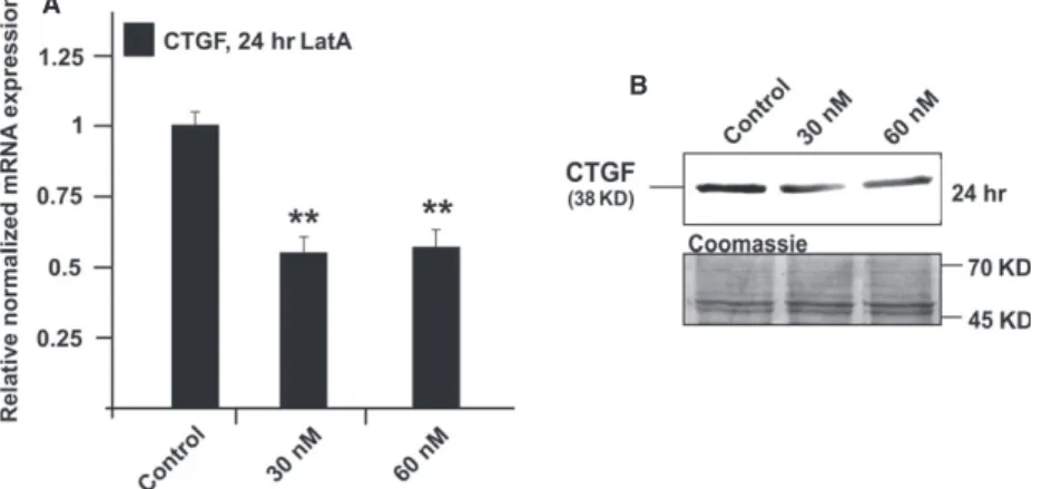
Treatment with LatA causes a reduction in the amounts of CTGF and its mRNA
Treatment of HTM cells with 30 nM LatA for 24 hrs led to a highly significant 0.55 0.1-fold reduction in CTGF mRNA expression when compared to untreated control cells (Fig. 3A;
P<0.01. A com- parable effect was observed after treatment with 60 nM LatA, which did also cause a reduction in the amounts of CTGF (P
<0.01, Fig. 3B).
Treatment with ET-1, Ang II or IGF-1 induces CTGF and its mRNA
To assess the effects of ET-1 on the expression of CTGF in the TM, HTM cells were treated with ET-1 at a concentration of 100 nM for 1
–24 hrs and 1 day to 3 days. The amounts of CTGF and its mRNA were analysed by real-time RT-PCR and Western blot analysis. Treat- ment for 1, 3 and 12 hrs did not cause significant changes in the expression of CTGF (data not shown). After 3 days of treatment, the expression of mRNA for CTGF increased significantly by 1.6 0.2- fold (Fig. 4A;
P<0.05). In addition, higher amounts of CTGF were detected in cell extracts of treated cells compared to untreated control cells (Fig. 4B).
To study the effects of Ang II, HTM cells were treated for 1, 3 or 6 hrs with concentrations of 10 nM and 1
lM of Ang II and analysed as described above. Following treatment with 10 nM Ang II, the amounts of CTGF and its mRNA remained unaffected (P
>0.05, Fig. 5B). In contrast, treatment with 1
lM of Ang II led to significant changes after 3 hrs of treatment. The expression of mRNA for CTGF increased up to 2.16 0.18-fold (Fig. 5A;
P<0.05), an effect that correlated with the presence of higher amounts of CTGF (Fig. 2B).
When cells were treated for 6 hrs, no significant changes were observed when compared with control cells (Fig. 5B).
For studies on the CTGF-inducing effect of IGF-1, HTM cells were treated with IGF-1 at concentrations of 5 and 50 ng/ml for 6 and 24 hrs respectively. After 6 hrs of incubation, there was no significant increase between IGF-1-treated and control cells with respect to the amounts of CTGF and its mRNA. In contrast, treatment with 50 ng/ml of IGF-1 for 24 hrs led to a significant 2.2 0.2-fold increase of CTGF mRNA when compared to untreated control cells (Fig. 6A;
P<
0.05). The increase in mRNA caused a significant increase in the amounts of CTGF (P
<0.05, 2.1 0.3-fold, Fig. 6B).
Discussion
We conclude that CTGF is a primary response gene after exposure to stress that protect HTM cells from injury. The protective effect of CTGF appears to be associated with a stabilization of the actin cyto- skeleton. This conclusion is based on (i) the findings that CTGF is immediately upregulated in HTM cells after treatment with different stressors and (ii) that CTGF induces the expression of the sHSP
aB- crystallin. (iii) The conclusion is supported by the observation that a knockdown of CTGF leads to a reduced cell viability in HTM cells after exposure to stress and to an attenuated up-regulation of
aB-crystal- lin, (iv) whereas a pretreatment with CTGF could prevent the stress- induced cell loss.
The cellularity of the TM decreases throughout life [37, 38]
and the loss of TM cells is more pronounced in patients with POAG [39]. The causes for the loss of TM cells are not clear, but it is known that TM cells are exposed to many different stressors during the ageing process [40, 41]. Among the stressors is mechanical stress mediated by ciliary muscle contraction and aqueous flow rate changes. Mechanical stress is thought to be elevated in POAG patients with increasing IOP [40], a major risk factor for glaucoma.
Besides mechanical stress, TM is constantly exposed to oxidative stress [41].
In many diseases, it is accepted that the ageing process is associ-
ated with a higher exposure rate to free radicals, which would lead to
a decline of physiological functions of various tissues [42] and sev-
eral studies suggest that an increased formation of ROS might be
associated with glaucoma [36, 43]. In POAG patients, a significant
reduction in mitochondrial respiratory activity and a significant
decrease in anti-oxidative capacity were detected [41, 44
–46]. Those
findings resulted in the hypothesis that oxidative stress could be
involved in onset and progression of POAG. Here, we show that CTGF
is a primary response gene after oxidative stress in TM cells. The data
Fig. 1Effect of heat shock on the expression of CTGF andaB-crystallin. (A) Western blot analysis for CTGF in cell extract of HTM-N cells after heat shock at 42°C and recovery phase of 15–60 min. After 30 min. the CTGF expression was increased up to 3.30.4-fold (P<0.05) and maintained at this level until 60 min. (B) Western blot analysis foraB-crystallin in cell extract of HTM cells after treatment with 2.5–50 ng/ml of CTGF for 24 hrs.aB-crystallin was significantly increased after CTGF treatment for 24 hrs (3.20.3-fold,P<0.05). (CandD) Western blot analysis for aB-crystallin in cell extracts of HTM-N (C) and pSiCTGF HTM-N cells (D) after heat shock at 42°C and recovery phases of 15–60 min. Densitometric evaluation of HTM-N cells showed a maximum of 4.10.3-fold after 30 min. (P<0.05) while there was only a slight induction of 2.10.5-fold observed after 60 min. in pSiCTGF HTM-N cells. Membranes were stained with coomassie blue to confirm equal loading of proteins. (E) Immunohis- tochemical staining ofaB-crystallin (green) and actin fibres (phalloidin, red) in HTM-N and pSiCTGF HTM-N cells 30 min. after heat shock. HTM-N cells showed a more intense staining foraB-crystallin and an increased formation of actin stress fibres after heat shock, while there was no increase in pSiCTGF HTM-N cells. Scale bar=50lm.are supported by
in vivoanalysis of early response genes after oxida- tive stress. In mice, the induction of oxidative stress in the cerebellum led to a substantial increase in CTGF within 6 hrs [47]. The immediate up-regulation of CTGF under stress conditions was further confirmed by our heat-shock experiments. Together with the known findings that also mechanical stress is able to induce CTGF expression [31], we conclude that CTGF might be a general primary response gene to various kinds of stressors in HTM cells.
The physiological function of the early up-regulation of CTGF seems to be a protective mechanism in HTM cells. The supplementa- tion of CTGF prior to H
2O
2treatment had a beneficial effect on the
viability of TM cells. A potential role for CTGF in cell survival was shown in gallbladder cancer cells, where silencing of CTGF led to a reduced cell viability [48]. We could observe a similar effect in TM cells, where reduced levels of CTGF led to a decline in cell viability rate after oxidative stress, whereas adding CTGF partially rescued the loss of TM cells. A protective function of CTGF was previously shown in the kidney, where supplementation of CTGF guarded puromycin- treated podocytes from cell death [49].
The protective effect of CTGF might be directly linked to the expression of the sHSP
aB-crystallin, as CTGF treatment led to a sig- nificant up-regulation of
aB-crystallin in HTM cells.
aB-crystallin belongs to the family of sHSPs, and it is known to be up-regulated in the TM of POAG patients [34]. The increased presence of sHSPs might have a protective effect, as TM cells respond to oxidative stress and heat shock by
aB-crystallin induction [50], whereas silencing of CTGF in TM cells blocked the stress-induced up-regulation of the
aB-crystallin. As both proteins are simultaneously regulated during the exposure to heat shock, we assume that CTGF acts as modulator of the
aB-crystallin synthesis, because of the matricellular character of CTGF [51]. sHSPs are able to protect cells by different mechanisms depending on their subcellular localization. Under stress conditions,
aB-crystallin can translocate to the mitochondria and thereby inter- acting with various components of the mitochondrial apoptotic machinery and preventing cell death [52, 53], whereas the cytosolic
aB-crystallin can inhibit actin depolymerization, thereby leading to an increased cell survival [54]. We assume that CTGF protects the cells against the oxidative stress-induced disruption of the cytoskeleton and disaggregation of actin fibres, a critical point for cell survival [54]. In an earlier study, we could already show the positive effect of CTGF on formation of actomyosin fibres and the contractility in HTM cells [11], whether the mitochondrial apoptotic events are also altered after CTGF treatment have to be investigated in the future.
Based on our observations, we wanted to address additionally the question whether CTGF regulation in HTM cells is also linked to other factors, which are present in the AH and/or are involved in the
AB
C
Fig. 2(A) Real-time RT-PCR analysis of CTGF mRNA expression in HTM cells after treatment with 50lM H2O2for 1–24 hrs. A 3-hrs treat- ment resulted in a significant increase in CTGF mRNA expression up to 1.80.2-fold which remained up-regulated for at least 24 hrs. The mean value obtained from untreated cells was set at 1. GNB2L and GAPDH were used as reference genes. MeansSD are shown. (B) Western blot analysis for CTGF in cell extract of HTM cells after treat- ment with 50lM H2O2 for 3 and 24 hrs. Densitometric evaluation showed a maximum of 2.20.3-fold after oxidative stress for 3 hrs.
Membranes were stained with coomassie blue to confirm equal loading of proteins. (C) Measurement of cell viability in HTM-N and pSiCTGF HTM-N cellsviaMTT assay after treatment with 50lM H2O2alone or in combination with 50 ng/ml of CTGF for 24 hrs. Treatment of HTM-N cells lead to a significant reduction to 75%5%. The decrease was more intense in pSiCTGF-HTM-N cells (55%2%). Cells treated with a combination of H2O2 and CTGF showed a significant higher viability (in HTM-N 103%2% and pSiCTGF-HTM-N 84%1%). The mean value obtained from untreated cells was set at 1. MeansSD are shown. Asterisks mark statistically significant (*P<0.05) and high sig- nificant differences (**P<0.01).
outflow facility regulation and are assumed to be involved in CTGF regulation in other tissues.
In the context of a CTGF-mediated induction of ECM synthesis, we also investigated the effect of IGF-1 on CTGF expression. IGF-1 stimu- lates CTGF to induce collagens
viabinding to the IGF-binding domain of CTGF [55]. IGF-1 is present in the AH [56] and is expressed in the TM together with its receptors [57]. In studies on the signalling path- ways of IGF-1, the RhoA/ROCK signalling pathway is among the most commonly highlighted [58]. In our study, physiological concentra- tions of IGF-1 led to an increased expression of CTGF in TM cells [56]. Little is known though about the function of IGF-1 and its recep- tor within the TM, an avenue that should be analysed in the future.
Clearly, the common pathway between the molecules investigated here appears to be the RhoA/ROCK pathway that is involved in the regulation of outflow facility by altering the actin cytoskeleton of the TM. Previous studies showed that Ang II and ET-1 induce CTGF expression
viathe small GTPase RhoA [59, 60]. In this study, we obe- served a late induction of CTGF after ET-1 treatment indicating that the increase in CTGF could be a secondary effect. On the other hand, Horstmeyer
et al.observed a pronounced late induction of CTGF after ET-1 in comparison to short time treatments. This finding was explained with different expression patterns of the two endothelin receptors A and B at the different points [61]. In contrast, a rapid increase in CTGF after Ang II treatment clearly implicates a direct
AB
Fig. 3Analysis of CTGF expression in HTM cells after treatment with 30 or 60 nM latrunculin A for 24 hrs. (A) Real-time RT-PCR analysis of CTGF mRNA expression. The mean value obtained from untreated cells was set at 1. GNB2L and GAPDH were used as reference genes. MeansSD are shown. Asterisk marks statistically significant differences between control and latrunculin A-treated cells (**P<0.01). (B) Western blot analysis for CTGF in HTM cell extract. Densitometric evaluation showed a minimum of 0.670.7-fold (*P<0.01) compared to the untreated control after treatment with 60 nM for 24 hrs. Membranes were stained with coomassie blue to confirm equal loading of proteins. Asterisks mark statistically significant (*P<0.05) and high significant differences (**P<0.01).
A
B
Fig. 4Analysis of CTGF expression in HTM cells after treatment with 100 nM endothelin-1 for different time periods. (A) Real-time RT-PCR analysis of CTGF mRNA expression. The mean value obtained from untreated cells was set at 1. GNB2L and GAPDH were used as reference genes.
MeansSD are shown. Asterisk marks statistically significant differences between control and endothelin-1-treated cells (*P<0.05). (B) Western blot analysis for CTGF in HTM cell extract. Densitometric evaluation showed a maximum of 1.80.2-fold compared to untreated control after 3 days. Membranes were stained with coomassie blue to confirm equal loading of proteins.
effect of the substance. The fast decline of the CTGF expression under the basal levels might be because of the short half-life of Ang II.
The CTGF-inducing effect of RhoA was shown by the treatment of HTM cells with a constitutive active form of RhoA, whereas the inhibi- tion of the Rho kinase, a downstream mediator of RhoA, lead to a substantial down-regulation of CTGF [62]. The CTGF expression is coupled to the stability of the actin cytoskeleton [63], the formation of F-actin stress fibres led to an increased CTGF expression. This regula- tion was dependent on the availability of G-actin. Actin disrupting agents like LatA and B, which cause an enhanced cellular content of G-actin, lead to a reduction in CTGF expression in different cell lines [64]. Quite similarly, the LatA treatment of HTM cells caused a signifi- cant down-regulation of CTGF. As latrunculin treatment of perfused anterior chambers lead to an increase in outflow facility [65, 66], we
speculate that, given the loss of contractility in TM cells, the decrease in CTGF-mediated ECM deposition might contribute in the long run to the outflow increasing effect of latrunculins. On the other hand, under stress, like increased mechanical load or oxidative stress, the TM cells react with increased F-actin stress fibres formation, leading to decreased G-actin levels and thereby to an enhanced CTGF expres- sion. Thereby, inducing a vicious circle, as CTGF by itself can activate the RhoA/ROCK signalling pathway [11]. If the stress last only for a short period, the expression of CTGF declines rapidly to a basal expression, which led to the assumption that compensatory mecha- nisms are activated to normalize the CTGF expression levels [67].
Under pathological conditions, which were shown in other tissues, those mechanisms seem to fail and the CTGF up-regulation maintains in those tissues leading to fibrotic changes [68, 69].
A B
Fig. 5Analysis of CTGF expression in HTM cells after treatment with 10 nM or 1lM angiotensin-II for different time periods. (A) Real-time RT-PCR analysis of CTGF mRNA expression. The mean value obtained from untreated cells was set at 1. GNB2L and GAPDH were used as reference genes.
MeansSD are shown. Asterisk marks statistically significant differences between control and angiotensin-II-treated cells (*P<0.05). (B) Western blot analysis for CTGF in HTM [71] cell extract. Densitometric evaluation showed a maximum of 2.60.8-fold compared to the untreated control after treatment with 1lM for 3 hrs. Membranes were stained with coomassie blue to confirm equal loading of proteins.
A B
Fig. 6Analysis of CTGF expression in HTM cells after treatment with 5 or 50 ng/ml IGF-1 for different time periods. (A) Real-time RT-PCR analysis of CTGF mRNA expression. The mean value obtained from untreated cells was set at 1. GNB2L and GAPDH were used as reference genes.
MeansSD are shown. Asterisk marks statistically significant differences between control and IGF-1-treated cells (*P<0.05). (B) Western blot analysis for CTGF in HTM cell extract. Densitometric evaluation showed a maximum of 2.10.3-fold (*P<0.05) compared to the untreated con- trol after treatment with 50 ng/ml for 24 hrs. Membranes were stained with coomassie blue to confirm equal loading of proteins.
We conclude that CTGF is an important regulatory molecule in the TM and that the RhoA/ROCK signalling pathway is involved in some CTGF-mediated TM effects [11, 62, 70]. The newly identified molecules and stressors, together with the findings that CTGF is induced by dexamethasone, TGF-
b1 and 2 in HTM cells [31, 32], brings up the idea that CTGF may play an important role in the path- ogenesis of glaucoma. Along this line, the reported constitutive basal expression of CTGF in the TM [25] could be necessary for mainte- nance of the TM actin cytoskeleton and ECM. An immediate upregu- lation of CTGF following short-term stress might be an important mechanism to protect TM cells from damage and death. In contrast, a continuous upregulation of CTGF after chronic cellular stress might significantly alter TM homoeostasis and lead to an increase in TM ECM, actin-mediated contractility and finally stiffness. Clearly, such
a scenario is likely to result in increased outflow resistance and IOP, and finally POAG.
Acknowledgements
The work was supported by a grant from the Deutschen Forschungsgemeins- chaft (FOR1075, TP3). We greatly appreciate the expert help of Corinna Unger and Eva Zitzelsperger with the cell culture experiments.
Conflicts of interest
The authors confirm that there are no conflicts of interest.
References
1. Quigley HA.Number of people with glau- coma worldwide.Br J Ophthalmol. 1996; 80:
389–93.
2. Gordon MO, Beiser JA, Brandt JD,et al.
The Ocular Hypertension Treatment Study - Baseline factors that predict the onset of pri- mary open-angle glaucoma. Arch Ophthal- mol. 2002; 120: 714–20.
3. Leske MC, Heijl A, Hussein M,et al.Fac- tors for glaucoma progression and the effect of treatment: the early manifest glaucoma trial.Arch Ophthalmol. 2003; 121: 48–56.
4. TheAGISInvestigators. The Advanced Glau- coma Intervention Study (AGIS): 7. The rela- tionship between control of intraocular pressure and visual field deterioration. The AGIS Investigators.Am J Ophthalmol. 2000;
130: 429–40.
5. Grant WM.Experimental aqueous perfusion in enucleated human eyes.Arch Ophthalmol.
1963; 69: 783–801.
6. Johnson M.What controls aqueous humour outflow resistance?Exp Eye Res. 2006; 82:
545–57.
7. Stamer WD, Acott TS.Current understand- ing of conventional outflow dysfunction in glaucoma.Curr Opin Ophthalmol. 2012; 23:
135–43.
8. Tamm ER, Fuchshofer R. What increases outflow resistance in primary open-angle glaucoma? Surv Ophthalmol. 2007; 52:
S101–4.
9. Lutjen-Drecoll E, Shimizu T, Rohrbach M, et al.Quantitative analysis of ‘plaque mate- rial’ in the inner- and outer wall of Schlemm’s canal in normal- and glaucoma- tous eyes.Exp Eye Res. 1986; 42: 443–55.
10. Rohen JW, Lutjen-Drecoll E, Flugel C, et al. Ultrastructure of the trabecular meshwork in untreated cases of primary
open-angle glaucoma (POAG).Exp Eye Res.
1993; 56: 683–92.
11. Junglas B, Kuespert S, Seleem AA,et al.
Connective tissue growth factor causes glaucoma by modifying the actin cytoskele- ton of the trabecular meshwork. Am J Pathol. 2012; 180: 2386–403.
12. Tian B, Gabelt BT, Geiger B,et al.The role of the actomyosin system in regulating tra- becular fluid outflow.Exp Eye Res. 2009; 88:
713–7.
13. Tian B, Geiger B, Epstein DL,et al.Cyto- skeletal involvement in the regulation of aqueous humor outflow.Invest Ophthalmol Vis Sci. 2000; 41: 619–23.
14. Tanihara H, Inoue T, Yamamoto T,et al.
Phase 2 randomized clinical study of a Rho kinase inhibitor, K-115, in primary open-angle glaucoma and ocular hyperten- sion. Am J Ophthalmol. 2013; 156: 731– 6 e2.
15. Inatani M, Tanihara H, Katsuta H, et al.
Transforming growth factor-beta 2 levels in aqueous humor of glaucomatous eyes.
Graefes Arch Clin Exp Ophthalmol. 2001;
239: 109–13.
16. Ochiai Y, Ochiai H.Higher concentration of transforming growth factor-beta in aqueous humor of glaucomatous eyes and diabetic eyes.Jpn J Ophthalmol. 2002; 46: 249–53.
17. Yamamoto N, Itonaga K, Marunouchi T, et al.Concentration of transforming growth factor beta2 in aqueous humor.Ophthalmic Res. 2005; 37: 29–33.
18. Picht G, Welge-Luessen U, Grehn F,et al.
Transforming growth factor beta 2 levels in the aqueous humor in different types of glaucoma and the relation to filtering bleb development. Graefes Arch Clin Exp Ophthalmol. 2001; 239: 199–207.
19. Fuchshofer R, Tamm ER.The role of TGF- beta in the pathogenesis of primary open- angle glaucoma.Cell Tissue Res. 2012; 347:
279–90.
20. Gottanka J, Chan D, Eichhorn M, et al.
Effects of TGF-beta2 in perfused human eyes.Invest Ophthalmol Vis Sci. 2004; 45:
153–8.
21. Junglas B, Yu AH, Welge-Lussen U,et al.
Connective tissue growth factor induces extracellular matrix deposition in human tra- becular meshwork cells.Exp Eye Res. 2009;
88: 1065–75.
22. Cicha I, Yilmaz A, Klein M,et al.Connec- tive tissue growth factor is overexpressed in complicated atherosclerotic plaques and induces mononuclear cell chemotaxisin vi- tro.Arterioscler Thromb Vasc Biol. 2005; 25:
1008–13.
23. Ito Y, Aten J, Bende RJ, et al. Expres- sion of connective tissue growth factor in human renal fibrosis. Kidney Int. 1998;
53: 853–61.
24. Yamamoto T, Sawada Y, Katayama I,et al.
Nodular scleroderma: increased expression of connective tissue growth factor.Derma- tology. 2005; 211: 218–23.
25. Tomarev SI, Wistow G, Raymond V,et al.
Gene expression profile of the human tra- becular meshwork: NEIBank sequence tag analysis. Invest Ophthalmol Vis Sci. 2003;
44: 2588–96.
26. Overby DR, Zhou EH, Vargas-Pinto R,et al.
Altered mechanobiology of Schlemm’s canal endothelial cells in glaucoma.Proc Natl Acad Sci USA. 2014; 38: 13876–81.
27. van Setten GB, Blalock TD, Grotendorst G, et al.Detection of connective tissue growth factor in human aqueous humor.Ophthalmic Res. 2002; 34: 306–8.
28. Browne JG, Ho SL, Kane R,et al.Connec- tive tissue growth factor is increased in pseudoexfoliation glaucoma.Invest Ophthal- mol Vis Sci. 2011; 52: 3660–6.
29. Ho SL, Dogar GF, Wang J,et al.Elevated aqueous humour tissue inhibitor of matrix metalloproteinase-1 and connective tissue growth factor in pseudoexfoliation syn- drome.Br J Ophthalmol. 2005; 89: 169–73.
30. Fahmy IA, Ismail SAM, Abd-el-hamid M.
Role of aqueous humor matrix metallopro- teinase-2 and its inhibitor and connective tissue growth factor in the pathogenesis of primary open angle glaucoma and pseud- oexfoliative glaucoma.Rawal Med J. 2008;
2: 179–83.
31. Chudgar SM, Deng P, Maddala R, et al.
Regulation of connective tissue growth fac- tor expression in the aqueous humor out- flow pathway.Mol Vis. 2006; 12: 1117–26.
32. Fuchshofer R, Yu AH, Welge-Lussen U, et al.Bone morphogenetic protein-7 is an antagonist of transforming growth factor- beta2 in human trabecular meshwork cells.
Invest Ophthalmol Vis Sci. 2007; 48: 715–
26.
33. Pang IH, Shade DL, Clark AF,et al.Preli- minary characterization of a transformed cell strain derived from human trabecular mesh- work.Curr Eye Res. 1994; 13: 51–63.
34. Lutjen-Drecoll E, May CA, Polansky JR, et al. Localization of the stress proteins alpha B-crystallin and trabecular meshwork inducible glucocorticoid response protein in normal and glaucomatous trabecular mesh- work.Invest Ophthalmol Vis Sci. 1998; 39:
517–25.
35. Welge-Lussen U, May CA, Eichhorn M, et al. AlphaB-crystallin in the trabecular meshwork is inducible by transforming growth factor-beta. Invest Ophthalmol Vis Sci. 1999; 40: 2235–41.
36. Gabelt BT, Kaufman PL.Changes in aque- ous humor dynamics with age and glau- coma.Prog Retin Eye Res. 2005; 24: 612–
37.
37. Alvarado J, Murphy C, Polansky J,et al.
Age-related changes in trabecular meshwork cellularity.Invest Ophthalmol Vis Sci. 1981;
21: 714–27.
38. Grierson I, Howes RC.Age-related depletion of the cell population in the human trabecu- lar meshwork.Eye. 1987; 1: 204–10.
39. Alvarado J, Murphy C, Juster R.Trabecular meshwork cellularity in primary open-angle glaucoma and nonglaucomatous normals.
Ophthalmology. 1984; 91: 564–79.
40. WuDunn D. Mechanobiology of trabecular meshwork cells. Exp Eye Res. 2009; 88:
718–23.
41. Sacca SC, Izzotti A, Rossi P,et al.Glauco- matous outflow pathway and oxidative stress.Exp Eye Res. 2007; 84: 389–99.
42. Wang CH, Wu SB, Wu YT,et al.Oxidative stress response elicited by mitochondrial dysfunction: implication in the pathophysiol- ogy of aging. Exp Biol Med (Maywood).
2013; 238: 450–60.
43. Tourtas T, Birke MT, Kruse FE,et al.Pre- ventive effects of omega-3 and omega-6 Fatty acids on peroxide mediated oxidative stress responses in primary human trabecu- lar meshwork cells. PLoS ONE. 2012; 7:
e31340.
44. Abu-Amero KK, Morales J, Bosley TM.
Mitochondrial abnormalities in patients with primary open-angle glaucoma.Invest Oph- thalmol Vis Sci. 2006; 47: 2533–41.
45. Ferreira SM, Lerner SF, Brunzini R,et al.
Oxidative stress markers in aqueous humor of glaucoma patients. Am J Ophthalmol.
2004; 137: 62–9.
46. Zanon-Moreno V, Garcia-Medina JJ, Gal- lego-Pinazo R, et al. Antioxidant status modifications by topical administration of dorzolamide in primary open-angle glau- coma. Eur J Ophthalmol. 2009; 19: 565–
71.
47. Bajo-Graneras R, Sanchez D, Gutierrez G, et al.Apolipoprotein D alters the early tran- scriptional response to oxidative stress in the adult cerebellum. J Neurochem. 2011;
117: 949–60.
48. Garcia P, Leal P, Ili C,et al.Inhibition of connective tissue growth factor (CTGF/
CCN2) in gallbladder cancer cells leads to decreased growthin vitro.Int J Exp Pathol.
2013; 94: 195–202.
49. Fuchshofer R, Ullmann S, Zeilbeck LF, et al.Connective tissue growth factor mod- ulates podocyte actin cytoskeleton and extracellular matrix synthesis and is induced in podocytes upon injury. Histochem Cell Biol. 2011; 136: 301–19.
50. Tamm ER, Russell P, Johnson DH,et al.
Human and monkey trabecular meshwork accumulate alpha B-crystallin in response to heat shock and oxidative stress.Invest Oph- thalmol Vis Sci. 1996; 37: 2402–13.
51. Wallace DM, Murphy-Ullrich JE, Downs JC, et al.The role of matricellular proteins in glaucoma.Matrix Biol. 2014; 37: 174–82.
52. Acunzo J, Katsogiannou M, Rocchi P.Small heat shock proteins HSP27 (HspB1), alp- haB-crystallin (HspB5) and HSP22 (HspB8) as regulators of cell death. Int J Biochem Cell Biol. 2012; 44: 1622–31.
53. Chis R, Sharma P, Bousette N,et al.alpha- Crystallin B prevents apoptosis after H2O2 exposure in mouse neonatal cardiomyo-
cytes. Am J Physiol Heart Circ Physiol.
2012; 303: H967–78.
54. Wettstein G, Bellaye PS, Micheau O,et al.
Small heat shock proteins and the cytoskel- eton: an essential interplay for cell integ- rity? Int J Biochem Cell Biol. 2012; 44:
1680–6.
55. Lam S, van der Geest RN, Verhagen NA, et al.Connective tissue growth factor and igf-I are produced by human renal fibro- blasts and cooperate in the induction of col- lagen production by high glucose.Diabetes.
2003; 52: 2975–83.
56. Koliakos GG, Schlotzer-Schrehardt U, Konstas AG,et al.Transforming and insu- lin-like growth factors in the aqueous humour of patients with exfoliation syn- drome.Graefes Arch Clin Exp Ophthalmol.
2001; 239: 482–7.
57. Cao Y, Pfaffl MW, Da B,et al.Insulin-like growth factor 1 (IGF-1) mRNA and IGF-1 protein. Expression in cells of the trabecular meshwork of the bovine eye.Ophthalmolog- e. 2002; 99: 555–8.
58. Qiang YW, Yao L, Tosato G,et al.Insulin- like growth factor I induces migration and invasion of human multiple myeloma cells.
Blood. 2004; 103: 301–8.
59. Igarashi A, Okochi H, Bradham DM,et al.
Regulation of connective tissue growth fac- tor gene expression in human skin fibro- blasts and during wound repair. Mol Biol Cell. 1993; 4: 637–45.
60. Rodriguez-Vita J, Ruiz-Ortega M, Ruperez M,et al.Endothelin-1,viaETA receptor and independently of transforming growth fac- tor-beta, increases the connective tissue growth factor in vascular smooth muscle cells.Circ Res. 2005; 97: 125–34.
61. Horstmeyer A, Licht C, Scherr G,et al.Sig- nalling and regulation of collagen I synthesis by ET-1 and TGF-beta1.FEBS J. 2005; 272:
6297–309.
62. Iyer P, Maddala R, Pattabiraman PP,et al.
Connective tissue growth factor-mediated upregulation of neuromedin U expression in trabecular meshwork cells and its role in homeostasis of aqueous humor outflow.
Invest Ophthalmol Vis Sci. 2012; 53: 4952– 62.
63. Ott C, Iwanciw D, Graness A,et al.Modula- tion of the expression of connective tissue growth factor by alterations of the cytoskele- ton.J Biol Chem. 2003; 278: 44305–11.
64. Chaqour B, Yang R, Sha Q. Mechanical stretch modulates the promoter activity of the profibrotic factor CCN2 through increased actin polymerization and NF-kap- paB activation. J Biol Chem. 2006; 281:
20608–22.
65. Ethier CR, Read AT, Chan DW.Effects of latrunculin-B on outflow facility and trabec- ular meshwork structure in human eyes.
Invest Ophthalmol Vis Sci. 2006; 47: 1991– 8.
66. Peterson JA, Tian B, Geiger B, et al.
Effect of latrunculin-B on outflow facility in monkeys.Exp Eye Res. 2000; 70: 307–
13.
67. Chaqour B, Goppelt-Struebe M.Mechanical regulation of the Cyr61/CCN1 and CTGF/
CCN2 proteins.FEBS J. 2006; 273: 3639– 49.
68. Chaqour B, Whitbeck C, Han JS, et al.
Cyr61 and CTGF are molecular markers of bladder wall remodeling after outlet obstruc- tion.Am J Physiol Endocrinol Metab. 2002;
283: E765–74.
69. Wahab NA, Mason RM. Connective tissue growth factor and renal diseases: some answers, more questions.Curr Opin Nephrol Hypertens. 2004; 13: 53–8.
70. Pattabiraman PP, Maddala R, Rao PV.
Regulation of plasticity and fibrogenic activ- ity of trabecular meshwork cells by Rho GTPase signaling.J Cell Physiol. 2014; 229:
927–42.
71. Rao VR, Krishnamoorthy RR, Yorio T.
Endothelin-1, endothelin A and B receptor expression and their pharmacological properties in GFAP negative human lamina cribrosa cells. Exp Eye Res. 2007; 84:
1115–24.