Original article:
THE EFFECTS OF SYNBIOTIC SUPPLEMENTATION ON SERUM INFLAMMATORY MARKERS AND EDEMA VOLUME IN
BREAST CANCER SURVIVORS WITH LYMPHEDEMA
Saeideh Vafaa, Shahpar Haghighatb,*, Leila Jananic, Ali Saneei Totmaja, Mehraban Navaeia, Ali Amirinejada, Hadi Emamatd, Zahra Salehie
,
Mitra Zarratia,*a Department of Nutrition, School of Public Health, Iran University of Medical Sciences, Tehran, Iran
b Breast Cancer Research Center, Motamed Cancer Institute, ACECR, Tehran, Iran
c Department of Biostatistics, School of Public Health, Iran University of Medical Sciences, Tehran, Iran
d Student Research Committee, Department of Clinical Nutrition and Dietetics, Faculty of Nutrition Sciences and Food Technology, National Nutrition and Food Technology Re- search Institute, Shahid Beheshti University of Medical Sciences, Tehran, Iran
e Immunology Department, School of Medicine, Tehran University of Medical Sciences, Tehran, Iran
* Corresponding authors: Mitra Zarrati, Faculty of Nutrition, School of Public, Iran Univer- sity of Medical Sciences, Tehran, Iran. Tel: +982186704814, Fax: +982186704814, E-mail: Zarrati_ms@yahoo.com, zarrati.m@iums.ac.ir
Shahpar Haghighat, Breast Cancer Research Center, Motamed Cancer Institute, ACECR, Tehran, Iran. Tel: +982122890030. E-mail: sha_haghighat@yahoo.com
http://dx.doi.org/10.17179/excli2019-1876
This is an Open Access article distributed under the terms of the Creative Commons Attribution License (http://creativecommons.org/licenses/by/4.0/).
ABSTRACT
Breast cancer-related lymphedema (BCRL) is one of the most common complications of breast cancer treatments, which may be exacerbated by obesity. Dysbiosis may negatively impact the management of obesity and lymphedema by increasing inflammation. The objective of this study was to assess the effects of supplementation with synbiotics on inflammatory markers, serum leptin concentration and edema volume in overweight and obese BCRL women following a low-calorie diet (LCD). In a randomized double-blind controlled clinical trial, 88 breast cancer survivors with lymphedema were supplemented once a day for 10 weeks with either a synbiotic or a placebo capsule. Both groups were under a low-calorie diet (LCD). At the end of the study, synbiotic supplementation resulted in a significant reduction in leptin (P=0.003) and TNF-α (P=0.039) between the study groups. Besides, edema volume was significantly reduced within the synbiotic group after the intervention. We did not observe any significant effects of the synbiotic supplementation in hs-CRP, and IL-1β between the study groups (P=0.550, P=0.118 respectively). Conclusively, synbiotic supplementation along with an LCD program in breast cancer sur- vivors with lymphedema had beneficial effects on the concentration of serum inflammatory markers and edema volume.
Keywords: Inflammation, breast cancer, lymphedema, probiotic, prebiotic, caloric restriction
INTRODUCTION
Breast cancer (BC) is an important health concern in women worldwide (Coughlin and Ekwueme, 2009). Due to advances in cancer treatment and early detection, the five-year relative survival rate for females diagnosed with BC has increased to 89.7 % (American Cancer Society, 2018). However, many of these patients face chronic complications of BC treatments, such as lymphedema (Erickson et al., 2001). Lymphedema is the most prevalent and distressing symptom for patients with breast cancer after surgery and/or radiation (Wanchai et al., 2016).
Breast cancer survivors may experience lymphedema complications for months or years after cancer treatment (Norman et al., 2009). Breast cancer-related lymphedema (BCRL) incidence has been reported to be 21.4 % (DiSipio et al., 2013) and is character- ized by swelling, heaviness, pain, limb use limitation, and decreased quality of life (Hormes et al., 2010; Dayan et al., 2018).The lymphatic system is critically important in maintaining tissue fluid homeostasis and reg- ulating the inflammatory response by improv- ing drainage of extravasated fluid, thus reduc- ing edema formation and levels of pro-inflam- matory mediators include tumor necrosis fac- tor-α (TNF-α) (Schwager and Detmar, 2019).
Lymphatic injury due to cancer treatments leads to the disruption of lymph flow. Lym- phatic fluid stasis may cause increased in- flammatory mediators such as TNF-α, fat deposition and progressive fibrosis (Gouso- poulos et al., 2016, Tashiro et al., 2017). Be- side, TNF-α plays the main role in inflamma- tion-induced lymphatic contractile dysfunc- tion (Chen et al., 2017). Studies have impli- cated that chronic inflammation is a hallmark in the pathophysiology of lymphedema (Ly et al., 2017; Rockson et al., 2018).
Many studies indicated that obesity and also overweight may predispose women to lymphedema and may affect the response to treatment (DiSipio et al., 2013; Ly et al., 2017; Helyer et al., 2010; Hespe et al., 2017;
Rockson et al., 2018). Moreover, obesity has
a positive correlation with lymphedema se- verity (Ridner et al., 2011; Helyer et al., 2010). In contrast, weight loss programs have effectively improved thetreatment of BCRL (Shaw et al., 2007a, b). Anincrease in serum leptin concentration has been reported in obese patients (Minocci et al., 2000; Van Dielen et al., 2001). Leptin enhances the ex- pression of pro-inflammatory cytokines, such as TNF-α (Bulló et al., 2003). Since obesity is a major risk factor for lymphedema (Vignes et al., 2007; Mehrara and Greene, 2014), the increased level of leptin in overweight and obese patients, can exacerbate inflammation and increase the edema severity (Ly et al., 2017). Anti-inflammatory and anti-fibrotic agents seem to be two key factors in prevent- ing the progression of this disorder (Schaverien and Aldrich, 2018; Hespe et al., 2017; Rockson et al., 2018).
Dysbiosis or gut microflora alteration is not uncommon among overweight and obese individuals (Brown et al., 2012), which may cause systemic inflammation (Le Chatelier et al., 2013; Verdam et al., 2013). Probiotics are living and non-pathogenic bacteria with help- ful effects for improving or maintaining the microbiome which can modify the microflora (Alard et al., 2016; Wischmeyer et al., 2016).
Synbiotics refer to food ingredients or dietary supplements combining probiotics and prebi- otics, non-digestible fiber compounds that stimulate the growth of beneficial bacteria, in the form of synergism (Pandey et al., 2015).
Further, probiotics may contribute to im- proved homeostasis and decreased side ef- fects related to anti-cancer therapies (Mego et al., 2013). Moreover, it has been shown that decreased inflammation may occur by reduc- ing the production of pro-inflammatory cyto- kines, including interleukin‐1 (IL-1) and TNF-α (McLoughlin et al., 2017). On the other hand, the intestinal microbiome dysbio- sis has been shown to affect the development of lymphedema through complicated biologi- cal mechanisms including downregulation of pro-inflammatory cytokines such as TNF-α (Amdekar et al., 2012; Archer et al., 2015;
Solanki et al., 2015).
We are aware of no study that has exam- ined the effect of synbiotic supplementation on inflammatory profiles and edema volume in BCRL. The current study was therefore performed to investigate the effects of synbi- otics on the inflammation status and edema volume of breast cancer survivors with lymphedema following a low-calorie diet.
The result of this research may provide new strategies for lymphedema management.
MATERIALS AND METHODS Study design
Women with BC related lymphedema who were treated by complex decongestive therapy (CDT) were recruited in this random- ized double-blind controlled clinical trial.
Lymphedema therapy was carried out in two phases, including an intensive phase that is performed in the clinic and a maintenance phase which is achieved by the patient and family at home to stabilize edema reduction.
This study was conducted in the maintenance phase to assess the effect of the interventions on changing limb volume.
Our study was approved by the Ethics Committee of Iran University of Medical Sci- ences and followed the Declaration of Hel- sinki and Good Clinical Practice guidelines.
All participants signed a written informed consent before study enrollment. This re- search was registered at the Iranian Registry of Clinical Trials (IRCT2017092023861N7).
Participants
Eighty-eight women with unilateral arm lymphedema diagnosed by a lymph therapist were recruited to the study at Seyed Khandan Rehabilitation Clinic at Tehran, Iran from Oc- tober 2017 to November 2018. Inclusion cri- teria were age between 18 and 65 years, BMI 25 - 40 kg/m2, stage 1 or 2 of lymphedema, completed surgery and adjuvant treatment completion (except hormone therapy/aroma- tase inhibitors) at least six months before en- rollment. Patients with a history of probiotic supplements or anti-inflammatory drugs con-
sumption and participating in weight loss pro- grams in the last 6 months, were excluded from the study.Other exclusion criteria were included BC recurrence or metastasis, infec- tion, smoking and alcohol consumption, auto- immune diseases, serious food allergies, men- tal illness, endocrine diseases including thy- roid disorders, diabetes, gastrointestinal prob- lems, taking multivitamin-mineral and omega-3 up to one month before the start of the study, continuous and heavy physical ac- tivity and history of diseases leading to edema such as heart, kidney, or liver failure.
Randomization and allocation
Participants were randomly assigned to the synbiotic supplementation group (n=44) or the placebo group (n=44). All participants received a low-calorie diet from the beginning to the end of the intervention.
For randomization, permuted-block ran- domization with a block size of 4 was per- formed by the statistician consultant using the online site (www.sealedenvelope.com). To apply the concealment in the randomization process, unique codes were assigned to the supplement boxes, which were generated by the software. Randomization and allocation were concealed from investigators, partici- pants, and analyzers until the statistical anal- ysis was completed.
Weight-reduction program
In this study, the individual dietary pro- gram was based on baseline weight and the subject’s food records to produce an energy deficit of 500 to 1000 kcal/day from estimated energy requirements throughout the study. No participant was recommended a daily intake
<1200 kcal. The participant’s energy require- ment was calculated according to the Mifflin- St. Jeor equations. The recommended compo- sition of the diet was 55 % to 60 % of the en- ergy from carbohydrates, 10 % to 15 % from protein and 20 % to 35 % from total fat.
Intervention
The synbiotic supplements (Lacto Care, Zist Takhmir Co., Tehran, Iran) contained 109 colony-forming units (CFU) per gr (CFU/g) beneficial bacteria such as Lactobacillus ca- sei, Lactobacillus acidophilus, Lactobacillus rhamnosus, Lactobacillus bulgaricus, Bifidobacterium breve, Bifidobacterium longum, Streptococcus thermophiles, and 38.5 mg fructo-oligosaccharides. The placebo was a capsule similar in appearance, weight, smell, and packaging, containing lactose (Zist Takhmir Co., Tehran, Iran).
The women in the synbiotic or placebo groups took a daily capsule for 10 weeks.
They were asked to keep the study capsules refrigerated (between 2 and 7˚ C) throughout the study. They were also requested not to take any probiotic-containing food, probiotic yogurt or its products in 14 days before (2 week run-in period) and throughout the study.
Compliance with consumption of capsules was monitored once a week through phone calls and confirmed by counting the capsules in face to face interviews after 5 weeks of in- tervention.
Assessment of variables
Demographic and clinical characteristics were collected by personal interview and the medical report. They were recorded in a checklist. Anthropometric indices were meas- ured for all participants at baseline and after 10 weeks of intervention. Height and weight were measured without shoes and in light clothing using a digital scale (Seca, Hamburg, Germany). Body mass index (BMI) was cal- culated using formula weight in kg divided by height in m2. Waist circumference was meas- ured at the narrowest part of the torso.
Lymphedema volume was measured us- ing the water displacement method (submerg- ing the healthy limb and then the affected limb in a water tank up to 2 cm below the arm- pit). Edema volume was calculated as the vol- ume difference between affected and unaf- fected arms in milliliters. All measurements conducted by an experienced examiner who was unaware of the study-group assignments.
Nutrient intake of the participants at the beginning and end of the study, based on a 3- day food record (two weekdays and one weekend day), were calculated by Nutritionist IV software (First Databank, San Bruno, Calif, USA) modified for Iranian foods.
Physical activity was assessed using an International Physical Activity Questionnaire (IPAQ) by interview (Vasheghani-Farahani et al., 2011).
Blood samples were taken after 8-12 hour fasting at baseline and the end of the 10 weeks intervention. Serum samples were separated by centrifugation (Hettich D-78532, Tuttlingen, Germany) at 3500 rpm for 10 min and stored at -80 °C until assayed. Serum con- centrations of leptin, IL-1β, and TNF-α were measured using ELISA kits (eBioscience, US) and hs-CRP was measured by an immu- noturbidimetric assay (Pars Azmoon kit, Teh- ran, Iran).
Sample size
The primary outcome of this study was TNF-α, since its effect had not been reported in similar studies. Based on the amount of Co- hen's standard effect, assuming probability of a type I error of 5 % (α = 0.05) and a type II error of 20 % (β = 0.2; power = 80 %) and the Cohen standardized effect value of 0.65 (one effect at moderate level), also considering up to 10 % losses to follow-up, to detect the de- sired effect, the sample size was calculated 44 patients for each group.
Statistical analysis
For continuous variables, the Shapiro- Wilk’s test and a histogram were applied to ensure normality. All participants who were randomly assigned and completed an initial assessment were included in the final results by using an intention-to-treat analysis. For quantitative variables, the mean ± standard deviation or the median (first quartile - third quartile) and for qualitative variables, fre- quency (percent) was used.
To compare quantitative outcomes be- tween the study groups, an independent sam- ples t-test (for age, age at cancer diagnosis, di- etary intakes, weight, BMI, fat percent, waist circumference and physical activity) or a non- parametric Mann-Whitney test (for time since cancer treatment, duration of lymphedema, tumor size, CDT course numbers, lymph nodes dissected, lymph nodes involved, edema volume, hs-CRP, IL-1β, TNF-α, and leptin) was used. Changes of outcomes within each group after the intervention compared to baseline values were assessed by a paired sample t-test or a non-parametric Wilcoxon rank signed test. Chi-square test or Fisher's exact test was used to compare qualitative factors (including lymphedema stage, surgery type, chemotherapy, radiotherapy, etc.) be- tween the study groups. Delta (Δ) was used to show the differences in the variables before and after the intervention. We used AN- COVA to compare post-intervention out- comes between the study groups by adjust- ment on baseline values of each outcome and baseline BMI as the covariate. Spearman’s rank correlation coefficient analysis was per- formed to evaluate the relationship between changes in body weight/waist circumference and the blood factors/edema volume differ- ence. The level of significance for all statisti- cal analyses was p < 0.05. All statistical anal- yses were performed using SPSS version 24 (IBM SPSS, Armonk, NY: 2016). Non-para- metric ANCOVA analysis was performed with R software (package ‘sm’ in R-3.5.1 for windows).
Our approach for analyses can be consid- ered as an intention-to-treat (ITT) analysis, since it includes every subject who was ran- domized according to randomized treatment assignment. It ignores noncompliance, proto- col deviations, withdrawal and anything that happens after randomization.
RESULTS Participants’ characteristics
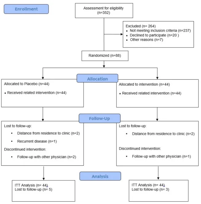
As demonstrated in the study flow dia- gram (Figure 1), we recruited 352 partici- pants. However, 264 subjects were excluded
from the study because of not meeting the in- clusion criteria. Of the 88 women enrolled in the study, 44 participants were randomized to the synbiotic group, and 44 were randomized to the placebo group. Eight participants (10 %) withdrew from the study following randomization: four patients because of dis- tance from residence to the clinic, one patient because of recurrent disease and three patients because of follow-up with other physicians.
Finally, 41 and 39 participants in the synbiotic and placebo groups completed the trial, re- spectively.
No side effects were reported following the supplementation of synbiotic in breast cancer survivors with lymphedema through- out the study.
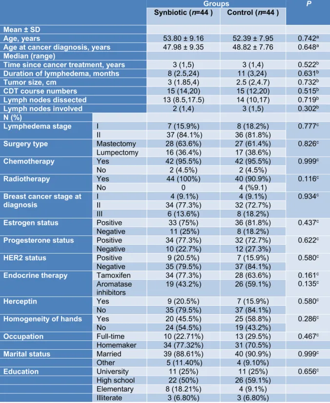
Baseline demographic and clinical char- acteristics of lymphedema patients have been presented in Table 1. The baseline mean (±
SD) age of participants was 53.80 (± 9.16) years in the synbiotic group and 52 (± 7.95) in the placebo group. There was no significant difference between the two groups regarding demographic and clinical characteristics.
Dietary intake
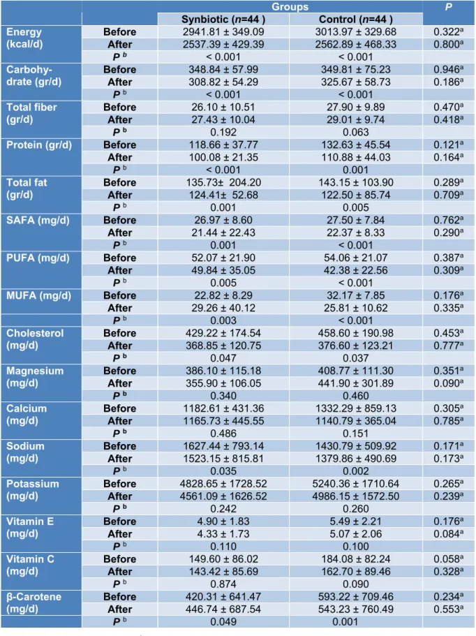
According to the 3-day food records ob- tained within each group before and after the intervention, there was no statistically signif- icant difference in terms of energy, dietary macro- and micro-nutrient intakes between the study groups in baseline and after 10 weeks of intervention (Table 2).
However, both synbiotic and placebo groups showed a significant within-group re- duction in daily energy intake (P < 0.001, P <
0.001), carbohydrate (P < 0.001, P < 0.001), protein (P < 0.001, P =0.001), total fat (P = 0.001, P = 0.005), SFA (P = 0.001, P < 0.001), PUFA (P < 0.005, P < 0.001), MUFA (P = 0.003, P < 0.001), cholesterol (P=0.047, P=0.037), sodium (P=0.035, P=0.002) and beta-carotene (P=0.049, P=0.001) respec- tively (Table 2).
Figure 1: Consort diagram
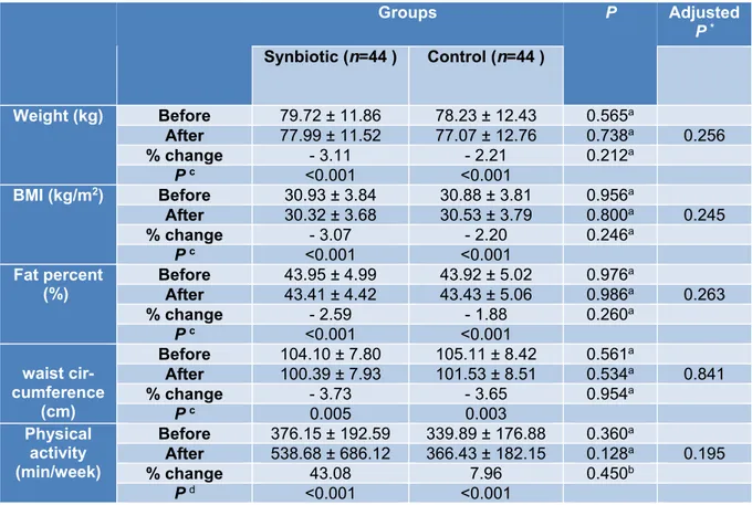
Anthropometry and physical activity
The mean values of weight (kg), BMI (kg/m2), body fat percent ( %), waist circum- ference (cm) and physical activity (min/week) at baseline and after 10 weeks in synbiotic and control groups are depicted in Table 3. The statistic tests showed no significant difference between the study groups after the interven- tion. Within groups changes showed signifi- cant statistical differences (P< 0.005) in syn- biotic and control groups regarding mean value of weight (-2.48 ± 3.03 kg, -1.73 ± 2.26
kg), BMI (-0.95 ± 1.14 kg/m2, -0.68 ± 0.90 kg/m2), body fat percent (-1.14 % ± 1.37, -0.83 % ± 1.09) and waist circumference changes (-3.89 ± 3.26 cm, -3.84 ± 3.60 cm), respectively.
The physical activity level significantly increased within each group [17.0 (3.25, 30.50) min/week and 21.0 (7.0, 33.0) min/week in synbiotic and control groups re- spectively, P< 0.001]. The intention-to-treat (ITT) analysis did not show different results between the study groups.
Table 1: Clinical and demographic characteristics of participants at baseline
Groups P
Synbiotic (n=44 ) Control (n=44 ) Mean ± SD
Age, years 53.80 ± 9.16 52.39 ± 7.95 0.742a
Age at cancer diagnosis, years 47.98 ± 9.35 48.82 ± 7.76 0.648a Median (range)
Time since cancer treatment, years 3 (1,5) 3 (1,4) 0.522b Duration of lymphedema, months 8 (2.5,24) 11 (3,24) 0.631b
Tumor size, cm 3 (1.85,4) 2.5 (2,4.7) 0.732b
CDT course numbers 15 (14,20) 15 (12,20) 0.515b
Lymph nodes dissected 13 (8.5,17.5) 14 (10,17) 0.719b
Lymph nodes involved 2 (1,4) 3 (1,5) 0.302b
N (%)
Lymphedema stage I 7 (15.9%) 8 (18.2%) 0.777c
II 37 (84.1%) 36 (81.8%)
Surgery type Mastectomy 28 (63.6%) 27 (61.4%) 0.826c Lumpectomy 16 (36.4%) 17 (38.6%)
Chemotherapy Yes 42 (95.5%) 42 (95.5%) 0.999c
No 2 (4.5%) 2 (4.5%)
Radiotherapy Yes 44 (100%) 40 (90.9%) 0.116c
No 0 4 (%9.1)
Breast cancer stage at
diagnosis I 4 (9.1%) 4 (9.1%) 0.934c
II 34 (77.3%) 32 (72.7%)
III 6 (13.6%) 8 (18.2%)
Estrogen status Positive 33 (75%) 36 (81.8%) 0.437c
Negative 11 (25%) 8 (18.2%)
Progesterone status Positive 34 (77.3%) 32 (72.7%) 0.622c
Negative 10 (22.7%) 12 (27.3%)
HER2 status Positive 9 (20.5%) 7 (15.9%) 0.580c
Negative 35 (79.5%) 37 (84.1%)
Endocrine therapy Tamoxifen 34 (77.3%) 28 (63.6%) 0.161c 0.135c Aromatase
inhibitors 19 (43.2%) 26 (59.1%)
Herceptin Yes 9 (20.5%) 7 (15.9%) 0.580c
No 35 (79.5%) 37 (84.1%)
Homogeneity of hands Yes 20 (45.5%) 25 (58.8%) 0.286c
No 24 (54.5%) 19 (43.2%)
Occupation Full-time 10 (22.71%) 13 (29.5%) 0.467c
Homemaker 34 (77.32%) 31 (70.5%)
Marital status Married 39 (88.61%) 40 (90.9%) 0.999c
Other 5 (11.40%) 4 (9.10%)
Education University 11 (25%) 11 (25%) 0.656c
High school 22 (50%) 26 (59.1%)
Elementary 8 (18.21%) 4 (9.1%)
Illiterate 3 (6.80%) 3 (6.80%)
a Independent-Samples t-test. b Mann–Whitney U test. c chi-square or Fisher exact test.
Parametric quantitative data reported as mean ± SD, non-parametric quantitative data reported as me- dian (Q1, Q3), qualitative data reported as number (%).
CDT, Complete decongestive therapy; TAC, Taxotere, Adriamycin and Cyclophosphamide; Tax, Taxol;
AC, Adriamycin and Cyclophosphamide; HER2, human epidermal growth factor receptor 2.
Table 2: Dietary intakes of participants throughout the study
Groups P
Synbiotic (n=44 ) Control (n=44 ) Energy
(kcal/d) Before 2941.81 ± 349.09 3013.97 ± 329.68 0.322a After 2537.39 ± 429.39 2562.89 ± 468.33 0.800a
P b < 0.001 < 0.001
Carbohy-
drate (gr/d) Before 348.84 ± 57.99 349.81 ± 75.23 0.946a
After 308.82 ± 54.29 325.67 ± 58.73 0.186a
P b < 0.001 < 0.001
Total fiber
(gr/d) Before 26.10 ± 10.51 27.90 ± 9.89 0.470a
After 27.43 ± 10.04 29.01 ± 9.74 0.418a
P b 0.192 0.063
Protein (gr/d) Before 118.66 ± 37.77 132.63 ± 45.54 0.121a
After 100.08 ± 21.35 110.88 ± 44.03 0.164a
P b < 0.001 0.001
Total fat
(gr/d) Before 135.73± 204.20 143.15 ± 103.90 0.289a
After 124.41± 52.68 122.50 ± 85.74 0.709a
P b 0.001 0.005
SAFA (mg/d) Before 26.97 ± 8.60 27.50 ± 7.84 0.762a
After 21.44 ± 22.43 22.37 ± 8.33 0.290a
P b 0.001 < 0.001
PUFA (mg/d) Before 52.07 ± 21.90 54.06 ± 21.07 0.387a
After 49.84 ± 35.05 42.38 ± 22.56 0.309a
P b 0.005 < 0.001
MUFA (mg/d) Before 22.82 ± 8.29 32.17 ± 7.85 0.176a
After 29.26 ± 40.12 25.81 ± 10.62 0.335a
P b 0.003 < 0.001
Cholesterol
(mg/d) Before 429.22 ± 174.54 458.60 ± 190.98 0.453a
After 368.85 ± 120.75 376.60 ± 123.21 0.777a
P b 0.047 0.037
Magnesium
(mg/d) Before 386.10 ± 115.18 408.77 ± 111.30 0.351a
After 355.90 ± 106.05 441.90 ± 301.89 0.090a
P b 0.340 0.460
Calcium
(mg/d) Before 1182.61 ± 431.36 1332.29 ± 859.13 0.305a After 1165.73 ± 445.55 1140.79 ± 365.04 0.785a
P b 0.486 0.151
Sodium
(mg/d) Before 1627.44 ± 793.14 1430.79 ± 509.92 0.171a After 1523.15 ± 815.81 1379.86 ± 490.69 0.173a
P b 0.035 0.002
Potassium
(mg/d) Before 4828.65 ± 1728.52 5240.36 ± 1710.64 0.265a After 4561.09 ± 1626.52 4986.15 ± 1572.50 0.239a
P b 0.242 0.260
Vitamin E
(mg/d) Before 4.90 ± 1.83 5.49 ± 2.21 0.176a
After 4.33 ± 1.73 5.07 ± 2.06 0.084a
P b 0.110 0.100
Vitamin C
(mg/d) Before 149.60 ± 86.02 184.08 ± 82.24 0.058a
After 143.42 ± 85.69 162.70 ± 89.46 0.328a
P b 0.874 0.090
β-Carotene
(mg/d) Before 420.31 ± 641.47 593.22 ± 709.46 0.234a
After 446.74 ± 687.54 543.23 ± 760.49 0.553a
P b 0.049 0.001
a Independent-Samples t-test. b Paired sample t-test.
Parametric quantitative data reported as mean ± SD
SAFA, saturated fatty acids; PUFA, Polyunsaturated fatty acids; MUFA, Monounsaturated fatty acids.
Table 3: Anthropometrics and physical activity characteristics in two groups before and after intervention
Groups P Adjusted
P * Synbiotic (n=44 ) Control (n=44 )
Weight (kg) Before 79.72 ± 11.86 78.23 ± 12.43 0.565a
After 77.99 ± 11.52 77.07 ± 12.76 0.738a 0.256
% change - 3.11 - 2.21 0.212a
P c <0.001 <0.001
BMI (kg/m2) Before 30.93 ± 3.84 30.88 ± 3.81 0.956a
After 30.32 ± 3.68 30.53 ± 3.79 0.800a 0.245
% change - 3.07 - 2.20 0.246a
P c <0.001 <0.001
Fat percent (%)
Before 43.95 ± 4.99 43.92 ± 5.02 0.976a
After 43.41 ± 4.42 43.43 ± 5.06 0.986a 0.263
% change - 2.59 - 1.88 0.260a
P c <0.001 <0.001
waist cir- cumference
(cm)
Before 104.10 ± 7.80 105.11 ± 8.42 0.561a
After 100.39 ± 7.93 101.53 ± 8.51 0.534a 0.841
% change - 3.73 - 3.65 0.954a
P c 0.005 0.003
Physical activity (min/week)
Before 376.15 ± 192.59 339.89 ± 176.88 0.360a
After 538.68 ± 686.12 366.43 ± 182.15 0.128a 0.195
% change 43.08 7.96 0.450b
P d <0.001 <0.001
a Independent-Samples t-test. b Mann–Whitney U test. c Paired sample t-test. d Wilcoxon signed-rank test. *ANCOVA test adjusted for baseline measures of each variable and baseline BMI.
Parametric quantitative data reported as mean ± SD BMI,Body Mass Index
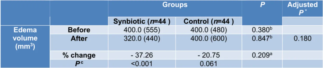
Edema volume measurement
As shown in Table 4, there was a signifi- cant reduction in edema volume in the synbi- otic group after the 10 week intervention (-37.26 percent, P< 0.001). However, accord- ing to the ANCOVA tests adjusted for base- line edema volume and pre-intervention BMI, there were no significant differences between the study groups after the intervention (P=0.180).
Inflammatory markers
Baseline measures of inflammatory mark- ers were not significantly different between the study groups, except hs-CRP [median (IQR): 3.20 (3.75), 1.85 (1.88) in synbiotic and placebo group respectively, P= 0.022]. In the synbiotic group after 10 weeks of inter- vention, hs-CRP (-3.12 percent, P=0.032), IL- 1β (-8.37 percent, P=0.018), and leptin
(-25.58 percent, P=0.026) decreased signifi- cantly (Table 4). Initial analysis showed no significant change in serum TNF-α levels fol- lowing supplementation with synbiotic (Ta- ble5) but ITT analysis revealed lower TNF-α values in the synbiotic group following inter- vention (P=0.039). Data in Table 5 demon- strates that 10 weeks of synbiotic supplemen- tation significantly reduced TNF-α andleptin between the study groups (Adjusted P= 0.039;
P= 0.003, respectively).
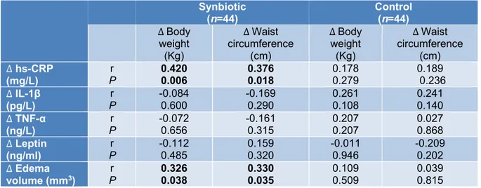
The correlation of changes in serum hs- CRP with body weight (r=0.420, P=0.006) and waist circumference (r=0.376, P=0.018) alterations in the synbiotic group was statisti- cally significant. Also, edema volume changes significantly correlated with body weight (r=0.326, P=0.038) and waist circum- ference (r= 0.330, P=0.035) in the synbiotic group (Table 6).
Table 4: Participants edema volume characteristics in two groups before and after intervention
Groups P Adjusted P * Synbiotic (n=44 ) Control (n=44 )
Edema volume
(mm3)
Before 400.0 (555) 400.0 (480) 0.380b
After 320.0 (440) 400.0 (600) 0.847b 0.180
% change - 37.26 - 20.75 0.209a
P c <0.001 0.061
a Independent-Samples t-test. b Mann–Whitney U test. c Wilcoxon signed-rank test. *ANCOVA test ad- justed for baseline edema volume and baseline BMI.
Edema volume reported as median (IQR).
Table 5: Inflammatory markers in two groups before and after intervention
Groups P Adjusted
P * Synbiotic (n=44 ) Control (n=44 )
hs-CRP
(mg/L) Before 3.20 (3.75) 1.85 (1.88) 0.022b 0.550
After 3.10 (2.65) 1.80 (2.3) 0.085b
% change - 3.12 - 2.70 0.021a
P c 0.032 0.526
IL-1β (pgL)
Before 382.0 (890.75) 339.0 (128) 0.140b 0.118 After 350.0 (816) 337.0 (285.5) 0.348b
% change - 8.37 - 5.01 0.528a
P c 0.018 0.030
TNF-α (ng/L)
Before 36.0 (19.5) 38.0 (107.75) 0.187b 0.039
After 34.0 (18.5) 40.0 (120) 0.058b
% change - 5.55 5.26 0.097a
P c 0.124 0.377
Leptin (ng/ml)
Before 86.0 (32) 105.0 (202.5) 0.411b 0.003
After 64.0 (32.5) 73.0 (218) 0.004b
% change - 25.58 -20.47 0.071a
P c 0.026 0.523
a Independent-Samples t-test. b Mann–Whitney U test. c Wilcoxon signed-rank test. * ANCOVA test ad- justed for baseline inflammatory markers and baseline BMI.
Non-parametric quantitative data reported as median (IQR).
hs-CRP, high-sensitivity C-reactive protein; IL-1, Interleukin-1; TNF-α, Tumor Necrosis Factor-α.
DISCUSSION
This is the first interventional trial that in- vestigates the possible beneficial role of syn- biotic supplementation on inflammatory markers (TNF-α, IL-1β, hs-CRP), serum lep- tin concentration, anthropometric parameters, and edema volume in BCRL patients.
Our results revealed that synbiotic supple- mentation for 10 weeks reduced edema vol- ume, the serum concentration of hs-CRP, IL- 1β and leptin in overweight and obese patients with BCRL. Furthermore, the synbiotics group had significantly lower levels of serum leptin and TNF-α concentration compared to placebo after the intervention.
Table 6: The correlation of inflammatory marker and edema volume with body weight and waist circum- ference in synbiotic and control groups
Synbiotic
(n=44) Control
(n=44)
∆ Body weight (Kg)
∆Waist circumference
(cm)
∆Body weight (Kg)
∆ Waist circumference
(cm)
∆ hs-CRP (mg/L)
r
P 0.420
0.006 0.376
0.018 0.178
0.279 0.189
0.236
∆ IL-1β
(pg/L) r
P -0.084
0.600 -0.169
0.290 0.261
0.108 0.241
0.140
∆ TNF-α (ng/L)
r P
-0.072 0.656
-0.161 0.315
0.207 0.207
0.027 0.868
∆ Leptin (ng/ml)
r
P -0.112
0.485 0.159
0.320 -0.011
0.946 -0.209
0.202
∆ Edema volume (mm3)
r
P 0.326
0.038 0.330
0.035
0.109 0.509
0.039 0.815 Significant correlations (P < 0.05) are shown in bold text.
Data from the two groups were pooled before analysis.
∆ Indicates change from baseline.
hs-CRP, high-sensitivity C-reactive protein; IL-1β, Interleukin-1; TNF-α, Tumor Necrosis Factor-α.
A significant improvement was seen in anthropometric indices including body weight, BMI, fat percent and waist circumfer- ence within groups at the end of the interven- tion which is thepredictable consequence of the low-calorie diet prescribed to all patients.
The anthropometric changes were not signifi- cant between the study groups because the simultaneous occurrence of cancer, lymphedema and obesity increased the pa- tient’s inflammation (Kolb et al., 2016, Zahid et al., 2016) to the level that we couldnot ob- serve any significant effects for synbiotics in these cases. Also, in a meta-analysis study was shown that probiotic supplementation (3 to 12 weeks) has a small size effect on the re- duction of anthropometric factors (Borgeraas et al., 2018).
Reduction in excess edema volume after synbiotic supplementation is a new finding that has not been reported elsewhere. The per- centage of edema reduction was 37.36 % and 20.75 % in the synbiotic and placebo groups, respectively. The lack of significant differ- ences between the study groups after the in- tervention could indicate the positive effects of the weight-loss diet on this marker. In the study by Shaw et al. 12 weeks prescription of
a weight-loss diet, led to a significant reduc- tion in weight, BMI, and edema volume in BCRL patients (Shaw et al., 2007a) Probiot- ics with their weight-lowering characteristics can promote this positive effect. Obesity is one of the well-known factors in the onset and progression of lymphedema and obese indi- viduals have a decreased lymphatic transport capacity (Yoon et al., 2018; Savetsky et al., 2014).
In different studies, the prognostic effect of high BMI in lymphedema incidence (Yoon et al., 2018; Iyigun et al., 2018; Helyer et al., 2010) and the predictive effect of weight loss accompanied by complete decongestive ther- apy in edema reduction (Shaw et al., 2007a, b; Duyur Cakit et al., 2019) have been con- firmed.
Alleviated hs-CRP serum levels in the synbiotic group was another finding of this study. This beneficial effect was also seen in another study performed on diabetic hemodi- alysis patients. After 12 weeks of interven- tion, serum hs-CRP was significantly lower in the synbiotics group (Soleimani et al., 2017).
Obesity by activation of the c-Jun N-terminal kinase, nuclear factor-kappa B, and protein kinase R pathways induce the inflammatory
response (Solinas et al., 2010; Nakamura et al., 2010). Lack of significant differences in hs-CRP serum levels between the study groups after the intervention may be because of the synergistic effects of cancer/chemo- therapy and obesity on inflammation in our study (Kolb et al., 2016; Zahid et al., 2016).
The results of the current study showed a significant reduction in serum IL-1β in both groups after the intervention (-8.37 %; control
= -5.01 %), but the non-significant differ- ences of this variable between groups are probably due to the beneficial effects of the weight loss program on this cytokine, owing to the fact that a decrease in body fat during weight loss reduces the production of IL-1β (Kirchner et al., 2014). Shadnoush et al. re- ported that 8 weeks of consumption of bifidobacterium and lactobacillus in the form of probiotic yogurt, decreased serum IL-1β concentration in inflammatory bowel disease patients compared to control (Shadnoush et al., 2013). No weight loss program was per- formed in their study that could explain why the differences in serum IL-1β were signifi- cant after probiotic intake.
Obesity leads to impairment and imbal- ance in gut microbiota called dysbiosis which weakens intestinal integrity accompanied by lipopolysaccharide (LPS)-related endotoxe- mia. LPS is a large molecule that exists in the cell wall of gram-negative bacteria. LPS stim- ulates the activation of the transcription nu- clear factor-kappa B (NF-κB) by binding to toll-like receptor 4 (TLR4) in intestinal epi- thelial cells. NF-κB upregulates gene expres- sion of pro-inflammatory cytokines such as TNF-α, IL-1β, and IL-6 which mediate a rise in hs-CRP, inflammation, and consequently edema (Neyrinck et al., 2016; Torres et al., 2018; Dinarello, 2009). Also, producing short-chain fatty acids by probiotics prevents the synthesis of hepatic CRP that results in a reduction in its serum levels (Badehnoosh et al., 2018).
We demonstrated that synbiotic supple- mentation for 10 weeks in BCRL patients led to a significant reduction in serum leptin lev- els compared to baseline and control group.
This effect was also seen in studies on over- weight and obese individuals (Sanchez et al., 2014; Zarrati et al., 2014), but it was not in line with the study by Mobini et al. Supple- mentation with Lactobacillus reuteri in dia- betic patients for 12 weeks, could not change leptin (Mobini et al., 2017). Our supplements contain diverse strains of probiotics compared to that study. Each strain of probiotics may exert a special function (Pandey et al., 2015), which may be responsible for the incon- sistency of the results. Several mechanisms can explain the beneficial effects of probiotics serum leptin levels. Probiotics have a critical role in leptin secretion regulation (Behrouz et al., 2017). Leptin increases the production of TNF-α in monocytes (Scarpellini and Tack, 2012; Zhou et al., 2011). Any factor that re- duces the leptin level can also reduce the lev- els of TNF-α. Therefore, in our study, a de- crease in the leptin serum level consequently reduced the TNF-α serum level in the synbi- otic group compared with the placebo. De- spite a significant decrease in TNF-α serum level, because of multiple outcomes and the possibility of significant correlation due to multiple testing, this factor should be inter- preted with caution. An adjusted significant level can be confirmed in the next and larger studies.
In spite of the potential strengths of our study, such as using a probiotic intervention for the first time among BCRL patients, this study has some limitations. Although the syn- biotics supplementation may affect fecal bac- terial load, we could not assess this correla- tion because of budget limitations. Also, we did not consider a control group receiving placebo capsules without any low-calorie diet. So, we can not conclude if the weight reduction program had a synergistic effect along with the synbiotic cap- sules or not. Evaluation of the gene expression of inflammatory cytokines in future re- searches may provide valuable evidence in this field.
CONCLUSION
In conclusion, 10 weeks of synbiotic sup- plementation along with an LCD program among BCRL patients resulted in a significant reduction in serum levels of TNF-α and leptin compared with the control group. Also, edema volume was significantly reduced within the synbiotic group after intervention.
However, no significant differences were found in either of the anthropometric varia- bles, IL-1β, and hs-CRP between the study groups. Weight reduction and synbiotic prod- uct consumption in the daily diet of lymphedema patients may be suggested as ef- fective and safe interventions in lymphedema management.
Acknowledgment
We would like to gratefully thank the par- ticipants for their support in the study. We are also thankful to Namdar laboratory and Zist Takhmir Company and all patients that partic- ipated in this study.
Funding
This study was supported by Iran Univer- sity of Medical Sciences (IUMS) with a 96- 02-27-31433 grant number.
Conflict of interest
The authors declare that they have no con- flict of interest.
REFERENCES
Alard J, Lehrter V, Rhimi M, Mangin I, Peucelle V, Abraham AL, et al. Beneficial metabolic effects of se- lected probiotics on diet‐induced obesity and insulin resistance in mice are associated with improvement of dysbiotic gut microbiota. Environ Microbiol. 2016;18:
1484-97.
Amdekar S, Roy P, Singh V, Kumar A, Singh R, Sharma P. Anti-inflammatory activity of lactobacil-lus on carrageenan-induced paw edema in male wistar rats.
Int J Inflam. 2012;2012:752015.
American Cancer Society. Cancer facts & figures:
American Cancer Society; 2018. Available from:
https://www.cancer.org/content/dam/cancer-org/re- search/cancer-facts-and-statistics/annual-cancer-facts- and-figures/2018/cancer-facts-and-figures-2018.pdf.
Archer AC, Muthukumar SP, Halami PM. Anti-inflam- matory potential of probiotic Lactobacillus spp. on car- rageenan induced paw edema in Wistar rats. Int J Biol Macromol. 2015; 81:530-7.
Badehnoosh B, Karamali M, Zarrati M, Jamilian M, Bahmani F, Tajabadi-Ebrahimi M, et al. The effects of probiotic supplementation on biomarkers of in-flam- mation, oxidative stress and pregnancy out-comes in gestational diabetes. J Matern Fetal Neonatal Med.
2018; 31:1128-36.
Behrouz V, Jazayeri S, Aryaeian N, Zahedi MJ, Hos- seini F. Effects of probiotic and prebiotic supplemen- tation on leptin, adiponectin, and glycemic parame-ters in non-alcoholic fatty liver disease: a randomized clin- ical trial. Middle East J Dig Dis. 2017;9:150-7.
Borgeraas H, Johnson LK, Skattebu J, Hertel JK, Hjelmesaeth J. Effects of probiotics on body weight, body mass index, fat mass and fat percentage in sub- jects with overweight or obesity: a systematic review and meta-analysis of randomized controlled trials.
Obesity Rev. 2018;19:219-32.
Brown K, DeCoffe D, Molcan E, Gibson DL. Diet-in- duced dysbiosis of the intestinal microbiota and the ef- fects on immunity and disease. Nutrients. 2012;4:
1095-119.
Bulló M, García‐Lorda P, Megias I, Salas‐Salvadó J.
Systemic inflammation, adipose tissue tumor necrosis factor, and leptin expression. Obesity Res. 2003;11:
525-31.
Chen Y, Rehal S, Roizes S, Zhu H-L, Cole WC, von der Weid P-Y. The pro-inflammatory cytokine TNF-α inhibits lymphatic pumping via activation of the NF- κB-iNOS signaling pathway. Microcirculation. 2017;
24(3):10.1111/micc.12364.
Coughlin SS, Ekwueme DU. Breast cancer as a global health concern. Cancer Epidemiol. 2009;33:315-8.
Dayan JH, Ly CL, Kataru RP, Mehrara BJ. Lymph- edema: pathogenesis and novel therapies. Annu Rev Med. 2018;69:263-76.
Dinarello CA. Immunological and inflammatory func- tions of the interleukin-1 family. Annual review of im- munology. 2009;27:519-50.
DiSipio T, Rye S, Newman B, Hayes S. Incidence of unilateral arm lymphoedema after breast cancer: a sys- tematic review and meta-analysis. Lancet Oncol.
2013;14:500-15.
Duyur Cakit B, Pervane Vural S, Ayhan FF. Complex decongestive therapy in breast cancer-related lymph- edema: does obesity affect the outcome negatively?
Lymphat Res Biol. 2019;17:45-50.
Erickson VS, Pearson ML, Ganz PA, Adams J, Kahn KL. Arm edema in breast cancer patients. J Natl Can- cer Inst. 2001;93:96-111.
Gousopoulos E, Proulx ST, Scholl J, Uecker M, Detmar M. Prominent lymphatic vessel hyperplasia with progressive dysfunction and distinct immune cell infiltration in lymphedema. Am J Pathol. 2016;186:
2193-203.
Helyer LK, Varnic M, Le LW, Leong W, McCready D.
Obesity is a risk factor for developing postoperative lymphedema in breast cancer patients. The Breast J.
2010;16(1):48-54.
Hespe GE, Nores GG, Huang J-J, Mehrara BJ. Patho- physiology of lymphedema - Is there a chance for med- ication treatment? J Surg Oncol. 2017;115:96-8.
Hormes J, Bryan C, Lytle L, Gross C, Ahmed R, Troxel A, et al. Impact of lymphedema and arm symptoms on quality of life in breast cancer survi-vors. Lymphology.
2010;43:1-13.
Iyigun ZE, Duymaz T, Ilgun AS, Alco G, Ordu C, Sarsenov D, et al. Preoperative lymphedema-related risk factors in early-stage breast cancer. Lymphat Res Biol. 2018;16:28-35.
Kirchner H, Nylen C, Laber S, Barres R, Yan J, Krook A, et al. Altered promoter methylation of PDK4, IL1 B, IL6, and TNF after Roux-en Y gastric bypass. Surg Obes Relat Dis. 2014;10:671-8.
Kolb R, Sutterwala FS, Zhang W. Obesity and cancer:
inflammation bridges the two. Curr Opin Pharmacol.
2016;29:77-89.
Le Chatelier E, Nielsen T, Qin J, Prifti E, Hildebrand F, Falony G, et al. Richness of human gut microbiome correlates with metabolic markers. Nature. 2013;500:
541.
Ly CL, Kataru RP, Mehrara BJ. Inflammatory mani- festations of lymphedema. Int J Mol Sci. 2017;18(1):
171.
McLoughlin RF, Berthon BS, Jensen ME, Baines KJ, Wood LG. Short-chain fatty acids, prebiotics, synbiot- ics, and systemic inflammation: a systematic review and meta-analysis. Am J Clin Nutr. 2017;106:930-45.
Mego M, Holec V, Drgona L, Hainova K, Ciernikova S, Zajac V. Probiotic bacteria in cancer patients under- going chemotherapy and radiation therapy. Comple- ment Ther Med. 2013;21:712-23.
Mehrara BJ, Greene AK. Lymphedema and obesity: is there a link? Plastic Reconstruct Surg. 2014;134(1):
154e.
Minocci A, Savia G, Lucantoni R, Berselli M, Ta- gliaferri M, Calo G, et al. Leptin plasma concentra- tions are dependent on body fat distribution in obese patients. Int J Obesity. 2000;24:1139.
Mobini R, Tremaroli V, Ståhlman M, Karlsson F, Lev- in M, Ljungberg M, et al. Metabolic effects of L acto- bacillus reuteri DSM 17938 in people with type 2 dia- betes: A randomized controlled trial. Diabetes Obes Metab. 2017;19:579-89.
Nakamura T, Furuhashi M, Li P, Cao H, Tuncman G, Sonenberg N, et al. Double-stranded RNA-dependent protein kinase links pathogen sensing with stress and metabolic homeostasis. Cell. 2010;140:338-48.
Neyrinck AM, Schüppel VL, Lockett T, Haller D, Delzenne NM. Microbiome and metabolic disorders related to obesity: Which lessons to learn from exper- imental models? Trends Food Sci Technol. 2016;57:
256-64.
Norman SA, Localio AR, Potashnik SL, Simoes Torpey HA, Kallan MJ, Weber AL, et al. Lymphede- ma in breast cancer survivors: incidence, degree, time course, treatment, and symptoms. J Clin Oncol. 2009;
27:390-7.
Pandey KR, Naik SR, Vakil BV. Probiotics, prebiotics and synbiotics- a review. J Food Sci Technol. 2015;52:
7577-87.
Ridner SH, Dietrich MS, Stewart BR, Armer JM. Body mass index and breast cancer treatment-related lymph- edema. Support Care Cancer. 2011;19:853-7.
Rockson SG. The lymphatics and the inflammatory re- sponse: lessons learned from human lymphedema.
Lymphat Res Biol. 2013;11:117-20.
Rockson SG, Tian W, Jiang X, Kuznetsova T, Had-dad F, Zampell J, et al. Pilot studies demonstrate the poten- tial benefits of antiinflammatory therapy in human lymphedema. JCI insight. 2018;3(20):e123775.
Sanchez M, Darimont C, Drapeau V, Emady-Azar S, Lepage M, Rezzonico E, et al. Effect of Lactobacillus rhamnosus CGMCC1. 3724 supplementation on weight loss and maintenance in obese men and wom- en. Brit J Nutr. 2014;111:1507-19.
Savetsky IL, Torrisi JS, Cuzzone DA, Ghanta S, Al- bano NJ, Gardenier JC, et al. Obesity increases in- flammation and impairs lymphatic function in a mouse model of lymphedema. Am J Physiol Heart Circ Phys- iol. 2014;307:H165-72.
Scarpellini E, Tack J. Obesity and metabolic syn- drome: an inflammatory condition. Dig Dis. 2012;30:
148-53.
Schaverien MV, Aldrich MB. New and emerging treat- ments for lymphedema. Semin Plastic Surg. 2018;32:
048-52.
Schwager S, Detmar M. Inflammation and lymphatic function. Front Immunol. 2019;10:308.
Shadnoush M, Shaker Hosseini R, Mehrabi Y, Delpisheh A, Alipoor E, Faghfoori Z, et al. Probiotic yogurt affects pro- and anti-inflammatory factors in pa- tients with inflammatory bowel disease. Iran J Pharm Res. 2013;12:929-36.
Shaw C, Mortimer P, Judd PA. A randomized con- trolled trial of weight reduction as a treatment for breast cancer‐related lymphedema. Cancer. 2007a;110:
1868-74.
Shaw C, Mortimer P, Judd PA. Randomized con- trolled trial comparing a low‐fat diet with a weight‐re- duction diet in breast cancer‐related lymphedema. Can- cer: 2007b;109:1949-56.
Solanki HK, Shah DA, Maheriya PM, Patel CA. Eval- uation of anti-inflammatory activity of probiotic on carrageenan-induced paw edema in Wistar rats. Int J Biol Macromol. 2015;72:1277-82.
Soleimani A, Zarrati Mojarrad M, Bahmani F, Taghi- zadeh M, Ramezani M, Tajabadi-Ebrahimi M, et al.
Probiotic supplementation in diabetic hemodialysis pa- tients has beneficial metabolic effects. Kidney Int.
2017;91:435-42.
Solinas G, Karin M. JNK1 and IKKbeta: molecular links between obesity and metabolic dysfunction.
FASEBJ. 2010;24:2596-611.
Tashiro K, Feng J, Wu SH, Mashiko T, Kanayama K, Narushima M, et al. Pathological changes of adipose tissue in secondary lymphoedema. Brit J Dermatol.
2017;177:158-67.
Torres S, Fabersani E, Marquez A, Gauffin-Cano P.
Adipose tissue inflammation and metabolic syn- drome. The proactive role of probiotics. Eur J Nutr.
2018;58: 27-43.
Van Dielen F, Van't Veer C, Schols A, Soeters P, Buur- man W, Greve J. Increased leptin concentrations corre- late with increased concentrations of inflamma-tory markers in morbidly obese individuals. Int J Obe-sity.
2001;25:1759.
Vasheghani-Farahani A, Tahmasbi M, Asheri H, Ash- raf H, Nedjat S, Kordi R. The Persian, last 7-day, long form of the International Physical Activity Question- naire: translation and validation study. Asian J Sports Med. 2011;2:106-16.
Verdam FJ, Fuentes S, de Jonge C, Zoetendal EG, Er- bil R, Greve JW, et al. Human intestinal microbiota composition is associated with local and systemic in- flammation in obesity. Obesity. 2013;21:E607-E15.
Vignes S, Arrault M, Dupuy A. Factors associated with increased breast cancer-related lymphedema volume.
Acta Oncol. 2007;46:1138-42.
Wanchai A, Armer JM, Stewart BR, Lasinski BB.
Breast cancer-related lymphedema: A literature re- view for clinical practice. Int J Nurs Sci. 2016;3:202- 7.
Wischmeyer PE, McDonald D, Knight R. Role of the microbiome, probiotics, and ‘dysbiosis therapy’in crit- ical illness. Curr Opin Crit Care. 2016;22(4):347.
Yoon JA, Shin YB, Shin MJ, Yun RY, Kim KY, Song YS, et al. An assessment of the relationship between abdominal obesity and the severity of upper extremi-ty lymphedema. Lymphat Res Biol. 2018;16:458-63.
Zahid H, Simpson ER, Brown KA. Inflammation, dysregulated metabolism and aromatase in obesity and breast cancer. Curr Opin Pharmacol. 2016;31:90-6.
Zarrati M, Salehi E, Nourijelyani K, Mofid V, Zadeh MJ, Najafi F, et al. Effects of probiotic yogurt on fat distribution and gene expression of proinflammatory factors in peripheral blood mononuclear cells in over- weight and obese people with or without weight-loss diet. J Am Coll Nutr. 2014;33:417-25.
Zhou W, Guo S, Gonzalez-Perez R. Leptin pro-angio- genic signature in breast cancer is linked to IL-1 sig- nalling. Brit J Cancer. 2011;104:128.