Characterization of differential Toll-like receptor function in human immune cells and association with susceptibility to recurrent HSV-1 reactivations
and gastric cancer
Dissertation
Zur Erlangung des akademischen Grades doctor rerum naturalium
(Dr. rer. nat) Im Fach Biologie eingericht an der
Mathematisch-Naturwissenschaftlichen Fakultät I der Humboldt– Universität zu Berlin
von
Chin-An Yang, MD
Präsident der Humboldt-Universität zu Berlin Prof. Dr. Dr. h.c. Christoph Markschies
Dekan der Fakultät Prof. Dr. Andreas Herrmann
Gutachter:
1. Prof. Dr. Hans-Dieter Volk 2. Prof. Dr. Günther Schönrich 3. Prof. Dr. Ralf Reiner Schumann
Datum der Promotion: 20. 01. 2011
Table of contents
Summary ...6
Zusammenfassung...7
Abbreviations...8
1 Introduction...9
1.1 Overview of Toll-like receptors...9
1.1.1 Toll-like receptors and viral infections...9
1.1.1.1 TLR 1, 2, 4, 6 ...9
1.1.1.2 TLR 3, 7, 8, 9 ...10
1.1.2 Toll-like receptors and cancer ...10
1.2 Clinical problems...10
1.2.1 Herpes simplex virus infections ...11
1.2.1.1 Clinical aspects ...11
1.2.1.2 Recognition of HSV-1 via TLRs...11
1.2.1.3 TLRs and susceptibility to recurrent HSV-1 diseases ...12
1.2.2 TLRs and susceptibility to gastric cancer ...13
1.3 Natural killer cell as an important player in viral infections and cancer ...13
1.3.1 Introduction of natural killer cells ...14
1.3.2 NK cells and herpes virus infections...14
1.3.3 Activation of NK cells via TLRs...14
1.3.4 NK cells and tumor immune surveillance...15
1.4 Evaluation of TLR function in different subsets of PBMCs...15
1.4.1 TLR expression on antigen-presenting cells...16
1.4.2 TLR expression on lymphocytes ...16
1.4.3 Previous assays for evaluating TLR functions...17
2 Aims and objectives...18
3 Materials and Methods...19
3.1 Subjects ...19
3.1.1 Subjects for the study of susceptibility to recurrent herpes labials ...19
3.1.2 Subjects for the study of susceptibility to gastric cancer...19
3.2 Reagents...20
3.2.1 TLR agonists ...20
3.2.2 Flow cytometry reagents ...21
3.2.2.1 Antibodies for detecting intracellular cytokine production in APCs..21
3.2.2.2 Antibodies for detecting intracellular cytokine production in NK cells and T cells...21
3.2.2.3 Antibodies for detecting levels of TLR expression...22
3.2.2.4 Buffers for FACS staining...22
3.2.3 Cytokines and mediums ...22
3.2.4 Cell isolation reagents ...23
3.2.5 HSV-1 viral lysate...23
3.2.6 Reagents for DNA extraction and PCR ...24
3.2.7 Reagents for RNA isolation and RT-PCR...24
3.3 Methods ...25
3.3.1 Cell isolation ...25
3.3.1.1 Isolation of peripheral blood mononuclear cells ...25
3.3.1.2 Isolation of NK cells...25
3.3.1.3 Isolation of monocytes and generation of monocyte-derived dendritic cells ...25
3.3.2 Toll-like receptor ligand stimulations...26
3.3.3 Flow cytometry analysis ...26
3.3.3.1 Staining procedures for extracellular cell markers and intracellular cytokines ...26
3.3.3.2 Gating strategies ...27
3.3.4 NK cell degranulation assay ...27
3.3.5 Total RNA isolation...28
3.3.6 cDNA synthesis ...28
3.3.7 Quantitative Real-Time PCR ...29
3.3.8 DNA extraction ...29
3.3.9 Melting curve analysis of TLR SNPs ...30
3.3.10 In vitro UV irradiation of PBMCs...30
3.3.11 HSV-1 infection of purified NK cells...31
3.3.12 HSV-1 viral lysate stimulation assay...31
3.3.13 Statistical analysis ...32
4 Results ...33
4.1 Establishment of a multi-color flow cytometric assay to evaluate TLR responses in different PBMC subsets ...33
4.1.1 Gating strategies ...33
4.1.2 Concentrations of the TLR ligands were optimized and TNF-α and IFN- γ production were selected as the read-out of the assay...34
4.1.3 IL-12 was added for IFN-γ profiling in NK cells and T cells ...35
4.1.4 NK cells and T cells could not be fully activated if BFA was added before TLR-ligand stimulation...36
4.1.5 Effect of cell cryopreservation ...39 4.2 TLR-ligand-induced cytokine production in different cell subsets detected
by the assay is in accordance with the literatures ...40
4.2.1 TLR-ligand-induced TNF-α response in monocytes, mDCs, pDCs and B cells...40
4.2.2 TLR-ligand-induced IFN-γ production in non-purified NK cells and T cells………..40
4.2.3 TLR responses in isolated NK cells ...42
4.3 Detection of TLR responses in different PBMC subsets of subjects with HL ... ………...46
4.3.1 The amounts of TLR ligand-induced cytokines detected in unfractionated APCs, NK cells and CD8+T cells were not different between HL subjects and asymptomatic controls...46
4.3.2 Poly(I:C)-triggered responses of purified NK cells are significantly lower in subjects with HL ...47
4.3.3 The percentage of NK cells is lower in people with recurrent HL ...49
4.3.4 The co-stimulatory function of accessory cells restored the impaired poly(I:C) response of isolated NK cells ...49
4.4 Mechanisms of poly(I:C)-hyporesponsiveness in NK cells...51
4.4.1 The amount of intracellular TLR3 expression did not correlate with the level of poly(I:C)-responsiveness in NK cells ...51
4.4.2 TLR3 412 F/F impairs TLR3 surface expression and IFN-γ response to poly(I:C) ...52
4.4.3 The poly(I:C)-induced IFN-γ response is highest in HSV-1-seronegative individuals ...54
4.4.4 Purified NK cells can be infected by HSV-1 when activated.. 55
4.4.5 The cytotoxic potential of NK cells in HL subjects is not impaired ...56
4.5 TLR-induced IFN-γ responses of CD56bright NK cells in HL subjects.. 58
4.5.1 Reduced IFN-γ response of CD56bright NK cells to R-848 in people with HL.. ………58
4.5.2 Co-stimulatory function of accessory cells restored the reduced IFN-γ response of CD56bright NK cells to R-848 ...60
4.6 IFN-γ response of NK cells to HSV-1...61
4.7 TLR1 602S/S genotype decreases IFN-γ response to Pam3Cys in NK- and CD8+T cells ...64
4.8 TLR1 I602S SNP and susceptibility to recurrent HL ...66
4.9 Association of TLR1 I602S and gastric cancer ...67
4.9.1 TLR1 602S/S genotype is associated with susceptibility to gastric cancer………...67
4.9.2 TLR1 602S/S genotype is not associated with susceptibility to H. pylori infection ...68
4.9.3 TLR1 I602S is associated with metastasis ...69
5 Discussions ...71
5.1 Establishment of a multi-color flow cytometry-based assay for the evaluation of TLR functions in different cell subsets of PBMC ...71
5.2 Direct TLR responses of NK cells ...72
5.3 TLR3- hyporesponsiveness of NK cells in subjects with recurrent HL ...73
5.3.1 NK cell hyporesponsiveness to poly(I:C) could be restored by co- stimulatory function of APCs ...73
5.3.2 Mechanisms of NK cell hyporesponsiveness to poly(I:C) in people with recurrent HL ...76
5.3.3 Possible influences of TLR3-hyporesponsiveness on the interplay of NK cells and T cells ...76
5.3.4 Highest NK cell TLR3 response in HSV-1-seronegative people...77
5.3.5 Other possible genetic susceptibilities to recurrent HL ...77
5.4 NK cells and HSV-1 ...78
5.5 TLR1 I602S SNP impairs IFN-γ secretion in NK cells and T cells...80
5.6 Association of TLR1 I602S and risk of gastric cancer...80
5.7 Association of TLR1 I602S and gastric cancer metastasis ...81
5.8 Conclusion ...82
6 References ...84
Acknowledgements...84
Publikationsliste ...97
Erklärung...98
Summary
Toll-like Receptors (TLRs) are essential innate receptors which recognize conserved structures of pathogens, or danger signals released from damaged cells. Alterations of TLR responses might result in severe viral infections or a higher risk of cancer.
Therefore, development of clinical assays to evaluate TLR functions could provide personalized information about susceptibility to these diseases. Since TLRs are differentially expressed on different subsets of human peripheral blood mononuclear cells (PBMCs), a multi-color flow cytometry-based assay was developed to detect TLR responses of individual cell types simultaneously. We observed that the magnitude of TLR responses largely varied between human subjects, but was highly reproducible over one month.
To evaluate the potential role of differences in natural killer (NK) cell TLR response we studied the association of NK cell TLR function and TLR single nucleotide polymorphisms (SNPs) with susceptibility to recurrent herpes labialis (HL) and gastric cancer. Using our assay, impaired TLR3 response of NK cells was found in people with recurrent HL. In addition, we have identified enhanced levels of homozygous TLR3 L412F SNP in people with recurrent HL, which results in lower surface expression and reduced NK cell response to poly(I:C).
TLR1 I602S, another common SNP, has been reported to decrease TNF- responses of monocytes toward TLR2/1 agonist, Pam3CSK4 (Pam3Cys), stimulation. In our study, we found that TLR1 I602S homozygosity also contributes to impaired IFN-γ responses of NK cells and CD8+T cells. Although we did not observe an association of TLR2/1 deficiency with recurrent HL, association of TLR1 I602S with risk for primary as well as metastatic gastric cancer was found in a cohort of 326 patients.
To sum up, our results suggest that genetic polymorphisms of TLRs can impair TLR function of NK cells, which contribute to the increased susceptibility to HSV-1 diseases and gastric cancer.
Keywords: Toll-like receptor; Natural killer cells; Herpes simplex virus; Gastric cancer;
Single nucleotide polymorphism
Zusammenfassung
Toll-like Rezeptoren (TLRs) sind essentielle angeborene Rezeptoren, die konservierte Strukturen von Krankheitserregern oder Gefahrsignale, die von beschädigten Zellen freigesetzt werden, erkennen können. Genetische Variationen in TLRs wie Einzel- Nukleotid-Polymorphismus (SNP) können die Funktion von TLRs beeinträchtigen und erste Studien zeigen, dass dies zu einer erhöhten Anfälligkeit gegenüber Virusinfektionen oder einem erhöhten Krebsrisiko führen kann. In dieser Studie haben wir einen Multicolor-Durchflußzytometrie-Test entwickelt, um die TLR-Funktionen in verschiedenen Subpopulationen unseparierter peripherer mononukleärer Blutzellen (PBMCs) simultan analysieren zu können. Wir konnten beobachten, dass das Ausmaß der TLR-Antworten zwischen den Probanden stark variierte, jedoch über einen Zeitraum von einem Monat gut reproduzierbar war.
Zunächst untersuchten wir TLR Reaktionen bei Patienten mit rezidivierenden Herpes labilalis (HL). Im Vergleich zu asymptomatischen Personen war eine HL- Anamnese mit einer signifikant verminderten TLR3-IFN-γ-Antwort nach Stimulation mit poly(I:C) in NK Zellen assoziiert. Weitere molekulare Untersuchungen zeigten eine mögliche Beteiligung von TLR3 L412F SNP, welcher die oberflächliche TLR3 Expression und die IFN-γ-Antworten in NK-Zellen reduzierte. Einige Studien zeigen, dassTLR1 I602S, ein weiterer sehr verbreiteter SNP, in der Lage ist die TNF--Antworten von Monozyten gegen den TLR2/1-Agonisten (Pam3Cys) zu verringern. In der hier vorliegenden Arbeit konnten wir zudem nachweisen, dass TLR1 I602S SNP auch die Funktion von NK- Zellen und CD8+ T-Zellen beeinträchtigt. Wir konnten keine Assoziation zwischen TLR2/1-Defizienz und reaktivierendem HL feststellen. Jedoch konnten wir an einer großen Kohorte von über 326 Patienten zeigen, dass der TLR1 SNP sowohl ein Risikofaktor für Magenkarzinomentstehung als auch für die Metastasierung ist.
Zusammenfassend weisen unsere Ergebnisse darauf hin, dass genetische Polymorphismen von TLRs die Funktion von NK-Zellen beeinträchtigen und zu einer erhöhten Anfälligkeit für HSV-1 Erkrankung und Magenkarzinom führen können.
Schlagwörter: Toll-like Rezeptoren; NK-Zellen; Herpes Simplex Virus; Magenkarzinom;
Einzel-Nukleotid-Polymorphismus
Abbreviations
TLR: Toll-like receptor HSV: Herpes simplex virus
HSE: Herpes simplex encephalitis HL: Herpes labialis
PBMC: Peripheral blood mononuclear cell NK: Natural killer cell
mDC: Myeloid dendritic cell pDC: Plasmacytoid dendritic cell APC: Antigen presenting cell
rhIL-12: Recombinant human interleukin-12 Pam3Cys: Pam3CSK4
BFA: Brefeldin-A
TNF-α: Tumor necrosis factor- α IFN-γ: Interferon- γ
MFI: Mean florescence intensity SNP: Single nucleotide polymorphism APOE: Apolipoprotein E
NF-κB: Nuclear factor- κB IKK: IκB kinase
1 Introduction
1.1 Overview of Toll-like receptors
Toll-like receptors (TLRs) are important innate immune receptors that recognize conserved features of microbes, or referred to as pathogen-associated molecular patterns (PAMPs). TLRs bind to PAMPs through solenoid ectodomains, which contain leucin-rich repeats; and exert inflammatory responses via pathways derived from the intracellular Toll-Interleukin-1 receptor (TIR) domain [1,2]. TLRs were named for their similarity to Toll, a receptor originally involved in embryonic development of the fruit fly Drosophila melanogaster discovered in 1985 by the German scientist Christiane Nüsslein-Volhard. In 1996, Jules A. Hoffmann and colleges further showed the importance of Toll in combating fungal infection in fruit flies [3]. Human TLRs were subsequently identified and shown to be involved in sensing specific structures of microbial origin and even endogenous danger signals (reviewed in [4]).
1.1.1 Toll-like receptors and viral infections
In humans, there are TLR1-10 in the TLR family. The activation of TLR during viral infection is essential for the induction of dendritic cell (DC) maturation, the production of interferons, and the initiation of the adaptive immune system [5].
1.1.1.1 TLR 1, 2, 4, 6
TLR 2 forms heterodimers with TLR1 or TLR6. Triacylated and diacylated lipoproteins, which are common gram-positive cell wall components, are ligands for TLR2/1 and TLR2/6. However, viral proteins of Herpes simplex virus -1, -2 (HSV-1, HSV-2), and Cytomegalovirus (CMV) have also been reported to be recognized by TLR2 [6,7].
Similarly, TLR4 is mainly activated by LPS, the component of the outer membrane of gram-negative bacteria, but it is also activated by the fusion protein of respiratory syncytial virus [8].
1.1.1.2 TLR 3, 7, 8, 9
TLR3, 7, 8 and 9 are specialized in the recognition of viral nucleic acids. These receptors reside in the endosome. TLR3 can be activated by double-stranded RNA (dsRNA) which is often produced during viral replication; TLR7 and 8 are stimulated by single-stranded RNA (ssRNA); and TLR9 is responsible for recognizing the nonmethylated CpG nucleotides in DNA viruses. For all TLR signaling except TLR3, the adaptor Myeloid differentiation primary response protein 88 (MyD88) is important for the activation of NF-kB pathways and thus the production of proinflammatory cytokines.
TIR-domain-containing adapter-inducing interferon-β (TRIF), on the other hand, is involved in TLR3- and TLR4- mediated MyD88-independent pathways, and is essential for the induction of both NF-kB and type I interferon signaling [9].
1.1.2 Toll-like receptors and cancer
TLRs play several roles in carcinogenesis. For example, engagement of TLR2/1 complex significantly increases the anti-tumor activity of CD8T cells [10]. TLRs can also recognize endogenous signals released from damaged or dying tumor cells, thus breaking down tumor-induced immune tolerance [11]. Therefore, hypofunction of TLRs may increase risks for cancer. Furthermore, polymorphism in a promoter sequence of TLR2 and a TLR4 variant allele have been reported to be involved in Helicobacter pylori (H. pylori)-associated gastric cancer [12,13]. Polymorphism in the TLR10-TLR1-TLR6 gene cluster, on the other hand, is associated with an increased risk for prostate cancer [14].
1.2 Clinical problems
An overview of the clinical importance of recurrent HSV infections and roles of TLRs in HSV diseases is described below. Since TLR functions can also contribute to cancer risks, roles of TLR in susceptibility to gastric cancer are addressed at the end of this section.
1.2.1 Herpes simplex virus infections
1.2.1.1 Clinical aspects
Herpes simplex viruses are common human pathogens, which include herpes simplex virus -1 (HSV-1) and herpes simplex virus-2 (HSV-2). Primary infection or reactivation of HSV can cause a broad spectrum of diseases, such as gingivostomatitis in childhood, cold sores (herpes labialis), keratitis, genitalis, and even encephalitis. While Herpes labialis (HL) is the most prevalent manifestation of HSV-1 diseases, herpes simplex encephalitis (HSE) is the most common cause of sporadic viral encephalitis [15], which results in high morbidity and mortality. Moreover, genital co-infection of HSV and human immunodeficiency virus (HIV) can accelerate the disease progression to AIDS [16].
Once infected, the virus becomes latent in the neural ganglions, and reactivates from time to time. Although the guanosine analogue acyclovir which interferes with viral DNA replication can alleviate the disease, currently there is no biological method to prevent the primary infection of HSV, to prevent the establishment of latency, or to eradicate the latently-infected HSV. Vaccine developments for HSV are so far inconclusive (reviewed in [17,18]).
Toll-like receptor (TLR) agonists have emerged as an alternative for HSV treatment. For example, delivery of the TLR3 agonist poly(I:C) has been shown to increase the survival rate in a mouse model of HSE [19], and to protect human neural cells from HSV-1 infection in an in vitro study [20]. In addition, the TLR7 and TLR8 agonist, Resiquimod (R-848), has been applied as a cream in a small randomized controlled clinical study of genital HSV-2 infection, which decreased viral shedding rates [21]. However, large clinical trials are needed to determine the efficacy of treatment with TLR agonists.
1.2.1.2 Recognition of HSV-1 via TLRs
HSV belongs to Herpesviridae, which are a family of double-stranded DNA (dsDNA) viruses. HSV is in the subfamily of alpha-herpes viruses, and its linear DNA genome is encased within an icosahedral capsid, covered by tegument and envelope. TLRs are involved in the pathogenesis of HSV infections (Scheme. 1). It has been shown that the HSV protein can be recognized by TLR2 and the viral DNA can activate TLR9 signaling
[6]. The stimulation of TLR2 by HSV leads to NF-kB activation, and the stimulation of TLR9 leads to the production of type I interferons, especially in plasmacytoid dendritic cells (pDCs) (Reviewed by Finberg et al. [22]). Although HSV-1 is a dsDNA virus, it can produce single- and double-stranded RNA during viral replication. Thus, endocytosis of HSV-1-infected cells could make the recognition of viral nucleic acids via TLR3, 7 and 8 possible.
Scheme. 1: TLR recognition of HSV-1. It is proposed that some proteins on the surface of the virion can be recognized by TLR2. The double-stranded HSV-1 DNA viral genome can be recognized by TLR9 in the endosome, and RNAs produced during viral replication can be recognized by TLR3, 7 and 8. Double- stranded viral RNA can also be sensed by another innate receptor, RIG-I, which resides in the cytoplasm (not shown). The stimulation of the TLRs leads to the activation of NF-κB. Activation of TLR3, 7, 8 and 9 especially triggers the secretion of type I interferons in DCs. Type I interferons can further activate IFN-γ production in NK cells.
1.2.1.3 TLRs and susceptibility to recurrent HSV-1 diseases
HSV-1 infects 80-90% of all individuals by mid-life [23]. While most of the HSV-1 carriers are asymptomatic, approximately one third suffer from recurrent mucocutaneous infections - herpes labialis (HL). It is well known, that in HL-prone individuals potential immunosuppressive factors like stress, sun-exposure or infections can trigger the outbreak of HL. However, mechanisms underlying the difference of susceptibility to HL have been only partially elucidated. Recent findings provide evidence that genetic
factors may predispose to HL. Hobbs et al. has mapped a HL susceptibility locus on human chromosome 21 through linkage analysis [24]. Furthermore, it has been suggested that polymorphisms of apolipoprotein E-(APOE)-ε4, a molecule involved in HSV-1 viral entry, is associated with HL [25].
TLR3 mutation as well as deficiency in a TLR adaptor molecule UNC-93B resulting in impaired TLR3, TLR7, and TLR9 activation, were shown in otherwise clinically immunocompetent patients with severe HSV-1 reactivation, HSE [26,27]. It is possible that defects in TLR signaling also play a role in the susceptibility to recurrent HL.
1.2.2 TLRs and susceptibility to gastric cancer
Gastric cancer is one of the most common causes of cancer-related death in the world [28] and exhibits a clear inflammatory etiology [29,30]. Inadequate elimination of H.
pylori leads to chronic gastritis and is the most recognized etiological risk factor for non- cardia gastric cancer [31,32]. However, H. pylori infection alone is not enough to cause gastric malignancy, since overall only 10% of infected people develop gastric or duodenal ulcers and less than 3% of the infected subjects develop gastric cancer [33].
Variations in host genetic factors involved in recognition and cytokine release upon H.
pylori infection, such as TLRs, IL-1 and TNF- have been reported to be associated with risks of gastric cancer (reviewed in [34]).
On the other hand, it has been shown recently that H. pylori induces innate immune response via NOD1 and the TLR-2/1 complex [35,36]. Moreover, dying tumor cells release high-mobility-group box 1 (HMGB1), which can stimulate the anti-tumor activity of immune cells via TLR2 [37]. Therefore, functional polymorphisms in TLR1 and TLR2 [38,39,40] might contribute to the susceptibility of gastric cancer.
1.3 Natural killer cell as an important player in viral infections and cancer
Natural killer cells (NK cells) play essential roles in the immunity to viral infections and malignant cells. NK cells are especially important in herpes virus infections, since NK- deficient patients usually have severe or frequent herpes diseases (as described below). Therefore, detecting potential defective TLR responses in NK cells derived from subjects with recurrent HSV-1 reactivations is one of the main focus in our study.
1.3.1 Introduction of natural killer cells
Natural killer cells (NK cells) are derived from lymphoid progenitors in the bone marrow and circulate in the peripheral blood. In the early phase of several intracellular infections, particularly with herpes viruses and Leishmania, NK cells are profiled by accessory cell-secreted cytokines, like type I interferons, IL-12, and TNF-α, to produce large amounts of IFN-γ. This is essential for controlling microbes before subsequent IFN-γ production of T cells. In addition, using a variety of invariant receptors, NK cells are able to recognize the changes of cell surface proteins induced by malignant transformation or viral infections, and kill the targets via the release of cytotoxic granules (summarized in [41]). There are two main subpopulations of NK cells: CD56dim NK cells consist of more than 90% of the NK cells, and are responsible for the cytotoxic activities; CD56bright NK cells are the minor subpopulation, and are mainly cytokine producers [42,43,44].
1.3.2 NK cells and herpes virus infections
NK cells play an important role in the control of chronic herpes virus infections (reviewed by Mossmann et al. [45]). For example, NK cells can directly recognize a protein of another herpes virus, mouse cytomegalovirus (MCMV) [46]. Defects in NK cell IFN-γ production or cytotoxicity have been reported to result in increased susceptibility to MCMV [47,48]. Furthermore, mice lacking NK cells are more susceptible to both HSV-1 and HSV-2 infections [49,50,51]. In human NK cell deficiency syndromes patients are particularly susceptible to recurrent or severe herpes virus diseases (including HSV, Epstein-Barr virus (EBV), Varicella zoster virus (VZV), and Cytomegalovirus (CMV) ) [52,53]. In fact, NK cells are not only important in the early innate immune control of HSV, but are also reported to augment effective adaptive immune response against HSV [49].
1.3.3 Activation of NK cells via TLRs
NK cells recognize their targets by interplay of activating and inhibitory signals. For example, decreased MHC class I expression or increased amount of stress-induced NKG2D-ligands can lead to the activation of NK cells (reviewed in [54]). TLRs could serve as activating receptors of NK cells. Human NK cells have been reported to
express all known TLR mRNAs (TLR1-10), with higher expression levels of TLR2, 3, 5, and 6 [55,56]. Ligands for TLR2, 3, 4, 5, and 7 were shown to stimulate IFN-γ secretion of NK cells in the presence of a maintenance dose of IL-2 or IL-12 [56,57]. As described in the previous section, TLR2 and TLR9 have been shown to trigger the DC response to HSV-1 [6], and one of the HSV-1 genes is involved in controlling TLR3 responses [58].
Therefore, impairments of TLR responses in NK cells may result in increased susceptibility to recurrent or severe HSV-1 diseases. Indeed, Zhang et al.[26] recently demonstrated that NK cells isolated from two TLR3-deficient patients with HSE displayed impaired IFN-γ response to TLR3 agonist (poly(I:C)) stimulation, while their blood DCs responded normally in terms of IFN-α, -β, -γ secretion. However, TLR responses of NK cells in UNC-93B-deficient HSE patients have not been studied.
1.3.4 NK cells and tumor immune surveillance
NK cells and T cells can recognize tumor cells via cancer-induced molecules (NKG2D ligands) or tumor-specific antigens. Malignant cells might be eliminated by direct cytotoxic activities of these lymphocytes. In addition, IFN-γ produced by NK/T cells is an essential anti-tumor cytokine, as mice deficient in IFN-γ have been reported to develop spontaneous lymphoma and have increased incidence of lung adenocarcinoma [59].
Furthermore, NK cells also contribute to tumor immune surveillance by the induction of efficient T cell-mediated anti-tumor immune responses via the release of IFN-γ and priming of DCs [60]. In our study, a TLR single nucleotide polymorphism (SNP) was found to affect the IFN-γ production of NK cells and T cells. The association of the TLR SNP with risk of gastric cancer was examined in the last part of my project.
1.4 Evaluation of TLR function in different subsets of PBMCs
TLRs are differentially expressed on different subsets of human PBMCs. Most of the data obtained so far were done by examining the TLR mRNA level on resting isolated cell populations. We should be aware of the possibility that the expression of some TLRs might be up-regulated via the interaction between immune cell subsets in the in vivo physiology. In order to evaluate TLR responses of each immune cell type simultaneously, a multi-color flow cytometric assay was developed in our study to assess TLR function in unseparated cells ex vivo.
1.4.1 TLR expression on antigen-presenting cells
Studies of purified human monocytes, myeloid dendritic cells (mDCs), and plasmacytoid dendritic cells (pDCs) revealed differential expression of TLRs among each subset [55,61,62,63]. Freshly isolated human monocytes express TLR1, 2, 4, 5, 6 and 8, and mDCs express TLR1, 2, 3, 5, 6, 8, and 10 (reviewed in [61]). Although TLR4 expression on mDC is very low, it has been reported that the TLR4 agonist LPS induces IL-12 production by most mouse and human CD11chigh DC subsets [63]. DCs are professional antigen-presenting cells (APCs), and in humans, mDC and pDC are the two main subtypes. Unlike mDCs, isolated human pDCs express only TLR1, 6, 7, 9, and 10 [55].
Both types of the DCs can respond to TLR stimulations by producing the inflammatory cytokine TNF-α, but pDC precursors, in particular, produce in addition a large amount of type I interferons in response to viral infections [64]. The two types of DCs do interact with each other. For example, type I interferons derived from pDCs are known to be instrumental in activating mDCs [65].
1.4.2 TLR expression on lymphocytes
TLR functions have been characterized in cells in the adaptive immune systems [66,67,68,69]. B cells express TLR1, 2, 6, 7, 9, 10, and possibly TLR4 and 8 [70]. On the other hand, B lymphocytes can be important APCs, and they respond to TLR2, 4, and 9 stimulations by producing IL-6, especially in conjunction with CD40 activation [71]. TLR 7 and 8 agonist R-848 has also been recognized as a potent B cell stimulator [68]. Similarly, T cells, another essential component of the adaptive immune system, express several TLRs [55]. MacLeod H and Wetzler LM have written in a perspective article [72] that the increasing data which demonstrate expression and activation of TLRs on T cells, provide evidence for a direct role for TLRs in the activation of an adaptive immune response. In fact, Imanishi et al. [73] have shown in mice that the stimulation of TLR2 on one type of effector T cells, Th1 cells, directly induced IFN-γ production, cell proliferation,and cell survival without T cell receptor (TCR) stimulation, and these effectswere greatly enhanced by IL-2 or IL-12. Human NK cells also express a broad range of TLRs as described in the previous paragraphs.
1.4.3 Previous assays for evaluating TLR functions
Genetic defects and polymorphisms in TLR signal transduction have been reported to be related to severe viral and bacterial infections [27,74,75,76]. However, people with these diseases can show normal quantitative cellular parameters in the current clinical assays for innate and adaptive immunity [26,27,77]. Therefore, it is necessary to develop a functional assay to detect potential impairments of TLR signaling in patients with recurrent viral infections.
Although scientists have been trying to establish a clinical assay to evaluate TLR functions of cells in human peripheral blood [78,79,80], most assays are somehow limited. Ida et al. studied the TLR-ligand stimulated cytokine production of peripheral blood DC subsets in whole blood [80]. Yet, the frequency of cytokine-positive DCs in whole blood was found to be lower when compared to that in freshly isolated PBMC.
TLR ligand neutralizing factor in serum could be the interference. Deering and Orange avoided the influence of soluble factors in whole blood by measuring TNF-α secretion in the supernatant of isolated PBMC after different TLR agonist stimulations [78].
Nevertheless, TLRs are differentially expressed on different subpopulations of PBMCs [55]. Thus, the detection of TLR-defect could have been missed by evaluating the cytokine level in the supernatant of the whole PBMC population.
Furthermore, since all TLRs can trigger NF-kB activation in the final pathways, TNF-α is mostly chosen to be the read-out parameter. However, previous assays mainly focused on TNF-α secretion in APCs only, without evaluating the antiviral responses in NK cells and lymphocytes [78,79,80]. NK cells, as mentioned in previous sections, are particularly important in innate anti-viral and anti-tumor defences. Therefore, we wanted to develop a practical multi-color flow cytometry-based assay to evaluate TLR functions in different subsets of PBMCs, especially focusing on TLR-induced IFN-γ production in NK cells, and its association with recurrent HSV-1 infections and risk of gastric cancer.
To further find out potential underlying genetic susceptibility mechanisms, subjects were genotyped for common TLR SNPs.
2 Aims and objectives
Toll-like receptors play important roles in recognizing conserved structures of pathogens and endogenous danger signals. TLR-mediated inflammatory responses are essential in controlling viral or bacterial infections. Deficiency or over-reaction in the TLR pathway results in susceptibility to a wide variety of human diseases and sometimes a poor clinical outcome. Polymorphisms in TLR signaling have been reported, which suggest heterogeneity of TLR responses among different individuals. TLRs are also expressed differentially in human PBMCs. In order to detect potential defects in TLR pathways which might be associated with disease susceptibility, we wanted to develop a clinical assay to evaluate TLR responses of each cell subset of PBMC.
Natural killer cells are indispensible in herpes virus infections, and exert vigorous anti- tumor activity. It has been shown that NK cells can directly respond to TLR stimulations.
Therefore, we evaluated TLR responses of NK cells and the inter-individual variation of these responses. We were interested in the roles of TLR responses of immune cells, especially NK cells, in two clinical scenarios: the susceptibility to recurrent orolabial HSV-1 reactivations, Herpes labialis (HL); and the susceptibility to gastric cancer.
The main objectives of our study are as follows:
To establish multi-color flow cytometric-assays for the evaluation of TLR responses of different PBMC subsets and TLR responses of isolated NK cells
To analyze the association of differential TLR responses and susceptibility to recurrent HL
To analyze the association of functional TLR polymorphisms and susceptibility to gastric cancer
To investigate potential molecular mechanisms of NK cell TLR- hyporesponsiveness
3 Materials and Methods
3.1 Subjects
3.1.1 Subjects for the study of susceptibility to recurrent herpes labials
Human peripheral blood was collected from 32 clinically immunocompetent volunteers, mean age 29.9±9.8years. 19 of the blood donors had a history of HL (2-4 times/ year), while the other 13 donors of similar age and sex had no history of HL. All blood donors were free of acute viral infections or HL at the time of evaluation. Serological evidence of previous HSV-1 infection was investigated by the Institute of Virology, Charité - Universitätsmedizin Berlin. 6 HSV-1-seronegative healthy donors were also recruited.
35 ml of fresh blood was drawn from each donor into heparin-containing BD Vacutainer tubes and used for functional studies. Additional blood donors were further collected for TLR3 SNP genotyping, including DNA samples collected from the Institute of Microbiology and Hygiene, Charite. A total number of 53 controls and 51 individuals with HL histories were genotyped. The research was approved by the Charité institutional ethic committee and informed consent was obtained from each participant.
3.1.2 Subjects for the study of susceptibility to gastric cancer
DNA samples of gastric cancer patients were extracted and genotyped for TLR1 I602S SNP by our collaborator, Dr. Lutz Hamann/Prof. Ralf Schumann (Institute for Microbiology and Hygiene, Charite, CCM).
The majority of the patients enrolled were treated at the Robert-Rössle Cancer Center, Charité University Medical Center, Berlin-Buch for histologically proven gastric adenocarcinoma following surgery. Exclusion criteria were previous gastric surgery, preoperative chemotherapy or unknown UICC stage. Patients were followed up in the institution’s outpatient clinic. DNA-testing was permitted by written consent including DNA-testing before surgery. A total of 291 patients were included and analyzed for TLR- 1 and -2 polymorphisms. In addition, 35 Patients were recruited from an ongoing case- control study by three different departments of the Charité University Medical Center Berlin: A) The Department of General, Visceral, Vascular and Thoracic Surgery; Division
of Molecular Biology, Campus Charité Mitte (CCM); B) The Department of Medicine I, Gastroenterology, Rheumatology and Infectious Diseases, Campus Benjamin Franklin (CBF); C) The Department of Hematology and Oncology, Campus Virchow-Klinikum (CVK). As control group, 410 H. pylori-positive patients, proven by urease breath test or histology, were recruited. Samples were included if gastric adenocarcinoma was excluded by histology [81]. All patients had histologically proven gastric adenocarcinoma. The study was approved by the Ethics committee of the Charité.
3.2 Reagents
3.2.1 TLR agonists
The TLR ligands used in this study are listed below (Table.1), along with the optimal concentrations for inducing maximal TNF-α responses in APCs (monocytes, mDCs, pDC and B cells) and IFN-γ responses in NK and T cells. According to manufacturer’s suggestions and the concentrations suggested in the literatures, Pam3Cys was titrated from10 ng-10 μg/ml, poly(I:C) was titrated from 100 ng-100 μg/ml, LPS was titrated from 10 ng-1 μg/ml, R-848 was titrated from 100 ng-10 μg/ml, and CpG was titrated from 1-5 μM.
Table. 1. TLR ligands used to stimulate cells in the study. Pam3CSK4 is an agonist for TLR1/2 heterodimer. Resiquimod stimulates both TLR7 and TLR8.
3.2.2 Flow cytometry reagents
3.2.2.1 Antibodies for detecting intracellular cytokine production in APCs
Productions of TNF-α and IFN-α were detected in monocytes, mDC, pDC and B cells with the following monoclonal antibodies:
Extracellular staining: For up to 2x10⁶ cells in 100 μl volume, 10 μl of PE-conjugated BDCA-1 (clone AD5-8E7, Miltenyi Biotec, Germany), 10 μl of APC-conjugated BDCA-2 (clone AC144, Miltenyi Biotec, Germany), 15 μl of PerCP-conjugated CD14 (clone MφP9, Becton Dickinson, Heidelberg, Germany), and 5 μl of APC-Cy7-conjugated CD19 (clone SJ25C1, Becton Dickinson) were added. (PE: phycoerythrin, APC:
allophycocyanin, PerCP: peridinin-chlorophyll-protein complex)
Intracellular staining: For up to 2x10⁶ cells/100 μl, 0.5 μl of FITC-conjugated TNF-α (clone MAb11, Becton Dickinson), 0.5 μl of PE-Cy7-conjugated TNF-α (clone MAb11, e- Bioscience), or 10 μl of FITC-conjugated IFN-α (clone MMHA-11, PBL Biomedical Laboratories) was added. (FITC: fluorescein isothiocyanate)
3.2.2.2 Antibodies for detecting intracellular cytokine production in NK cells and T cells
Productions of TNF-α and IFN-γ were detected in NK cells and T cells with the following antibodies:
Extracellular staining: For up to 2x10⁶ cells/100 μl, 10 μl of PerCP-conjugated CD3 (Clone SK7, Becton Dickinson), 5 μl of APC-conjugated CD8 (clone RPA-T8, Becton Dickinson), and 15 μl of PE-conjugated CD56 (clone B159, Becton Dickinson) were added.
Intracellular staining: For up to 2x10⁶ cells/100 μl, 15 μl of FITC-conjugated IFN-γ (clone 25723.11, Becton Dickinson) and 0.5 μl of PE-Cy7-conjugated TNF-α (clone MAb11, e-Bioscience) were added.
3.2.2.3 Antibodies for detecting levels of TLR expression
TLR expression levels of PBMCs of high and low responders to the respective TLR stimulation were evaluated via flow cytometry by using monoclonal antibodies against TLR1 (FITC-conjugated, clone GD2.F4, abcam), TLR2 (PE-coupled, clone TLR2.1, e- Bioscience), TLR3 (PE-labeled, clone TLR3.7, e-Bioscience), and TLR8 (PE-labeled, clone44C143, e-Bioscience). The same fluorochrome-labeled isotype-matched monoclonal antibodies were used as controls. The mean fluorescence intensities (MFI) of the TLR signals were compared to the MFI of the respective isotype controls.
3.2.2.4 Buffers for FACS staining
PBS+2% Flebogamma (washing buffer, to prevent unspecific binding), 1mM EDTA, BD FACS Lysing Solution, BD FACS Permeabilizing Solution (for intracellular staining)
3.2.3 Cytokines and mediums
a. Co-stimulatory cytokines for TLR stimulations of NK cells and T cells:
rhIL-12 (1 ng/ml, R&D Systems) , rhIL-15 (1 ng/ml, R&D Systems) b. Cytokines for generation of monocyte-derived dendritic cells:
rhIL-4 (20 ng/ml, R&D Systems), GM-CSF (50 ng/ml, Leukine, Berlex) c. Positive control for NK cell and T cell stimulation:
PMA (phorbol myristate acetate): 100 ng/ml (Sigma, Deisenhofen, Germany) Ionomycin: 1 μg/ml (Sigma, Deisenhofen, Germany)
d. Chemicals to stop cytokine secretion from the cell:
Brefeldin-A (BFA): 7.5 μg/ml (Sigma, Deisenhofen, Germany) Monensin: 2 mM (Sigma, Munich, Germany)
e. Cell culture medium:
RPMI 1640 Medium (Biochrom AG, Germany), containing 10% heat-inactivated fetal bovine serum, 1 U/ml Penicillin / Streptomycin, and 2 mM L-alanyl-L- glutamine; all reagents have been tested for very low endotoxin existence by the company. The RPMI medium was used for ex vivo incubation of PBMCs.
f. Freezing medium:
RPMI 1640 Medium (Biochrom AG, Germany), containing 10% DMSO and 36%
serum; 1 ml of the freezing medium was used to resuspend 107 cells. Freezing container (Nalgene Cryo Container) was used to store the ice-cold cells in -80 ۫C before transferring to the nitrogen tank.
3.2.4 Cell isolation reagents
Biocoll (Ficoll) separating solution (density 1.077 g/ml, Biochrom AG, Germany) was used after adding RosetteSep NK cell/monocyte enrichment cocktails or before the application of MACS NK cell isolation kit.
RosetteSep NK cell enrichment cocktail (StemCell Technologies): Containing antibodies directed against cell surface antigens on human non-NK cells (CD3, CD4, CD19, CD36, CD66b) and glycophorin A on red blood cells (RBCs). After centrifugation, the non-NKs along with RBCs were at the bottom of the tube.
RosestteSep monocyte enrichment cocktail (StemCell Technologies): unwanted cells were linked to antibody complexes recognizing CD2, CD3, CD8, CD19, CD56, CD66b and glycophorin A on RBCs.
NK isolation kit (MACS, Miltenyi Biotec): untouched NK cells were isolated from PBMCs by depleting magnetically-labeled non-NK cells (T cells, B cells, stem cells, DCs, monocytes, granulocytes, and erythroid cells).
3.2.5 HSV-1 viral lysate
The recombinant HSV-1 used in this assay was kindly provided by Yasushi Kawaguchi.
The UV-inactivated HSV-1 viral lysate was produced by the Charite Institute of Virology (kindly provided by Dr. Martin J. Raftery and Prof. Günther Schönrich). HSV-1 (KOS strain) was propagated and titered on Vero cells. After 15 minutes of UV inactivation, the remaining viral activity was < 10 PFU/μl. HSV-1 viral lysate also contained Vero cell debris. The freeze-thaw lysate of Vero cells were used as the control lysate. Both the viral lysate and control lysate were titrated and 10 μg/ml was the optimal concentration for the activation of 2.5 x 10⁵ NK cells.
3.2.6 Reagents for DNA extraction and PCR
a. DNA extraction kit: QIAamp DNA Blood Mini Kit ( QIAGEN)
b. DNA was extracted from whole blood or frozen PBMC and the concentration was determined by measuring OD260 on a spectrometer. The purity of the DNA was confirmed by calculating the OD260/OD280 ratio.
c. PCR: Taq PCR kit (USB Corporation, Cleveland, OH USA); MgCl2, PCR buffer and Taq-Polymerase purchased from Applied Biosystems, Rodgau-Jügesheim.
d. Primers: Primers detecting TLR SNPs are described later in the section “melting curve analysis of TLR SNPs”
e. Gel: 2% Tris-acetate-EDTA /ethidium bromide agarose gels
3.2.7 Reagents for RNA isolation and RT-PCR
a. Total RNA isolation kit: Nucleospin RNA II extraction kit (Macherey-Nagel) b. cDNA synthesis: Quanti Test reverse transcription kit (QIAGEN)
c. RT-PCR: TaqMan Universal PCR Master Mix (Applied Biosystems)
d. Primers and probes: Primers and probes for detecting IFN-γ and the house keeping gene hypoxanthine-guanine phosphoribosyltransferase (HPRT) were kindly provided by Birgit Sawitzki (Institute of Medical Immunology, Charite, CCM). TLR3 TaqMan Gene Expression Assay (Applied Biosystems) was used to detect basal TLR3 mRNA expressions.
3.3 Methods
3.3.1 Cell isolation
3.3.1.1 Isolation of peripheral blood mononuclear cells
35 ml fresh blood was collected from blood donors. 20 ml of the blood was used for PBMC isolation, and the other 15 ml underwent NK cell isolation. PBMCs were separated by using Biocoll solution, centrifugation, and were resuspended in 1 ml RPMI culture Medium, at 2x10⁶ cells for TLR ligand stimulation.
3.3.1.2 Isolation of NK cells
NK cells were isolated by adding the RosetteSep NK cell enrichment cocktail (StemCell Technologies) to fresh blood. The antibody cocktail connected CD3, CD4, CD19, CD36 and CD66b, to glycophorin A on red blood cells. The blood-antibody-cocktail mixture then underwent the same Ficoll separation steps as PBMC isolation, and the unwanted cell populations were discarded with the red blood cells. Purified NK cells were resuspended in 1 ml RPMI medium, at 3 x10⁵cells for TLR ligand stimulations. The contamination of monocytes and CD3+T cell in NK cell fractions was below 1% (n = 3 analyses). In some experiments, NK cells were also isolated by MACS NK cell isolation kit (Miltenyi Biotec, Bergisch Gladbach, Germany); the purity of NK cells was 95-97%.
3.3.1.3 Isolation of monocytes and generation of monocyte-derived dendritic cells
Monocytes in some individuals were also isolated by using RosestteSep monocyte enrichment cocktail (StemCell Technologies). DCs were further generated from isolated monocytes: enriched monocytes were cultured in 96 well flat-bottom plate for 7 days in complete RPMI medium with IL-4 (R&D Systems) 20 ng/ml and GM-CSF (Leukine, Berlex) 50 ng/ml. In co-incubation experiments with 1.5x10⁵ isolated NK cells, 1.5x10⁵ isolated monocytes or 1.5x10⁴ monocyte-derived DCs were added.
3.3.2 Toll-like receptor ligand stimulations
a. Detection of TLR ligand-induced TNF-α/ IFN-α production in monocytes, mDCs, pDCs, and B cells (Antigen presenting cell panel / APC panel):
PBMCs (2x10⁶cells/ ml) were treated with Pam3CSK4, Poly(I:C), LPS, Resiquimod, OND M362, and the ODN M362 control (Invivogen). 7.5 μg/ml brefeldin A was added together with or in some experiments 30 minutes after the addition of the above TLR ligands.
Cells were then stimulated for 4 hours at 37۫C incubator.
TNF-α / IFN-α secretion in monocytes, mDC, pDC and B cells was detected with the fluorescence-conjugated antibodies for flow cytometric analysis.
b. Detection of TLR ligand-induced IFN-γ production in T cells and NK cells (NK / T cell panel):
PBMCs (2x10⁶cells/ ml) were treated with rhIL-12 (1 ng/ml) alone or with TLR ligands Pam3CSK4, Poly(I:C), LPS, and Resiquimod for 24 hours.
7.5 μg/ml of brefeldin A (Sigma, Deisenhofen, Germany) was added for the final 6 hours of incubation.
IFN-γ secretion in NK cells and T cells were detected with fluorescence- conjugated antibodies for flow cytometric analysis.
3.3.3 Flow cytometry analysis
3.3.3.1 Staining procedures for extracellular cell markers and intracellular cytokines
After TLR-ligand stimulations, cells were washed with PBS+2% Flebogamma. 1mM EDTA was added for 10 minutes at room temperature and washed before the addition of antibodies for extracellular staining. The staining of extracellular markers was performed on ice for 15 minutes. Cells were then washed and 1x BD Lysis buffer was added for the removal of RBCs. Afterwards, cells were permeabilized with 1x BD FACS Permeabilizing buffer and stained intracellularly with antibodies against cytokines for 30
minutes on ice. Finally, the cells were washed and prepared for flow-cytometric analysis.
3.3.3.2 Gating strategies
a. Detection of TLR ligand-induced TNF-α/ IFN-α production in monocytes, myeloid DCs, plasmacytoid DCs, and B cells: PBMCs were first gated according to the forward scatter and the sideward scatter. Then monocytes and B cells were identified as CD14+ CD19-cells and CD14-CD19+ cells. The mDCs and pDCs were identified as CD14-CD19-BDCA1+BDCA2- and CD14-CD19-BDCA1- BDCA2+ cells, respectively. The proportion of TNF-α-positive cells (%) as well as the geometric mean fluorescence intensity (MFI) of TNF-α in TNF-α-positive population were determined.
b. Detection of TLR ligand-induced IFN-γ production in T cells and NK cells:
Lymphocyte-gate was first determined according to the forward scatter and the sideward scatter. Then T cells were identified as CD3+CD56- population, while NK cells were identified as CD3-CD56+ population. The NK cells were further divided into CD56bright and CD56dim subpopulations. The percentage of IFN-γ- positive cells and geometric MFI of IFN-γ staining in IFN-γ-positive cells were calculated. All data were acquired on BD FACSCanto II flow cytometer, and analyzed using BD FACSDiva software v6.0 and FlowJo software v8.8.6.
3.3.4 NK cell degranulation assay
As previously described by Penack et al. [82], the detection of the lytic granule membrane protein CD107a on the surface of NK cells represents degranulation and secretion of cytotoxic granules, and its expression showed a good correlation with NK cell cytotoxicity. Therefore, we have performed the CD107a assay on 4 individuals to determine the ability of their NK cells to degranulate.
Isolated NK cells were pre-incubated with 1 ng/ml of IL-12 or with medium only for 20 hours. 2.5 x 10⁴ isolated NK cells were resuspended in 1ml RPMI medium. The NK- sensitive cell line K562 cells (derived from patient with CML in blast crisis, Braunschweig, Germany) were added to isolated NK cells in effectors / target (E/T)
ratios of 1:1 and 1:5, in a total volume of 1 ml. Negative controls in the experiments were NK cells without the addition of target cells, and positive controls were samples treated with PMA/ ionomycin (PAM 100ng/ ml, ionomycin 1 μg /ml; Sigma, Deisenhofen, Germany). 20 μl PE-conjugated anti-CD107a antibody (clone H4A3, Becton Dickinson) was added during the co-incubation of NK cells and K562 cells. After 1 hour, 20 μl of the secretion inhibitor 2 mM monensin (Sigma, Munich, Germany) was added. After another 5 hours, cells were washed and stained with PerCP-labeled CD3, APC- conjugated CD56 (Becton Dickinson), and LIVE/DEAD dead cell stain kit (invitrogen).
CD107a expression on viable CD3-CD56+ NK cells was analyzed by flow cytometry.
3.3.5 Total RNA isolation
1 million NK cells purified by MACS human NK cell isolation kit (Miltenyi Biotec) were either unstimulated or stimulated with 1ng IL-12 and 50 μg/ml poly(I:C) in 1ml medium for 6 hours. Total RNA was isolated using the Nucleospin RNAII extraction kit (Macherey-Nagel). Briefly, Cells were washed with PBS and lysed in 350 μl lysis buffer (RA1) plus 3.5 μl β-mercaptoethanol. The cell lysate was vortexed vigorously and was frozen immediately in liquid nitrogen. Lysed cells were stored at -80 ۫C until more samples were collected. Further steps began with lysate filtration, homogenization with 70% ethanol, and DNA was binded to the column and was digested by rDNase at room temperature for 30 minutes. Afterwards, the column membrane was washed and RNA was eluted by 22 μl RNase-free H2O, and stored in nuclease-free collection tube at -80
۫C.
3.3.6 cDNA synthesis
12 μl RNA (<1 μg) was reverse transcribed by using Quanti Tect reverse transcription kit (QIAGEN). Briefly, RNA was denatured at 65 ۫C for 5 minutes, and possible contaminating genomic DNA was further cleared by gDNA wipeout buffer at 42 ۫C for 10 minutes. RNAs were then mixed with reverse transcriptase, RT buffer, and RT primer mix. The reaction was performed at 42 ۫C for 30 minutes and the reverse transcriptase was inactivated at 95 ۫C for 3 minutes. The synthesized cDNA was stored at -20 ۫C or 4
۫C for subsequent PCR reaction.
3.3.7 Quantitative Real-Time PCR
IFN-γ and TLR3 mRNA expressions were detected by qRT-PCR. The expression of the house-keeping gene HPRT was used as an endogenous control for normalization of the amount of sample RNA. The forward and reverse primer mix and TaqMan Probes for detecting IFN-γ and HPRT were kindly provided by Birgit Sawitzki (Institute for Medical Immunology, Charite, Berlin). Primers and probe for TLR3 was purchased from Applied Biosystems as TLR3 TaqMan Gene Expression Assay (amplifying exon 3-4). FAM and TAMRA were linked to the 5’end and the 3’end of the probe as reporter and quencher dyes, respectively. The TaqMan Universal PCR Master Mix (Applied Biosystems) were added to the primer and probe mixture, and together with 1 μl of cDNA resulted in a final reaction volume of 13 μl. The reaction mixture were placed in MicroAmp optical tubes and caps (Applied Biosystems), and after a short centrifugation, PCR was performed on 7500 Real-Time PCR System (Applied Biosystems). The PCR reaction started with 50
۫C for 2 minutes, 95 ۫C for 10minutes, and followed by 40 cycles of 95 ۫C for 15 seconds and 60 ۫C for 1 minute. Data were analyzed by using the Sequence Detector System (SDS) software (Applied Biosystems) according to manufacturer’s instructions. All real- time PCR assays were done in duplicates, and amplification of non-template controls and an intron control were included to exclude genomic DNA contaminations.
Relative IFN-γ mRNA expression level induced by IL-12+poly(I:C) treatment was calculated using the ∆∆Ct method using HPRT as reference gene and unstimulated cells as baseline. Fold induction of IFN-γ after poly(I:C) treatment = 2-∆∆Ct. ∆∆Ct = (Ct IFN-γ (stimulated) – Ct HPRT (stimulated))-(Ct IFN-γ (unstimulated) – Ct HPRT (unstimulated)).
TLR3 mRNA basal expression levels were compared between poly(I:C)-high responders and -low responders: TLR3 mRNA expression in donor A is x fold of the level in donor B, x = 2-∆∆Ct . ∆∆Ct = (Ct TLR3 (donor A unstimulated) – Ct HPRT (donor A unstimulated))-(Ct TLR3 (donor B unstimulated) – Ct HPRT (donor B unstimulated)).
3.3.8 DNA extraction
Genomic DNA was extracted from whole blood using QIAamp Blood Mini Kit (QIAGEN).
Briefly, 200 μl blood stored in EDTA tube at -80 ۫C was equilibrated to room temperature, mixed with protease and lysis buffer, and was incubated at 56 ۫C for 10 minutes. 100%
ethanol was added, and the mixture was spun through the column. The column membrane was washed and the DNA was eluted by 100 μl elution buffer (AE). The concentration of extracted DNA was determined by using NanoDrop spectrophotometer (Thermo Scientific).
3.3.9 Melting curve analysis of TLR SNPs
Genotyping of TLR single nucleotide polymorphism (SNP) was performed by using 50- 100ng genomic DNA and fluorescence-labeled hybridization FRET probes, followed by melting curve analysis on a LightCycler (Roche Diagnostics, Mannheim, Germany) as described before [40,83].
The primers and hybridization probes were purchased from TIB MOLBIOL (Berlin, Germany). In principle, the ‘sensor’ probe covers the position of the single nucleotide variance, and the adjacent ‘anchor’ probe has a much higher melting temperature.
When samples are steadily heated, the ‘target-probe’ hybrid melts dependent on the melting point of the ‘sensor’ probe. Melting of the hybrid causes a spatial separation of the dye molecules and a drop in fluorescence, which is detected by the LightCycler. A single nucleotide difference will result in a decrease of the melting temperature.
Primers and probes for detecting TLR1 I602S SNP (rs5743618) were:
TLR-1 forward: tgtgactacccggaaagttataga, and TLR-1 reverse: cccagaaagaatcgtgcc;
TLR-1 sensor: ccatgctggtgttggctgtgactgtg-FL, TLR-1 anchor: Red640- cctccctctgcatctacttggat-pH
Primers and probes for detecting TLR3 L412F SNP (rs3775291) were:
TLR-3 forward: ggaagataatgatattccaggcat, and TLR-3 reverse: gcaaaggagttcctagtcagc;
TLR-3 sensor: tcttggttaggttgaatatgtgtaag-FL, TLR-3 anchor: Red640- gagaatgagcaagtgatacaaatgtttcat-pH
3.3.10 In vitro UV irradiation of PBMCs
PBMCs were isolated via Ficoll separation and washed by PBS. 5x10⁵ cells were placed in 2 ml PBS per well of the 6-well plate. Half of the plate was covered by foil paper as ‘non-UV-irradiated’ controls. The plate was placed on top of a UV-illuminator,
which emits UV-light in the UVB range (312 nm) and provides 0.001J/cm2/sec. Cells were pulse-irradiated for 20 x 1 second, and received in total 20mJ/ cm2. Afterwards, both UV-irradiated and non-irradiated PBMCs were washed with PBS and placed in 1 ml cell culture medium/ tube. Cells were further stimulated with IL-12 + poly(I:C), or with IL-12 alone for 24 hours. IFN-γ production of NK cells was detected by intracellular staining and flow cytometry.
In order to avoid direct UV-irradiation of the NK cells, PBMCs were further separated in some experiments by MACS (Miltenyi Biotec) into NK and non-NK populations. The non-NK subsets were either UV-irradiated or un-irradiated, and were added back to isolated NK cells for poly(I:C) stimulation.
3.3.11 HSV-1 infection of purified NK cells
The HSV-1 used in this assay was kindly provided by Yasushi Kawaguchi [84]. It contains the marker gene venus green fluorescent protein (VGFP), which has been inserted into the HSV-1 Us3 gene. 8 x 10⁴ purified NK cells were cultured in 250 μl culture medium/ well on a 96-well round bottom plate. NK cells were pre-activated by 500 IU/ml IL-2 and 15μg/ml PHA for 24 hours before infection. Afterwards, live HSV-1 was inoculated onto NK cells at a multiplicity of infection (MOI) of 25 for another 24 hours by Dr. Martin J. Raftery (AG Schönrich, Institute of Virology, Charite, CCM).
Infected NK cells were detected by examining intracellular GFP signals using fluorescence microscope and flow cytometry.
3.3.12 HSV-1 viral lysate stimulation assay
HSV-1 viral lysate was UV-inactivated and titrated from 2 μg/ml to 100 μg/ml. The control lysate (uninfected Vero cells) was also titrated for the same concentration as the HSV-1 lysate. 10μg/ml was the optimal concentration for inducing maximal amount of IFN-γ production in T cells/ NK cells and the same concentration of the control lysate stimulated a low background of IFN-γ production in these cells.
Human PBMC (2x10⁶cells/ ml) or isolated NK cells (3 x10⁵ cells/ml) were stimulated with HSV-1 viral lysate or control lysate in the presence or absence of 1 ng/ml IL-12 for 24 h.
BFA was added at the last 6 hours. IFN-γ secretions in the NK cells/ T cells were then
analyzed via flow cytometry.
3.3.13 Statistical analysis
a. Detection of specific TLR-ligand-induced cytokine production in different cell subsets in the human PBMC: Non-parametric test was chosen to analyze the data. Data of cytokine production in monocytes, mDCs, pDC, B cells and T cells were analyzed from 8 donors and data of TLR ligand-induced IFN-γ secretion in non-purified and purified NK cells were collected from 10 donors. TLR ligand-triggered response was compared to the unstimulated samples (paired data). Wilcoxon’s signed-ranks test was applied for analyzing the difference between the cytokine production in each cell population in the presence or absence of each TLR-ligand.
b. Comparison of different levels of TLR-ligand-induced cytokine in cells of asymptomatic healthy subjects and individuals with recurrent herpes labialis: Mann- Whitney U tests were performed to determine significant differences of the TLR- responses between the two groups (independent samples).
c. Fisher’s exact test and chi-square test were used to analyze differences of frequencies of TLR3 L412F/TLR1 I602S variant allele and proportions of TLR3/TLR1 genotypes between individuals with and without recurrent HL, respectively.
d. The non-parametric Spearman’s test was applied for correlation studies.
All statistical tests were performed on GraphPad Prism v.5.
4 Results
Part I Development of multi-color flow-cytometric assay
4.1 Establishment of a multi-color flow cytometric assay to evaluate TLR responses in different PBMC subsets
4.1.1 Gating strategies
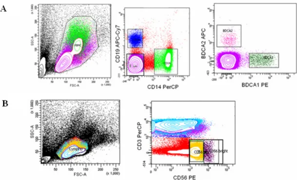
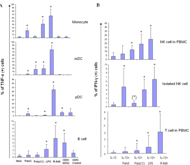
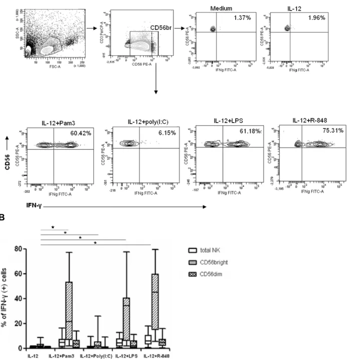
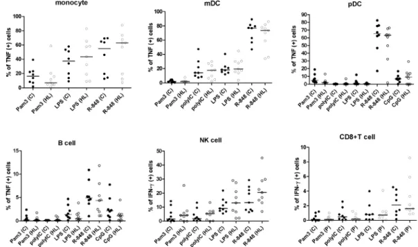
A multi-color flow cytometric assay was developed to evaluate TLR responses in different PBMC subsets. The assay was divided into two panels: the antigen-presenting cell panel (the APC panel) for detecting TLR-ligand-induced cytokines (TNF-α/ IFN-α) in monocytes, mDCs, pDCs, and B cells; and the NK cell and T cell panel (NK/T cell panel) for detecting TLR-ligand-induced cytokines (IFN-γ/ TNF-α) in NK cells and T cells. For detecting the APCs, PBMCs were first gated according to the forward and sideward scatters (Fig. 1A). Monocytes were defined as CD14+ CD19- cells, and B cells were gated on CD14-CD19+ cells. Cells negative for both CD14 and CD19 were further divided into BDCA-1+ cells and BDCA-2+ cells, and were defined as myeloid DCs and plasmacytoid DCs, respectively. For detecting NK and T cells, lymphocyte gate was first determined by forward and sideward scatters (Fig. 1B). T cells were detected by gating the lymphocytes on CD3+CD56- cells, while NK cells were defined as CD3- CD56+ cells. The subpopulation of NK cells which express a higher level of CD56 as detected by PE-conjugated CD56 antibody (BD Biosciences) was gated for the evaluation of the functions of CD56bright NK cells. In some experiments, T cells were further gated on CD8 to distinguish the CD8+ from the CD8- subsets. TLR-triggered cytokine- producing cells were detected by gating on specific cell surface markers against the intracellularly-stained cytokines (e.g. CD14+CD19-TNF-α+ cells represented TNF-α-producing monocytes, and CD3-CD56+IFN-γ+ cells represented IFN-γ-secreting NK cells).
Fig. 1. Gating strategies. (A) Antigen-presenting cell panel. PBMCs were first gated, and divided into CD14+ (monocytes) and CD19+ (B cells). The double negative population was then gated on BDCA1 (marker for mDC) and BDCA2 (marker for pDC). (B) NK/T cell panel. CD3+ cells derived from the lymphocyte gate are T cells, and CD3-CD56+ population are NK cells, which were further divided into CD56bright (<10% of total NK cells) and dim subsets.
4.1.2 Concentrations of the TLR ligands were optimized and TNF-α and IFN- γ production were selected as the read-out of the assay
Using PBMCs, the TLR ligand concentrations were titrated in the range suggested by the manufacturers and previous studies. Pam3Cys was titrated from 10 ng-10 μg/ml, poly(I:C) was titrated from 100 ng-100 μg/ml, LPS was titrated from 10 ng-1 μg/ml, R- 848 was titrated from 100 ng-10 μg/ml, and CpG was titrated from 1-5 μM. In case of the Pam3Cys-, LPS- and R-848- stimulation, the lowest concentration of the ligand to achieve the maximal TNF-α production in monocytes after 4 hours of incubation was selected as the optimal concentration for the assay (demonstrated in Table 1).
Moreover, the optimal concentrations of poly(I:C) and CpG oligonucleotide were determined as the ones to induce the maximal cytokine production of myeloid DCs and plasmacytoid DCs, respectively. Both IFN-α and TNF-α secretions in pDCs were detected after 18 hours of R-848 and CpG ODN stimulations. However, the TNF-α response was more prominent than the IFN-α response (with a 4-fold higher response to CpG ODN and a 2-fold higher response to R-848), and could be detected in all cell
types of the APC panel 4 hours after all TLR-ligand stimulations. Therefore, TLR-ligand induced TNF-α production was determined as the final read-out of the APC panel of the assay.
Similarly, the TLR ligand concentrations in the NK/T cell panel were titrated. Instead of 4 hours, TLR agonists enhanced significantly the IFN-γ production in NK cells after 24 hours of stimulation. In the presence of 1 ng/ml IL-12, the fold of poly(I:C)-enhanced IFN-γ production in NK cells (12.5-fold) was greater than the fold of increased TNF-α (3.4-fold), compared to cells stimulated with IL-12 alone. Furthermore, IFN-γ secretion in NK and T cells represents an important effector function in antiviral defences.
Therefore, IFN-γ was selected as the read-out for the NK/T cell panel.
4.1.3 IL-12 was added for IFN-γ profiling in NK cells and T cells
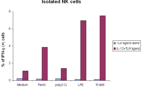
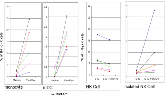
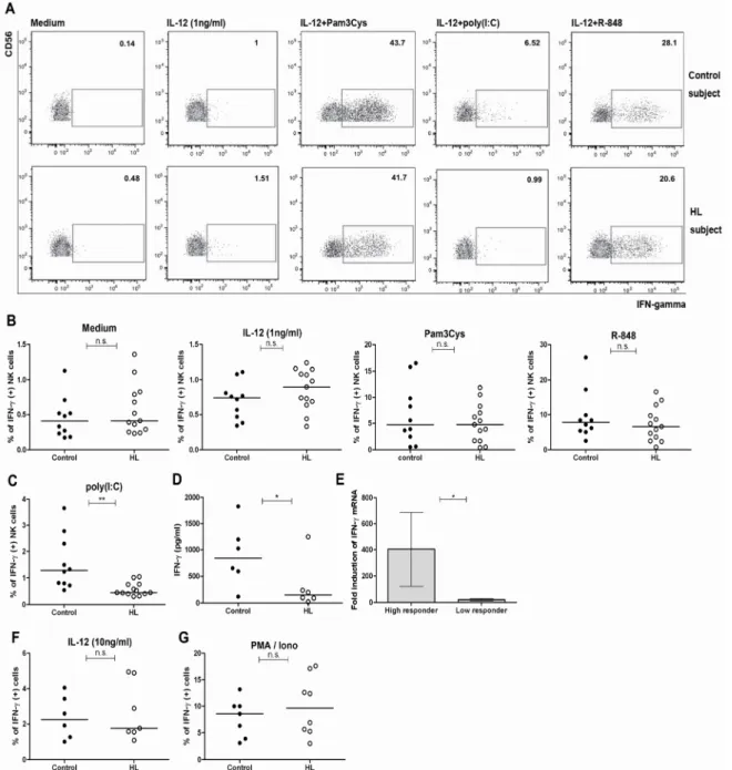
Although the NK cells and both CD8+ and CD8- T cells stimulated in the PBMC produced significant amount of IFN-γ in response to TLR-ligands (except to type C CpG and in some donors to Pam3Cys), isolated NK cells did not produce IFN-γ in response to any of the TLR ligands in the absence of co-stimulatory cytokines (Fig. 2). Therefore, for the profiling of IFN-γ production in NK cells, 1 ng/ml of IL-12 was added together with the TLR ligands for 24-hour stimulations.
After the addition of IL-12, we observed that all the TLR agonists, except type C ODN M362, enhanced substantially the IFN-γ production in isolated NK cells (Fig. 2). IL-12 also promoted the TLR-ligand-triggered IFN-γ response in non-purified NK cells. The response to poly(I:C) of NK cells in the PBMC increased almost 2-fold following IL-12 priming.
Fig. 2. TLR-ligand-stimulated response in isolated NK cells. The IFN-γ responses of isolated NK cells from one donor stimulated with TLR-ligand alone or with the addition of IL-12 for 24 hours are demonstrated as a representative. TLR-ligands triggered a substantial amount of IFN-γ production in isolated NK cells in the presence of 1 ng/ml IL-12.
4.1.4 NK cells and T cells could not be fully activated if BFA was added before TLR-ligand stimulation
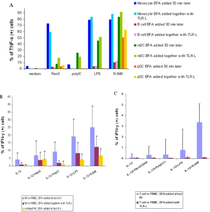
Brefeldin-A (BFA) is known to block most of the cytokine secretions. In order to decrease the influence of cytokines secreted by bystander cells and at the same time measure the TLR response that is closest to in vivo biology, we added BFA to the whole PBMC population along with TLR ligands in some experiments. Instead of adding BFA 30-60 minutes after TLR stimulation in the antigen-presenting cell panel, BFA was added with TLR ligands at the beginning of incubation for 4 hours. Instead of adding BFA at the last 6 hours of TLR stimulation in the NK/T cell panel, PBMCs were first primed by IL-12 for 6 hours, and stimulated with TLR agonists in the presence of BFA for another 18 hours.
In monocytes, mDCs, pDCs, and B cells, the TNF-α responses to TLR ligands were similar when BFA was added immediately prior or half an hour later to TLR agonists (Fig. 3A). Interestingly, when BFA was added together with TLR stimulators, the responses of NK cells to TLR ligands were much lower (Fig. 3B) and the responses in T cells were completely abrogated (Fig. 3C).
Taken together, the results suggest that although TLR-ligands can stimulate the IFN-γ
production in NK cells and T cells in the presence of IL-12, other cytokines or direct accessory cell-contact signals are required to fully activate the TLR-induced IFN-γ responses of NK cells, and especially of T cells. However, the antigen-presenting cells produced TNF-α directly after TLR-ligand stimulation, showing that these cells are independent of autocrine/ paracrine signals produced by the PBMC populations in response to TLR activation.
Therefore, in our assay, we have decided to add BFA together with TLR ligands for APC panel, and to add BFA at the final 6 hours of 24-hour TLR-ligand stimulation in NK/T cell panel.
Fig. 3. BFA added together with TLR-ligands decreased bystander cell co-stimulations. BFA was added directly with TLR-ligands in these experiments. (A) Compared to the TLR-induced cytokine response when BFA was added 30 minutes later, the responses in the APC panel when BFA was added together with the TLR ligands were similar (the result shown here is one representative of 3 donors). (B) The response of NK cells detected in the PBMCs when BFA was added together with TLR-ligands was decreased (n = 4), as compared to the response when BFA was added at last 6 hours (n = 14), although the level of response was still higher than the response of isolated NK cells (n = 14), except the response to Pam3CSK4. The bar chart represents mean ± SD. (C) The response in T cells when BFA was added with TLR-ligands was almost not detectable (n = 5, mean ± SD), as compared with responses of T cells when BFA was added at the last 6 hours (n = 5, mean ± SD).
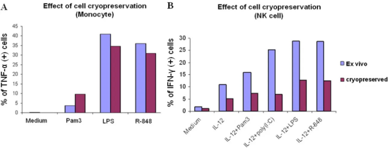
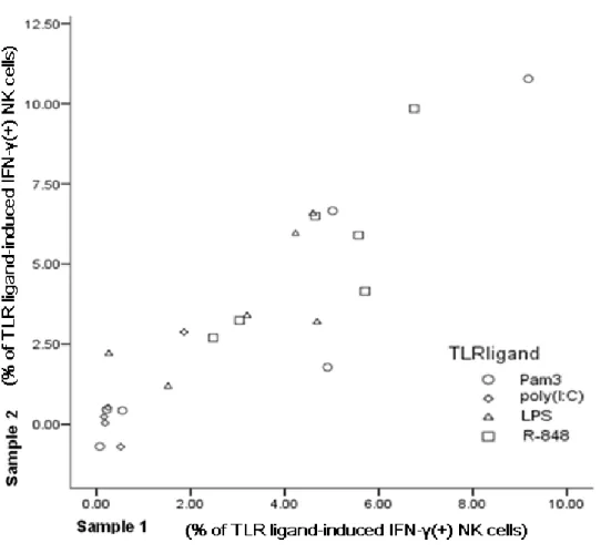
4.1.5 Effect of cell cryopreservation
Next, we sought to validate the assay for cryopreserved cells, since it could be applied to more clinical specimens that were properly cryopreserved. Fresh blood was taken from the same individual, and part of it underwent cryopreservation, the other part was stimulated directly with TLR-ligands. The cryopreserved PBMCs were kept at least one day in the nitrogen tank, and were thawed immediately on the day of experiment. The thawed cells were then rested overnight in the 37 ۫ C incubator and stimulated with the TLR agonists on the next day. The cryopreserved monocytes produced a similar amount of TNF-α in response to TLR-ligand stimulations as the freshly prepared monocytes (Fig. 4A); however, the cryopreserved NK cells produced less than half the amount of IFN-γ in response to TLR stimulations as compared to fresh NK cells (Fig.
4B). Therefore, we decided to work with fresh blood in our assay.
Fig. 4. Effect of cryopreservation in monocytes (A) and NK cells (B). TLR-ligand-induced cytokine productions of cells isolated from fresh blood (ex vivo) were compared with the TLR responses of cryopreserved cells from the same donor. One representative of 2 experiments is shown.