NK cells in tumor immune evasion: Role of tumor- associated ligands that regulate NK cell function and
therapeutical implications
Inaugural-Dissertation
zur
Erlangung des Doktorgrades
der Mathematisch-Naturwissenschaftlichen Fakultät
der Universität zu Köln
vorgelegt von
Jörg Keßler aus Siegen
Köln März, 2012
1. Berichterstatterin: PD Dr. Roswitha Nischt
2. Berichterstatter: Prof. Dr. Matthias Hammerschmidt
3. Berichterstatter: Prof. Dr. Elke Pogge von Strandmann
Prüfungsvorsitzender: Prof. Dr. Siegfried Roth
Tag der mündlichen Prüfung: 14. Mai 2012
Table of contents
1 Zusammenfassung ... 1
2 Abstract ... 3
3 Introduction ... 5
3.1 Natural killer cells ... 5
3.1.1 NK cells and their function in immunity and immunotherapies ... 5
3.1.2 Regulation of NK cell activity ... 6
3.1.3 NK cell receptors and their ligands ... 8
3.1.4 NK cells and their role in hematopoietic stem cell transplantation ... 10
3.2 HLA-B associated transcript 3 (BAT3) ... 13
3.3 Exosomes ... 13
3.4 Malignancies suitable for NK cell based immunotherapies ... 15
3.4.1 Acute and chronic leukemias ... 15
3.4.2 Hodgkin lymphoma ... 16
3.5 Bispecific antibodies and immunoligands ... 18
3.6 Aim of the current study ... 20
4 Material and Methods ... 21
4.1 Materials ... 21
4.1.1 Cell Lines ... 22
4.1.2 Vectors ... 22
4.1.3 Antibodies ... 23
4.2 Methods ... 25
4.2.1 Standard molecular biological methods ... 25
4.2.2 Cell biological, biochemical and immunological methods ... 27
5 Results ... 34
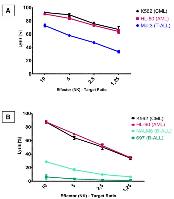
5.1 NK cell mediated in vitro GVL effects on AML and ALL cells ... 34
5.1.2. Expression of ligands for activating NK receptors on leukemic cell lines.... 36
5.1.3 Blocking of NKp30 signaling leads to reduced lysis of AML cells ... 38
5.1.4 HLA expression on ALL target cell lines contributes to their resistance against NK cells ... 39
5.1.5 Increased cytotoxicity towards ALL cells upon external NKp30 stimulation via BAT3 expressing exosomes ... 42
5.1.6 NK cell mediated cytokine release in a mixed lymphocyte reaction upon NKp30 triggering ... 44
5.2 Impaired functions of peripheral NK cells in Hodgkin Lymphoma patients ... 46
5.2.1 Activating receptor NKG2D is down-regulated on NK cells in Hodgkin Lymphoma patients ... 46
5.2.2 Soluble ligands for NK cell receptors are elevated in HL sera ... 47
5.2.3 Hodgkin Lymphoma serum impairs NK cell activity ... 49
5.2.4 Cytotoxic functionality of HL patients’ NK cells is impaired ... 52
5.2.5 HL NK cell cytotoxicity can be restored ... 53
5.2.6 αCD16-αCD30 immunoligand activates NK cells in vivo ... 58
6 Discussion ... 61
6.1 Balance of activating and inhibitory signaling contributes to the distinct lysing ability of NK cells towards leukemic cell lines. ... 61
6.2 NK cell inhibition contributes to the immune escape in Hodgkin lymphoma ... 64
6.3 A αCD16-αCD30 immunoligand as an effective tool for NK cell based immunotherapy in HL ... 66
References ... 69
Abbreviations ... 76
Danksagung... 77
Erklärung ... 78
Lebenslauf ... 79
____________________________________________________________________________________
1 Zusammenfassung / Abstract
1 Zusammenfassung
NK Zellen sind Lymphozyten des angeborenen Immunsystems und spielen eine wichtige Rolle bei der körpereigenen Erkennung und Zerstörung von Tumorzellen, bei Anti-Tumor Effekten nach Knochenmarktransplantationen (HSCT) und somit als Effektorzellen bei Immuntherapien. Neben inhibierenden Rezeptoren steuern aktivierende Rezeptoren wie NKp30, NKG2D und CD16 die Aktivität von NK Zellen. Ziel der Arbeit war es zum molekularen Verständnis der unzureichenden Bekämpfung von Tumorzellen durch NK Zellen beizutragen. Dies wurde anhand von Leukämiezelllinien myeloiden und lymphatischen Ursprungs untersucht, da diese von NK Zellen nach Stammzelltransplantationen je nach Ursprung der Leukämie des Empfängers (myeloid, lymphatisch) sehr unterschiedlich bekämpft werden. Im Rahmen dieser Arbeit konnte gezeigt werden, dass der zytotoxische Rezeptor NKp30 maßgeblich dazu beiträgt, dass NK Zellen myeloide leukämische Zelllinien besser lysieren als Zelllinien lymphatischen Ursprungs. So können Exosomen, die Liganden für NKp30 auf der Oberfläche exprimieren, die Resistenz der lymphatischen Leukämiezellen gegen NK-Zellerkennung und Killing überwinden. Zusätzlich verhindert ein verstärktes inhibitorisches signaling durch HLA-Moleküle auf lymphatischen Zelllinien eine effektive Lyse.
Darüber hinaus wurde am Beispiel des Hodgkin Lymphoms (HL) gezeigt, dass NK Zellen der Patienten anders als NK Zellen gesunder Spender nicht in der Lage sind Hodgkin Zelllinien zu eliminieren. Dies konnte auf lösliche Faktoren im Serum der Patienten zurückgeführt werden, die auch die Aktivität gesunder NK Zellen beeinträchtigen. Die Ergebnisse der Arbeit zeigen, dass hierbei die löslichen Liganden MICA für den NKG2D Rezeptor und BAT3 für den NKp30 Rezeptor eine wichtige Rolle spielen. Diese Liganden lösen in membrangebundener Form NK Zell Zytotoxizität aus, während sie in löslicher Form eine inhibitorische Wirkung haben und sogar zu einer verringerten Expression von NK-Zellrezeptoren führen. In der Tat zeigen NK Zellen von HL-Patienten eine niedrigere Expression von NKG2D als NK Zellen gesunder Spender.
Nach der Therapie des Lymphoms waren die Serumlevel dieser beiden Liganden wieder auf dem Niveau gesunder Probanden.
NK Zellen in Hodgkin Patienten konnten ex vivo und in vivo erfolgreich mit einem bispezifischen Antikörper-Konstrukt aktiviert werden, das die NK Zellen via CD16
2 stimuliert und CD30 als Antigen auf Zielzellen bindet. So konnte experimentell eine sehr effiziente Lyse von Hodgkin Zellen erreicht werden.
Zusammenfassend zeigen die Daten, dass die Expression aktivierender Rezeptoren (NKp30 und NKG2D) und der korrespondierenden Liganden Phänotyp und Funktion der NK Zellen beeinflusst und zur Immun-Evasion beiträgt. Die Inhibition der NK Zellen ist jedoch reversibel und kann mit therapeutischen Antikörpern überwunden werden.
NK Zellen stellen somit einen vielversprechenden Ansatz dar, Immuntherapien auf zellulärer Ebene als Kombination oder Alternative zu Standardtherapien zu entwickeln.
____________________________________________________________________________________
3 Zusammenfassung / Abstract
2 Abstract
NK cells are lymphocytes of the innate immune system and play a crucial role in tumor immune surveillance, in beneficial graft versus tumor effects upon hematopoietic stem cell transplantations (HSCT) and thereby as effectors in cellular immunotherapies. NK cells are regulated by inhibitory receptors mostly binding to HLA molecules and activating receptors like CD16, NKG2D and NKp30. Aim of this work was to contribute to the molecular understanding of the insufficient control of tumor cells through NK cells.
This was examined on the basis of leukemic cell lines with myeloid or lymphatic origin, since these cells, according to their origin, are recognized and killed very differently by donor NK cells upon stem cell transplantation. In this study we could show that the cytotoxic receptor NKp30 contributes decisively to a better lysis of myeloid leukemic cell lines compared to lymphatic leukemic cell lines. Thus, exosomes expressing ligands for NKp30 can help to overcome the resistance of lymphatic leukemia cells against NK cell recognition and killing. Additionally an increased inhibitory signaling via HLA molecules on lymphatic cell lines prevents a more efficient lysis.
Moreover, it was shown in the setting of Hodgkin lymphoma (HL) that patients´ NK cells in contrast to healthy NK cells are not able to lyse Hodgkin cell lines. Soluble factors in the patients´ sera were identified to be responsible for that. Of note, patient sera also suppressed cytotoxic activity of NK cells from healthy donors by downregulation of activating receptors. Results indicate that the soluble ligands MICA for NKG2D and BAT3 for NKp30 play an important role. While these ligands trigger cytotoxicity in their membrane bound form, as soluble factors they have inhibitory impact and can downregulate NK cell receptors. Indeed, NK cell derived from HL patients showed a lower expression level of NKG2D than healthy NK cells. After therapy and clearance of the tumor, serum levels of these soluble ligands were comparable to healthy donors again.
Furthermore, NK cells in HL patients were successfully reactivated in vivo and ex vivo with a bispecific antibody construct. This construct stimulated NK cells via CD16 and binds CD30 as an antigen on target cells. Thereby, a very efficient lysis of Hodgkin target cells was achieved in experiments.
Taken together, the data indicate that the increased expression of tumor associated ligands for NKG2D and NKp30 in the serum of HL patients influences the phenotype and the function of HL NK cells and hence contributes to a tumor immune evasion. However,
4 the inhibition of NK cells is reversible and can be overcome with therapeutical bispecific antibodies.
Thus, NK cells seem to be a promising approach for cellular based immunotherapies as an alternative to standard therapies.
_____________________________________________________________________________________________________________________________
5 Introduction
3 Introduction
3.1 Natural killer cells
3.1.1 NK cells and their function in immunity and immunotherapies
Natural killer cells (NK cells) belong to the group of lymphocytes (T and B cells) and as such they arise from progenitor cells in the bone marrow. In contrast to T cells and B cells, NK cells do not have several maturation steps in a primary lymphoid tissue. As they also lack antigen specific receptors, they are attributed to the innate immune system (Vivier, Tomasello et al. 2008). NK cells are located largely in the spleen, but also in lymph nodes and in the peripheral blood (Witte, Wordelmann et al. 1990;
Fehniger, Cooper et al. 2003). They contribute with approximately 10 % to the peripheral blood mononuclear cells (PBMCs) and were originally defined through their capability of killing virally and tumor transformed cells (Lanier 2005). They play a crucial role in tumor immune surveillance, as they monitor self-cells for any aberrant expression patterns, that indicate stressed or transformed cells.
With T-lymphocytes NK cells share two functions that are crucial for an intact immunity:
toxicity against target cells and the secretion of cytokines (Biron, Nguyen et al. 1999).
Via their receptors, NK cells can distinguish healthy from non-healthy cells which they can lyse without prior antigen stimulation (Smyth, Cretney et al. 2005). A unique way of lysing transformed cells is the antibody dependent cellular cytotoxicity (ADCC). CD16 (FcγRIII) on NK cells binds the Fc-part of antibodies coating target cells and thereby leads to an activation and lysis (Newman and Riley 2007). For both ways, NK cells use receptors and adhesion structures to build an immunological synapse. This enables the NK cell to exert a directed secretion of cytolytic vesicles containing perforins and granzymes (Orange 2008; Vivier, Tomasello et al. 2008).
Besides that, NK cells can be activated and recruited by antigen presenting cells (APCs) via various chemo- and cytokines (Walzer, Dalod et al. 2005; Lucas, Schachterle et al.
2007). In addition, a crosstalk between NK cells and dendritic cells (DCs) has shown an influence on adaptive immune reactions (Ferlazzo, Tsang et al. 2002; Vitale, Della
6 Chiesa et al. 2005), as well as the NK cell based secretion of pro inflammatory cytokines like IFN-γ and TNF-α (Cooper, Fehniger et al. 2001).
In some malignancies, NK cells fail to exert their function of surveying stressed and transformed cells. Hodgkin Lymphoma (HL) (see 3.4.2) is a good example for this failure, as NK cells are present in the surrounding tumor tissue, but are obviously not able to control emergence and development of the tumor
Thus, NK cells have become a popular target, when it comes to cellular based immunotherapies. There are several strategies of harnessing and improving NK cell functions in cancer. Some of them aim at trafficking of NK ells, others at susceptibility of target cells towards NK cell mediated apoptosis. But most of those strategies, like in this study, try to modulate the balance between activating and inhibiting receptor signaling of NK cells (see 3.1.2).
3.1.2 Regulation of NK cell activity
With the ability to kill target cells without any antigen specific stimulation or priming, it is important that NK cells can distinguish between healthy and stressed cells. The basic principle is to avoid killing of normal cells, while transformed cells are recognized and eliminated (Lanier 2005). The two regulatory mechanisms comprise “missing-self recognition” and “induced-self recognition” (Fig 3.1). Signal integration of both pathways determines the regulation of the NK cell (Bryceson and Long 2008).
“Missing self” describes the lack of protection of self-cells through a downregulation of HLA class I molecules. Normally, these molecules provide a protective signaling via binding to inhibitory receptors (mainly Killer immunoglobulin like receptors, KIRs) on the surface of the NK cells. However, on transformed cells class I molecules are less expressed on the surface and therefore do not deliver an appearance of a healthy self- cell (Ljunggren and Karre 1990; Bryceson and Long 2008).
“Induced self” in contrast delivers triggering signals via a variety of activating NK receptors, e.g. NKG2D, CD16 and NCRs (NKp30, NKp44 and NKp46, see 3.1.3), that leads subsequently to the elimination of the target cell. On transformed cells, the ligands
_____________________________________________________________________________________________________________________________
7 Introduction
for these activating receptors are often upregulated (Groh, Rhinehart et al. 1999;
Moretta, Bottino et al. 2001). Since the “missing self” is not necessarily sufficient for lysis of the target cells, the “induced self” signaling finally leads to the activation of the NK cell.
Besides the integration of “missing self” and “induced self”, also synergistic effects among activation receptors seem to be important, the binding of one receptor is not necessarily sufficient. (Bryceson, Ljunggren et al. 2009).
Figure 3.1: Regulation of NK cell activity (Simhadri, 2008)
“Missing-self” and “Induced-self” contribute to the regulation of lysing activity toward a target cell.
Inhibitory receptors recognize self MHCs. This negative signaling protects the cell from being killed (middle), unless it is “overruled” by the binding of inducible ligands to activating receptors (bottom). A lack of inhibitory signaling leads to an activation of NK cells (top).
8 3.1.3 NK cell receptors and their ligands
Decisive for the protection of normal self-cells from NK cell based attacks is the recognition and binding of inhibitory receptors to mainly the polymorphic HLA class I molecules (Ljunggren and Karre 1990) but also other surface molecules like sialic acid or cadherins. Killer cell immunoglobulin-like receptors (KIRs) provide the biggest and obviously most important group of inhibitory receptors that bind to different alleles of HLA class I molecules. (Vivier, Tomasello et al. 2008). They belong to the C-type lectin superfamily and are characterized by immune receptor tyrosine-based inhibitory motif (ITIM) in the cytoplasmic moiety. The tyrosine phosphorylation by src kinases subsequent to the ligand interaction leads to the recruitment of SHP-1 and-2 phosphatases. Eventually this results in suppressed cytotoxicity and secretion of cytokines (Lanier 1998).
Mandatory for triggering NK cell effector functions are the activating NK cell receptors.
The apparently most important and best examined receptors are the low affinity Fc receptor CD16 or FcγRIII, NKG2D as well as NKp30, NKp44 and NKp46, termed Natural Cytotoxicity Receptors (NCRs).
CD16 binds to the Fc part of IgG antibodies that coat antigens on target cells. Upon ligation, CD16 binds to adapter proteins, whose phosphorylated tyrosines in the immune receptor tyrosine-based activation motif (ITAM) become a target for src family tyrosine kinases (Vivier, Morin et al. 1991). This activates downstream signaling and leads to cytokine secretion and cytotoxicity. Besides that, CD16 is also reported to have broader activating potential in direct NK cell cytotoxicity, independent of antibody binding (Mandelboim, Malik et al. 1999). CD16 also plays an important role in classifying NK cell into subsets: CD56brightCD16dim NK cells show an enhanced cytokine production, while CD56dimCD16bright NK cells produce more cytolytic factors and are describes to be mainly responsible for cytotoxicity (Vivier, Tomasello et al. 2008). Furthermore, CD16 seems to be a receptor that needs little or no support from other activating NK receptors in terms of overcoming the threshold for activation (Bryceson, March et al. 2005; Bryceson and Long 2008; Bryceson, Ljunggren et al. 2009). This makes CD16 a very feasible target for immunotherapeutic approaches and therefore a key receptor in this work.
NKG2D, another well described activating receptor, is expressed on NK cells, but also on CD8+ T cells as a co-receptor (Moretta, Bottino et al. 2001). Its intracellular domain
_____________________________________________________________________________________________________________________________
9 Introduction
has no ITAM motif, therefore transmembrane adapter molecules like DAP10 are required. DAP10 is phosphorylated by the phosphatidylinositol-3- kinase (PI-3K) that leads to a cascade ending up with calcium flux and triggered cytotoxicity (Upshaw, Arneson et al. 2006). Known ligands for NKG2D are the MHC class I related molecules MICA and MICB induced upon cellular stress and the UL-16 binding proteins, which are upregulated on some tumor transformed cells (Bauer, Groh et al. 1999; Cosman, Mullberg et al. 2001). By shedding these ligands for NKG2D from the surface, tumor cells exhibit an evasion strategy to escape the immune surveillance of NK cells (Waldhauer and Steinle 2006; Waldhauer, Goehlsdorf et al. 2008; Nuckel, Switala et al.
2010).
More recently, the group of Natural Cytotoxicity Receptors (NCRs) has been defined, comprising NKp46 (NCR1), NKp44 (NCR2) and NKp30 (NCR3). These three receptors are exclusively expressed on NK cells, among which NKp44 only appears on activated NK cells (Sivori, Vitale et al. 1997; Vitale, Bottino et al. 1998; Pende, Parolini et al.
1999). In contrast to NKG2D, but similar to CD16, NCRs associate with ITAM-carrying factors like CD3δ or FcεRIγ. Via phosphorylation of these ITAMs through Zap70 or Syk, a cascade leads to degranulation of cytokines or cytolytic factors (Moretta, Bottino et al.
2001; Andre, Castriconi et al. 2004). Only few ligands for the NCRs have been identified to date. Viral hemagglutinins bind to NKp46 and NKp44 and the cytomegalovirus tegument protein pp65 binds to NKp30 having an inhibitory effect. (Mandelboim, Lieberman et al. 2001; Arnon, Achdout et al. 2005). Only little information on the cellular ligands for the NCRs has been revealed until now. Both the HLA-B associated transcript 3 (BAT3) and the B7 family member B7-H6 binds to NKp30 and so far unknown ligands for NKp46 are expressed on pancreatic β-cells (Pogge von Strandmann, Simhadri et al.
2007; Brandt, Baratin et al. 2009; Gur, Porgador et al. 2010).
Interestingly, NK cells have a “cross-talk” with Dendritic cell (DCs) via NKp30. They can either stimulate or kill DCs and hence have a direct influence on adaptive immunity (Ferlazzo, Tsang et al. 2002; Vitale, Della Chiesa et al. 2005).
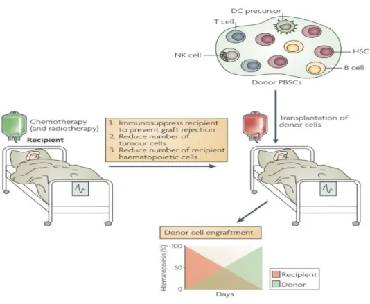
10 3.1.4 NK cells and their role in hematopoietic stem cell transplantation
In order to replace the hematopoietic system, a leukemic patient often gets a transfer of stem cells following chemotherapy and irradiation. If the donor is for example a sibling or even an unrelated donor, the process is termed allogeneic hematopoietic stem cell transplantation (HSCT). In this setting two reactions can occur: the welcome Graft- versus-leukemia effect (GVL) and the adverse Graft-versus-host disease (GVHD).
Figure 3.2: Allogeneic stem cell transplantation (Shlomchik 2007), modified
The recipient receives chemo- and radiotherapy in order to kill tumor and to destroy the old
“defect” hematopoietic system. This is replaced by the transplant containing hematopoietic stem cells (HSC) and various immune cells including NK cells. After several weeks, hematopoiesis is completely done by cells and tissue of the donor´s transplant.
GVL is mediated by cytotoxic CD8+ T lymphocytes (CTLs) as well as alloreactive NK cells (Ruggeri, Capanni et al. 1999; Pegram, Ritchie et al. 2011). These donor cells recognize tumor cells as foreign in the host and kill them due to their cytotoxic ability.
_____________________________________________________________________________________________________________________________
11 Introduction
This can support the treatment thereby making making less severe regimen (irradiation and chemotherapy) possible. More importantly the probability of a relapse can be reduced. (Parham and McQueen 2003).
The main adverse reaction of allogeneic HSCT leads to the GVHD. Donor`s alloreactive CD8+ T cells not only recognize malignant cells in this case, but also attack healthy tissue. In acute GVHD, most affected organs are the liver and skin. With immune suppressive agents or depletion of T cells from the graft, this side effect can be diminished. But T cell mediated GVL is also abrogated (Shlomchik 2007).
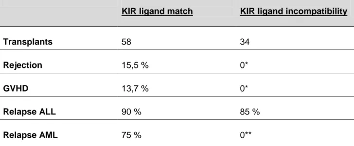
The group of L. Ruggeri et al could elucidate the role and the efficacy of alloreactive NK cells in the setting of allogeneic HSCT (Table 3.1). Here, patients with either acute myloid leukemia (AML) or acute lymphoid leukemia (ALL) received transplants mismatched in one HLA haplotype. Due to typing of recipients` HLA-C locus, donor- recipient pairs could be divided into two groups: either with or without KIR-ligand incompatibility. Only in the latter setting, alloreactive NK cell clones occur (Fig. 3.2). Of recipients with AML and a KIR-ligand incompatibility, none experienced a relapse within five years. In contrast, three of four AML patients without that incompatibility and thus not benefiting from NK cell alloreactivity sustained a relapse. This positive effect was
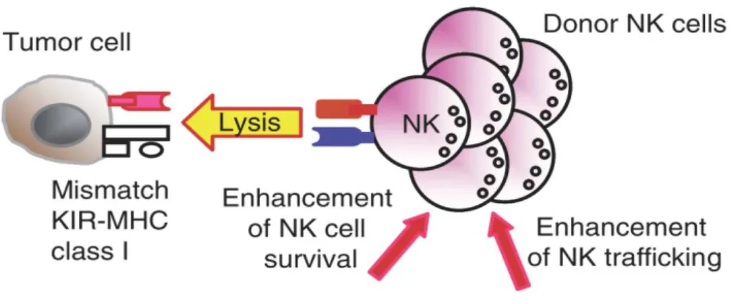
Figure 3.3: Alloreactive NK cells after HSCT (Terme, Ullrich et al. 2008) Through the mismatch of the KIR on donor´s NK cells and recipient´s MHC on the tumor cells, NK cells become activated. They can kill the tumor cell and proliferate for alloreactive NK cell clones with enhanced trafficking.
12 restricted to AML patients. For recipients with ALL, no impact on relapse rates could be observed.
One of the aims of this work is to find putative reasons for these differing cytotoxic abilities of NK cells towards distinct leukemic cells.
Interestingly, NK cell alloreactivity also seems to be responsible for the reduction of GVHD incidence. The reason for this might be the crosstalk and subsequent killing of DCs through NK cells. This might interrupt the cascade of cytokine signaling and interaction between DCs and T cells that normally leads to severe Graft versus host reactions (Ruggeri, Capanni et al. 2002; Parham and McQueen 2003).
Table 3.1: Transplantation outcome in HLA haplotype mismatched transplants with and without KIR ligand incompatibility. (Ruggeri, Capanni et al. 2002), modified
KIR ligand match KIR ligand incompatibility
Transplants 58 34
Rejection 15,5 % 0*
GVHD 13,7 % 0*
Relapse ALL 90 % 85 %
Relapse AML 75 % 0**
_____________________________________________________________________________________________________________________________
13 Introduction
3.2 HLA-B associated transcript 3 (BAT3)
BAT3 is a gene located in the MHC III cluster on chromosome 6 together with a variety of immunological important factors like the heat shock protein HSP70 or the tumor necrosis factors α and β (TNF α, β). Also the already mentioned NCRs and the NKG2D ligands MICA and MICB are encoded in this cluster. The full length transcript of BAT3 has 3.4 kb and a molecular weight of approximately 130 kDA. Due to its nuclear localization sequence, the protein is predominantly located in the nucleus. Under circumstances and by mechanisms, which are not completely understood yet, it can be released from cells to the extracellular environment. Important for this translocation might be the BAG domain, encoded next to the NLS at the C-terminus. This domain indicates that BAT3 interacts with heat shock proteins of the HSP70 family. (Banerji, Sands et al. 1990; Sasaki, Gan et al. 2007)
For this work, the most important function of BAT3 is the ability to bind and to trigger the activating NK receptor NKp30. For this BAT3 must be released from tumor cells (or also DCs). This happens via the secretion of exosomes (see below), small membrane vesicles that express BAT3 on their surface. Besides this membrane bound form, BAT3 can also appear in a soluble manner, for example in sera of CLL patients. Only the surface-associated protein can activate NK cells, while the unbound BAT3 seems to have a contrary effect and decreases NK cell activity (Pogge von Strandmann, Simhadri et al. 2007; Simhadri, Reiners et al. 2008).
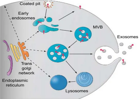
3.3 Exosomes
Exosomes are 50-100 nm small membrane vesicles originating in the endosomal system and secreted by probably all cell types.
Formation of exosomes occurs in so called multi vesicular bodies (MVBs) developing from late endosomes. These MVBs fuse with the plasma membrane to release their exosomes to the extracellular environment.
14 A broad range of molecules is hereby expressed on the surface of the exosomes for example immunological important molecules MHC class I, adhesion molecules like ICAMs and integrins or chaperones like HSP70. Interestingly, after a stress induction like a non-lethal heat shock, release of exosomes can be increased (Simhadri, Reiners et al. 2008) (van Niel, Porto-Carreiro et al. 2006). Depending on the cell type, the expression pattern varies. For instance the tetraspanine CD63 serves as a control marker for the purification of exosomes from the HEK cell line 293T, however it is absent in exosomes of other cell lines.
Functions of exosomes are poorly understood. A main task seems to be the transport of intracellular factors to the extracellular environment. Here, several aims are assumed, ranging from a simple transport and removal of “old” proteins to cell-cell communication.
In the case of CD8+ T cells, exosomes can even convey cell death via transporting cytotoxic granules. (Herz, Pardo et al. 2009; Thery, Ostrowski et al. 2009). Exosomes
Figure 3.4: Formation of exosomes (Fevrier and Raposo 2004)
Multivesicular bodies (MVBs) are part of the endosomal system. Through budding into the inside, exosomes are generated from the membrane of these MVBs. Later the MVBs merge with the plasma membrane and exosomes are released.
_____________________________________________________________________________________________________________________________
15 Introduction
from tumor cells can carry molecules that account for the recognition of immune cells, for example NK cells. According to their different expression of surface markers, functions can be completely contrary. Exosomes with a high expression of HSP70 can cause enhanced lysis compared to exosomes expressing less HSP70. Other cancer cells like the breast cancer cell line T47d are even capable of diminishing cytolytic activity of NK and T cells via their exosomal release (Clayton and Tabi 2005; Gastpar, Gehrmann et al. 2005).
Ligands for activating NK cells receptors have also been found on exosomes, for instance as the above mentioned BAT3 as a ligand for NKp30 (Simhadri, Reiners et al.
2008). In this thesis, exosomes serve as a tool for carrying membrane associated BAT3.
Since for this ligand it is crucial whether it is soluble or not, exosomes are the appropriate vehicle to activate NK cells via NKp30.
3.4 Malignancies suitable for NK cell based immunotherapies
3.4.1 Acute and chronic leukemias
Leukemic diseases are subdivided into a big number of different malignancies. They are often differentiated by their origin and their course. Four of the most common leukemias are the acute myeloid leukemia (AML), the chronic myeloid leukemia (CML), the acute lymphoblastic leukemia (ALL) and the chronic lymphocytic leukemia (CLL). ALL and CLL arise from aberrant cells of the lymphoid side of the hematopoietic system: T cells, B cells and their progenitors. ALL cells are fast proliferating progenitors of B or T cells in the bone marrow. Depending on the stage and characteristics of the malignant cell population, further subdivisions are done. One peak of incidences occurs in childhood at an age of 2-5. AML derives from myeloid blood cells like granulocytes or monocytes.
There are several subcategories, basically depending on the cell type the leukemia originated from (Bennett, Catovsky et al. 1976). Its incidence increasess with age and the main symptom is a replacement of healthy bone marrow by the myeloblasts, the malignant cells in AML. Like for ALL patients, an HSCT is a common treatment to replace the defect hematopoietic system.
16 3.4.2 Hodgkin lymphoma
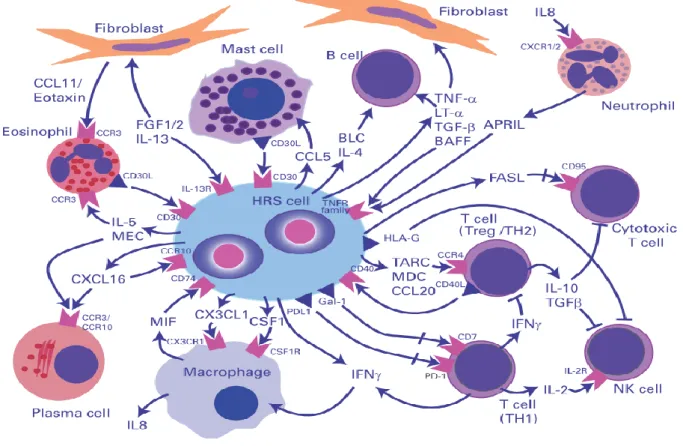
Hodgkin lymphoma (HL) is a subtype of malignant lymphomas that occurs to a great extent at two peaks: at an age of 15-35 years and over 55 years. Main characteristic is the presence of Reed-Sternberg cells (HRS cells). These large cells contain several nuclei and are usually derived from B lymphocytes. A further hallmark of HL is a large number of immune cells surrounding the HRS cells. All types of lymphocytes plus other cells like macrophages, stromal cells, eosinophils or mast cells contribute to approximately 98-99 % of the tumor (Pileri, Ascani et al. 2002). The main reason for the great number of immune cells seems to be the secretion of a variety of chemokines and cytokines that provide a micromilieu of a maintaining inflammation (Fig 3.4). Despite this great majority of potential tumor threatening cells and a persisting inflammatory milieu, the HRS cells are able to fully evade immune responses.
Figure 3.5: Microenvironment and crosstalks in Hodgkin lymphoma (Steidl, Connors et al. 2011)
The binucleated HRS cell is surrounded by bystander cells that are attracted by chemokines.
These cells themselves secret further chemokines and cytokines to shape the homeostasis of the infiltrating immune cells.
_____________________________________________________________________________________________________________________________
17 Introduction
This attenuation of immunological reactions is not only evoked by the tumor cells, but also by a complex and not fully understood crosstalk among the immune cells themselves (Steidl, Connors et al. 2011). Thus, T helper 2 cells and regulatory T cells account for the most bystander cells in HL, especially in direct proximity to the HRS cells (Ma, Visser et al. 2008). HRS cells also attract macrophages via the secretion of for example colony stimulating factor (CSF1) and other chemokines. Macrophages are believed to be responsible for suppressing antitumor immunity and therefore supporting tumor progression (Qian and Pollard 2010). They also attract further immune cells like neutrophils through IL-8 secretion (Foss, Herbst et al. 1996).
In general, soluble and also shed factors therefore seem to play an important role in the mechanisms of HL tumor formation. One example for this is the macrophage migration inhibitory factor (MIF). MIF is involved in angiogenesis, tumor cell migration and apparently suppresses p53 activity (Hudson, Shoaibi et al. 1999; Fingerle-Rowson, Petrenko et al. 2003). In HL, on the one hand it binds CD74 (the invariant chain of pre MHC class II) on the surface of HRS cells. This protein is implicated in signaling for lymphocyte activation processes (Leng, Metz et al. 2003; Stein, Qu et al. 2004). On the other hand with regard to the role of NK cells in HL, MIF might be involved in tumor escape mechanisms. In ovarian cancer for example, it has been shown, that MIF leads to a downregulation of NKG2D on NK cells and thus might affect anti-tumor activities.
Accordingly, one aspect in this work is the influence of soluble factors or serum of HL patients respectively on NK cell activity and hence a putative reason for impaired anti- tumor efficacy.
With respect to NK cell based immunotherapies, the (re-)activation via activating NK receptors is the second focus of this part.
18
3.5 Bispecific antibodies and immunoligands
The concept of bispecific antibody and immunoligands therapies is almost as old as the idea and the application of monoclonal antibody therapies. It is based mainly on the principle of retargeting and activation of immune effector cells. For this aim, one moiety of the structure binds to an activating receptor on an effector cell, the other one to an antigen on a target cell. In case of bispecific antibodies, one of the two different antigen determining sites recognizes the target cell, the other one the effector cell. For cytotoxic T lymphocytes, CD3 has become a popular target for bispecific antibodies to be addressed.
Unfortunately, bispecific antibodies go along with a rather low efficacy and some adverse reactions like immunogenicity. (Muller and Kontermann 2010).
As an advanced approach while keeping the principle way of function, bispecific antibody engineering created a broad range of different molecules distinguishable in size and number of binding domains.
So called immunoligands share their basic task with bispecific antibodies, but consist of different structures. Usually the antigen binding moiety is a single chain variable fragment (scFv), consisting of the variable domains of the light and the heavy chain derived from an antibody. The effector cell binding moiety can also be a scFv or a natural ligand to the targeted receptor like in the “ULBP2-BB4” construct. In this bispecific protein, ULBP2 engages NKG2D while the scFv BB4 binds to CD138 on multiple myeloma cells. This led to an activation of NK cells and an effective lysis of CD138 positive multiple myeloma cells (von Strandmann, Hansen et al. 2006).
_____________________________________________________________________________________________________________________________
19 Introduction
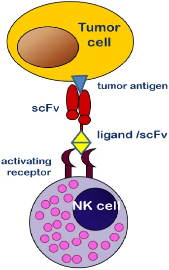
Figure 3.6: Function of immunoligands
Immunoligands bind to target cells and NK cells simultaneously through their two distinct binding moieties. A ligand a single chain variable fragment (scFv), respectively binds an activating receptor on the surface of the effector cell while another scFv binds specifically an antigen on the target cells. Due to that, NK cells are relocated to target cells and trigger cytotoxic activity.
20
3.6 Aim of the current study
NK cells play an important role in both immune surveillance of tumors and cellular based immunotherapies including the adoptive transfer of allogeneic NK cells in HSCT (Smyth, Hayakawa et al. 2002; Terme, Ullrich et al. 2008; Waldhauer and Steinle 2008). In some settings and under not fully understood preconditions, NK cells fail to execute their function in killing malignant and transformed cells.
For instance, NK cells have the ability to destroy leukemic cells with a lymphatic origin (ALL) upon allogeneic stem cell transplantation. In contrast, myeloid leukemic cells (AML) are resistant to NK cell mediated cytotoxicity.
In the autologous setting, Hodgkin lymphoma is a good example for absent immune surveillance as NK cells are present in the HL microenvironment but fail to control malignant HRS cells.
Aim of this study was (I) to identify molecules involved in the impaired receptor-ligand based target cell recognition for example contributing to different lysis capability against AML and ALL cells in HSCT settings. The main focus laid on differences in target cell phenotypes and their influence on NK cell activity and strategies to overcome tumor cell dependent defects of NK cells.
Furthermore (II) potential reasons for an obvious failure of tumor immune surveillance of NK cells should be illuminated in the setting of HL. Here the state, phenotype and activity of peripheral NK cells was examined and compared to healthy NK cells as well as possible causes for impairments of HL NK cells.
(III) Additionally to the task of finding underlying reasons for deficient NK cell activity, approaches to overcome these handicaps were identified and investigated in order to develop innovative NK cell based immunotherapies.
_____________________________________________________________________________________________________________________________
21 Material and Methods
4 Material and Methods
4.1 Materials
All salts, chemicals and reagents were purchased from Roth (Karlsruhe), Merck (Darmstadt), Sigma-Aldrich (Munich), Invitrogen (Karlsruhe) and BD Bioscience (Heidelberg). Media for cell culture was purchased from GIBCO®-Invitrogen (Karlsruhe).
Technical devices are mentioned at dedicated sites.
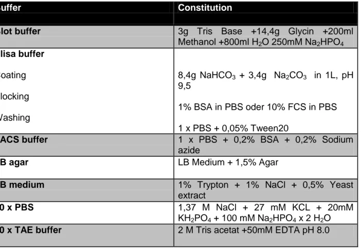
Table 4.1: Standard buffers
Buffer Constitution
Blot buffer 3g Tris Base +14,4g Glycin +200ml
Methanol +800ml H2O 250mM Na2HPO4 Elisa buffer
Coating Blocking Washing
8,4g NaHCO3 + 3,4g Na2CO3 in 1L, pH 9,5
1% BSA in PBS oder 10% FCS in PBS 1 x PBS + 0,05% Tween20
FACS buffer 1 x PBS + 0,2% BSA + 0,2% Sodium
azide
LB agar LB Medium + 1,5% Agar
LB medium 1% Trypton + 1% NaCl + 0,5% Yeast
extract
10 x PBS 1,37 M NaCl + 27 mM KCL + 20mM
KH2PO4 + 100 mM Na2HPO4 x 2 H2O
50 x TAE buffer 2 M Tris acetat +50mM EDTA pH 8.0
22 4.1.1 Cell Lines
Table 4.2: Cell lines and their physiological origin.
Cell Line Origin
293T human fibroblast kidney cell line
697 human B cell precursor leukemia (B-ALL)
HL-60 human acute myeloid leukemia (AML)
K562 human chronic myeloid leukemia (CML)
L428 human hodgkin lymphoma
L540 human hodgkin lymphoma
MOLT-3 human T cell leukemia (T-ALL)
NALM-6 human B cell precursor leukemia (B-ALL)
4.1.2 Vectors
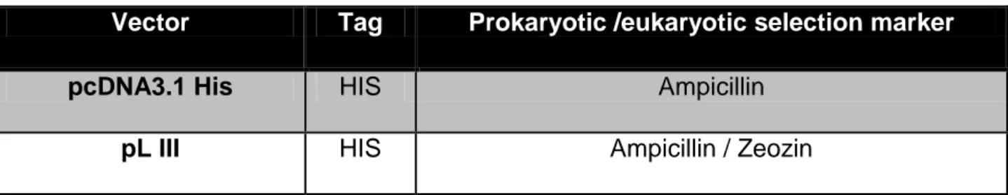
Table 4.3: Vectors for DNA cloning
Vector Tag Prokaryotic /eukaryotic selection marker
pcDNA3.1 His HIS Ampicillin
pL III HIS Ampicillin / Zeozin
_____________________________________________________________________________________________________________________________
23 Material and Methods
4.1.3 Antibodies
Table4.4: Antibodies
Antigen Fluorchrome Company Reactivity Species
CD3 APC BD Biosciences human mouse
CD3 PE BD Biosciences human mouse
CD9 purified BD Biosciences human mouse
CD16 PE BD Biosciences human mouse
CD16 APC BD Biosciences human mouse
CD45 PacificBlue Biolegend human mouse
CD56 PE BD Biosciences human mouse
CD56 APC BD Biosciences human mouse
CD56 AlexaFl.-647 Biolegend human mouse
CD86 purified Immuno Tools human mouse
CD86 FITC BD Biosciences human mouse
CD86 PE BD Biosciences human mouse
CD107a PE BD Biosciences human mouse
NKp30 PE Beckman Coulter human mouse
NKp44 PE Beckman Coulter human mouse
NKp46 purified BD Biosciences human mouse
NKp46 PE Beckman Coulter human mouse
NKp46 APC BD Biosciences human mouse
NKp46 AlexaFl-.647 Biolegend human mouse
hNKG2D PE BD Biosciences human mouse
24
NKG2D Fitc Abcam human mouse
hNKG2A PE BD Biosciences human mouse
HLA-ABC purified BD Biosciences human mouse
HLA-DR purified BD Biosciences human mouse
HLA-ABC PE BD Biosciences human mouse
HLA-DR FITC BD Biosciences human mouse
IgG Fc purified Dianova human goat
IgG purified Dianova goat
IgG H+L purified Dianova mouse goat
IgG PE BD Biosciences human mouse
human IgG1 PE BD Biosciences mouse goat
IgG APC BD Biosciences human mouse
7-AAD 7-AAD BD Biosciences
Annexin V PE BD Biosciences
anti-mouse FITC BD Biosciences mouse goat
anti-mouse PE Dako mouse goat
_____________________________________________________________________________________________________________________________
25 Material and Methods
4.2 Methods
4.2.1 Standard molecular biological methods
DNA digestion using restriction enzymes
For the cloning of DNA fragments as well as for the testing of plasmids, DNA was digested with appropriate restriction enzymes. For that, few µg were incubated with reaction buffer and 10 units of one or two enzymes and incubated for two hours at a temperature appropriate for the used enzyme. After that, successful cleavage of DNA was tested in an agarose gel.
For cloning scCD16-Immunoligands, scCD16 was designed with 5` NheI restriction site (GCGCTAGC) and a 3` SfiI restriction site (GGCCCAGCCGGCC). The enzymes NheI and SfiI (Fermentas) were used to excise scCD16opt DNA fragment from its origin pBluescript II SK+ vector. Destination vector was “pMS III” already containing the single chains BB4 and RFT5 respectively. This vector was also cut with these two enzymes to open a gap 5` of those single chains for ligating scCD16.
Purification of DNA fragments in agarose gels
Through restriction enzymes digested DNA fragments were separated in a standard 1%
agarose gel (TAE buffer, ethidium bromide) with 120 Volts for approximately 30 min.
Bands were then excised from the gel with a clean scalpel and eluted with GeneJet™
Gel extraction kit of Fermentas and resolved in water.
For ligations of DNA fragments into vectors, the Rapid DNA ligation Kit (Fermentas) was used according to the manufacturer’s protocol
26 Bacteria and media
The chemocompetent E.coli strain XL10 Gold was used for all cloning strategies.
Standard LB media was used with Ampicilin (100 µg/ml) according to the resistance given by the vector. Bacteria were incubated over night at 37°C and 150 rpm.
Transformation of chemocompetent bacteria
To transform chemo competent Bacteria, either 5µl ligation product or 1 µl plasmid were added to 50 µl thawed (on ice) bacteria solution and incubated on ice for 20 minutes.
Following a 45 seconds heat-shock at 42°C, bacteria were transferred back on ice and subsequently plated on plates for selection and incubation over night at 37°C.
The next day, clones were picked and used for inoculation of 3ml LB medium for Mini- preparations or 100 ml for Maxi-preparations of plasmid DNA.
Preparation of plasmid DNA
For the preparation of plasmid DNA, either the GeneJET™ Plasmid Miniprep Kit for Minipreps (Fermentas) or the NucleoBond® Xtra Maxi kit for Maxipreps (Macherey- Nagel) were used according to the manufacture`s protocols.
Concentrations of purified plasmids were determined and the DNA was used for transient transfection of 293T cells (human embryonic kidney cell line).
Sequencing of clonings
Samples of DNA were sent to Starseq® in Mainz according to default requirements of the company.
_____________________________________________________________________________________________________________________________
27 Material and Methods
4.2.2 Cell biological, biochemical and immunological methods
Cultivation of mammalian cell lines
All tumor cell lines (table 3.1) used in this study were cultivated in RPMI supplemented with 50 µg/mL penicillin, 50 µg/mL streptomycin, 2mM L-Glutamin and 10% fetal calf serum at 37°C with 5% CO2. 293T cells were kept in DMEM with the same additions. The viability and the cell count were determined by trypan blue staining.
Cultivation of primary mammalian cells
Primary mammalian cells (NK cells and PBMCs) purified from Buffy Coats or fresh blood samples were cultivated in in IMDM supplemented with 50 µg/mL penicillin, 50 µg/mL streptomycin, 2mM L-Glutamin and 10% fetal calf serum at 37°C with 5% CO2. When serum-free cultivation was needed, cells were kept in AIM-V lymphocyte medium. For experiments including autologous serum from HL patients, NK cells were kept in IMDM with 10 % autologous serum. Medium for NK cells contained usually 10 U IL-2. For activation experiments, NK cells purified from HL patients` blood were cultivated without IL-2 for one night at most.
Purification of Natural Killer cells via MACS (Magnetic Activated Cell Sorting)
Human NK cells were separated from peripheral blood mononuclear cells (PBMC).
PBMCs were purified using Ficoll-Paque density gradient centrifugation with leucosep columns from Greiner (Solingen) or normal falcons from BD (Heidelberg).
For experiments with leukemic cell lines, PBMCs / NK cells were gained from healthy donors` buffy coats. Buffy coats were obtained from the blood bank of the university hospital and were always used the same day.
PBMCs / NK cells from HL patients were purified from EDTA blood. For experiments comparing lysing ability of HL NK cells and healthy NK cells, those healthy cells were also purified from EDTA blood.
28 Depletion of all non NK cells was performed with the NK cell isolation kit (human) and the autoMACS Pro device (Miltenyi, Bergisch Gladbach). PBMCs were incubated with biotin-labeled antibodies targeting different markers on all peripheral blood cells except NK cells. Subsequently, streptavidin coupled magnetic beads were added to bind the cell surface attached biotinylated antibodies. After washing and centrifugation, NK cells were then separated from labeled non NK cells with the autoMACSpro® (Miltenyi Biotec).
The purity of NK cells should have been at least 90% and this was tested using flow cytometry analysis.
Transfection of 293T cells
For transfection with recombinant BAT3, 1 x 10⁶ 293T cells were plated in small cell culture flasks (25 cm2) the day before transfection for a final confluence of approximately 70-90 %. For gaining high amounts of the cloned scCD16-Immunoligands from the supernatants of transfected cells, 4x10⁶ 293T cells were plated in medium cell culture flasks.
For the transfection, the reagents LipofectamineTM Reagent (Invitrogen) and jetPrime™
(Polyplus transfection) were used and cells were cultivated either in Opti-MEM™
(Invitrogen) for the purification of BAT3 exosomes or DMEM (Gibco) and CD293 (Gibco) for the secretion of scCD16-Immunoligands, respectively .
According to manufacturer’s protocol, the dedicated amount of cDNA was incubated with the transfection reagent for 10-20 min and then added to the cells. BAT3 transfected and control cells were cultivated 48h before purification of BAT3 exosomes (see below).
For the expression of scCD16-Immunoligands, 293T were cultivated for two days, then the success of transfection was controlled by expression of GFP in transfected cells.
Purification of BAT3 exosomes
293T cells untransfected and transfected with recombinant were cultivated for 48 h in small cell culture flasks in OptiMEM (Gibco). Then, cells were dissociated with trypsin
_____________________________________________________________________________________________________________________________
29 Material and Methods
and transferred into falcons, in which they were exposed to a nonlethal heat shock at 42°C for 30 minutes followed by a recovery period of 60 minutes at 37°C with 5% CO2.
Exosomes from the supernatants were then purified by three successive centrifugations:
300 x g (5 min) and 1200 x g (20 min) to eliminate cells and debris, and 100 000 x g for 90 minutes to pellet the exosomes. Before ultracentrifugation, cells were passed through a 0,45 µm filter to deplete larger particles. The exosomal pellet was washed once in approximately 8 ml PBS, centrifuged again at 100 000 x g for 90 minutes and resuspended in 250 µl PBS. Exosomes were stored at -80°C and tested in flow cytometry for BAT3 expression.
Coupling of exosomes and FACS analysis of exosome coated beads
Exosomes were incubated with 20 µl 4.5 micron microsphere polybead carboxylate latex beads (5x10⁷/ml, Polysciences) for 90 min at 25° C and 700 rpm. The beads were washed once with PBS and then incubated with 1% BSA / PBS for 30 min at 25° C and 700 rpm. After multiple washing steps with PBS and centrifuging 10 sec at 13000 rpm, the beads were resuspended in 100 µl PBS per tube and stained for CD9 (293T cell line) for quantity control and BAT3 to verify the overexpression of BAT3 on the exosomes purified from the BAT3 transfected cells. Beads alone were gated and isotype control antibodies were used as controls for the flow cytometry analysis.
Flow cytometry (FACS analysis)
In principal, all cells were incubated with either unconjugated or with fluorochrome (PE, APC, Cy3, AlexaFluor 647) conjugated primary antibodies (Table 4.4) for 20 min at 4°C and washed three times with FACS buffer (Table 4.1). In case of unconjugated primary antibodies, labeled secondary antibodies against the species of the primary antibody were used, for example goat-anti-mouse-PE.
In case of cells consisting of different types of cells, the population of interest was gated in the SSC (granularity) and FSC (size) dot plot.
30 Purification of MACS separated NK cells was determined by checking lymphocyte antigens like NKp46 for NK cells, CD3 for T-cells or CD19 for B-cells. Only NK-cells with more than 90 % purity have been used for subsequent experiments.
The expression of ligands for the activating NK cell receptors NKG2D, NKp30 and NKp46 was also checked in the flow cytometer. ALL and AML cell lines and primary cells (blasts from leukemia patients) were incubated with soluble NKG2D, NKp30 and NKp46 receptors that were tagged with an antibody Fc moiety. Those receptors bind ligands (e.g. in case of NKG2D: ULBP1-3, MICA or MICB) on the surface of the cells. As secondary antibody, α-FC-Cy3 was used.
Blasts of fresh patients’ blood samples were gated (after lysis of erythrocytes) in a dot plot indicating SSC (granularity) versus CD45. CD45 is a lymphocyte marker that is less expressed on blasts compared to lymphocytes. With these two parameters blasts could be distinguished from lymphocytes and granulocytes and analyzed for expression of ligands for activating NK cell receptors.
Competition binding assay of the αCD16-αCD30 immunoligand on NK and target cells (Flow cytometry)
To validate the binding of αCD16-αCD30 on CD16 and CD30 on target cells, NK cells (CD16+) and CD30+ L428 cells (HL cell line) were incubated with a monoclonal antibody against CD16 and CD30 respectively. Simultaneously, αCD16-αCD30 was added at two different concentrations to compete with the monoclonal antibodies for the binding to the antigens. Goat-α-mouse antibody labeled with PE was used to stain bound monoclonal antibodies and measurement was done with a FACSCalibur (BD).
Detection of immunoligand in Western blot
For verification of expression and secretion of scCD16-Immunoligands, SDS-Page (10
% acrylamide gel) and Western blot was performed with lysates and His-purified supernatants of transfected and non-transfected control cells. Samples were heated at 95°C in Laemmli buffer before loading. Separation was performed at 170 V and with the