Impact of innate and adaptive immune cells in tumor immune surveillance
Dissertation
zur Erlangung des Doktorgrades der Naturwissenschaften (Dr. rer. nat.)
der Fakultät für Biologie und Vorklinische Medizin der Universität Regensburg
vorgelegt von Stephanie Blaimer
aus Kelheim
im Jahr 2020
2 Das Promotionsgesuch wurde eingereicht am: 21.02.2020
Die Arbeit wurde angeleitet von: Prof. Edward K. Geissler, PhD
Unterschrift Doktorandin:
3
Table of Contents
Acknowledgement ... 9
1 Abstract ... 11
2 Introduction ... 13
2.1 Cancer and metastasis ... 13
2.1.1 Development and characteristics ... 13
2.1.2 Treatment of cancer ... 15
2.2 Tumor immune surveillance and immunoediting ... 20
2.2.1 Elimination ... 20
2.2.2 Equilibrium ... 21
2.2.3 Escape ... 21
2.3 Natural Killer (NK) cells ... 23
2.3.1 General characteristics and functions ... 23
2.3.2 Inhibitory and activating NK cell receptors ... 25
2.3.3 Human NK cell subpopulations ... 27
2.3.4 Mouse NK cell subpopulations ... 27
2.3.5 NK cells in cancer and metastasis ... 28
2.3.6 NK cells in immunotherapies ... 28
2.4 CD8
+T lymphocytes ... 29
2.4.1 General characteristics and functions ... 29
2.4.2 Activation of CD8
+T cells ... 29
2.4.3 Human CD8
+T cell subsets ... 30
2.4.4 Mouse CD8
+T cell subsets ... 30
2.4.5 CD8
+T cells in cancer and metastasis ... 31
2.4.6 CD8
+T cells in cancer immunotherapy ... 32
2.5 Interleukin 15 (IL-15) ... 33
2.5.1 Characteristics and signaling pathways ... 33
2.5.2 Pro and contra of IL-15 immunotherapy ... 34
2.5.3 Improvements of IL-15 agents... 35
4
2.5.4 IL-15 combination therapies ... 35
2.6 Spontaneous melanoma mouse model – Grm1 ... 36
2.7 BalbNeuT breast cancer model ... 37
2.8 Glutamate – ligand of the Grm1 receptor ... 38
2.9 Background and aim of this project ... 40
2.10 Hypotheses ... 41
2.10.1 Spontaneous melanoma mouse model – Grm1 ... 41
2.10.2 BalbNeuT breast cancer model ... 41
2.10.3 Glutamate ... 42
3 Materials and methods ... 43
3.1 Materials ... 43
3.1.1 Mice ... 43
3.1.2 Reagents, cytokines and kits... 44
3.1.3 Buffers, Media ... 45
3.1.4 Cell lines ... 45
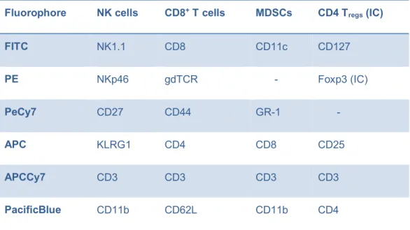
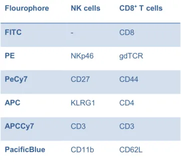
3.1.5 Flow cytometry antibodies ... 46
3.2 Methods... 49
3.2.1 Evaluation of tumor formation and – progression in Grm1 mice... 49
3.2.2 IFNγ- Elispot of NK cells ... 50
3.2.3 Treatment of Grm1 mice with NK cell-depleting antibody PK136 ... 50
3.2.4 Preparation of soluble IL-15 pre-complexed to IL-15Rα ... 51
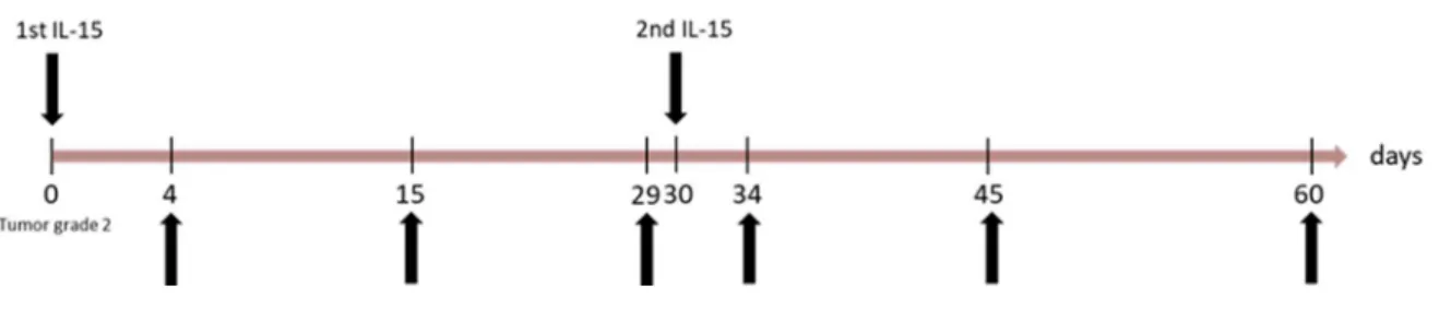
3.2.5 Treatment of Grm1 mice with pre-complexed IL-15 ... 51
3.2.6 Counting of metastases in lung and liver sections of Grm1 mice ... 51
3.2.7 Phenotypical analysis of different immune cell subpopulations in Grm1 mice on day 4 and day 60... 52
3.2.8 Cytotoxicity assay of NK cells from Grm1 mice ... 53
3.2.9 IncuCyte® ... 53
3.2.10 Analysis and sorting of immune cell subsets at different time points after IL-15 treatment in Grm1 wt and B6 wt mice ... 54
3.2.11 Whole transcriptome amplification (WTA) ... 55
5
3.2.12 RT² Profiler and single qPCR ... 55
3.2.13 Transplantation of mammary glands of Balb-NeuT mice to Balb/c wt mice ... 57
3.2.14 Monitoring of primary tumor development and curative surgery ... 57
3.2.15 Phenotypical analysis of different immune cell subpopulations in the Balb- NeuT model ... 57
3.2.16 CD107a degranulation assay ... 58
3.2.17 Measurement of glutamate concentrations in serum and skin samples of B6 wt and Grm1 Tbet
-/-mice... 59
3.2.18 Analysis of differentiation and proliferation of naïve CD8
+T cells after addition of different concentrations of glutamate ... 59
3.2.19 Calcium (Ca
2+) influx ... 60
3.2.20 Annexin V / PI assay ... 60
3.2.21 Analysis of activation marker expression of CD8
+T cells after addition of different concentrations of glutamate ... 61
3.2.22 Analysis of Eomesodermin (Eomes) and Tbet expression of CD8
+T cells after addition of different concentrations of glutamate... 61
3.2.23 Mixed leukocyte reaction (MLR) of B6 CD8
+T cells and Balb/c splenocytes ... 61
3.2.24 Analysis of activation marker expression of CD4
+T cells after addition of different concentrations of glutamate ... 62
3.2.25 Statistics ... 62
4 Results ... 65
4.1 Spontaneous melanoma mouse model - Grm1 ... 65
4.1.1 Activity of Grm1 NK cells was similar to B6 wt NK cells ... 65
4.1.2 NK cell depletion in Grm1 mice had no influence on tumor onset and tumor progression ... 66
4.1.3 IL-15 treatment in Grm1 mice did not prevent tumor progression and metastasis formation ... 67
4.1.4 IL-15 effects on CD8
+T cells, but not NK cells, four days after treatment . 69 4.1.5 IL-15 treatment induces significant changes in CD8
+T cell and NK cell subsets 60 days after treatment ... 70
4.1.6 IL-15 treatment led to higher NK cell cytotoxicity in vitro and ex vivo ... 71
6 4.1.7 IL-15-stimulated NK cells from Grm1 mice tended to have higher killing capacity compared to IL-15-stimulated NK cells of B6 wt mice ... 72 4.1.8 IL-15 shortly boosted NK cells, but significantly reduced NK cell numbers after dual treatment ... 74 4.1.9 IL-15 led to a proportional shift to CD27
lowmNK cells in Grm1 mice ... 75 4.1.10 IL-15 led to a significant increase in central memory CD8
+T cells (TCMs) ... 79 4.1.11 IL-15 treatment led to decreased expression of Eomes and NKG2D in NK cells several days after stimulation... 82 4.2 BalbNeuT model ... 85 4.2.1 Monitoring of primary tumor growth and curative surgery in Balb/c wt mice transplanted with BalbNeuT mammary glands ... 85 4.2.2 Transplanted mice developed lung metastases after curative surgery of the primary tumor ... 86 4.2.3 Presence of primary breast tumors or lung metastasis did not lead to a rise of effector immune cells in spleen ... 87 4.2.4 Changes in NK cell subsets were observed in lungs five weeks after
curative surgery of the primary tumor... 89 4.2.5 NK cell activity was enhanced in the presence of primary tumors... 91 4.3 Glutamate ... 95
4.3.1 Grm1 Tbet
-/-mice did not show higher glutamate concentrations in serum and skin samples compared to B6 wt mice ... 95 4.3.2 High glutamate concentrations led to impaired differentiation of naïve CD8
+T cells ... 96 4.3.3 High glutamate concentrations led to impaired proliferation of naïve CD8
+T cells after stimulation... 97 4.3.4 Ca2
+influx after addition of different glutamate concentrations ... 98 4.3.5 Cell death is not responsible for effects of glutamate on CD8
+T cells ... 99 4.3.6 Higher glutamate concentrations led to decreased expression of
transcription factors Eomes and Tbet... 99
4.3.7 Expression of different activation markers in CD8
+T cells is decreased with
higher glutamate concentrations ...101
4.3.8 Higher glutamate concentrations impaired alloreaction of CD8
+T cells ...102
4.3.9 Glutamate does not affect expression of CD4
+T cell activation markers .103
7
5 Discussion ...105
6 Sources ...115
7 Appendix ...131
7.1 List of abbreviations ...131
7.2 List of figures ...135
8
9
Acknowledgement
First of all, I am grateful to Prof. Dr. med. Hans J. Schlitt, the director of the Clinic and Polyclinic for Surgery, for providing a PhD position in the Department of Experimental Surgery.
I would like to express my very great appreciation to my supervisor Prof. Edward K.
Geissler. He patiently guided me through my thesis and encouraged me day by day to never give up and do my best.
My sincere thanks go to Prof. Dr. Gunter Meister and Prof. Dr. med. Markus Guba for being my mentors and giving me good advice at my annual research report meetings.
I would like to offer my special thanks to Dr. med. Philipp Renner for enabling the PhD position at the Experimental Surgery and for guiding my PhD project.
Many thanks go to Dr. rer. nat. Elke Eggenhofer, who always had an open ear for me and always gave me good advice.
I am particularly grateful for the assistance given by Lydia Schneider. She taught me every experimental method and I could not have done my work without her experience and help.
I would like to thank my colleagues Tatjana Libeld and Manfred Haas for actively supporting me in the laboratory and helping me with my experiments.
I would like to thank all of my colleagues in the Department of Experimental Surgery for the friendly and supportive atmosphere, which made my work even more fun. My special thanks are extended to Christian Mulas and Regina Heindl, colleagues of my collaboration with the Department of Experimental Medicine, for helping me with one of my mouse projects and performing all transplantation work.
Finally, I wish to thank my family for their support and encouragement throughout my
study.
10
11
1 Abstract
Cancer is one of the leading causes of death worldwide. In 2018, the International Agency for Research on Cancer (IARC) recorded 18.1 million new cancer cases and a mortality rate of 9.6 million cancer deaths with rising incidence. In 2040, an increase of 61.7% is expected compared to 2018. Treatment of cancer depends on type and stage, and treatment modalities range from surgery, radiotherapy, chemotherapy and hormone therapy to targeted therapy which can be used as single treatments or in combination. Most recently, immunotherapy has shown great promise to treat or cure cancer, immune cells can reach tissues where surgery is impossible and can even treat microscopic diseases or disseminated metastases. There are several types of immunotherapies including ones using tumor-specific immune cells (“cell therapy”) to attack cancer cells, or those which enhance already existing tumor-specific immune responses in the body.
In my thesis, two different tumor mouse models mimicking natural tumor development
were used to investigate the role of innate and adaptive immune cells, like NK cells
and CD8
+T cells, in natural tumor immune surveillance. We observed that tumor
development in mice generally led to activation of the differentiation and activity of NK
cells. However, sustained stimulation of NK cells by tumor cells resulted in the
generation of terminally-differentiated CD27
lowNK cells with diminished anti-tumor
capacities. While IL-15 treatment of mice promoted the development of these specific
immune cell subsets, it led to the exhaustion of NK cells. In turn, tumor development
did not alter the distribution of CD8
+T cell subsets and naïve CD8
+T cells were the
predominant subpopulation over time. Only the treatment of mice with IL-15 led to
accumulation of terminally-differentiated central memory T cells which were also
characterized by low cytotoxicity and reduced cytokine production. Taken together,
tumor cells that develop naturally in a spontaneous cancer model are able to evade
immune destruction by promoting the generation of immune cells with low anti-tumor
activities. Treatment of mice with IL-15 boosts the immune system, but accumulation
of terminally-differentiated immune cells with an exhausted phenotype lead to the
ultimate inefficiency of this cytokine treatment and to uncontrolled development of
tumors and metastases.
12
13
2 Introduction
2.1 Cancer and metastasis
2.1.1 Development and characteristics
Noncommunicable diseases (NCDs) are the main cause of death worldwide. They are defined as persistent medical conditions resulting from genetic, environmental and physiological alterations. Besides cardiovascular and respiratory diseases, as well as diabetes, cancer is one of the main four types of NCDs
1. In 2018, the International Agency for Research on Cancer (IARC) recorded 18.1 million new cancer cases and a mortality rate of 9.6 million cancer deaths worldwide. Lung, breast, colorectal, prostate and stomach cancer are the five most commonly diagnosed cancer types.
Breast cancer represents the highest incidence in females (24.2%) in comparison to lung (14.5%) and prostate (13.5%) cancer in males
2. Many factors can lead to cancer development. Genetic disorders, chemical and physical carcinogens, like ionizing radiation and UV light, but also bacteria, viruses or parasites can cause a malignant neoplasm
3.
Hanahan and Weinberg proposed six significant characteristics of cancer cells in “The
Hallmarks of Cancer” in 2000
4. They comprise sustained proliferative signaling,
evasion from growth suppressors, resistance to cell death, replicative immortality,
induction of angiogenesis and activation of invasion and metastasis
5. Tumor
metastases are actually the primary cause of cancer morbidity and mortality (90%)
versus the primary tumor itself. In 2011, Hanahan and Weinberg extended these
hallmarks with two additional characteristics: cancer cells are able to reprogram their
energy metabolism and can evade destruction by the immune system (Fig.1)
5.
In a process, called angiogenesis, tumors can trigger the formation of new blood
vessels. Detached cells can develop a mesenchymal-like phenotype (epithelial
mesenchymal transition, EMT) and use the formed blood or lymphatic system to
intravasate from tumor sites to surrounding tissues
6,7. After evading the immune
system and surviving in the circulation, these potentially “metastatic” cells adhere to
vessel walls and extravasate in distant organs. Only <0.01% of circulating tumor cells
overcome selection and are able to reactivate their proliferation and form new blood
14 vessels to provide the growing macroscopic tumor
8-11. The most common sites of cancer spreading are lung, liver and bone. In 1889, Steven Paget already postulated that the spread of metastasis is not random into any organ. The “seed” (tumor cells) and the “soil” (organ) have to be compatible. The target organ or tissue where tumor cells with metastatic potential migrate must provide a proper microenvironment
5,12,13.
Since many years there is discussion as to how metastases form, resulting in two different models: the linear and the parallel progression models. In the linear model, disseminated tumor cells (DTCs) spread from late stage primary tumors, acquire genetic mutations and migrate into distant sites of the body. On the contrary, in the parallel progression model, DTCs detach early from first primary tumor lesions and develop as metastases in parallel with the primary tumor
14.
Figure 1: Hallmarks of Cancer postulated by Hanahan and Weinberg in 2000 and 2011(modified from 4,5).
15 2.1.2 Treatment of cancer
Treatment of cancer is dependent of the type, location and stage of disease and has to be decided individually. There are many forms of treatment like surgery, radiotherapy, chemotherapy, hormone therapy or targeted therapy. The general problem of cancer treatment is that many patients remain asymptotic in early stages of the disease, and early signs and symptoms of cancer when they do occur are ignored.
Thus, the tumor can spread undisturbed in different parts of the body, called
“secondary or metastatic sites”. Treatments of patients with metastatic cancer often fail while because primary tumors are indeed sensitive to radiotherapy or chemotherapy, metastases are more resistant to cytotoxic agents or cannot be precisely localized to be cut out
15. Moreover, chemotherapy and radiotherapy have many severe side effects including non-specific effects on surrounding healthy tissues
16.
Immunotherapy became attractive and promising over the last few years. Endogenous immune cells can reach tissues where surgery is impossible and can even treat microscopic diseases and disseminated metastasis. Furthermore, immunotherapy offers long-term treatment in comparison to chemotherapy or radiotherapy that have effects only at the time they are being applied; the immune system has memory and often persists for a lifetime. The functions and activities of relevant immune cells is the main subject of this thesis work and will be concentrated on in the remaining part of this introductory section. There are several types of immunotherapies including ones using tumor-specific immune cells to attack cancer cells, or ones which generally enhance already existing tumor-specific immune responses in the body. Discussed below are some of the main immunotherapy approaches:
Monoclonal antibodies (mABs) made in laboratories are specific antibodies
recognizing proteins, such as cancer cells. After binding tumor-associated proteins,
immune cells are attracted and can bind the mABs via their Fc receptors. Afterwards,
immune cells are capable of killing cancer cells in a process called antibody-dependent
cell-mediated cytotoxicity (ADCC)
17. Classically, natural killer (NK) cells are the main
immune cell population which recognizes Fc domains of mABs. Secretion of perforin
and granzymes by activated NK cells lyse tumor cells resulting in release of tumor
antigens which are taken up by antigen-presenting cells (APCs).
16 Tumor antigens are presented on major histocompatibility complex (MHC) molecules to cytotoxic T cells (CTLs) which leads to activation of the CTLs and destruction of tumor cells
18. Furthermore, B cells are stimulated by the presented tumor antigens, resulting potentially in the development of tumor-specific antibodies. mABs can also be used to therapeutically target cancer cells. For example, the mAB Trastuzumab recognizes the extracellular domain of the Her2/neu receptor, which belongs to the EGFR family and is overexpressed in 25% of breast cancers. Dimerization of the receptor leads to cell proliferation, cell survival and metastases
19. By targeting these overexpressing tumors, a combination of Trastuzumab therapy with cytotoxic chemotherapy can promote survival in women with metastatic breast cancer compared to chemotherapy alone
20.
Another form of immunotherapy in use today involves checkpoint inhibitors. Cancer
cells can evade immune destruction through expression of ligands that bind to
checkpoint proteins on immune cells. Such proteins, like cytotoxic T-lymphocyte
antigen 4 (CTLA-4), programmed death-1 (PD-1) and lymphocyte antigen gene 3
(LAG-3) regulate the immune system (generally inhibitory), and are important for self-
tolerance. Attachment of tumor ligands to checkpoint proteins can potentially dampen
the anti-tumor immune response. Therefore, antibodies against checkpoint proteins or
against tumor ligands which bind to checkpoints (PD-L1) have been developed to
achieve long-lasting immunity
21. In more detail, T cell activation occurs via two signals,
where the first signal involves the interaction between antigens that are presented on
MHC molecules of APCs and the T cell receptor. T cells then require a second, co-
stimulatory, signal that often involves the interaction of CD28 on T cells and
CD80/CD86 on APCs. CTLA-4 is a CD28 homolog which rather transmits signals to
inhibitory pathways in T cells after binding of CD80/CD86
22. Already in 1996, it was
shown that blockade of CTLA-4 leads to the promotion of anti-tumor responses in
mice
23. In 2011, Ipilimumab was approved by the FDA as the first CTLA-4 checkpoint
inhibitor that improved the overall survival of patients with metastatic melanoma
24,25.
PD-1 is also a CD28 homolog and binds to the ligands PD-L1 and PD-L2 that can be
upregulated by IFN-γ
26,27. Interaction between PD-1 and PD-L1/PD-L2 leads to
immune suppression and is correlated with poor prognosis in some cancers like
17 melanoma and gastric cancer
28-30. Pembrolizumab was the first antibody that was approved by FDA as a PD-1 inhibitor and is used for treatment in different cancer types
31. Nivolumab is another PD-1 inhibitor that showed therapeutic responses of 44% and progression-free survival rates of 6.9 months in patients with melanoma.
Combination therapy of nivolumab and the CTLA-4 inhibitor ipilimumab enhanced response rates to 58% and progression-free survival to 11.5 months
32,33. In many cancer types, PD-L1 is constitutively expressed on tumor cells or the expression is induced by inflammatory signals
34,35. Besides binding to PD-1, PD-L1 binds to CD80/CD89 which leads to dampening effects on T cell stimulation and anti-tumor activity
36. The FDA has approved three different PD-L1 inhibitors at the moment:
atezolizumab, durvalumab and avelumab
37-39.
Another promising cancer therapy uses chimeric antigen receptor (CAR) T cells
(Fig.2). CARs are made by first harvesting T cells from patient blood. T cell receptors
are genetically engineered in these cells ex vivo to improve the recognition of tumor-
associated antigens (TAAs) without prior presentation of these antigens through MHC
molecules
40-42. Thus, it is no longer possible for tumor cells to escape immune
destruction through downregulation of MHCs
43. Moreover, since CAR T cells are not
dependent of MHC class I or II, CD8
+T cells as well as CD4
+T cells can be modified
40.
T cell receptors are engineered in different ways to become CARs. Besides viral
transduction and use of transposons, T cell receptors are altered by CRISPR/Cas9
vectors or through non-viral transfer methods. Currently, there is already the 4
thgeneration of CARs which are able to simultaneously recognize a broad range of tumor
antigens with high specificity and activate themselves due to signaling and co-
stimulatory domains in the intracellular part of the receptor
44. After modification of T
cell receptors, CAR T cells are expanded in culture with IL-2 and undergo extensive
quality control before reinfusion into the cancer patient. During CAR T cell production
in the laboratory, patients receive lympho-depleting chemotherapy to “make space” for
the cells to be infused. Also, immune suppressive cells such as regulatory T cells
(Tregs) and myeloid-derived suppressor cells (MDSCs) are depleted to improve the
efficiency and persistence of CAR T cells
45,46.
18 Once the cells are back in the patient and after the recognition of TAAs, CAR T cells kill tumor cells via secretion of perforins and granzymes and/or via the expression of Fas ligands to activate the Fas death signaling in malignant cells. The repertoire of ligands that can be recognized by CAR T cells is huge and diverse. However, presently, the most promising results of CAR T cell therapy are observed against B cell malignancies by treatment with anti-CD19 CARs
47-49. 65% of the current 113 registered CAR T cell trials are targeting hematological malignancies
50. However, CAR T cell therapy of solid tumors is more challenging due to a lack of tumor-specific antigens.
Additionally, in hematological diseases, modified T cells have easy access to cancer cells and their tumor antigens. In contrast, migration into solid tumors requires attraction by chemokines, overcoming of tumor stroma and specific recognition of TAAs.
Figure 2: Principle of CAR T-cell therapy (modified from 41).
19 Cytokines regulate the development and maturation of innate and adaptive immune cells and their communication. In cancer therapy, cytokines are used to activate or maintain patients’ immune responses against tumors. Mouse models first showed that interleukins (ILs) or interferons (IFNs) like IL-2, IL-12, IL-15, IL-21, GM-CSF (granulocyte-macrophage colony-stimulating factor) and IFN-α increase the number of effector immune cells and enhance their anti-tumor immunity
51,52. Early on, IFN-α and high-dose IL-2 were approved for treatment of different human cancer types
53. IFN-α is a type I IFN which upregulates the expression of MHC molecules on tumor cells
54-56
. Moreover, IFN-α activates different types of immune cells including dendritic cells
(DCs), macrophages, CD8
+T cells and NK cells
57,58. Besides indirect effects of IFN-α
on tumor cells through stimulation of anti-tumor immune cell activity, IFN-α directly
interferes with proliferation and angiogenesis associated with malignant cells
59-61. IL-2
is used to stimulate T cell activation and expansion
62; furthermore, IL-2 leads to
secretion of cytokines and enhanced anti-tumor activities of NK cells, as well as B cell
expansion and differentiation. However, efficacy of cytokine therapy with IL-2 is
overshadowed by severe side effects. Besides life-threatening toxicities, IL-2 not only
activates effector immune cells but also immune suppressive cells like Tregs and
MDSCs
63. Moreover, the expression of inhibitory checkpoint proteins is enhanced by
IL-2 and the secretion of immune suppressive factors like IL-10 and TGF-β is
stimulated. IL-15 shares many features with IL-2 including the activation of
development, differentiation and anti-tumor activity of CD8
+T cells and NK cells
64.
However, it has some advantages in comparison to IL-2, since IL-15 is not as toxic and
does not stimulate immune suppressive cells
65. In Chapter 2.5 I will discuss IL-15 in
more detail and the relevance for this thesis. Cytokines are not only used as in vivo
adjuvants but also in vitro for stimulation and expansion of immune cells in adoptive
cell therapy. Dendritic cells are stimulated with GM-CSF, IL-4, TNF-α, IL-6 and IL-1β,
whereas IL-15 and IL-21 are the most prominent cytokines for activating and
expanding T cells. Patients receive low-dose IL-2 after reinfusion of T cells to improve
the survival of transferred cells leading to better clinical outcomes
66.
20 2.2 Tumor immune surveillance and immunoediting
The hypothesis that cancer development is monitored and controlled by immune cells was created decades ago. In the 20
thcentury Frank MacFarlane Burnet and Lewis Thomas created the immune surveillance theory
67. Different mouse models and also human studies confirmed this hypothesis and showed that the immune system, especially lymphocytes and their secreted cytokines, are able to recognize and eliminate primary tumors (reviewed in
68). However, doubts gradually arose about this hypothesis because of the development of cancer in immunocompetent mice with an intact immune system. Different further experiments showed the formation of tumors with reduced immunogenicity or the acquirement of mechanisms to evade or suppress the immune system. Therefore, the term “cancer immunoediting” was created including the host-protecting and tumor-sculpting processes of the immune system
68. Dunn et al. divided the cancer immunoediting in three phases (three Es) which will be described in the following chapters: Elimination, Equilibrium and Escape (Fig.3).
2.2.1 Elimination
The elimination phase is known as the conventional cancer immune surveillance raised
by Burnet and Thomas. In this phase, the immune system recognizes and eliminates
transformed tumor cells before they become clinically apparent. For example, APCs
like dendritic cells are activated by tumor-secreted danger signals (DAMPs, danger-
associated molecular patterns). In addition, T cells recognize tumor antigens with their
T cell receptor provoking the induction of adaptive anti-tumor responses
69,70. There are
two different sorts of tumor antigens. A distinction is made between tumor-specific
antigens (TSA) and tumor-associated antigens (TAA). TSAs are antigens that are only
present on tumor cells but not on normal cells. In comparison, TAAs are expressed on
tumor as well as on normal cells, however normally at lower levels on normal cells
71,72.
Besides APCs and T cells, NK cells and their receptors bind ligands (to be explained
in a later chapter) expressed on tumor cell surfaces resulting in the release of
immunomodulatory and pro-inflammatory cytokines which can kill tumor cells directly
or again promote the development of tumor-specific adaptive immune responses
73-75.
21 At that time, researchers thought that the immune system is able to completely destroy tumor cells. However, there are rare tumor cell variants surviving the elimination phase and entering the next phase of the immunoediting process, the “equilibrium”.
2.2.2 Equilibrium
Some tumor cells which were not recognized and destroyed by immune cells in the aforementioned elimination phase go into dormancy
76. These cells stop their proliferation, development and activity for periods of time up to decades in length. In this equilibrium phase tumor cells adapt to constant selection pressure of adaptive immune cells, especially by T cells or via genetic instability. T cells even sculpt the immunogenicity of the tumor, as was confirmed in different transplant models. For instance, tumors from mice with immunodeficiency were more immunogenic than tumors from immunocompetent mice, presumably because tumor cells with stimulatory antigens were not as likely to be eliminated. Transplantation of these tumors into wildtype mice showed a higher elimination rate of tumors raised in an immunodeficient background
76. Only highly immunogenic cells were eliminated and non-immunogenic cells could grow
77. Moreover, deficiencies in IL-12 and IFNγ signaling pathways lead to progressive tumor growth when transplanted into wildtype mice. This work served to demonstrate the role of adaptive immune components in changing (sculpting) tumor immunogenicity
78.
2.2.3 Escape
In the last phase of the immunoediting process, called the “escape” phase, edited tumor cells with low immunogenicity start growing again and appear as visible tumors.
They are resistant to detection and elimination by the immune system. The entry into
this phase can be caused by different events. Tumor cells are able to change the
expression of tumor antigens and ligands (e.g. MHC components)
79. Downregulation
allows tumor cells to be invisible to immune cells and further tumor progression can
occur
80,81. Moreover, tumor cells producing immunosuppressive cytokines like tumor
growth factor β (TGF-β), vascular endothelial growth factor (VEGF), indoleamine 2,3-
dioxygenase (IDO) or IL-10 can favor the microenvironment towards tumor growth
82,83.
22 Recruitment of Tregs and MDSCs can also inhibit anti-tumor responses as another escape mechanism for tumor cells to evade immune surveillance.
Figure 3: Three E´s of cancer immunoediting: Elimination, Equilibrium and Escape (modified from68,84).
Abbreviations: NKR= natural killer cell receptor, DAMPs= danger-associated molecular patterns, NK cell=
natural killer cell, DC= dendritic cell, NKT= natural killer T cell, MHC= major histocompatibility complex, IL=
interleukin, TGF= transforming growth factor, IDO= Indolamin-2,3-Dioxygenase, VEGF= Vascular Endothelial Growth Factor, PD-L1= programmed cell death ligand 1, Tregs= regulatory T cells, MDSCs=
myeloid-derived suppressor cells.
23 2.3 Natural Killer (NK) cells
2.3.1 General characteristics and functions
The innate immune system includes physical epithelial barriers, phagocytic leukocytes, dendritic cells and natural killer (NK) cells. The latter NK cells are large granulocytes which are widely distributed in minor fractions (2-10% in mouse, 2-18% in human) in lymphoid and non-lymphoid tissues. NK cells are capable to recognize and kill pathogen-infected or neoplastic cells without prior sensitization
85. There are different ways how NK cells can act:
Firstly, NK cells secrete two main cytotoxic proteins. Effector NK cells release perforin resulting in disruption of cell membranes followed by secretion of granzymes.
Granzymes are serine proteases that can pass cavernous cell membranes of target cells and induce apoptosis (Fig 4; 1)
86,87. Different studies using perforin-deficient mice show the importance of this protein in NK cell cytotoxicity and tumor immune surveillance, whereas the role for granzymes is less well described
88,89.
Secondly, NK cells activate the Fas death receptor pathway by expressing FasL and tumor necrosis factor-related apoptosis-inducing ligand (TRAIL) on NK cell surfaces leading to programmed cell death
90,91(Fig. 4; 2). Furthermore, secretion of IFN-γ by NK cells induces the expression of death receptors on target cells
92. However, signaling through IFN-γ is slower than the perforin/granzyme pathway, occurring only after several hours.
Thirdly, for example, after treatment of patients with a monoclonal antibody therapy, NK cells recognize antibody-coated tumor cells with their Fc receptors (e.g. CD16).
These receptors cross-link and activate NK cells and induce ADCC (Fig.4; 3).
After NK cell activation, secretion of cytokines like IFN-γ, TNF and GM-CSF leads to activation of further innate immune cells such as DCs, macrophages and neutrophils.
Moreover, NK cells can activate the adaptive arm of the immune system, especially T
cells
93(Fig.4; 4). Different chemokines like IFN-γ, GM-CSF and TNF produced by NK
cells attract the aforementioned effector cells to sites of inflammation. Especially IFN-
γ can initiate the upregulation of MHC class I and II on other immune cells resulting in
stimulation of antigen presentation and orchestration of immune cell interactions
85.
24 Tumor cell proliferation and growth can be dampened with the antiangiogenic interferon gamma-induced protein 10 (IP-10), also induced by IFN-γ
94.
Figure 4: Different mechanisms of NK cell killing of tumor cells. 1. Secretion of perforin and granzymes; 2.
Expression of FasL and TRAIL to initiate Fas death pathway; 3. Antibody-dependent cell cytotoxicity; 4.
Cytokine secretion for activation of other innate and adaptive immune cells. Abbreviations: IFN= interferon, TRAIL= Tumor Necrosis Factor Related Apoptosis Inducing Ligand, TNF= tumor necrosis factor, GM-CSF=
Granulocyte-macrophage colony-stimulating factor, DC= dendritic cell, ΜΦ= macrophages.
25 2.3.2 Inhibitory and activating NK cell receptors
A wide range of inhibitory and activating cell surface receptors regulates NK cell activity. These receptors are genetically encoded in comparison to ones of B and T cells which are made by somatic recombination
85. Expression of different combinations of receptors leads to heterogeneity within the NK cell population and offers the opportunity to respond to a diversity of stimuli.
Inhibitory receptors such as killer immunoglobulin-like receptors (KIRs, human) and C-type lectin-like receptors (Ly49, mouse) mainly bind to MHC class I molecules, which are human leukocyte antigen (HLA ) class I molecules in humans
95,96. Another group of inhibitory receptors such as CD94/NKG2A receptors recognize non-classical MHC class I molecules (HLA-E in human; Qa-1 in mouse)
97,98. The receptors have cytoplasmic immune receptor tyrosine-based inhibitory motifs (ITIMs), which are phosphorylated after binding of these ligands. The phosphorylation results in recruitment and activation of tyrosine phosphatases SHP-1 and SHP-2, leading to downstream signaling and inhibition of NK cells
99,100. Absence of MHC class I molecules on target cells (“missing-self”) and/or stimulation of activating receptors activates NK cell cytotoxicity. Therefore, healthy cells that express high levels of MHC class I molecules are spared from NK cell activity. Killer cell lectin-like receptor G1 (KLRG1) binds cadherins and acts through an ITIM, like the receptors mentioned above
101,102. In Table 1, inhibitory receptors of human and mouse NK cells with their corresponding ligands are listed.
Additionally to inhibitory receptors, NK cells also express a wide range of activating
receptors. The signaling occurs through immune-receptor tyrosine-based activating
motifs (ITAMs). Tyrosine residues are phosphorylated after binding of a ligand followed
by recruitment of the kinases. Afterwards, downstream signaling leads to degranulation
of cytokines and chemokines. However, stimulation of more than one activating
receptors is required to activate NK cells
103, and strong stimuli are needed to overcome
inhibitory signals. Besides signaling through activating KIRs and Ly49 receptors,
stimulation of NKG2D leads to proliferation, cytotoxicity and cytokine production by NK
cells
104.
26 Natural cytotoxicity receptors (NCRs), e.g. NKp30, NKp44 or NKp46 bind a wide range of bacterial-, virus- and parasite-originating ligands and are also involved in NK cell cytotoxicity against tumor cells
105-107. CD16 recognizes specific antigens on tumor cells and induce tumor cell death by ADCC
108. In Table 2, activating receptors of human and mouse NK cells with their corresponding ligands are listed.
Table 1: Inhibitory receptors of human and mouse NK cells
Inhibitory receptors Receptor Ligand
Human KIR HLA-A/B/C
CD94/NKG2A HLA-E
KLRG1 Cadherin E/N/R
Mouse Ly49A/C/I/P MHC class I
CD94/NKG2A Qa-1
KLRG1 Cadherin E/N/R
Table 2: Activating receptors of human and mouse NK cells
Activating receptors Receptor Ligand
Human KIR HLA-A/B/C/G
NKG2C/E HLA-E
NKG2D MICA/MICB/ULBP
NKp30, NKp40, NKp46 (NCRs) Viral-, bacterial, parasite-, cellular- ligands
CD16 -
Mouse Ly49D/H MHC class I
NKG2C/E Qa-1
NKG2D H-60/Rae-1/Mult1
27 2.3.3 Human NK cell subpopulations
Human lymphocytes consist of 2-18% NK cells that are distinguishable from T cells due to the absence of CD3 expression. They can be divided into two subpopulations identified by the cell surface marker CD16 and CD56
109. On the one hand, there are CD16
brightCD56
dimNK cells that are highly cytotoxic immune cells with poor cytokine production
110,111. They constitute 90-95% of NK cells and are present in blood and at sites of inflammation
112. The high expression of CD16 binding to immunoglobulins leads to killing of target cells through ADCC. On the other hand, CD16
dim/negativeCD56
brightNK cells predominantly located in lymph nodes are less cytotoxic but are potent producers of cytokines like IFN-γ, TNFβ, IL-10, IL-13 and GM- CSF after obtaining different activation signals
109. NK cells respond to cytokines like IL-1, IL-2, IL-12, IL-15 or IL-18 released by other immune cells such as APCs or through signals recognized by their activating receptors CD16 and NKG2D
109,113. 2.3.4 Mouse NK cell subpopulations
Mouse NK cells do not express CD56, and are rather divided by their expression of surface markers CD11b and CD27 representing their differentiation status
114. They develop from CD11b
negCD27
highimmature (iNK), over CD11b
posCD27
highmature NK cells to CD11b
posCD27
lowterminally-differentiated NK cells
115. CD27
highare comparable to CD56
brighthuman NK cells also present in lymph nodes with a high capacity to produce cytokines like IFN-γ. They have the ability to kill tumor cells which express MHC I, which is consistent with the lower levels of inhibitory receptors for MHC I on CD27
highNK cells
110. Terminally-differentiated CD27
lowNK cells show low cytotoxicity and reduced production of cytokines. Concerning proliferation there are also differences between subsets. Immature and CD27
highNK cells have high proliferation capacity in comparison to CD27
lowcells with a limited ability to divide.
Mature NK cells also express KLRG1 which is an inhibitory receptor and marker of terminal differentiation
116. NK cell development is dependent of different cytokines recognized by receptors containing a common gamma (γ
c) chain. Signaling through γ
cis required for their differentiation, homeostasis and function. This subunit is part of the
recognition complex for cytokines such as IL-2, IL-4, IL7, IL-9, IL-15 and IL-21
117. IL-
12 and Type I IFNs activate NK cells leading to antitumor activity
118.
28 2.3.5 NK cells in cancer and metastasis
As described in chapters above and shown in many in vitro and in vivo studies, NK cells play an important role in tumor immune surveillance by spontaneous killing of tumors without prior sensitization
119,120. In the course of immune evasion, malignant cells downregulate their expression of MHC class I to escape recognition by receptors of CD8
+T cells. Consequently, NK cells are able to detect these tumor cells (“missing self”) which results in NK cell proliferation and cytotoxicity including cytokine production and release of lytic granules
121,122. Furthermore, expression of stress-induced molecules (MICA, MICB and UL-16) on tumor cells leads to stimulation of activating NKG2D receptor
122,123. Studies showed that patients with high levels of peripheral blood NK cells have a longer metastasis-free survival
85. However, mice with deficiencies in NK cell cytotoxicity components like perforin and IFN-γ show an increased incidence of tumors
75,124. Moreover, perforin is important in controlling the formation of metastasis
125,126.
2.3.6 NK cells in immunotherapies
NK cells have been used in different therapy options against cancer. Autologous NK
cell therapy includes extraction of NK cells from patients, followed by in vitro
manipulation to expand them and increase their cytotoxicity before reinfusing into
patients where they are then better able to seek out and destroy tumor cells
127,128.
However, this therapy has limitations, such as through inhibition of NK cells by tumor
cell expression of self-HLA molecules
129. Hence, allogeneic NK cell therapy is
performed with transplantation of allogeneic hematopoietic stem cells or infusion of
mature allogeneic NK cells. Administration of monoclonal antibodies is another method
to induce ADCC by NK cells. These antibodies bind to tumor cells that are then
recognized by the activating receptor CD16 on NK cells
130,131. Besides use of different
cytokines to expand and activate immune cells, modified NK cells called chimeric
antigen receptor (CAR) NK cells have been utilized. These CAR NK cells are
genetically engineered for more efficient and specific binding to tumor antigensf
132-134.
29 2.4 CD8
+T lymphocytes
2.4.1 General characteristics and functions
CD8
+T cells are cytotoxic T lymphocytes (CTLs) that are generated in the thymus and belong to the adaptive arm of the immune system. Their major role is the clearance of intracellular pathogens and tumors. After recognition of foreign MHC class I antigens presented by APCs, CD8
+T cells perform effector functions comparable with those of NK cells. CD8
+T cells release perforin and granzymes to kill malignant cells. Secretion of IFN-γ and TNF-α strengthens their immune response and activates other cells of the innate and adaptive immune system
135. Like NK cells, CD8
+T cells express FasL on the cell surface binding to the Fas receptor on target cells. After activation of the caspase cascade, targets cells to go into apoptosis.
2.4.2 Activation of CD8
+T cells
Naïve CD8
+T cells migrate from the thymus to secondary lymphoid organs, like lymph nodes and spleen, where they are activated to mediate immune responses
136. The activation of CD8
+T cells requires two different signals: Signal 1 is the binding of a MHC protein by specific T cell receptors (TCRs) which is provided by APCs like dendritic cells
137. Importantly, CD8
+T cells recognize the main structure of MHC class I molecules, rather than the specific peptide in the MHC molecule cleft which is recognized by CD4 helper T cells. Signal 2 is mediated via stimulation through a co- receptor interaction with e.g. CD28 receptor on the T cell surface and B7 proteins (CD80/CD86) on APCs. Signaling through CD28 enhances signal 1 and is necessary for proliferation and activation of effector functions. Absence of signal 2 leads to apoptosis of T cells and represents one mechanism of self-tolerance. After binding of MHC and CD80/CD86 by T cells, downstream signaling leads to IL-2 production and stimulation of CD8
+cell proliferation in an autocrine and paracrine feedback loop.
Effector cells produce cytokines like IFN-γ and TNF-α as well as perforin and
granzymes. After performing their effector functions, 95% of the CD8
+T cells die and
only a small fraction of memory CD8
+T cells persist. With the presence of specific
cytokines like IL-15 these memory cells can survive in the host
138.
30 2.4.3 Human CD8
+T cell subsets
Human CD8
+T cell development occurs in the following order with increasing cell differentiation and effector functions and decreasing memory function and proliferation:
naïve T cells (T
N), stem cell memory cells (T
SCM), T central memory cells (T
CM), T effector memory cells (T
EM) and T effector cells (T
eff)
42. Human CD8
+T memory and effector cells can be divided by the markers CD27 and CD45RA. Naïve CD8
+T cells express both markers in comparison to memory cells only expressing CD27 and effector cells only expressing CD45RA
139. CCR7 is another marker distinguishing CD8
+T subsets. Memory cells express this gene in contrast to effector cells which do not show CCR7 expression
140. CCR7 is a chemokine receptor which is responsible for T cell trafficking and homing within secondary lymphoid organs
141.
2.4.4 Mouse CD8
+T cell subsets
Mouse CD8
+T cells differentiation is similar to that of human CD8
+T cells. Naïve T cells differentiate to effector, T
EMand finally to T
CMcells
142. CD8
+T cell subsets in mice can be identified by the expression markers CD44 and CD62L. They differentiate from naïve CD44
lowCD62L
posT cells (TN) to CD44
highcells that can be further distinguish into CD62L
negeffector T cells (Teff) and CD62L
poscentral memory T cells (TCM).
These two effector cell subsets differ in location and effector functions (Fig. 5). Teff
cells are found in inflamed peripheral tissue with high effector functions like production
of high amounts of perforin, IL-4, IL-5 and IFN-γ after antigen stimulation
143. On the
contrary, TCM cells are in secondary lymphoid organs displaying less or no effector
functions. However, TCM cells secrete IL-2 quickly after stimulation resulting in a
feedback loop leading to their proliferation and differentiation into Teff cells with IFN-γ
secretion
140. TCM cells can be activated by antigen stimulation as well as by response
to different cytokines. For example, in vivo mouse studies show proliferation of T cells
after stimulation with IL-7 and IL-15
144,145.
31
Figure 5: Mouse CD8+ T cell subsets with different locations and effector functions: effector CD8+ T cells (Teff); central memory CD8+ T cells (TCM)
2.4.5 CD8
+T cells in cancer and metastasis
T cells entering tumors are called tumor-infiltrating lymphocytes (TILs) and are considered as a positive prognostic marker in many different solid tumors
146-148. CD8
+T cells kill target cells in different ways comparable to the capabilities of NK cells.
In vivo studies show earlier tumor onset in mice lacking lymphocytes or the IFN-γ
receptor after injection of methylcholanthrene (MCA)-induced sarcoma in comparison
to wildtype mice. Similar results are observed in mice developing spontaneous
epithelial tumors
124. However, during tumor progression CD8
+T cells can become
exhausted and tumor cells evade tumor immune surveillance through different
mechanisms
149. CD8
+T cell-secreted IFN-γ enhances expression of PD-L1 on cancer
cells which binds to PD-1 on T cells leading to suppression of their activity. Moreover,
Tregs in the tumor microenvironment induce CTLA-4 expression on T cells by releasing
adenosines, also resulting in immunosuppression after binding of the ligand
CD80/CD86 on tumor cells
150. Cancer cells stimulate MDSCs to produce TGF-β
enhancing the activity of immunosuppressive Tregs
151.
32 Besides adenosines, many other immunosuppressive compounds are released by cancer cells to escape killing by CD8
+T cells. Therefore, CD8
+T cells can either destroy or spare cancer cells, depending on the exact circumstances in the tumor microenvironment.
2.4.6 CD8
+T cells in cancer immunotherapy
To avoid peripheral tolerance of tumor cells by immune cells, immune-based therapy has become a standard or experimental procedure for treatment of many cancers
152,153. The aim of such therapies is the reactivation and expansion of tumor- specific cytotoxic T cells resulting in more targeted killing of tumor cells.
A very promising therapy is the use of monoclonal antibodies against checkpoint proteins on T cells. Many solid and hematopoietic tumors are treated with antibodies against CTLA-4 and/or PD-1/PD-L1 which prevent T cell inhibition by tumor cells and boost the activation of effector T cells to kill these malignant cells
154. Earlier in this review, the need for co-stimulation (signal 2) of T cells to activate or inhibit their activity was covered. Tumor cells can evade T cells by expressing inhibitory molecules, so the antibodies mentioned above serve to block these inhibitory ligands/receptors.
Currently, there are different clinical studies evaluating the efficiency of checkpoint inhibitors. However, only a small portion of patients show benefits from these therapies because of the development of resistances against the inhibitors
155. Combination therapies with e.g. chemotherapy or CAR T cell therapy show higher response rates than monotherapies
156.
Another approach is by CAR T cell therapy. In CAR T cell therapy, T cells from patients
are isolated from blood and genetically engineered in a biotechnology laboratory to
express a chimeric antigen receptor that specifically recognizes tumor antigens. The
receptor consists of an extracellular domain binding to tumor cells and intracellular co-
stimulatory and signaling domains responsible for proliferation and activation of
effector functions of CAR T cells after binding of tumor antigens
157. By changing the
combinations of co-stimulatory and signaling domains the anti-tumor activity of these
CAR T cells is improved significantly over time
158.
33 Presently, only CD19-specific CAR T cell therapies are approved by the FDA.
However, the advantages of this therapy in comparison to the aforementioned use of monoclonal antibodies are a high response rate and small numbers or lack of minimal residual cancer. CAR T cells also express checkpoint proteins after contact with tumor cells, giving them long term activity. Therefore, combination therapies with checkpoint inhibitors are performed to improve the cytotoxicity of CAR T cells and their secretion of IFN-γ
159. Moreover, CAR T cell efficiency is enhanced by use of interleukins like IL- 2, IL-4, IL-7, IL-12, IL-15 and IL-21.
2.5 Interleukin 15 (IL-15)
Immunotherapy takes advantage of the immune system to kill malignant cells without affecting healthy tissue. However, tumor cells can evade immune surveillance by interfering with the development and function of anti-tumor responses. Cytokine therapy was one of the earliest approaches to stimulate immune responses and develop long-lived tumor immunity. In early studies decades ago, treatment with IL-2 sometimes caused impressive tumor regression responses. However, only few patients responded to this therapy and there were very severe side effects
160,161. Interleukin 15 (IL-15), has more recently been used in cancer immunotherapy by the US National Cancer Institute and deserves further introduction especially with regard to this thesis
162.
2.5.1 Characteristics and signaling pathways
IL-15 is a four-α-helix protein belonging to the same family as IL-2. IL-15 can be produced by APCs. For example, dendritic cells produce IL-15 in the endoplasmic reticulum after their activation. Bound to an IL-15 receptor alpha subunit (IL-15Rα), IL- 15 is transferred through the cytoplasm to the cell surface of APCs to be presented to effector immune cells
163. A receptor complex on responding effector cells consisting of an IL-2/IL-15 receptor β (IL-15Rβ; CD122) subunit and a common gamma chain (γc;
CD132) receptor subunit bind to IL-15 with high affinity (Fig. 6). This binding results in
the activation of Janus kinases followed by initiation of the STAT5 pathway
164.
Moreover, IL-15 additionally stimulates the RAS-MAPK and PI3K-AKT pathway in
effector immune cells
165. Downstream signaling leads to proliferation, activation,
34 migration and decreased apoptosis of immune cells carrying the IL15 receptor complex like CD8
+T and NK cells
64,144,166,167. IL-15 in complex with the α subunit is also found in soluble form after cleavage from cell surfaces upon inflammatory signals
168. The binding of IL-15 to IL-15Rα is transient and short-lived but it prolongs the half-life of IL- 15 and enhances the affinity to the other subunits
169.
Figure 6: IL-15 receptor complex. IL-15 in complex with the IL-15α subunit is presented by APCs to the IL- 2/IL-15β and IL-15γ subunit expressed on effector immune cells (modified from 170).