zur
Erlangung der Doktorwürde der
Fakultät für Pharmazie der
Ruprecht-Karls-Universität Heidelberg
vorgelegt von
Diplom-Chemiker Ragen Pfeiffer
aus Berlin
Tag der mündlichen Prüfung: 17.12.2001
Development of Methods to Investigate Poly(ADP- ribose) polymerase-1 activity and DNA Base- Excision Repair in Relation to Cancer and Ageing
Erstgutachter: Prof. Dr. Manfred Wießler
Zweitgutachter: Priv.-Doz. Dr. Alexander Bürkle, Senior Lecturer
Contents
page
Abbreviations 5
I. Introduction 6
I. 1. Poly(ADP-ribose) polymerase-1 6
I. 2. Homologues of PARP-1 8
I. 3. Physiological and pathophysiological functions of PARP-1 9
I. 4. DNA excision repair in mammalian cells 11
I. 5. The mechanism of base-excision repair and the involvement of PARP-1 13 I. 6. The involvement of DNA repair and PARP-1 in the ageing process 17 I. 7. Current methods to detect and quantify poly(ADP-ribose) 21 I. 8. Current methods to measure DNA damage and DNA repair 24
I. 9. Aim of the project 28
II. Materials 31
II. 1. Appliances 31
II. 2. Chemicals 33
II. 3. Oligonucleotides 33
II. 4. Antibodies 34
II. 5. Membranes 34
II. 6. Buffers and solutions 34
II. 7. Cell culture medium 35
II. 8. Cell lines 35
II. 9. Animals and human blood donors 35
III. Methods 37
III. 1. Poly(ADP-ribose) immuno-dot-blot assay 37
III. 2. Automated fluorescence-detected alkaline DNA unwinding assay 38
IV. Results 40
IV. 1. Setting up a new immuno-dot-blot procedure to assess cellular
poly(ADP-ribosyl)ation capacity. 40
IV. 2. Evaluation of the new poly(ADP-ribose) immuno-dot-blot assay 44 IV. 3. Setting up an automated version of the fluorescence-detected
alkaline unwinding (FADU) assay to quantify DNA strand
breaks and repair in live cells. 46
IV. 4. Comparison of DNA strand break repair in a long-lived
(human) and a short-lived (rat)mammalian species. 54 IV. 5. Comparison of DNA strand break repair in PARP-1-/- and PARP-1+/+ cells. 57
V. Discussion 59
VI. Summary 63
VII. References 65
VIII. Own publications 73
IX. Acknowledgements 75
Abbreviations
b base
bp base pair
BER base-excision repair ß-MeEtOH ß-mercaptoethanol DMSO dimethylsulfoxid
Ex excitation
Em emission
ECL+ enhanced chemoluminescence+
EDTA ethylenediaminetetraacetate
FADU fluorescence-detected alkaline DNA unwinding
Gy Gray
EtBr ethidium bromide FcS foetal calf serum MNC mononuclear cells MMR mismatch repair
MNU N-methyl-N-nitrosourea NER nucleotide-excision repair NLS nuclear location signal p(ADP)r poly(ADP-ribose)
PARP-1 poly(ADP-ribose) polymerase-1; (EC 2.4.2.30) ROI reactive oxygen intermediates
SDS sodium dodecylsulfate TCA trichloroacetic acid
TEMED N,N,N',N'-tetramethylethylendiamine Tris Tris(hydroxymethyl)aminomethane
VPARP Vault-associated poly(ADP-ribose) polymerase
I. Introduction
I. 1. Poly(ADP-ribose) polymerase-1
Catalytic activation of poly(ADP-ribose) polymerase-1 (PARP-1; EC 2.4.2.30) is one of the immediate early reactions of eukaryotic cells to DNA-damaging treatment (for review: de Murcia & Shall, 2000; Bürkle, 2001a,b). PARP-1 is a nuclear 113-kDa enzyme that consists of three domains (Fig. 1. ): (1) A DNA-binding domain with two Zn2+ finger motifs (Gradwohl et al., 1990) and a nuclear location signal (NLS) (Schreiber et al., 1992); (2) an automodification domain, which serves as the major site for covalent attachment of (ADP- ribose) polymer; and (3) an NAD+-binding domain comprising the catalytic centre.
Fig. 1. Domain structure of PARP-1. The 113-kDa enzyme possesses at its N-terminal DNA binding domain two Zn fingers (F1 and F2)[module A] as well as a nuclear location signal (NLS) [module B]
that consists of two parts. In between these two parts there is a caspase-3 cleavage site. The second domain is the automodification domain [module D]. The third and C-terminal domain [modules E and F] carries the active site. Modified from de Murcia & Shall, 2000.
PARP-1 is a nuclear protein of very high abundance, with 500.000 to one million copies per cell (Ludwig et al., 1988), but is absent from the cytoplasm. PARP-1 activation is one of the first cellular responses to exposure to genotoxic agents.
PARP-1 specifically recognises DNA single and double-strand breaks via its zinc-fingers. The first zinc-finger binds to double-strand breaks, whereas the second zinc-finger is specific for single-strand breaks (de Murcia & Shall, 2000). However, the first zinc-finger is essential for the catalytic activity of PARP-1 by both double and by single strand breaks (Ikejima et al., 1990). PARP-1 is acting as a catalytic dimer (Mendoza-Alvarez & Alvarez-Gonzales, 1993), i.e. two PARP-1 molecules are binding to one strand break, with one enzyme molecule
NH2 COOH
DNA-binding Automodification Catalytic domain domain domain
Zn Fingers NLS BRCT Active site
F1 F2
A B C D E F
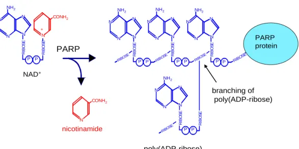
serving as the catalyst and the other as “acceptor” protein (i.e. substrate). On either side of the strand break 7 nucleotides are involved in the DNA-PARP-1 interaction and the typical V- shape of nicked DNA is stabilised (LeCam et al., 1994). The binding is very specific and of high affinity and leads to a 500-fold enhancement of the activity of the catalytic centre, probably mediated by some as yet unknown conformational change. Fig. 2 depicts the chemical structure of poly(ADP-ribose) [p(ADPr)], which is a biopolymer characterised by unique O-glycosidic ribose-ribose bonds (Chambon et al., 1966).
N
N N
N NH2
RIBOSE
P P
RIBOSE
N
N N
N NH2
RIBOSE
P RIBOSE N
N N
N NH2
RIBOSE
RIBOSE
N
N N
N NH2
RIBOSE
RIBOSE RIBOSE
N
poly(ADP-ribose) nicotinamide
NAD+
CONH2
RIBOSE N
N N
N NH2
RIBOSE
P P
RIBOSE
CONH2
N +
PARP
P P P P P
PARP protein
branching of poly(ADP-ribose)
Fig. 2. Chemical structure of poly(ADP-ribose). PARP-1 splits β-NAD+ thus releasing nicotinamide.
The ADP-ribosyl units are covalently linked to each other via α-glycosidic bonds to form the polymer.
The substrate for polymer formation is β-NAD+. Nicotinamide is released from β-NAD+ and the resulting ADP-ribosyl moiety is used to form ADP-ribose polymer (Althaus and Richter, 1987). Polymerisation starts with the formation of an ester bond between the first ADP-ribose unit with a glutamate or aspartate residue of an acceptor protein (mostly PARP-1 itself).
During the elongation reaction up to 200 additional ADP-ribose units can be added via formation of unique 1"-2’-O-glycosidic bonds between the 2’ carbon atom of the adenine- proximal ribose of an already coupled ADP-ribose unit and the 1’’ carbon of the adenine- distal ribose of a new ADP-ribose unit. Branching of the polymer occurs at around every 40th
unit by linking two adenine-distal ribose moieties. Electrostatic repulsion of PARP-1 from DNA via the highly negatively charged polymer causes the termination of the polymerisation (Durkacz et al., 1980). The polymer may then attract various repair enzymes and/or removes histones from the DNA (Naegeli and Althaus, 1992), thus orchestrating the sequential steps in DNA base-excision repair.
Degradation of the ADP-ribose polymer is carried out by p(ADPr) glycohydrolase, which rapidly splits the polymer into eicosamers. These are then degraded into monomers at a lower rate (Braun et al., 1994).
Orthologues of PARP-1 can be found in most eukaryotes, but not in yeast nor in prokaryotes.
The primary structure of PARP-1 in mammalians is highly conserved, as revealed by sequence comparisons of mouse (Huppi et al., 1989), rat (Beneke et al., 1997), cow (Saito et al., 1990), and human (van Gool et al., 1997) PARP-1 cDNA. Between the human and mouse PARP-1 the overall homology is 92 % at the amino acid level, and the homology of the catalytic site is 100 %.
I. 2. Homologues of PARP-1
It was found that cells from PARP-1-/- mice surprisingly do possess some residual p(ADPr) formation activity (Shieh et al., 1998). This finding triggered the discovery of a number of homologues of the PARP-1, encoded by separate genes. The current knowledge about the new members of the “PARP family” can be summarised as follows.
The 65-kDa PARP-2 enzyme is activated by DNA damage, as has long been known for PARP-1, but surprisingly is devoid of any zinc finger motif and the molecular basis for its DNA binding is not understood. Its function is most closely related to that of PARP-1. The phenotype of PARP-2-/- mice is very similar to PARP-1-/- mice i.e., mice are viable and fertile but hypersensitive to ionising radiation or alkylating agents. Double knockout mice are not viable. PARP-2 probably functions as a back-up enzyme for PARP-1 with respect to DNA repair and the maintenance of genomic stability under normal conditions, even though no up- regulation of the PARP-2 gene could be found in PARP-1-/- cells (Amé et al., 1999).
PARP-3, a 60-kDa protein, does not possess any DNA-binding site at all (Johansson, 1999).
Its function is totally unknown.
The 193-kDa PARP-4, also called VPARP (V standing for Vault), was found to be one of the three proteins present in Vault-particles (Jean et al., 1999;Kickhoefer et al., 1999). The function of Vault-particles still remains unclear. They may perhaps play a role in intracellular transport. VPARP is interacting with the mitotic spindle microtubules in HeLa cells, suggesting a role for this enzyme at the end of cellular division.
Tankyrase (PARP-5) is a 142-kDa protein containing 26 ankyrin repeats and a PARP catalytic fragment of limited size (Smith et al., 1998). It has been localised to human telomeres.
Tankyrase can poly(ADP-ribosyl)ate the telomere-binding protein TRF1 (de Lange, 1998 and van Steensel and de Lange, 1997). TRF1 then loses its affinity to DNA. It is possible that Tankyrase plays an important role in the regulation of human telomeres.
I. 3. Physiological and pathophysiological functions of PARP-1
PARP-1-/- mice show impaired DNA repair after N-methyl-N-nitrosourea (MNU) treatment, as determined by the alkaline comet assay (Trucco et al, 1998). MNU is an agent that introduces damage into the DNA that is mostly repaired by the DNA base-excision repair (BER) pathway. In addition PARP-1 has been found to have a binding site to XRCC1, which seems to play a crucial role in the BER pathway (Masson et al, 1998). More detail is provided in Fig. 4. In this context, it is interesting to note that PARP-1-/- mice have a largely normal phenotype and do not show a higher incidence of spontaneous tumours. However, if exposed to genotoxic agents the formation of cancer is enhanced (Tsutsumi et al, 2001).
A strong, positive correlation between maximal life span of mammals and maximal PARP-1 activity in permeabilised mononuclear blood cells has been established (Pero et al., 1985;
Grube & Bürkle, 1992). Furthermore, in permeabilised lymphoblastoid cell cultures derived from centenarians higher maximal PARP-1 activity was observed than in controls (Muiras et al., 1998). How PARP-1 may possibly be involved in the ageing process is illustrated below in Figs. 6 and 7.
Meyer et al. (2000) have shown that DNA damage-induced sister-chromatid exchange is inhibited if PARP-1 is overexpressed. These and other results implicate that PARP-1 plays an active role in the maintenance of genomic stability in cells under genotoxic stress (Bürkle, 2001c).
Tumour cells treated with chemotherapy or radiotherapy often are selected to become resistant against the treatment. Few resistant cells, which survive an initial treatment, are then able to grow up to a drug-resistant untreatable new tumour mass. Reducing the mutability of cancer cells via overexpressing PARP-1 might therefore slow down the emergence of resistant tumour cells, yielding fewer or no resistant cells escaping the initial treatments. New therapies against cancer might be possible using viral vectors transducing the PARP-1 gene.
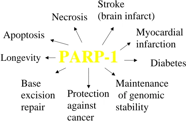
Fig. 3. Involvement of PARP-1 in diverse biological processes. PARP-1 seems to be involved in a variety of important physiological and pathological processes, such as DNA base-excision repair, maintenance of genomic stability of cells under genotoxic stress, longevity assurance, protection from cancer formation, cell death, type-1 diabetes, and tissue infarction. For details see text.
If severe DNA damage is inflicted to cells, there can be massive activation of PARP-1. This may cause a depletion of cellular NAD+ pools. As a consequence large amounts of cellular ATP will then be needed for de-novo synthesis of NAD+. At the same time, a severe NAD+ depletion will inhibit both glycolysis and respiration, leading to a complete energy depletion of the cell. There is no possibility for a cell to recover from such a situation and the cell is bound to die of necrosis (Berger, 1985). This scenario has been confirmed in a number of pathophysiological conditions. For instance, experimentally induced ischemia-reperfusion in brain, which serves as a model for stroke, is associated with massive release of endogenous
PARP-1
Apoptosis
Necrosis
Longevity
Stroke
(brain infarct)
Diabetes Base
excision repair
Maintenance of genomic stability
Protection against cancer
Myocardial
infarction
DNA-damaging compounds, including ROI, and leads to brain infarct. It has been shown that abrogation of PARP-1 activity, either by PARP-1 gene disruption or administration of low- molecular weight inhibitors, leads to an increased survival of the neurones at risk and dramatic reduction of brain infarct size (Eliasson et al., 1997). Very similar effects of PARP-1 inhibition on NO-induced necrosis have been observed in pancreatic islet cells (Radons et al., 1995). These data as well as other data obtained in different organs clearly indicate that PARP-1 is involved in the pathogenesis of tissue infarcts in heart and brain and of juvenile diabetes (type 1). Therefore new therapies might arise based on the use of specific PARP-1 inhibitors.
During apoptosis PARP-1 is cleaved by caspase-3 in the NLS region into two defined fragments of 24 kDa (N-terminal) and 89 kDa (C-terminal). This PARP-1 cleavage is widely used as a specific marker for apoptosis (Kaufmann et al., 1993, Alvarez-Gonzalez et al., 1999) and might also serve as a clinical diagnostic tool, e.g. in cancer treatment.
I. 4. DNA excision repair in mammalian cells
In mammalian cells DNA excision repair consists of three major pathways: base-excision repair (BER), nucleotide-excision repair (NER) and mismatch repair (MMR). Several other repair pathways that have been detected in lower eukaryotes or in prokaryotes apparently do not exist in mammals. One might speculate that only very reliable forms of DNA repair, which make very few errors, proved suitable for animals with very long life spans, and thus unreliable, mutation-prone pathways of DNA repair were lost during evolution.
The first step in BER is the excision of a single base carrying a lesion. Then the sugar- phosphate backbone is opened and the residual sugar-phosphate moiety is removed. In addition a varying number of nucleotides can be removed as well (for a more detailed description, see below). Typical lesions which are repaired by BER are “small” alterations of bases (e.g. oxidation, alkylation). Oxidation is mediated by oxygen free radicals, which arise both from various endogenous processes such as mitichondrial respiration, and from exposure to exogenous chemicals such as peroxides or bleomycin, or physical agents such as γ- radiation (see below). Another substrate for BER are abasic sites directly arising from spontaneous hydrolysis of the glycosidic bond connecting the base with C1’ of deoxyribose.
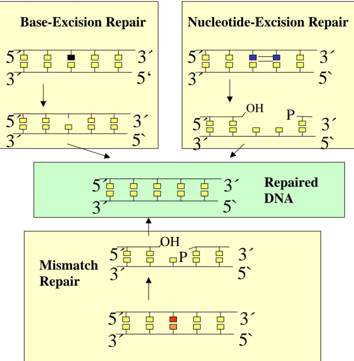
Fig. 4. Major functional differences between the three major DNA excision repair pathways in mammalian cells. In base excision repair the altered base is first excised without breakage of the sugar-phosphate backbone. Only subsequently is the sugar-phosphate backbone opened, the rest of the nucleotide removed and the missing nucleotide replaced. For more detail see Fig. 5. The function of mismatch repair is identification and correction of mismatched bases, mostly introduced as DNA replication errors. Typically, large segments (up to several kilobases) of the strand carrying the misincorporated base are excised followed by resynthesis using the cells DNA replication machinery.
In nucleotide excision repair oligonucleotides of about 30 nucleotides, incorporating the lesion site, are cut out followed by resynthesis.
5‘
3´
5´
3´
5`
3´
5´
3´
5`
3´
5´
3´
5`
3´
5´
3´
5`
3´
5´
3´
OH
5`
3´
5´
3´
P
OH
5`
3´
5´
3´
P
Base-Excision Repair
Mismatch Repair
Nucleotide-Excision Repair
Repaired
DNA
γ-Radiation can damage DNA either via direct energy transfer to this macromolecule or via radiolysis of water, leading to formation of ROI causing oxidative damage. The wide spectrum of radiation-induced lesions ranges from damaged bases or deoxyribose moieties to direct formation of DNA single and double strand breaks. All of these can be repaired via base excision repair, except double strand breaks, which require repair via “non-homologous end joining” (NHEJ) or recombination pathways. It should be noted that unrepaired double strand breaks are extremely cytotoxic lesions, with a single break of this kind being sufficient to induce apoptosis.
NER is characterised by the excision of an oligonucleotide from damaged DNA as an initial step. Typical kinds of damage that will be removed by nucleotide excision repair are pyrimidine photodimers and other photoproducts caused by UV-B and UV-C radiation, as well as “bulky” chemical adducts, induced for instance by polycyclic aromatic hydrocarbons (e.g. benzo[a]pyrene), and DNA crosslinks. NER has its own set of enzymes operating at any stage of the process.
MMR is defined by the kind of damage introduced into DNA rather than by the pathway of the repair. Typical kinds of “damage” to be recognised by the MMR machinery are mismatching pairs of normal bases resulting from misincorporation of nucleotides during normal DNA synthesis or base pair mismatches induced by alkylation of one partner (e.g. O6- methylguanine formation). Different sub-pathways exist, which have their individual sets of enzymes.
I. 5. The mechanism of base-excision repair and the involvement of PARP-1
The first step of BER is recognition of a damaged base by a DNA base glycosylase and base removal without breaking the sugar-phosphate backbone. In mammals there exist a whole variety of glycosylases, all of which recognise and remove bases that carry specific kinds of lesions from DNA. As mentioned above, there is yet another mechanism of abasic site formation, i.e. spontaneous hydrolysis of the relatively weak glycosidic bond linking the base with the sugar-phosphate backbone of the DNA.
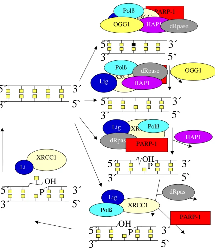
Fig. 5. Scheme of the DNA base-excision repair pathway and the key enzymes involved. Different kinds of damaging agents can cause different kinds of damages, which can enter the base-excision repair pathway at different stages. Chemical alterations of DNA bases, such as oxidation or alkylation, will be recognised by DNA glycosylases, one of which is OGG1. Glycosylase activity then generates an apurinic / apyrimidinic (AP) site.
5‘
3´
5´
3´
5`
3´
5´
3´
OH
5`
3´
5´
3´
P
OH
5`
3´
5´
3´
P
5`
3´
5´
3´
OH
5`
3´
5´
3´
P
XRCC1 Li
g
XRCC1
HAP1
Lig Polß
dRpase
PARP-1 PARP-1 OGG1 XRCC
Lig 1
Polß dRpase
HAP1
PARP-1 XRCC
1 HAP1 Lig
Polß
dRpase OGG1
PARP-1 XRCC1
Lig Polß
dRpas
e
It is important to note that even in the absence of base damage, DNA can undergo spontaneous hydrolysis of the N-glycosydic bonds linking the bases with the deoxyribose, resulting in direct formation of AP sites.
AP sites are recognised by an AP endonuclease (e.g. HAP1), catalysing hydrolysis of the phosphodiester bond. This results in 5´terminal deoxyribose-phosphate moiety, which is then excised by a DNA deoxyribose phosphodiesterase (dRpase).
At this stage PARP-1 binds to the strand break and gets automodified with p(ADPr). It is thought that automodified PARP-1 can cause local relaxation of chromatin, due to the extremely high affinity, by which histones bind to p(ADPr).
As a next step, DNA polymerase ß (Polß) fits in the missing nucleotide and ligation by ligase I or III (Lig) takes place.
γ-Radiation leads to several different kinds of damage that can be removed by DNA BER, including damage to bases or deoxyribose moieties as well as direct formation of DNA single strand breaks. By contrast, double strand breaks, which can also arise as a direct result of γ-irradiation, are not a substrate for BER, but have to be repaired via “non-homologous end joining” (NHEJ) or recombination pathways rather.
As a next step the residual sugar-phosphate moiety is cut out by AP endonuclease (HAP1) leaving a gap of just one nucleotide. This is followed by DNA repair synthesis, which is either of the “short patch” type, representing the major pathway and filling in only one nucleotide, or “long patch” type, filling in 2-6 nucleotides.
As a next step PARP-1 binds to the DNA strand break and gets automodified with ADP- ribose polymer. The precise function of PARP-1 at the molecular level in this process remains elusive. It has been shown that PARP-1 is able to interact with DNA polymerase-β but not with DNA polymerases δ or ε (Dantzer et al., 2000). DNA polymerase-β has been shown to be involved in both long patch and short patch BER (Klungland and Lindahl, 1997; Dianov et al., 1999). In the absence of both PARP-1 and DNA-polymerase-β BER is extremely inefficient and one may speculate on a functional synergy between the two proteins (Dianov et al., 1999).
There is apparently not just a single function PARP-1 has to fulfil in BER. PARP-1 has been shown to interact with the tumour suppressor protein p53, thus perhaps playing a role in signalling DNA damage to cell-cycle checkpoint proteins and influence the decision of whether or not to progress in cell cycle (Trucco et al., 1998) or even to trigger the apoptotic programme in the presence of massive DNA damage.
There is substantial evidence that PARP-1 plays a role in chromatin remodelling either via covalently modifying histones (de Murcia et al., 1988; de Murcia et al., 1986) and/or via histones binding non-covalently to p(ADPr) automodifying PARP-1 (Althaus et al., 1994).
The resulting local and reversible decondensation of DNA could facilitate the DNA repair process.
DNA polymerase-β fills in the missing nucleotides, and the function of ligases I or III is finally to covalently close the interrupted DNA strand.
XRCC1 can be viewed as an organising or “scaffolding” protein. It is present throughout the whole BER process, having binding sites for nicked DNA itself and and for most proteins involved in the base excision repair process like PARP-1, OGG1, DNA polymerase-β and ligase III.
I. 6. The involvement of DNA repair and PARP-1 in the ageing process
One of the central predictions of the Disposable Soma Theory of ageing (Kirkwood, 1977;
Kirkwood & Austad 2000) is that critical limitations exist in macromolecular maintenance and repair of somatic cells. Such limitations are thought to have evolved in accordance with the extrinsic mortality level an animal species is facing in a given habitat, as a result of a trade-off between allocating of available bioenergy to somatic maintenance and repair on the one hand and reproducing as well as other energy-consuming activities (e.g. muscular activity) on the other. Given the constant attack of biological macromoloecules by endogenous and exogenous damaging compounds such as ROI, it is thought that somatic maintenance and repair act as critical determinants of cellular stress resistance and organismal longevity.
Specifically, in the adult body one may group the cells in two different classes, i.e. cells that are post-mitotic and have stopped dividing, and cells that are proliferative. Examples for the first group are muscle cells, neurones and cells that form the eye lens. Most neurones of the body are generated during the embryonic and foetal phase of development and will persist for the rest of the body’s lifetime, although in some regions of the brain, stem cells have recently been identified from which new neurones can arise as a replacement of single degenerated neurones, but not of those lost in larger numbers as a result of brain trauma or disease processes. Likewise, most of post-natal striated muscle fibres are thought to persist throughout lifetime, although there is seemingly some regenerative potential provided by muscle stem cells. Terminally differentiated cells of the eye lens even lose most of their organelles and to become transparent. The potential problems arising from ageing in such cells that may persist for many decades if not a century are very different from those in proliferating cells (Fig. 6.). Cancer formation is extremely rare in postmitotic cell types.
Instead there is the problem of waste products accumulating over time, like lipofuscin, and of accumulation of mitochondrial DNA mutations, ultimately leading to severe deficiency in bioenergy metabolism, which cause the characteristic problems in these cells. Another major problem for post-mitotic cells might be represented in cascades of deteriorating information- storage or information-transmitting molecules, leading to error accumulation at all levels of the metabolic machinery of the cell (Fig. 6) and finally functional impairment and even cell death.
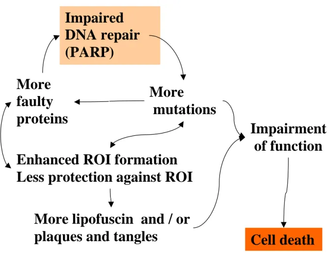
Fig. 6. Simplified hypothetical scenario of the ageing process in non-dividing, post-mitotic cells.
ROI formed by the cell’s normal metabolic activity cause damage to proteins and DNA, and the latter, if not accurately repaired, may represent pre-mutagenic lesions. In mitochondrial DNA, which replicates also in postmitotic cells, this may lead to mutation. In the nuclear genome, accumulated unrepaired DNA damage may lead to transcriptional errors across the damaged site altogether.
Mutations occurring in open reading frames of genes can cause synthesis of faulty proteins. Faulty proteins themselves involved e.g. in DNA synthesis or DNA repair would then allow the accelerated formation of further mutations. Faulty proteins involved in the mitochondrial respiratory chain will cause the enhanced formation of ROI. Other proteins, if impaired, lose their function to protect against these ROI. An enhanced occurrence of ROI will then again cause an increased amount of impaired proteins, which in turn can cause mutations. In addition to these cycles insoluble deposits, such as lipofuscin, amyloid plaques and tangles, can begin to form. These deposits then can cause functional impairment at all levels of cellular metabolism, which may as well accelerate the cycles described above, until finally the overall impairment of cellular functions leads to cell death.
More
mutations More
faulty proteins
Enhanced ROI formation Less protection against ROI
More lipofuscin and / or plaques and tangles
Impaired DNA repair (PARP)
Impairment of function
Cell death
Proliferative cells an the other hand, can further be sub-divided into differentiated cells, stem cells and germ cells.
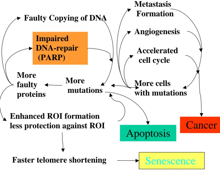
Fig. 7. Simplified hypothetical scenario of the ageing process in dividing cells. Initially, there are few errors at any stage of the cell function. The first inevitable errors then cause the subsequent degeneration of all systems that are vital for the cell. Reactive oxygen intermediates (ROI) cause the first mutations. Mutations occurring in open reading frames of genes can cause faulty proteins to be synthesised. Faulty proteins involved e.g. in DNA synthesis or DNA repair would then allow the accelerated formation of further mutations. Faulty proteins involved in the mitochondrial respiratory chain will cause the enhanced formation of ROI. Other proteins, if impaired, lose their function to protect against these ROI. More frequent occurrence of ROI would, in turn, lead to increased amounts of non-functional or dysfunctional proteins and accelerated telomere shortening which then can cause
More
mutations More
faulty proteins
Impaired DNA-repair (PARP)
Faulty Copying of DNA
Enhanced ROI formation less protection against ROI
More cells
with mutations Accelerated cell cycle Angiogenesis Metastasis Formation
Cancer
Senescence
Faster telomere shortening
Apoptosis
earlier senescence. If the integrity and stability of the cellular genome decays beyond a certain limit, the cell will have to undergo apoptosis. Another kind of degeneration can involve the cell-cycle control of the cell. A possible consequence of faster cell division may be an increased risk of mutational error accumulation and therefore an increased risk of malignant transformation, with cells acquiring more and more features of the malignant phenotype such as angiogenesis or metastatic growth. In conclusion, cellular senescence, apoptosis and cancer can all be viewed as consequences of degeneration arising during, or being facilitated by, the ageing process. Interestingly stem cell compartments seem to be better protected against such degeneration.
In dividing cells different mechanisms of deterioration can be envisaged (Fig. 7). Dividing cells are at all times at risk to lose cell cycle control and to enter the multi-step pathway of carcinogenesis. Cellular senescence and apoptosis are presumably mechanisms protecting against cancer formation, but at the same time have to be viewed as mechanisms of degeneration.
Germ cells must be particularly well protected against accumulating errors. It might well be the case that entirely different mechanisms are active to protect them from deteriorating, which are not represented in Fig. 7.
Over the last decade, data have accumulated suggesting a scenario of how DNA repair, and PARP-1 in particular, may be involved in the control of such cascades of error accumulation and thus are emerging as longevity-assurance factors (Grube et al., 1992; Bürkle et al., 1994;
Muiras et al., 1998, Meyer et al., 2000; Beneke et al., 2000; Bürkle, 2001 b,c,d).
I. 7. Current methods to detect and quantify poly(ADP-ribose)
Over the past three decades, a wide variety of analytical procedures have been developed to detect and quantify p(ADPr) formed in living cells or in subcellular systems, respectively (de Murcia & Shall 2000).
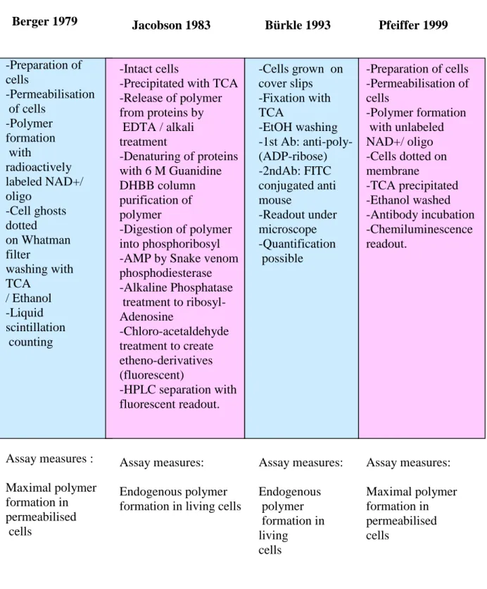
Jacobson and colleagues (1984) have established a method that detects and quantifies p(ADPr) formed endogenously in living cells and comprises biochemical extraction, purification and enzymatic degradation of p(ADPr) to yield unique monomeric nucleosides, followed by fluorescent derivatisation and finally reversed-phase HPLC separation and fluorescence-based detection and quantification of polymer-derived nucleosides. This method still serves as the “gold standard” in the field, but is expensive, laborious and requires very large cell numbers, which is prohibitive for analyses of large numbers of samples.
Another, much simpler, method to estimate endogenous polymer formation in living cells has been developed by Bürkle and colleagues (1993; Schlicker et al., 1999; Seker et al., 2000). In this method cells are grown on or adhered to cover slips and then treated with damaging agents. Thereafter cells are fixed with TCA and washed with ethanol (Fig. 8). As a first antibody the anti-p(ADPr) monoclonal 10H is used. As a second antibody fluorescein isothiocyanate-labelled anti-mouse immunoglobulin is used. The readout is done under the microscope comparing cells subjected to various treatment conditions with controls.
Quantification of the result is possible but very laborious. This assay would also be inappropriate for very large sample sizes.
An assay originally developed by Berger and colleagues (1979) measures p(ADPr) formation in permeabilised cells (Fig. 8). It is based on radioactively labelled NAD+, the ADP-ribosyl moiety of which is incorporated into the polymer and thus becomes acid-insoluble. The readout is done by liquid scintillation counting of TCA insoluble material. Addition of a short double-stranded oligonucleotide to the reaction buffer leads to dose-dependent stimulation of polymer formation above background (Grube et al., 1991).
Many interesting results have already been obtained using this “Berger” assay e.g. a correlation between longevity in mammals an maximal p(ADPr) formation (Grube & Bürkle 1992) and higher maximal poly(ADP-ribosyl)ation capacity in centenarian-derived cells than in controls (Muiras et al, 1998). However, major drawbacks are the use of radioactivity, including expensive radioactive tracers, the relatively large cell numbers needed, and problems with complete removal of any unincorporated NAD+ from TCA precipitates. In this dissertation a new, non-isotopic and simple method for the assessment of poly(ADP- ribosyl)ation capacity is presented (Fig. 8).
Fig. 8. Scheme summarising available assays to detect and quantify poly(ADP-ribose) Berger 1979
-Preparation of cells
-Permeabilisation of cells
-Polymer formation with
radioactively labeled NAD+/
oligo -Cell ghosts dotted on Whatman filter
washing with TCA
/ Ethanol -Liquid scintillation counting
Assay measures : Maximal polymer formation in permeabilised cells
Pfeiffer 1999
-Preparation of cells -Permeabilisation of cells
-Polymer formation with unlabeled NAD+/ oligo -Cells dotted on membrane
-TCA precipitated -Ethanol washed -Antibody incubation -Chemiluminescence readout.
Assay measures:
Maximal polymer formation in permeabilised cells
Jacobson 1983
-Intact cells
-Precipitated with TCA -Release of polymer from proteins by EDTA / alkali treatment
-Denaturing of proteins with 6 M Guanidine DHBB column purification of polymer
-Digestion of polymer into phosphoribosyl -AMP by Snake venom phosphodiesterase -Alkaline Phosphatase treatment to ribosyl- Adenosine
-Chloro-acetaldehyde treatment to create etheno-derivatives (fluorescent)
-HPLC separation with fluorescent readout.
Assay measures:
Endogenous polymer formation in living cells
Bürkle 1993
-Cells grown on cover slips -Fixation with TCA
-EtOH washing -1st Ab: anti-poly- (ADP-ribose) -2ndAb: FITC conjugated anti mouse
-Readout under microscope -Quantification possible
Assay measures:
Endogenous polymer formation in living
cells
I. 8. Current methods to measure DNA damage and repair
The two currently most popular methods to measure DNA damage and repair at highest levels of sensitivity are the ‘comet’ assay (reported detection limit 0.05 Gy; Singh, 2000) and the
Fig. 9. Schematic overview describing the steps of the experimental procedure of the comet assay.
Cells resuspended in a buffer are added to a first layer of a gel. Then a second layer of gel is cast enclosing the single cells. Cells are exposed to DNA-damaging agents in the gel, which may be followed by repair period. Then cells are lysed in situ and treated with alkali. After electrophoresis, the gel has to be stained and washed. The readout of the stained DNA of the single cells has to be done under a microscope. The tail moment of comets can be evaluated by quantitative digital imaging.
Preparation of cells
Incorporation of cells in the gel
Damaging procedure/ followed by repair Lysis
Alkaline treatment Running of gel
Fluorescent staining of nuclear DNA with EtBr Readout under microscope
Software-assisted evaluation
of single cells (typically 100 per data point)
Cells inbedded
in microgel double layer
fluorescence-detected DNA unwinding assay (FADU) (reported detection limit 0.1 Gy; Singh, 2000).
The mechanisms underlying these two assays differ from each other. The principle of the comet assay is based on different migration velocities of DNA fragments of different size in agarose gels. The more single-strand breaks have been introduced into the DNA, the smaller the single-stranded fragments of the DNA will be and the faster these fragments will migrate in an electric field applied to the gel. Therefore, whole cells are embedded in an agarose gel, lysed and treated in situ with alkali to render the DNA single-stranded prior to running the gel. (N.B. A neutral version of this assay can be used to quantify double strand breaks). In an appropriate electrical field, the genomic DNA migrates out of the nucleus into the agarose and is then stained with the intercalating fluorescent dye ethidium bromide, allowing visualisation of the DNA. Viewed microscopically the combination of the DNA that has stayed within the confines of the nucleus and the “tail” of DNA that has migrated makes individual cells look like comets. Quantitative microscopic evaluation is done by measuring the length and intensity of the comet in relation to the signal of the non-migrating nuclear DNA in comparison with standards (Fig. 9.).
The principle of the FADU assay is based on the fact that double-stranded DNA exposed to defined, moderate alkaline conditions unwinds at a constant rate, starting at its natural ends as well as at internal single or double-strand breaks. As a result, there will be more extensive unwinding of DNA carrying strand breaks (Fig.10.). This effect can be quantified using chemical probes that intercalate preferentially or exclusively into double-stranded DNA and become fluorescent by being intercalated. Thus the fluorescent signals obtained are an (inverse) increase of different levels of DNA breakage presentin the cells at the time of lysis.
The manually performed FADU assay as originally published by Birnboim and Jevcak (1981) is very tedious and difficult to standardise, hence hardly used any more.
The comet assay has been improved in recent years (Sing, 2000), e.g. by establishing evaluation software to assist the readout procedure. Nevertheless, it is also still very tedious, as gels have to be prepared and processed manually in a multi-step procedure (Fig. 9).
In this dissertation, an automated version of the FADU assay is presented. Using an appropriately designed laboratory robot, all the critical and tedious pipetting steps have been automated and are performed under strict temperature control and protection from light.
Furthermore the readout has been radically simplified by using a 96-well fluorescence reader instead of single cuvettes (Fig. 11).
Fig. 10. Principle of the fluorescence-detected alkaline DNA unwinding. Double stranded DNA is represented as double lines. DNA strand breaks are indicated by arrows. Following cell lysis by detergents and disruption of chromatin by high concentrations of urea, cellular DNA is exposed to alkali under rigorously controlled conditions with regard to pH, temperature and time. Alkali exposure is achieved via diffusion from a separate layer placed on top of the lysate, rather than by
Lysis
Lysis
Lysis
Lysis NaOH
NaOH
NaOH
NaOH Neutr.
Neutr.
Neutr.
Neutr.
EtBr or Sybrgr.
EtBr or Sybrgr.
EtBr or Sybrgr.
EtBr or Sybrgr.
T
P 0
P x
B
direct mixing, which would introduce artificial strand breaks. To the “T-samples, neutralisation buffer is added before the alkali so that the solution never reaches a pH necessary for unwinding. Alkaline unwinding in the P and B values starts only at the ends of chromosomes or at single and double-strand breaks. The more damage has been introduced into the DNA the less double-stranded DNA will remain after the unwinding phase. To stop unwinding, neutralisation buffer is added to the P and B values. For detection ethidium bromide or Sybr green (open circles) is added to all samples. These dyes intercalate preferentially into double stranded DNAinducing their fluorescence (filled green circles). The more double stranded DNA has remained, the more intense the fluorescent signal will be.
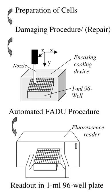
Fig. 11. Schematic overview depicting the experimental procedure of the automated FADU assay.
Only the preparation of cells and the damaging procedure/ (repair) have to be done manually. The assay itself is fully automated. The readout is done in a 1-ml 96-well plate.
Preparation of Cells
Damaging Procedure/ (Repair)
Nozzle
1-ml 96- Well plate
Encasing cooling device
Automated FADU Procedure
Readout in 1-ml 96-well plate
Fluorescence reader x
y
z
As a result, the throughput, accuracy, reliability and operator convenience have been increased dramatically, thus enabling large-scale analyses that should be of interest to a wide range of laboratories engaged in basic biomedical research or applied toxicology as well as to pharmaceutical companies.
I. 9. Aim of the project
Quantitative assessment of exogenous DNA damage and also of DNA repair activities is of utmost importance not only for a broad range of basic scientific research fields but also for routine tasks, such as medical monitoring of patients or probands to assess individual genotoxic exposures and the body’s response to them, as well as toxicological screening in the chemical and pharmaceutical industry or monitoring of environmental pollution.
However, there is also a growing demand to assess endogenous DNA damage and its repair.
The rapidly increasing fraction of elderly people in almost all countries worldwide and the increased awareness of the additional morbidity and the forthcoming strain to health-care systems resulting from this demographic change highlight the importance of understanding the process of ageing at the molecular level. The latter is indispensable for the development of urgently needed novel, rational modalities of prophylaxis or therapy of ageing-associated pathologies. Over the past few years much evidence has already accumulated supporting a critical role for endogenous macromolecular damage (including DNA damage) as a driving force of the ageing process at the cellular level, leading to decreased stress resistance and increased vulnerability of cells and tissues (Bürkle, 2001 d). In particular, the past and current work of Dr Bürkle’s group has been focussed on the impact of poly(ADP-ribosyl)ation and BER on the ageing process in mammals.
Grube & Bürkle (1992) have described a correlation between maximal lifespan of mammalian species and poly(ADP-ribosyl)ation capacity. Muiras et al (1998) have observed higher specific poly(ADP-ribosyl)ation capacity in lymphoblastoid cell lines derived centenarians compared to controls. Meyer et al (2000) have found that PARP-1 overexpression protects against DNA damage-induced sister-chromatid exchange. Beneke et al (2000) have discovered that purified PARP-1 of humans possesses twice as much automodification activity as purified PARP-1 of rats. These observations are very much in line with a report by
Kapahi et al. (1999) describing that fibroblasts from long-lived mammalian species are more resistant to a variety of stresses, including genotoxic stress.
The above-mentioned results as well as data from other laboratories lead to the present working hypotheses:
• Somatic cells from long-lived organisms should be genetically more stable, mediated at least in part by their higher poly(ADP-ribosyl)ation capacity.
• Long lived organisms should possess more proficient DNA repair systems (already shown for NER, but as yet unknown for BER).
To be able to address these questions experimentally, two technologies had first to be further developed. One of them is a non-radioactive immuno-dot-blot assay to assess poly(ADP- ribosyl)ation capacity (Pfeiffer et al, 1999). This assay should help establish whether PARP-1 can serve as a marker of human ageing. Furthermore, with the recent discovery of PARP homologues, it was extremely interesting to check if this type of assay is specific for PARP-1 or will also reflect PARP-2 activity.
The second technology that needed substantial improvement beyond the present state of the art was the FADU assay. This assay was first described in 1981 but was seldom used, even though it had been reported to possess very high sensitivity. The reason for this is that it is very difficult to perform and even very skilled and highly motivated lab workers were unable to run it in a reproducible manner (Dr Alexander Bürkle, personal communication). Another significant disadvantage was the large number of cells required.
The aim of the present PhD work was to set up automation of the FADU assay, along with miniaturisation by downscaling of the procedure to the 96-well format. It was desired that apart from preparation of cells and the DNA-damaging treatment, all pipetting steps (i.e.
sequential additions of defined buffers to the primary cell lysates) should be carried out by a laboratory robot, with a single 96-well plate serving as the basic matrix for the whole procedure, including readout of the samples in an appropriate fluorescence reader. Predictably such an assay format should be much more convenient to operate than other current methods including the comet assay.
Given the laboratory’s specific research interests in biogerontological research, one of the first applications (out of a vast range of possible applications) then was to determine BER capacity as a function of lifespan in mammals (see above).
From the above considerations, it is also clear that successful establishment of improved methods for the assessment of DNA damage and repair should also facilitate research on, or routine monitoring of, exogenous genotoxic exposures.
II. Materials and Methods
II. 1 Appliances
60Co-gamma-irradiation device (Gammacell 1000 Elite, Nordion Inc, Canada);
1-ml 96-Well plates. These were made from commercial 2-ml 96-well plates (Masterblock, Greiner Labortechnik) by cutting them down to a height of 2.0 cm thus accommodating 1 ml in each well. The plate is levelled thus enabling sealing of all wells simultaneously with Parafilm. The plates can be re-used after washing with 1M NaOH and then with DMSO and ethanol. A volume of 1 ml per well and a height of 2.0 cm of the plate are the largest dimensions suitable for use in the 96-well fluorescence reader.
To control temperature in the 1-ml 96-well plate a self-made cooling device (Fig. 12) was assembled from commercially available polypropylene plates, in which water tubing encases the 1-ml 96-well plate. For each of the 96 well positions, a hole was drilled from the top between the tubing thus enabling the diluter of the robot to access all 96 wells, while the plate is inserted in the cooling device. An ethanol-water mixture (1:3) of desired temperature is circulated through the tubing using a Lauda 2000 water bath.
Pipetting robot, Miniprep I (Fig. 12) (Tecan, Crailsheim, Germany) with one arm, carrying a single stainless steel nozzle
Fluorescence reader (Spectrafluor Plus, Tecan, Crailsheim, Germany);
Multi-channel pipette (Sealpette 1200, 12 channels, Jencons, UK)
Heraeus Biofuge 3 (Heraeus, Germany)
Thermomixer 5436 (Eppendorf, Hamburg, Germany)
24-well dot-blot manifold, custom-made with well diameter of 1.2 cm (Steinbrenner Laborsysteme GmbH, Eberbach, Germany).
LAS-1000 chemoluminescence detection system (Fuji; Raytest, Straubenhardt, Germany) in conjunction with „Aida“ software (Raytest)
.
Fig. 12. Pipetting robot set up for the FADU assay. (1) Transparent custom-made 1-ml 96-well plate, which is inserted in (2a) the lower part of the self-made cooling device. (2b) The upper part of the cooling device, with 96 holes for the nozzle to pass through, has been turned over and during operation will be placed on top of the lower part (2a) and fixed with six bolts (3) and nuts. Encasing the 1-ml 96-well plate, the cooling device is then placed on its stand (4) i.e. in a position within the working area of the robot defined at a precision of 0.2 mm. A sister plate (5) is already in its stand.
The nozzle (6) precisely finds its target with regard to X, Y, Z coordinates. The water tubing (7) is connected to the water bath (8) for temperature control. Ice cold buffers are stored in a little ice box (9) accessible to the nozzle from the top. For protection against light the working area is enclosed by cardboard wrapped in aluminium foil (10). It can be closed with a lid (not visible). Washing liquid for the nozzle (11).
10
1
2a
2b
3 4
5 6
7
8
9
11
II. 2. Chemicals
„ECL plus“ kit Amersham-Pharmacia ( Germany).
β-Mercaptoethanol Sigma (Germany) NAD+ (grade V) Sigma (Germany)
Digitonine Sigma (Germany)
3-Aminobenzamide Sigma (Germany)
TCA Roth (Germany)
Sodium pyrophosphate BDH (UK)
NaCl Merck (Germany)
Tris Merck (Germany).
Magnesium sulfate Merck (Germany).
DMSO BDH (UK)
Ethanol BDH (UK)
Ethidium bromide Merck (Germany)
EDTA Merck (Germany)
Glucose Merck (Germany)
Urea BDH (UK)
Sybr green Raytek (Germany)
Skimmed Milk Powder Fluka (Germany) Sodium hydroxide BDH (UK)
Penicillin Sigma (Germany)
Percoll Amersham Pharmacia (Germany)
Trypsin (0.5 µ g / ml) BRL (Germany)
Tween 20 Gerbu (Germany)
II. 3. Oligonucleotides
The PARP-1 activator deoxyoligonucleotide (GGAATTCC) was dissolved in 15 mM NaCl at 385 µg/ml. The 3’- and 5’-termini of the oligonucleotide were unphosphorylated.
II. 4. Antibodies
Mouse monoclonal antibody recognising p(ADPr) was purified from culture supernatant of 10H hybridoma cells (kind gift of M. Miwa and T. Sugimura, Tokyo, Japan) by using a protein-A column chromatography kit (Sigma). This was done by Mr Marcus Müller in the laboratory.
Peroxidase-conjugated anti-mouse secondary antibody (Dianova, Hamburg, Germany)
II. 5. Membranes
--Gene Screen membrane (NEN, Brussels, Belgium)
--Nitrocellulose membrane (Biometra, Germany)
III. 6. Buffers and solutions
--Permeabilisation buffer: 10 mM Tris-HCl pH 7.8; 1 mM EDTA; 4 mM MgCl2; 30 mM 2- mercaptoethanol, with or without 0.015% (w/v) digitonine supplementation as indicated
-- PBS-MT: PBS pH 7.4; 5 % semi skimmed milk powder; 0.05 % Tween 20
--PBS-T: PBS pH 7.4; 0.05 % Tween 20
--Percoll solution: 63 % Percoll [Amersham Pharmacia]; 0.15 M NaCl in H2O
--EDTA solution: 10 mM ethylenediaminetetraacetate, pH 8.4
--Suspension buffer: 0.25M meso-inositol, 10 mM sodium phosphate pH 7.4, 1 mM MgCl2
--Lysis buffer: 9 M urea; 10 mM NaOH; 2.5 mM cyclohexyl-diaminetetraacetate; 0.1%
sodium dodecylsulfate
--Alkali solution: 0.425 parts lysis buffer in 0.2 M NaOH
--Neutralisation buffer: 1 M glucose, 14 mM β-mercaptoethanol
--Sybr green solution: 13.3 mM NaOH; Sybr green (1:25.000)
II. 7. Cell culture medium .
RPMI 1640 medium (Sigma) supplemented with 100 U/ml penicillin, 100 µg/ml streptomycin, 2 mM glutamine, and 10% heat-inactivated foetal calf serum (Sigma). Cultures were incubated at 37°C/5% CO2.
DMEM medium (Gibco) supplemented with 100 U/ml penicillin, 100 µg/ml streptomycin, 2 mM glutamine, and 10% heat-inactivated foetal calf serum (Sigma). Cultures were incubated at 37°C/5% CO2.
II. 8. Cell lines
IARC 273, Epstein-Barr virus immortalised human B-lymphoblastoid cell line (kind gift of Dr M Pawlita, DKFZ, Heidelberg, Germany)
3T3 Embryo fibroblast cell line from PARP-1 -/- mice
3T3 Embryo fibroblasts cell line from PARP-1 +/+ mice (wild-type control)
(kind gift of Dr Gilbert de Murcia, ESBS, Université Louis Pasteur, Strasbourg, France)
II. 9. Animals and human blood donors
The rat strain used was Fischer 133.
Blood was obtained from nine months old rats (4 females and 2 males) after killing. This was performed by highly trained and authorised animal care staff, in compliance with the relevant national laws and regulations.
Human blood donors were healthy volunteers from diverse ethnic groups. All human blood donors were adult but not older than 41 years, which corresponds to one third of the maximum life span of humans of 123 years.
III. Methods
III. 1. Poly(ADP-ribose) immuno-dot-blot assay
Cell permeabilisation and p(ADPr) formation assay. IARC 273 cells were washed in PBS, resuspended in ice-cold permeabilisation buffer supplemented with 0.015% (w/v) digitonine at a density of 3 x 10⁶ cells/100 µl and left on ice for 1 min. Then 4 ml of ice-cold permeabilisation buffer was added. Cells were centrifuged at 1000 x g, 0°C for 10 min and resuspended in ice-cold permeabilisation buffer at a density of 5 x 10⁵ cells per 53 µl. To samples of 5 x 10⁵ cells on ice was added 13 µl of oligonucleotide solution and 34 µl of 3x reaction buffer (100 mM Tris-HCl, pH 7.8, 1 mM NAD+, 120 mM MgCl2), respectively, resulting in a total volume of 100 µl per reaction. The reaction was carried out for the times indicated at 30°C on a shaker and stopped by adding 400 µl of 6.25 mM 3-aminobenzamide in PBS on ice.
Detection procedure. Volumes of the reaction mixture as indicated were vacuum aspirated onto a Gene Screen membrane by using a 24-well dot-blot manifold. Before the membrane dried completely, 400 µl of 10% TCA (w/v), 2% sodium pyrophosphate (w/v) was filled into the manifold, and 800 µl of 70% ethanol was carefully layered on top of this. The two layers were vacuum aspirated. Then the membrane was rinsed in PBS, blocked in PBS-MT and incubated with the first antibody (10H; 2.5 µ g/ml in PBS-MT) overnight at 4°C, with constant agitation. Thereafter, the membrane was washed with PBS-T and incubated with peroxidase- conjugated anti-mouse secondary antibody (1:10,000 in PBS-MT) for 1 h at room temperature. The blot was washed with PBS prior to chemoluminescence detection by using the „ECL plus“ kit.
Standardisation of p(ADPr) quantity. ADP-ribose polymer was purified from TCA precipitates of some of the samples by dihydroxyboronate chromatography, followed by enzymatic digestion to nucleosides, fluorescent derivatisation and quantification of polymer- specific nucleoside derivatives during reversed-phase HPLC separation, as described (Jacobson et al., 1984).
III. 2. Automated fluorescence detected alkaline DNA unwinding assay
Separation of mononuclear lymphocytes
Six ml of whole blood from human donors was collected with a syringe, with 60 µl EDTA solution present as an anti-coagulant. Then the blood was diluted 1:2 with PBS, layered on top of 15 ml Percoll solution and centrifuged at 1500 g using a Heareaus Biofuge.
Mononuclear cells were recovered, washed once with PBS, centrifuged at 1000 g and resuspended in suspension buffer at a concentration of 106 cells per ml. For measuring repair, the cells were resuspended in DMEM without FCS and other supplements at a concentration of 106 cells per 150 µl.
DNA damaging procedure
150 µl cell aliquots in Eppendorf tubes were cooled down to 0°C and irradiated on ice for different time periods with a 60Co-gamma source at a dose rate of 3.6 Gy per min in air.
DNA repair
To assess repair, cells damaged at 2.7 Gy were incubated at 37°C for various time periods on a Thermomixer, with a shaking frequency of 50 rpm to allow strand break repair to proceed.
To stop repair 850 µl ice cold suspension buffer was added on ice.
Lysis and DNA unwinding procedure
70 µl of all cell samples, which had been treated differently, was added to the 1-ml 96-well plate, respectively, and kept at 0°C in the dark working space of the robot. Then automated addition to all samples of 70µl of lysis buffer at a rate of 150µl / s was triggered.
Subsequently the alkali solution was added on top of the cell lysate in such a way that a second layer was forming, thus avoiding any mixing with the lysate. To do this the robot positioned the nozzle precisely 1.5 mm above the level of the lysate and added the alkali solution at a very low rate of 10µl / s. This was done in such a way as to allow exactly 12 min time for lysis (i.e. the preceding step) for each well. Then 15 min were allowed for the diffusion of the alkali into the lysate at 0°C. Then the temperature was shifted to 30°C for 90 min. Prior to the addition of 140 µl of neutralisation buffer at a rate of 200 µl / s the temperature was shifted to 22°C. For T-samples, as an internal standard representing cells with 100 % double stranded DNA, 140µl of neutralisation buffer was added prior to the
alkaline solution. In some experiments B-samples were included as an internal standard representing cells with 0 % double stranded DNA as a result of extensive of the lysate prior to addition of alkali, i.e. by passing the lysate 20 times through a 0.5 mm cannula. In course of the work it was noted that there was an excellent correlation between T and B values, and since the B values were quite low in the case of Sybr green, B samples were omitted from the final FADU version.
Readout
To perform the readout, 470 µl Sybr green solution was added with an automated multi channel pipette (Sealpette). The wells of the 1-ml 96-well plate were then sealed with Parafilm and the plate turned upside down 10 times. To reach an equilibrium the solution was allowed to rest precisely 10 min before scanning by a 96-well plate fluorescence reader at Em 492 nm and Ex 520 nm.
IV. Results
IV. 1. Setting up a new immuno-dot-blot procedure to assess cellular poly(ADP- ribosyl)ation capacity.
Assessment of the maximal cellular p(ADPr) formation (termed “poly[ADP-ribosyl]ation capacity”) has proven very interesting in a wide range of studies performed by many laboratories over the last three decades (Althaus & Richter 1987; de Murcia & Shall 2000, Bürkle 2001a,b). Unfortunately the available standard technique required use of expensive radioactively labeled tracers (3H- or 32P-NAD+) and, in addition, was fraught with problems arising from insufficient removal of unincorporated tracer from acid-insoluble product to be quantified (i.e. p[ADPr]), thus requiring multiple parallel determinations in view of the frequent outliers (cf. Muiras et al., 1998).
Assuming that an appropriate immuno-dot-blot procedure that would rely on the excellent binding specificity of monoclonal anti-p(ADPr) antibody 10H (Kawammitsu et al, 1984;
Bürkle et al, 1993) could be viable alternative, a set of preliminary experiments was performed to test a range of commercially available blotting membranes for their potential usefulness (not shown). Membranes were loaded with permeabilised cells that had been incubated under conditions allowing maximal accumulation of p(ADPr) [see below]. One of the most promising candidates turned out to be the nylon membrane ‘Gene Screen’. To determine more accurately its binding capacity, permeabilised IARC 273 lymphoblastoid cells were incubated with unlabeled NAD+ as substrate and “activator” oligonucleotide (GGAATTCC) for 6 min at 30°C, allowing maximal p(ADPr) accumulation to occur (Grube et al, 1991). The reaction was stopped by adding the competitive ADP-ribosylation inhibitor 3-aminobenzamide and cooling on ice. The cell ghosts were then directly dot-blotted onto the membrane, followed by TCA precipitation in situ, washing with ethanol and immunodetection of p(ADPr) by using monoclonal antibody 10H in conjunction with a peroxidase-based chemoluminescence detection system.
As a first step the reaction kinetics of the polymer formation under the conditions of the dot blot procedure was examined (Fig. 13.). It was found that there is an almost linear increase during the first four minutes followed by a plateau and a slight decrease at around 8 minutes.
Six minutes were chosen as a standard reaction time for all subsequent experiments.
Fig. 13. p(ADP)r formed in IARC 273 cells as a function of reaction time.60,000 permeabilised IARC 273 cells were incubated in reaction buffer at 30°C for varying time periods as indicated. Note that there is an almost linear increase in p(ADPr) formed during the first 4 minutes, followed by a plateau and a slight decrease at around 8 minutes.
In Fig. 14 is shown the intensity of chemoluminescence signals obtained as a function of cell number. The curve is linear up to 120,000 cells per blotting area (113 mm2), while above this
p(ADPr) [pmole]
0 2 4 6 8 10
0 1 2 3 4 5 6 7 8 9 10 11 12
Reaction time (min
)Fig. 14. Binding capacity of Gene Screen nylon membrane for permeabilised IARC 273 cells.
40,000 to 240,000 permeabilised cells that had been incubated with NAD+ and activator oligonucleotide for 6 min at 30°C were loaded on the membrane, followed by TCA precipitation in situ and immunodetection of p(ADPr) as described in Materials and Methods. Quantification of chemoluminescence signals was performed by using a Fuji LAS1000 detection system. Mean values ±1 SD (if larger than symbol size) are represented. Note that chemoluminescent signals are linear up to 120,000 cells loaded per blotting area (113 mm2).
cell number saturation of the membrane is apparent. No significant differences between an uncharged (Gene Screen) and a positively charged (Hybond N+; Amersham-Pharmacia) nylon membrane were found (data not shown). Nitrocellulose proved unsuitable, as it does not withstand exposure to 10% TCA.
0 50 100 150 200 250 300
0 500 1000 1500 2000 2500 3000
Cell number x 10
-3Signal intensity (arbitrary units)
![Fig. 1. Domain structure of PARP-1. The 113-kDa enzyme possesses at its N-terminal DNA binding domain two Zn fingers (F1 and F2)[module A] as well as a nuclear location signal (NLS) [module B]](https://thumb-eu.123doks.com/thumbv2/1library_info/5518789.1686826/6.892.145.716.490.673/domain-structure-possesses-terminal-binding-fingers-nuclear-location.webp)