INAUGURAL-DISSERTATION
zur
Erlangung der Doktorwürde der
Fakultät für Biologie der
Ruprecht-Karls-Universität Heidelberg
Vorgelegt von:
Diplom-Biologin Jinhyun Kim aus Suwon, Korea Tag der mündlichen Prüfung:
Improvement and establishment of the tTA- dependent inducible system in the mouse brain
Gutachter: Prof. Dr. Peter H. Seeburg Prof. Dr. Klaus-Armin Nave
Ich erläre hiermit, daß ich die vorliegende Dissertation selbst verfaßt und mich dabei keiner anderen, als der von mir ausdrücklich bezeichnten Quellen und Hilfen bedient habe. Desweiteren erkläre ich hiermit, daß ich an keiner anderen Stelle ein Prüfungsverfahren beantragt, beziehungsweise die Dissertation in dieser oder anderen Form bereits anderweitig als Prüfungsarbeit verwendet oder einer anderen Fakultät als Dissertation vorgelegt habe.
Heidelberg, September, 2001
Meinem Vater, dem Helden
Prof. Dr. Peter H. Seeburg danke ich für die Aufgeschlossenheit und Großzügigkeit, mich in seinem Labor aufzunehmen. Damals klopfte ich ohne Voranmeldung und Referenzen an seine Tür, der deutschen Sprache noch kaum mächtig und mit einem unpassend zurechtgelegten Satz beginnend: “Ich interessiere mich für
Krebsforschung….” Ich danke ihm für die immer herzliche Betreuung meiner Arbeit.
Dr. Rolf Sprengel danke ich für das Gefühl, immer hinter mir zu stehen und für den Freiraum, den er mir bei meiner Arbeit eingeräumt hat.
Rita Pfeffer und Axel Erhardt danke ich für die wertvolle Hilfe mit meinen Mäusen.
Allen meinen Mitarbeitern danke ich für die Zusammenarbeit und unser gemeinsames Lachen.
Summary
A genetic regulatory system employing the tetracycline-controlled transactivator (tTA) that can induce individual gene expression reversibly in a temporal and spatial manner could permit a more precise analysis of gene functions on animal physiology and behaviour. In this study we have described the optimization of the tTA coding sequence (humanized tTA, htTA) for efficient expression in the mouse and the establishment of the htTA-dependent inducible system by conditional means in transgenic mice. The coding sequence of the htTA was altered concerning mammalian codon usage, putative splicing signals, CG dinucleotide content, and toxicity of the transactivation domain VP16. These sequence changes on tTA resulted in the three- fold increase of the inducible activity in transiently transfected HeLa cells, compared to the prokaryotic one. Activity of the htTA was efficiently suppressed by addition of doxycycline.
To express the htTA in a neuron- and region-specific manner in mouse brain, the region-specific promoter of the high-affinity kainate receptor subunit KA1 was chosen. By using KA1 bacterial artificial chromosomes (BAC), it was expected that expression of htTA under the control of the KA1 promoter is directed to the areas expressing KA1 itself, namely in CA3 and dentate gyrus (DG) of the adult hippocampus. The functional activity of the htTA, however, was not observed in KA1-htTA transgenic mice, in spite of expression of the htTA mRNA as determined by RT-PCR. The htTA gene under the KA1 promoter appears to be expressed too low to induce tTA responsive genes (e.g. lacZ) in these transgenic mice. An alternative approach for region specific htTA expression was to use the CaMKIIα promoter but to repress the forebrain-specific promoter in regions other than hippocampus by the neuron-restrictive silencer element (NRSE) of the N-methyl-D-aspatate receptor 2C (NR2C) subunit. The expression pattern and activity of the htTA under control of the NR2C silencer-containing chimeric promoter were observed restrictively in hippocampus of two lines (sCN1 and CN10), namely only in DG, and in CA1 and DG, respectively. Activity of the htTA in the CN10 line was detected at postnatal stage and was turned off completely by doxycycline.
It is demonstrated here that the humanized tTA (htTA) provides efficient expression and reversible inducibility in vitro and in vivo and that the NR2C silencer-containing chimeric promoter was successfully applied in transgenic mice. The hippocampus- specific genetic switch by the htTA should make it possible to study genes of interest involved in hippocampal functions and should advance the research of gene function in the CNS (central nervous system).
1. INTRODUCTION ... 1
1.1. INDUCIBLE GENE EXPRESSION SYSTEMS... 1
1.1.1. Irreversible system by Cre or FLP recombinase... 1
1.1.2. Reversibly inducible systems derived from eukaryotic regulatory system ... 3
(1) Heat shock ... 3
(2) Heavy metal ion ... 3
(3) Interferon... 4
(4) Hormones ... 4
(5) Ecdysone... 4
1.1.3. Inducible systems based on prokaryotic regulatory elements ... 6
(1) lac Repressor/Operator-based inducible system ... 6
(2) Tetracycline-controlled transactivator (tTA) system... 7
1.2. WHY ‘HUMANISED TTA (HTTA)’ IS NEEDED... 10
1.3. GENERATION OF TRANSGENIC MICE EXPRESSING HTTA IN A SUBREGION-SPECIFIC MANNER IN BRAIN COMPARED TO PREVIOUS CAMKII-TTA MICE... 10
2. RESULTS ... 14
2.1. DESIGN AND CONSTRUCTION OF THE HUMANIZED TTA (HTTA)... 14
2.2. ACTIVATION POTENTIAL AND COMPARISON OF THE HTTA IN TRANSIENTLY TRANSFECTED CELLS... 14
2.3. GENERATION OF TRANSGENIC MICE EXPRESSING THE HTTA IN A REGION (HIPPOCAMPUS)-SPECIFIC MANNER IN BRAIN... 18
2.3.1. GENERATION OF KA1.HTTA TRANSGENIC MICE... 18
2.3.1.1. Genomic structure of the mouse KA1 gene ... 19
2.3.1.2. Screening of BACs library for mouse KA1 gene ... 19
2.3.1.3. Construction of targeting vector for homologous recombination of the KA1 BAC ... 21
2.3.1.4. Modification of KA1 BAC ... 21
(1) Shuttle vector and first recombination ... 25
(2) Second recombination of the cointegrates ... 25
(3) Flp recombination of the resolved KA1-htTA/tTA.KM BAC ... 25
2.3.1.5. Analysis of KA1.htTA and KA1.tTA founders... 26
2.3.2.GENERATION OF CAMKII-NR2C.HTTA TRANSGENIC MICE... 29
2.3.2.1. Characterization of the NR2C gene... 29
2.3.2.2. Construction of the minigene of CNhtTA and sCNhtTA ... 32
2.3.2.3. Analysis of founders of CNhtTA and sCNhtTA... 33
2.3.2.4. Developmental expression and activity of the htTA in the CN10 line ... 37
2.3.2.5. Expression pattern and activity of the htTA in CN10 in detail ... 38
2.3.2.6. Inducible regulation of gene expression by Dox in CN10 ... 43
2.3.2.7. Two different transcripts of the htTA under the chimeric promoter in the CN10 line ... 46
3. DISCUSSION ... 48
3.1. The humanized tTA ... 48
3.3. Suppression and reinduction in the CN10 by doxycycline... 50
3.4. Future applications... 51
3.5. Conclusion ... 52
4. MATERIALS AND METHODS ... 53
4.1. Synthesis and cloning of the htTA ... 53
4.2.Cell culture, transient transfections and luciferase assay ... 53
4.3. Screening of BACs library for isolation of mouse KA1 BACs ... 53
4.4. Pulsed field gel eletrophoresis (PFEG)... 54
4.5. Construction of targeting cassettes of the htTA and tTA ... 54
4.6. Modification of BACs through homologous recombination ... 54
(1) Subcloning into shuttle vector and first recombination... 54
(2) Second recombination of cointegrates ... 55
(3) Flp recombination... 55
4.7. Purification of the linear BAC DNA for pronuclear injection and identification by tail biopsy PCR ... 55
4.8. cDNA synthesis and RT-PCR ... 56
4.9. Construction of minigenes, sCNhtTA and CNhtTA ... 56
4.10. Purification and identification of transgenes, sCNhtTA and CNhtTA... 56
4.11. X-gal staining for vibratome sections ... 57
4.12. Immunocytochemistry (ICC) ... 57
4.13. Doxycycline treatment to mice ... 58
4.14. Cryostat section ... 58
4.15. RT-PCR and RACE (rapid amplification of cDNA ends) ... 58
4.16. Sequences of used oligonucleotides (5´-3´) ... 59
4.17. Abbreviations ... 61
5. REFERENCES... 62
Introduction
1. Introduction
It is now possible to introduce a defined, cloned gene into germline of an animal and to analyze its role and regulation within the whole transgenic organism. The transgenic mouse is the model system of choice in many physiological and biological studies. In particular, transgenic mice have been chosen as models for neuronal gene analysis and neurodegenerative diseases. Contemporary analysis of gene function in the whole organism rests predominantly on the technology of genomic manipulations of mice. Cloned gene delivery to mouse oocytes or embryonic stem cells permits stable alteration of the mouse genome. However, strategies of inducible somatic mutagenesis are required so that gene function can be studied in a chosen cell type at a chosen time. Our goal of this study is to optimize an inducible system for its use in mice and to generate transgenic mice expressing the improved inducible system in a more spatial manner in the central nervous system (CNS) for the investigation of neurobiological questions.
1.1. Inducible gene expression systems
Gene function during development has been often studied by making stable, uninducible changes to the genome. This approach has a major drawback in that a mutant organism may compensate for the loss of a gene product without an apparently altered phenotype or may yield a complex, uninterpretable phenotype. Moreover, if the genetic modification or complete loss of a gene by conventional genetic manipulation engenders embryonic or neonatal lethality, gene function at late stage of development cannot be analyzed. It would be highly valuable if the expression of a particular gene could be both temporally and spatially restricted through the use of an inducible genetic system. Various inducible genetic expression systems, by which gene expression could be turned on or off at will, have been developed. Irreversibly and reversibly inducible gene expression systems derived from simple eukaryotes and prokaryotes are described below.
1.1.1. Irreversible system by Cre or FLP recombinase
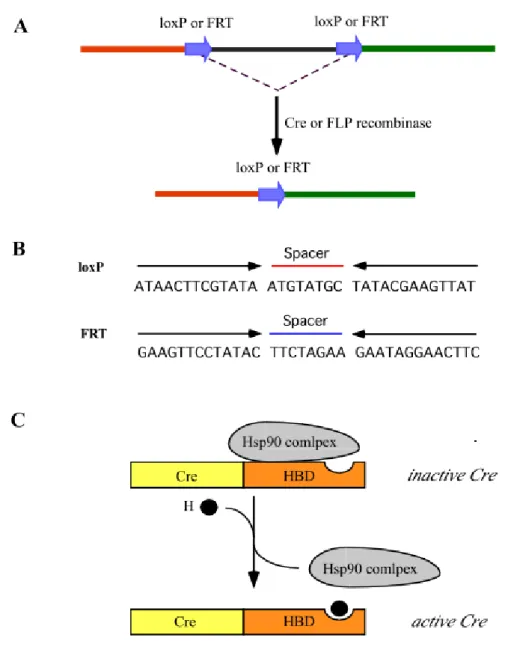
Somatic mutagenesis resulting from precise genetic modification can be efficiently achieved by using P1 phage Cre recombinase to catalyze excision or inversion of DNA flanked with specific 34 bp sequences termed loxP site (Hoess et al. 1986 and Sauer 1998). An alternative system for site-specific recombination is yeast Flp recombinase and FRT (Flp recombinase target) sites; however its efficiency is relatively low in mammalian cells because of different physiological temperature of yeast and mammals (Senecoff et al. 1988) (Figure 1A and B).
Introduction
Figure 1. Cre or FLP-mediated recombination and schematic representation of the regulation of Cre recombinase activity. (A) The genomic region flanked with two directly repeated loxP or FRT can be excised by the Cre or FLP recombinase. (B) lox P and FRT sites are composed of the spacer and inverted repeat indicated by thin arrows. (C) An activity of Cre recombinase can be regulated by fusion to the hormone-binding domain (HBD) of a steroid receptor. The fused Cre recombinase along with the hormone-reversible heat-shock protein 90 (Hsp90) complex is poattranslationally inactive. By hormone binding Hsp90 complex can be released from the Cre fusion protein and the Cre recombinase is in active state.
The genetic switch mediated by the Cre-loxP system is irreversible. Therefore temporal regulation of Cre recombinase expression has been attempted during past years. For this, Cre recombinase was fused with mutated steroid hormone-binding
Introduction domain (HBD) of progesterone or estrogen receptor, which is responsive not to endogenous hormones, under a control of tissue-specific promoter (Pcard 2000) (Figure 1C). The fusion protein becomes active by the synthetic ligands RU486 or tamoxifen, however, both RU486 and tamoxifen are prone to potential side effects resulting from interference with the endogenous hormone circuit such as processes of mammary gland or reproductive development (Kellendonk et al. 1996 and 1999, Brocard et al. 1997 and 1998, Danielian et al.1998). Second, local administration of adenovirus expressing Cre recombinase demonstrated toxicity and immunogenicity mediated by adenoviral infection (Akagi et al. 1997 and Anerson et al. 1998). The combination of Cre-loxP system and tetracycline-controlled regulatory system, which will be subsequently described, could be promising for Cre-based gene regulatory system in a conditional manner (Utomo et al. 1999).
1.1.2. Reversibly inducible systems derived from eukaryotic regulatory system The inducible systems, based on endogenous system from simpler eukaryotic organisms (e.g. Drosophila melanogaster, Caenorhabditis elegans), usually suffer from high basal level of activity in non-induced state and divers side effects, because the inducer activates not only a responsive transgene under a study but also undesired endogenes. Additionally there is a lack of a spatial control of a transgene and thus a non-specifically regulated transgene is expressed in all cell types. Such drawbacks of theses systems limited utility in eukaryotic organism for example in mice.
(1) Heat shock
Some heat shock protein promoters (D. melanogaster hsp70, C. elegans hsp68) which rely on endogenous heat shock transcription factors for their activity were used in cell lines (Schweinfest et al. 1988) and transgenic mice (Kothary et al.1989). The activity of these promoters has no basal level of gene expression at 37oC and induced with fast kinetics (within minutes) by a temperature shift from 37oC to 42oC. In spite of a stringent regulation, the induction system has drawbacks such as a poor induction ratio (~10 fold) with many pleiotropic effects through heat stress and no spatial regulation of a transgene.
(2) Heavy metal ion
The inducible system based on promoters responsive to heavy metal ions, particularly Cd2+ and Zn2+ showed a significant leakiness, the modest levels of induction, no cell type-specific control, and the toxicity associated with administration of heavy metal (Filmus et al. 1992).
Introduction (3) Interferon
Gene expression under a control of interferon-inducible regulatory element e.g. a promoter of Mx1 was activated rapidly in mice by injection, double-stranded RNA, or virus (Arnheiter et al. 1990, Kühn et al. 1995). However, the level of induction and background was variable in different tissues as well as in from mouse to mouse, because of different interferon availability in various tissues and variable production of endogenous interferon during viral infections and other illness. Additionally, an induction leads to biological side effects by treatment with interferon α/β or dsRNA.
(4) Hormones
The system by using steroid hormone (e.g. glucocorticoid, estrogen, and progesterone) inducible promoter exhibits a high leakiness of the inactive state and pleiotropic effects as above systems (Friedman et al. 1989, Braselmann et al. 1993, and Wang et al. 1994). Because a number of mammal endogenous promoters have a hormone-responsive element, undesired endogenes as well as a transgene could be expressed by hormone induction. It might make difficult to figure out definite conclusion of effects of a transgene under a study.
(5) Ecdysone
The insect steroid hormone ecdysone-inducible gene expression system has been described as an alternative and promising system for use in mammalian cells and transgenic mice (Yao et al. 1993). The ecdysone triggers metamorphosis in D.
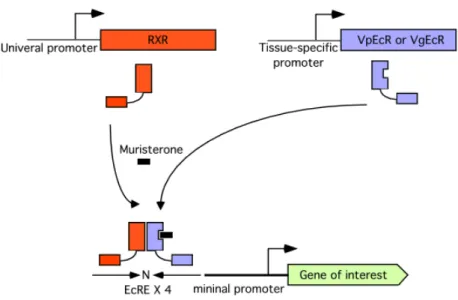
melanogaster mediated via a heterodimer of the ecdysone receptor (EcR) and ultraspiracle (USP). The heterodimer transactivates the ecdysone response elements (EcREs)-containing promoter composing of two inverted half-sites of the sequence AGGTCA spaced by 1 nucleotide. To improve sensitivity of an ecdysone-inducible system in mammalian cells, the USP was substituted by the retinoid X receptor (RxR), the mammalian homologue, and the EcR was modified. The N-terminally truncated EcR was fused with the herpes simplex virus protein VP16 activating domain (VpEcR). The DNA-binding specificity of VpEcR was also altered to mimic that of the glucocorticoid receptor (GR), which binds to an inverted repeat of the sequence AGAACA spaced by 3 nucleotides. The DNA-binding specificity was improved by mutating 3 amino acids residues of the DNA binding domain of VpEcR and by modifying the tandemly arranged ecdysone response elements (EcREs) containing two different half-sites, AGGTCA and AGAACA spaced by 1 nucleotide.
This modified receptor is called the VgEcR and combined with the RxR (Figure 2).
Thereby induction levels were increased to four orders of magnitude by treatment
Introduction with ecdysone, the synthetic analogue muristerone A (murA) or ponasterone A, which is found by screening natural plant compounds for improvement of inducibility, while maintaining a very low basal activity (No et al. 1996, Albanese et al. 2000, Yu et al.
2000). In this system, tissue-specificity can be conferred by choice of promoters that direct expression of the ecdysone receptors. The ecdysone-inducible system had no apparent side effect through an administration of ecdysone or murA, high inducibility with a very low leakiness. In addition, the lipophilic nature of ecdysteroid allows penetrating into all tissues, including the brain with negligible storage and expeditious clearance. Nevertheless the ecdysone-inducible system has a limitation of the requirement for three transgenes.
Figure 2. Schematic diagram of the ecdysone-inducible gene expression system. The retinoid X receptor (RxR), the mammalian homologue of ultraspiracle (USP), and the modified ecdysone receptor VpEcR (or VgEcR) can be expressed under a control of tissue-specific promoter and heterodimerize. The heterodimer can transactivate the ecdysone response elements (EcREs)-containing promoter in the presence of hormone or synthetic analogue muristerone. The EcREs are placed upstream of a minimal promoter, which can drive the expression of gene of interest. Then an ecdysone-responsive promoter can control expression of gene of interest. The EcRE is composed of tandemly arranged inverted sequences spaced by 1 nucleotide (indicated by arrows and N for spaced nucleotide).
Introduction 1.1.3. Inducible systems based on prokaryotic regulatory elements
Highly specific control of gene activity in higher eukaryotic cells has been achieved by the utilization of the well-characterized prokaryotic regulatory elements because regulatory elements and inducer do not rely on endogenous control elements. The inducible systems based on two well-studied regulatory elements from Escherichia coli, lac and tet operones, have been shown as monospecific regulatory circuits in mammalian cells and transgenic mice. The use of tissue-specific promoter for expression of these regulatory elements can contribute ability of a spatial regulation in transgenic mice.
(1) lac Repressor/Operator-based inducible system
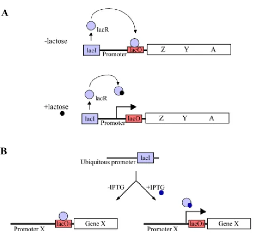
The E. coli lac operon-based system composes of two components; a lac repressor (lacR) and a DNA sequence called lac operator (lacO). In this system, the lac operator and repressor provide tight, reversible transcriptional control of genes involved in the uptake and metabolism of lactose (Figure 3A). The lac repressor binds to operator (lacO) and prevents formation of an initiation complex RNA polymerase and the promoter. This repression can be specifically reversed by the synthetic and nonmetabolizable inducer isopropyl β-D-thiogalactopyranoside (IPTG). The most straightforward approach followed the prokaryotic paradigm: lac operator sequences were placed near the TATA-box or start site of mammalian promoter. Thereby, a lacR-O complex within the promoter region would directly interfere with initiation of transcription of an endogene. The transcription from promoters containing lacO sequences could indeed be regulated in mammalian cells by IPTG (Hu and Davidson 1987 and Brown et al. 1987). During past years, few studies were established by the lac-operon based inducible system, no in transgenic mice, because of some limitations e.g. cytotoxic levels of IPTG required for induction, low intracellular concentration of the repressor protein to achieve a efficient induction in the mouse. Currently, Caronin and colleagues (2001) described the successful application of a functional lac operon- regulatory system in the mouse (Figure 3). In their study, the lac repressor was optimized concerning splicing, translational codon, and methylation-associated silence. Three lac operators were inserted into the tyrosinease promoter so that it is directly regulated by the ubiquitously expressed lac repressor. The lac repressor could repress the activity of a reporter gene, the mouse tyrosinease coding sequence, which subsequently could be derepressed by IPTG at a noncytotoxic level. The successful transfer of a lac operator-repressor gene regulation system to the mouse promises reversible control of the endogenous genome.
Introduction
Figure 3. The lac operon-based inducible system. (A) The lac operon in E. coli operates by a repression mechanism. In the absence of lactose (upper panel), the lac repressor (lacR) binds to lac operator (lacO) and prevents gene expression. In the presence of lactose (down panel), the lacR undergoes a conformational change and results in the derepression of genes. (B) Schematic representation of lac operon-based inducible system in transgenic mice. The lac repressor is ubiquitously expressed. The lac repressor represses the expression of gene X by binding to the lac operator sequences, which are were placed near the start site of promoter of gene X. This repression can be specially by the synthetic inducer isopropyl β-D- thiogalactopyranoside (IPTG); lacI, gene encoding lac repressor; lacO, operator sequence; Z, Y, A, genes encoding β-galactosidase, permease, and transacetylase, respectively, needed for uptake and utilization of lactose.
(2) Tetracycline-controlled transactivator (tTA) system
The tetracycline-controlled transactivator (tTA) is the fusion protein between the repressor (tetR) of the Tn10 tetracycline resistance operon of E. coli and the C-
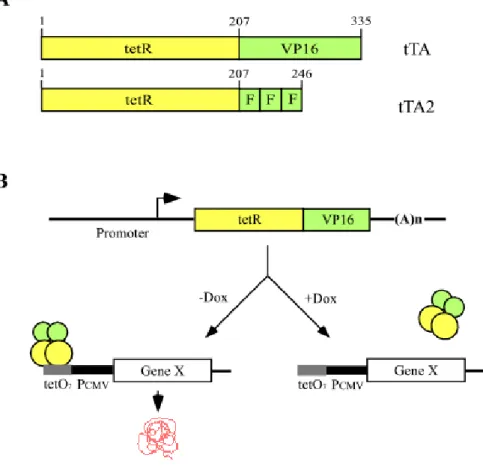
Introduction terminal transcriptional activating domain of VP16, known to be essential for the transcription of the immediate early viral gene (Gossen et al. 1992) (Figure 4A). This transactivator binds to operator sequence (tetO) of the tet operon in a tetracycline- dependent manner: When tTA-responsive promoter (Ptet), fused between seven tetO sequences and a minimal RNA polymerase II promoter sequences derived from the human cytomegalovirus immediate-early (IE) promoter (PhCMV), is placed in front of a gene, the tTA mediates on / off situation of gene transcription in presence or absence of tetracycline or its analogue (Baron U. et al. 2000, Mansuy et al. 2000) (Figure 4B). The bi-directional promoter, in which the seven tetO sequences (tetO7) are flanked with two minimal promoters, allows co-regulation of two genes (Baron et al.1995, Krestel et al. 2001).
Figure 4. The tetracycline-controlled transactivators (tTA) and schematic outline of tTA regulatory system. (A) The tTA is fusion protein between tet Repressor (tetR), consisting of 207 amino acids, and the 128 – amino acids transactivation domain of VP16. In tTA2, VP16 domain is changed with three minimal activation domains (F domain). (B) Mechanism of the tTA regulatory system. The tTA binds in absence of the effector molecule doxycycline (Dox) to an array of seven tet operator sequences (tetO7) placed upstream of a minimal promoter and
Introduction activates transcription from a minimal cytomegalovirus promoter (PCMV), which itself is inactive. But not in presence of Dox.
The tTA system is one of the most efficient inducible gene regulatory systems for use in mammalian cells, D. melanogaster (Girard et al. 1998), plant (Weinmann et al.
1994, Zeidler et al. 1996) and transgenic mice (Kistner et al. 1996, Ewald et al.1996, Mayford et al.1996). The distinct advantages of tTA system are (1) the high induction ratio (~105), combined with very low basal leakiness, (2) the specificity of the tetR for its operator sequence, the high affinity of tetracycline for the tetR, (3) well- characterized pharmacological properties of tetracycline and its analogues, which is able to penetrate most cell boundaries (Hillen and Berns 1994), (4) low toxicity of tetracycline and its derivatives, and (5) a genetic switch that would permit the control of individual gene activities reversibly in a temporal manner.
With these advantages, the tTA system has been extensively applied in different tissues, organs of transgenic mice. In neuroscience, the conditional gene expression system by using tTA has been employed to outline the modelling, therapeutic strategies of neuropathology and to study the molecular mechanisms underlying the processes of learning and memory. For instance in prion disease, the cellular prion protein (PrPc) is converted into the pathogenic isoform (PrPsc) via a posttranslational modification (Prusiner 1997). The expression of PrPc in transgenic mice was regulated by tTA under a control of the PrP gene promoter (Tremblay et al. 1998). It was shown that repression of PrPc expression on doxycycline in young adult transgenic mice (tTA:PrP) is not deleterious, whereas accumulation of PrPsc in same line of animal is lethal. Mice expressing a mutated huntingtin fragment controlled by tTA also lead to neuropathologic signs characteristic of Huntington’s disease and these symptoms could be reversed by suppression of the transgenic expression with doxycycline (Yamamoto et al. 2000). In the study of synaptic plasticity concerning learning and memory, transgenic mice carrying tTA under a control of the α-type calcium-calmodulin-dependent kinase II (CaMKIIα) gene promoter has been constructed and the tTA directed the expression of mutated Ca2+-inddependent form of CaMKIIα in transgenic mice (Mayford et al. 1996). Inducible expression of mutant CaMKIIα resulted in a loss of hippocampal LTP and a deficit in a spatial memory.
Suppression of transgene expression by an administration of doxycycline reversed both the physiological and the behavioural phenotype. Currently, Krestel et al. (2001) showed that the tTA-sensitive bidirectional expression module is well suited to express genes of interest in a co-regulated manner in mouse. In the study of Mack et al. (2001), expression of green fluorescent protein (GFP)-tagged GluR A could be controlled in GluR A-deficient mice by using CaMKII-tTA mice (Mayford et al.
1996). It was reported that hippocampal LTP was rescued in CA1 pyramidal cells of
Introduction
GFPGluR A. Theses studies strongly indicated that the tTA-mediated gene regulatory system has become a powerful tool for utility in mice.
1.2. Why ‘humanised tTA (htTA)’ is needed
Despite of a number of trials to create tTA mice, so far the useful and functional tTA mice in brain are the CaMKII-tTA (Mayford et al. 1996), enolase-tTA (Chen et al.
1998) and prion-tTA mice (Tremblay et al. 1998). It is difficult to generate transgenic mice expressing the tTA to direct efficiently a gene of interest in a region specific manner in brain. Unfortunately the trials of construction of tTA mice failed to present us useful tTA mice even by knock-in approach using GluR B, NMDA receptor 1 and ADARA2 (Jerecic et al. 1999). The tTA gene appear to be incorrectly spliced or expressed too deficient to induce tTA responsive gene (e.g. lacZ) in these transgenic mice. Therefore we attempt to increase the translation efficiency and mRNA stability of tTA derived from prokaryotic element in eukaryotic environment, termed
‘humanised’ tTA (htTA). The humanised Green Fluorescent Protein (GFP) from jellyfish (Zolotukhin et al. 1996), Cre recombinase and the currently reported lac repressor (Cronin et al. 2001) have been already shown stability and high level expression of these adapted protein in mammalian cell. In order to increase the tTA expression in eukaryotic environment, ‘humanized tTA (htTA)’ was designed on the DNA sequence level without change of amino acid sequence regarding eukaryotic codon usage, elimination of potential splicing signal, avoidance of CG sequence, and introduction of kozak sequence. In addition the tTA 2 gene, whose VP16 moiety is replaced by three minimal critical activating domains (F domain) was used as a template of a design of htTA (Baron et al. 1997, Regier et al. 1993). The modification of the activating domain of tTA 2 allows tolerance at higher intracellular concentrations, whereas overexpression of transcription in general results in squelching (Gill and Ptashne 1988).
1.3. Generation of transgenic mice expressing htTA in a subregion- specific manner in brain compared to previous CaMKII-tTA mice Next, we tried to achieve transgenic mice expressing the optimized htTA in a more spatially specific manner in brain for investigation of brain function at molecular and cellular level. The brain subregion-restricted gene regulation should allow a more precise analysis of gene function under a study.
Previously, the CaMKIIα promoter has been the most frequently used the promoter for control of expression of several inducible elements in transgenic mice showing its activity in relatively widespread forebrain of transgenic mice. The forebrain-specific
Introduction CaMKIIα promoter has been used to direct tissue-specific expression of the tTA (Mayford et al. 1996), rtTA (Malleret et al. 2001, Mansuy et al. 1998) and Cre recombinase (Minichiello et al.1999, Tsien et al. 1996) in rodent brain. The expression pattern of tTA under a control of CaMKIIα promoter has been determined in the neurons of the neocortex, the hippocampus, the striatum and septum, the amydala, and the basal ganglia by in situ hybridization (Mayford et al. 1996) (Figure 5). Additionally, tTA activity was found in thalamus, pons, medulla oblongata, spinal cord, and eyes with weaker signals by generating new sensitive tTA-reporter GFP lines (Krestel et al. 2001). In the αCaMKII-rtTA transgenic mice, it expression was found in CA1 and CA2 area of the hippocampus with almost no signal in CA3 region, in dentate gyrus, in superficial layers and deeper layer of cortex and in the striatum and septum by lacZ staining (Mansuy et al, 1998). The Cre recombinaes under a control of CaMKIIα promoter was observed in the hippocampus with strong signal in CA1, lighter signal in CA3 and dentate gyrus, the neocortex, the striatum, and amydala (Minichiello et al, 1999). Tonegawa group generated αCaMKII-Cre transgenic mice, however, the Cre expression was relatively late in development and mainly restricted in the hippocampus due to different condition of transgene integration (Tsien et al. 1996).
Figure 5. The expression of transgenes under a control of CaMKIIα promoter. (A) Schematic draws of the CaMKIIα promoter-controlled transgenes. The arrow indicates the transcriptional start of CaMKIIα promoter. Triangle represents synthetic intron sequences.
pA, polyadenylation signal. (B) Examples of CaMKIIα promoter-controlled expression patterns in brain. Left; In situ hybridization of CaMKII-Asp286 mRNA directed by CaMKII- tTA. Right; Spatial pattern of Cre activity in double transgenic mice of CaMKII-Cre:lox-lacZ by X-gal staining.(published by Mayford M. et al.1996 and Minichiello L. et al. 1999);Cx, cortex; CA1/3, areas of Ammon’s horn; DG, dentate gyrus; Amy, amygdala; H, hippocampus;
Cb, cerebellum; Str, striatum; Th, thalamus; ic, inferio colliculus; O, olfactory bulb; Bs, brain
Introduction Together, the CaMKIIα promoter has been useful to express transgenes in forebrain, but ultimately a transgenic mouse containing different tissue-specific or more specific expression patterns of the inducible regulatory elements could be needed depending on numerous genes of interest. The hippocampus has been one of the most frequently employed areas of the central nervous system as a model system for the study of neurobiological questions (Figure 6). There are two main reasons. First, its distinctive and identifiable structure. The apparent simplicity of the hippocampal neuronal circuitry is attractive at anatomical, physiological, and molecular biological level.
Second, investigations of a role of hippocampus. The hippocampus may affect some neurodegenerative diseases (e.g. Alzheimer’s disease), certain pathological conditions (e.g. epilepsy) and play a fundamental role in some forms of learning and memory.
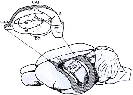
Figure 6. Simplified drawing of the location and the intrinsic connection of hippocampus of rat brain. The hippocampus is a banana-shaped structure that extends from the septal nuclei rostrally to temporal cortex, caudally. The long axis is called the septotemporal axis (indicated by S-T) and the orthogonal axis is the transverse axis (TRANS). A slice cut perpendicular to the long axis of the hippocampus (above left) shows several fields of the hippocampal formation and several of the intrinsic connections. Abbreviation: DG, dentate gyrus; CA1, CA3, areas of Ammon’s horn (cornu Ammonis); S, subiculum; pp, perforant path fibers from enthorhinal cortex; mf, mossy fibers from the granule cells; sc, Schaffer collateral connections from CA3 to CA1. (published by Amaral D.G: and Witter M.P. 1989)
For the study of hippocampal functions, affects from the other region of brain should be eliminated by expressing the inducible element in a hippocampus-specific manner.
Introduction For that we had two approaches by using KA1 bacterial artificial chromosome (BAC) and applying neuron-restrictive silencing element (NRSE). By using KA1 bacterial artificial chromosomes (BAC), it was expected that expression of the htTA under the control of KA1 promoter is directed in the areas expressing KA1 itself, namely in CA3 and dentate gyrus (DG) of hippocampus. An alternative approach was to repress extra hippocampal expression of the CaMKIIα promoter by the neuron-restrictive silencer element (NRSE) of the N-methyl-D-aspatate receptor 2C (NR2C) subunit.
Results
2. Results
2.1. Design and construction of the humanized tTA (htTA)
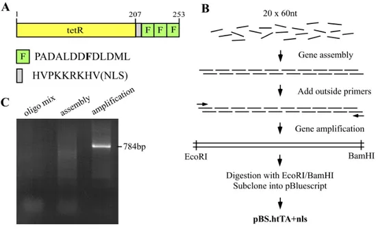
To increase the expression level of tTA in eukaryotic cells, the humanized tTA was designed on the DNA level in terms of (1) substitution of codons by their preferentially used degenerate codons in eukaryotic cell, (2) removal of potential splicing signals, (3) avoidance of CG dinucleotide sequences, which tend to be methylated and possibly prevent active gene expression, and (4) introduction of a Kozak consensus sequence, which could enhance translation efficiency of the mRNA (Kozak M. 1984), next to the start codon AUG. The tTA2, which has 100 % relative activation potential with a lower cytotoxicity, was used as template because it is tolerated at 3-fold higher concentration in cells. This is due to its VP16 activation domain being reduced to three acidic minimal activation domains (F domains), each consisting of only 13 amino acids (Baron et al. 1997) (Figure 7). To maximize the functional efficacy of the transactivator on the responsive gene in the nuclear compartment, the nuclear localization signal (NLS) was introduced between tetR and three F domains. The htTA was synthesized by assembly PCR with overlapping oligos according to the method of Stemmer et al. (1995) (Figure 8). The synthesized htTA fragment (784 bp) was cloned into pBluscriptIISK(+) and verified by sequence analysis.
2.2. Activation potential and comparison of the htTA in transiently transfected cells
To asses the activation efficiency of the htTA without and with the nuclear localization signal (NLS) compared with the prokaryotic tTA, the tTA, htTA-NLS and htTA+NLS genes were subcloned into the expression vector pRK5 (Schall et al. 1990).
HeLa X1/6 cells containing a chromosomally integrated tTA-responsive luciferase gene as a reporter (Gossen et al. 1992), were transiently co-transfected with plasmids encoding the tTA, htTA-N L S and htTA, respectively and β-galactosidase (pCMVpnlacF, Mercer et al. 1991) for a normalization. The transfected HeLa X1/6 cells were incubated in the absence or presence of doxycycline (1µg/ml) for 30 hrs.
Luciferase activity was detectable by use of the luminescent assay (luciferase gene assay kit, Dual-Light, TROPIX) and normalized to β-galactosidase activity (Table 1).
An increase of induction efficiency of the humanized tTA measurable by luciferase activity was observed and this activity was effectively abolished by doxycycline: the htTA-NLS reaches 133% and the htTA+NLS 298% of the activity conferred by prokaryotic original tTA in HeLa cells. The htTA+NLS generates almost 3-fold higher
Results
Figure 7. Alignment of DNA sequences of the tTA2 and humanized tTA gene. Nucleotides differing from the prokaryotic tTA2 were marked with box on the htTA sequences. Two blue boxes indicate translational start and stop codon, and the nuclear localization signal (NLS) is in a grey box. Underlines show restriction sites, which were used for subcloning.
Results
Figure 8. Design and synthesis of the humanized tTA. (A) Representation of the htTA fusion protein containing three F domains and nuclear localization signal (NLS) between humanized tetR and three F domains. The number indicates position of amino acids and capital letters are codons with single letter amino acids. (B) Schematic procedure for the htTA gene synthesis by oligo shuffling. Twenty overlapping oligos (approximately 60 nucleotides in length) on DNA sequences of the htTA gene were assembled by PCR and subsequently amplified with two outside primers (arrows). The outside primers contained EcoRI endonulclease site for 5’
end and BamHI for 3’ end. The amplified fragment was subcloned into pBluscriptIISK(+) via EcoRI and BamHI digestion. (C) 1 % agarose gel shows oligo mixture before the PCR assembly (left), smear assembly PCR products in variable length (middle) and the 748 bp fragment amplified with the outside primers.
induction while maintaining a very low basal activity and tolerance at higher intracellular concentration. Thereby the htTA+NLS could be confirmed as one of the best tetracycline-controlled transactivators for an application in transgenic mice.
Results Table 1. Comparison of induction efficiency of transcriptional activation between the synthesized htTAs and prokaryotic tTA
Luciferase activity
(RLU/µg protein)
+dox -dox
Relative Activity
(%)
Plasmid
tTA 0.1 1580 100 PRK5.tTA htTA-NLS 0.26 2107 133 PRK5.htTA-nls htTA+NLS 0.12 4716 298 PRK5.htTA+nls Rates were obtained light emission/µg protein. The luciferase activities were measured by use of the luminescent assay in three independently transfected HeLa cells, HeLa X1/6, which contain the luciferase gene under the transcriptional control of the chromosomally integrated tTA-dependent promoter PhCMV-1 (Gossen and Bujard 1992). HeLa X1/6 cells were transiently transfected with plasmids encoding the tTA, htTA-NLS, and htTA+NLS, respectively and cultured in the absence or presence of doxycycline (1 µg/ml) for 30 hrs. These values were normalized by co-transfection of plasmid encoding β-galactosidase (pCMVpnlacF, Mercer et al. 1991), which were assayed either simultaneously by luminescent assay or by standard liquid O- nitrophenyl β-galactoside assay (ONPG, Sigma), and related to the activity of previously described tTA (100 %); RLU, relative light units; dox, doxycycline.
Results
2.3. Generation of transgenic mice expressing the htTA in a region (hippocampus)-specific manner in brain
In our attempt to develop a subregion-restricted regulatory system in the mouse brain for study of molecular mechanisms of brain function, the improved htTA was used.
To achieve more region-restricted expression patterns of the tTA inducible system, especially in hippocampus, compared with the forebrain-specific CaMKII-tTA mouse (Mayford et al. 1996), we had two approaches: (1) using bacterial artificial chromosome (BAC) harbouring the 5’ regulatory region of the high-affinity kainate receptor subunit KA1 gene, and (2) applying the silencer region of N-methyl-D- aspatate receptor subunit 2C (NR2C).
2.3.1. Generation of KA1.htTA transgenic mice
To generate transgenic mice expressing the tTA-inducible system in a region-specific manner similar to the expression pattern of the high-affinity kainate receptor subunit KA1, the htTA+NLS was inserted into the ATG-including exon of the KA1 gene through homologous recombination of a KA1 BAC clone. In addition, the tTA was directed via the same way for comparison of mRNA stability and activities in vivo between the htTA and the tTA.
The KA1 gene has a highly restricted expression mainly in pyramidal cells of the hippocampal CA3 and granule cells of the dentate gyrus of adult rat brain determined through in situ hybridiztion (Werner et al. 1991). Kask et al. (2000) generated transgenic mice expressing Cre recombinase under a control of the KA1 promoter by using 550 kb YAC (yeast artificial chromosomes). It was shown that expression pattern of Cre recombinase controlled by the KA1 promoter carried on 550 kb YAC resembles that of endogenous KA1. The 5´ regulatory elements of the KA1 gene could accommodate to direct the htTA gene in a region-specific manner as the KA1 gene itself in transgenic mice. Now we used bacterial artificial chromosomes (BACs) carrying the mouse KA1 gene because there are several advantages of using BACs, compared to YACs. (1) BACs have a high stability in terms of their propagation in recombination deficient E. coli host cells, (2) isolation, purification and handling of BACs are less difficult since they exist as supercoiled circular plasmids, (3) a direct sequencing can be applied to BACs, (4) BACs can be modified in E. coli to insert transgenes at a desired position. and (5) a large size of the insert in BACs approximately 150 kb ~ 200 kb might provide for less integration dependency of transgene expression. Recently, transgenes were obtained successfully by modifying
Results BACs (Antoch et al. 1997, Jessen et al. 1998, Nielsen et al. 1997, and Probst et al.1998).
2.3.1.1. Genomic structure of the mouse KA1 gene
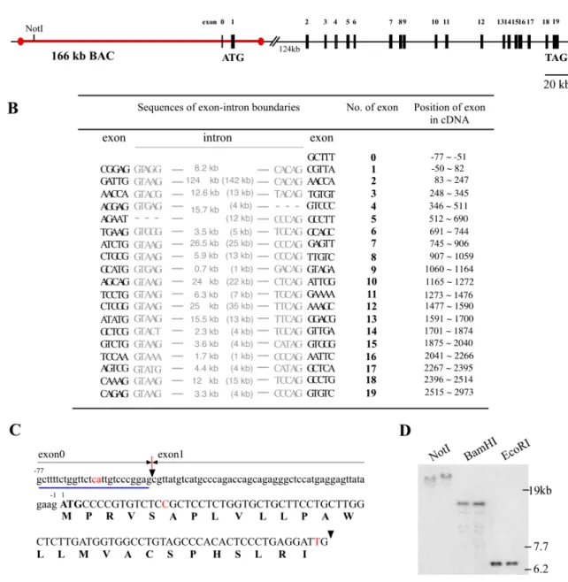
The cDNA from rat brain and human encoding the KA1 subunit have been cloned (Werner et al. 1991, Herb et al. 1992 and Kamboj et al. 1994) but its detailed genomic structure is not delineated. The Celera data bank search provided genomic sequence information of the mouse and human KA1 gene. The comparison of amino acid sequences at the KA1 cDNA level of mouse and human shows high percent of identity among species; 99.5 % of similarity of amino acid sequences between rat and mouse, and 97.5 % between mouse and human. The Celera data bank mouse genome accession x5J8B7W3WPH and human genome accession x54KRE8WCJ9 revealed that the gene of KA1 encoding 956 amino acids exists as a single copy on mouse chromosome 9 and on human chromosome 11, respectively and is divided into 19 translated exons both in mouse and human (Figure 9A). The mouse KA1 gene spans about 300 kb between the first ATG exon (132 bp) and the last exon 19 and within are some very large introns contained: one of 124 kb in a size between the first ATG exon and exon 2, three of approximately 25 kb between exon 6-7, exon 9-10, and exon 11- 12. The human genome Celera data revealed that the organization of exons and introns of the KA1 gene is highly conserved between human and mouse. The locations of exon-intron boundaries are at the exactly same positions on the mouse and human KA1 open reading frame (OFR) and the length of introns is very little variable (Figure 9B). It is reported that there is one untranslated exon (27 bp) upstream of the ATG exon in the rat cDNA sequence. The 27 bp untranslated exon (exon0) was identified 8.2 kb upstream of the ATG exon1 by southern blot analysis of mouse genome BACs and by Celera mouse genomic data with 2 different nucleotides compared to the rat cDNA sequence (Figure 9C and D). In human genome, no untranslated exon was yet identified.
2.3.1.2. Screening of BACs library for mouse KA1 gene
To isolate BACs containing the mouse KA1 gene, in particular 5’ regulatory region and the ATG exon, the 682 bp of PCR amplified fragment covering the partial introns surrounding the ATG exon was used for screening of the BAC library of the mouse C57BL/6 strain (Genome Systems, St. Louis, MO, USA). Two KA1 BAC clones were isolated and pulsed-field gel electrophoresis (PFEG) of NotI-digested BAC DNA revealed that two BAC clones contain inserts of approximately 166 kb and 60 kb in a size, respectively. Direct BAC sequencing and the Celera data bank search
Results
Figure 9. The exon-intron organization of the mouse KA1 gene. (A) Schematic representation of the mouse KA1 genomic structure according to the Celera data bank search. Exons of the KA1 gene are shown by filled boxes and numbered (exon0-19). Open box indicates the exon4, which was not found in mouse genomic sequences because of short segments unidentified by the Celera. Translational start codon (ATG) on the exon1 and stop codon (TAG) on exon 19 are indicated. Red line shows the location of 166 kb BAC harbouring the mouse KA1 gene. There is one NotI site within the 500 kb gene. (B) the nucleotide sequences of the exon-intron boundaries of the mouse KA1 gene. Intron sequences and length of the mouse KA1 gene are typed in grey. Additionally the length of introns of the human KA1 gene is in brackets. Position of exons is numbered on the mouse KA1 cDNA starting from translational start site. (C) The DNA sequences of exon0 and exon1 on the mouse KA1 cDNA and amino acid sequences (capital single letter under DNA sequences) of exon1. Black arrowheads indicate the boundary between exon0 and exon1; Red letters, different nucleotide
Results from rat; number, position of nucleotides. Blue underline shows the exon0 and was used as a probe for southern blot analysis of the BAC containing the 166kb mouse KA1 gene (D). NotI, BamHI and EcoRI are restriction enzymes used for digestion of BACs. Each left line is the 166 kb KA1 BAC and right one is the modified KA1 BAC inserted the htTA gene (see 2.3.1.4 in part of results).
KA1 gene containing 148 kb upstream of translational start codon ATG, 132 bp ATG harbouring exon (exon 1), 18 kb downstream of ATG exon, and exon0 (Figure 9A and C). The existence of the 27 bp of untranslated exon (exon0) on the BACs was also confirmed by Southern blot analysis with the 27 nucleotides (Figure 9D).
2.3.1.3. Construction of targeting vector for homologous recombination of the KA1 BAC
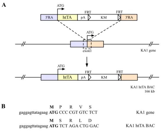
The 166 kb KA1 BAC clones were modified to express the htTA and prokaryotic tTA gene under the KA1 promoter control through homologous recombination. To insert the htTA and prokaryotic tTA respectively into the exon1 harbouring the translational start of the 166 kb KA1 BAC, two amplified recombinogenic arms (A; 630 bp, B; 560 bp) by PCR were on both sides of the targeting cassette, which contains the htTA or tTA gene, a polyadenylation sequence of human growth hormone gene and a selection marker gene (Kanamycine resistant gene, KM) (Figure 10). To eliminate the selection marker gene before microinjection, the KMgene is flanked by FRT (Flp recombinase target) sites, because this selection marker is needed only in E. coli during homologous recombination and the BAC plasmid (pBeloBAC11) harbours already one loxP site.
2.3.1.4. Modification of KA1 BAC
To modify BACs containing the KA1 gene to express the tTA gene under the KA1 promoter control, a temperature-sensitive shuttle vector based system was used for homologous recombination in E. coli, because BACs are propagated in the recombination deficient E. coli host cell (recA-), DH10B. There is often failure in obtaining intact size of BACs during a transfer of BACs to the other strain, which can be used for homologous recombination. To overcome the deficit of recombination in BAC host cells, the E. coli recA was introduced into BAC host cell DH10B (recA-) via the temperature-sensitive shuttle vector (pSV1.recA), which replicates at the permissive temperature (30oC) but is lost at the restrictive temperature (43oC).
Thereby it is possible to modify BACs directly in the recombination deficient host cell and to return to a recombination deficient condition for the stability of the
Results
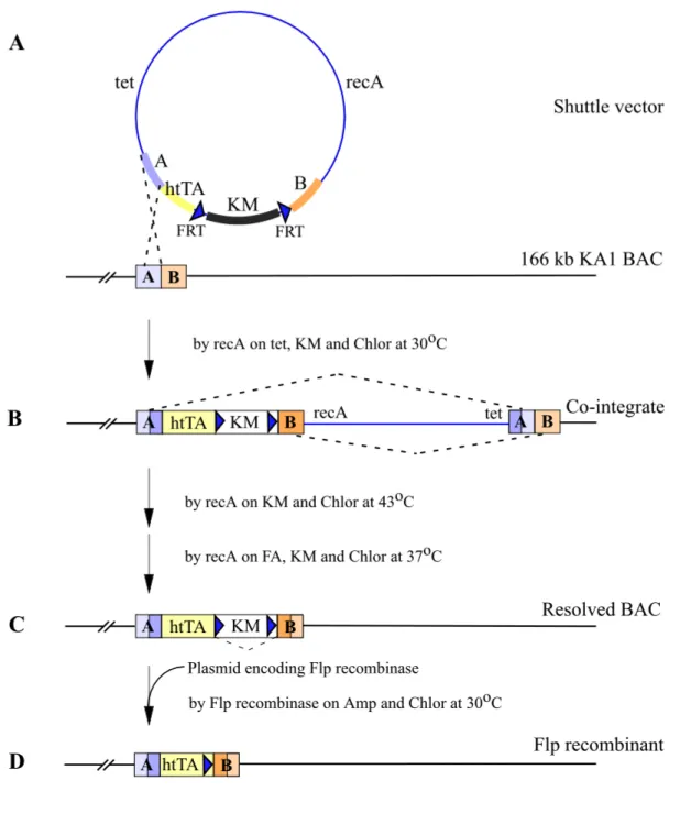
Figure 10. Generation of BAC carrying the KA1 gene to express the htTA under a control of the mouse KA1 promoter (illustrated only in case of the htTA but for the prokaryotic tTA via the exact same way). The htTA gene was inserted into the exon1 harbouring translation start codon of the KA1 gene on the 166 kb BAC by homologous recombination. (A) The targeted htTA cassette and locus of insertion on the KA1 BAC. Open lined box on the KA1 gene indicates the 132 bp exon1 containing translational start codon ATG; RA, recombinogenic arm amplified by PCR; htTA, the humanised tTA gene; pA, polyadenylation signal of human growth hormone; KM, Kanamycine resistance gene; FRT, Flp recombinase target site. (B) Small letters show the 5’ untranslated sequences and capital letters are codons with single letter amino acids for the KA1 and the htTA from modified KA1 BAC.
Results
Figure 11. The strategy to modify the 166 kb KA1 BAC. (A) Targeting cassette is integrated into the 166 kb KA1 BAC by recA via shuttle vector (pSV1.recA-htTA) based on temperature-sensitive origin (illustrated only in case of the htTA but for the prokaryotic tTA via the exact same way). (B) Cointegrate can be formed by homologous recombination through either A or B (only A case is illustrated) at 30oC. (C) Second recombination through B and subsequent temperature- and antibiotic-selection can produce resolved BAC by recA integrated into BAC. (D) The selection marker flanked with FRT can be eliminated by Flp recombinase. The Flp recombinase plasmid (pMAK-705FLP.amp) should be transformed into the cells containing the resolved BAC and incubated at 30oC. (Amp, ampicilin; Chlor, chloramphenicol; FA, fusaric acid; KM, kanamycine; tet, tetracycline; and FRT, Flp recombinase target site)
Results
Figure 12. Generation of the modified KA1.htTA BAC. (A) Schematic representation of expected BamHI-digested fragments in the KA1 BAC, cointegrate, resolved BAC and Flp recombinant by southern analysis with recombinogenic arm A probe (thin line) and recombinogenic arm B probe (open line) Two arrows indicate outside primers for colony PCR; FRT, Flp recombinase target sites; B, BamHI restriction site. (B) Conlony PCR of the KA1 BAC resolved BAC and Flp recombinants with outside primers. All 5 Flp recombinant, which were randomly picked show correct PCR fragment. (C) Southern blot analysis of the KA1 BAC, cointegrate, resolved BAC and Flp recombinant. BamHI-digested BAC DNAs of each step were hybridized with recombinogenic arm A probe (630 bp) and recombinogenic arm B probe (560 bp), respectively.
Results (1) Shuttle vector and first recombination
The htTA and tTA gene targeting cassettes were subcloned into a temperature- sensitive shuttle vector (pSV1.recA.NotI) (Figure 11A). The targeting cassettes containing shuttle vectors (pSV1.recA-htTA and –tTA, respectively) were then transformed into KA1 BAC host cells. The transformants were selected by tetracycline resistance (tet, carried by pSV1.recA,NotI), kanamycine (KM, carried by the targeting cassettes) and chloramphenicol (Chlor, carried by the KA1 BAC vector).
The first homologous recombination occurred by recA expressed via the pSV1.recA between the shuttle vector and KA1 BAC through either the 5’ recombinogenic arm (A) or 3’ recombinogenic arm (B) in cells growing at 30oC on plates containing tetracycline, kanamycine and chloramphenicol. Thereby this whole part of the shuttle vector as well as the targeting cassette were integrated into the KA1 BAC, termed co- integration (Figure 11B). Three of 24 clones were determined by southern blot analysis as correct cointegrates that have occurred through A.
(2) Second recombination of the cointegrates
Correct cointegrates underwent the second homologous recombination in cells growing on kanamycine and chloramphenicol plates at 43oC, called resolution (Figure 11C). This recombination event should result from the recA carried by only integrated shuttle vector, either into the KA1 BACs or bacterial chromosome, because the temperature of 43oC is nonpermissive for replication of the shuttle vector. The resolved BACs were subsequently selected by growing on kanamycine, chloramphenicol, and fusaric acid (FA) plates at 37oC, resulting from the fusaric acid causing negative selection for the loss of tet resistance (Maloy et al. 1981). Through the second homologous recombination and subsequent selections, the recA on the KA1 BAC was eliminated and thus a recombination deficient host cell condition obtained for the resolved KA1.htTA.KM BACs. During cloning of the targeting cassette into the shuttle vector and second recombination, the selection marker helped easily to get correct clones. Otherwise there are two possible recombinants after the second recombination, such as a resolved BAC and original BAC because of two- repeated recombinogenic arms. Almost 100 % clones, however, were correctly recombined with a selection of kanamycine. Colony PCR and southern blot analysis identified resolved BACs (Figure 12).
(3) Flp recombination of the resolved KA1-htTA/tTA.KM BAC
To eliminate the FRT-flanked kanamycine resistant gene, the Flp recombinase plasmid (pMAK-705FLP.amp), which is based on the pSC101 with temperature
Results sensitive replication origin to permit simple elimination of the plasmid after site- specific recombination (Hashimoto-Gotoh and Sekiguchi 1997), was transformed into the DH10B cell containing the resolved BAC (Figure 11D). The cells transformed with pMAK-705FLP.amp were incubated at 30oC and subsequently at 37oC on ampiciline and chloramphenicol plates. After Flp recombination any colony grew neither in the presence of kanamycine nor ampiciline, and 100 % site-specifically recombined. The finally modified BACs were also verified by colony PCR, southern blot and sequencing (figure 12). Pulsed-field electrophoresis gel (PFEG) and restriction maps with several edonucleases and southern blot pattern revealed no change of BACs after the final modification. By NotI digestion the modified KA1 BACs were released from the BAC plasmid (pBeloBAC11) and additionally lost 13 kb of 5’ regulatory region via endogenous NotI site. However, the NotI-linearized KA1.htTA BACs and KA1.tTA BACs still contain 153 kb of the KA1 gene, i.e. 135 kb 5’ regulatory region upstream of exon1 (Figure 9A). Thus NotI-linearized KA1.htTA BACs and KA1.tTA BACs, respectively, were purified by Sepharose CL- 4B gel fraction and injected into the male pronuclei of fertilized mouse oocytes.
2.3.1.5. Analysis of KA1.htTA and KA1.tTA founders
By tail biopsy of genomic PCR, the existence of htTA and tTA transgenes in potential founder animals was assessed: 7 founders out of 49 pups for KA1.htTA and 8 founders of 51 for KA1.tTA. To determine if the intact BACs had been integrated into the mouse genome, one of the BAC terminals was amplified by PCR, because BACs linearized with NotI contain some vector sequence. Incomplete integration was found in one of 15 founders, including KA1.htTA and tTA. By unidentified reasons some of those founders lacked transgene transmission or the ability to reproduce. To visualize expression pattern and activity of the htTA and tTA in these transgenic mice, the founders were bred with lacZ indicator lines (M3 and MNL), which contain a tTA- dependent minigene encoding the NMDA receptor subunit NR1 and lacZ in a bidirectional module (Jerecic et al. 1999) (Figure 13).
The double-transgenic animals of TgKA1.htTA/MNL or M3
and TgKA1.tTA/MNL or M3
were analyzed by staining for β-galactosidase activity on brain sections at ages postnatal day 7 (P7) and P42. Unfortunately, no blue staining was observed in brain sections in all animals even after 30 hours incubation for β-galactosidase activity. Nevertheless the existence of the htTA or tTA mRNA under the control of the KA1 promoter in brain was verified by reverse transcriptase-PCR (RT-PCR). 5 of 6 founders of KA1.htTA lines expressed htTA mRNA in mutant brains (figure14). For KA1.tTA lines only 1 out of 4 founders proved RT-PCR positive. With most of analyzed founders expressing RT-PCR detectable levels of tTA, the failure in induction of the
Results
Figure 13. Genetic cross to visualize the activity and expression pattern in the KA1-htTA (or KA1-tTA) transgenic mice. The KA1-htTA lines were bred with the lacZ indicator line (M3 or MNL) containing tTA-dependent NR1 and lacZ gene in a bidirectional module. In double transgenic animals of TgKA1-htTA/MNL or M3
, the expressed htTA can activate expression of lacZ gene and then activity and expression pattern of the htTA can be visualized by X-gal staining:
tetO7, seven tet repressor binding sites; rNR1, rat NMDA receptor subunit 1.
tTA-responsive gene in these mice might be explained by insufficient amount of tTA expression under the KA1 promoter driving from BACs. For the functional activity of tTA, a large supply of tTA seems to be required in brain and expression via BACs containing the KA1 promoter might not be strong enough. However, in terms of frequency of expression of the htTA and tTA gene in transgenic mice, the results of RT-PCR demonstrated that the utility of humanized tTA for transgenic mice is much more promising than that of the prokaryotic tTA.
Results
Figure 14. Reverse transcriptase-PCR (RT-PCR) verify the existence of the htTA and tTA mRNA in transgenic lines. (A) Total RNAs from brains of KA1-htTA transgenic lines (Kh1- 6) and wild type were amplified with htTA1/2 sitting on the htTA gene and NR1m1/2 sitting on different exons of the NR1 gene as a control. Mock was performed without reverse transcriptase. (B) RT-PCR of the KA1-tTA lines (Kt1-4), wild type, and TgCaMKIItTA (Mayford et al. 1996) with tta1/4 for the tTA gene and with NR1m1/2.
Results
2.3.2.Generation of CaMKII-NR2C.htTA transgenic mice
We next attempted to generate transgenic mice expressing the htTA by an alternative approach. Neurospecific gene expression can generally be achieved using a neuron- specific promoter such as the CaMKIIα promoter (Mayford, M. et al. 1996), the neuron–specific enolase promoter (Peel et al. 1997) or YAC/BAC containing KA1 promoter (Kask et al. 2000). An alternative approach was to repress ubiquitous expression using a negative regulatory element; neuron-restrictive silencer element (NRSE). The regionally restricted expression of the htTA in brain could be obtained by using the 8.5 kb fragment of CaMKII promoter that is transcriptionally suppressed by the 1.0 kb fragment of NR2C including NRSE to prevent widespread expression of the htTA in brain.
2.3.2.1. Characterization of the NR2C gene
To increase the knowledge of how the NR2C gene expression is controlled with a developmental and cell environmental specificity, the gene structure has been characterized and variable fragments up and downstream of the transcriptional start site were used to direct reporter gene in neuronal/non-neuronal cells and transgenic mice (Suchanek et al. 1995 and 1997). The NR2C subunit is mainly expressed in the cerebellar granule cells starting from the second week of postnatal life (Monyer et al.
1992). The 5’ untranslated region of the NR2C gene (Grin2c) includes three exons (exon 1-3) with two transcriptional start sites at –772 and –754 bp from the translational state site: 254 bp of exon 1, 172 bp of exon 2, and 235 bp of exon 3, interrupted by introns of 398 bp and 106 bp respectively (Figure 15). It was demonstrated that the 0.4 kb segment upstream of the transcriptional start site of the NR2C gene (Grin2c) has a general promoter activity in transgenic mice determined by the β-galactosidase expression pattern under the control of this 0.4 kb fragment of NR2C gene. On the other hand, the 1.0 kb segment (exons 1-3) downstream of the transcriptional start site directed the specific expression in cerebellar granule cells with weaker additional expression in other brain regions, i.e. dentate gyrus, olfactory bulb and cortex in transgenic mice. This 1.0 kb region of exon 1, 2, 3 of the NR2C gene seems to negatively regulate the transcription in NR2C negative cells for NR2C- specific expression. The 1.0 kb region contains a neuron-restrictive silencer element (NRSE)-like sequence near the 5’ end of exon 1. This 21 bp conserved negative transcriptional element (NRSE) has been identified in many neuronal genes such as N-methyl-D-aspatate receptor (NMDA1, NR2C), brain-derived neurotrophic factor (BDNF), γ-aminobutyric acid (GABA), nicotinic acetylcholine receptor (nACh) and
Results
Figure 15. The proximal promoter region and silencer region of the NMDA receptor subunit 2C. (A) The gene structure of proximal promoter region of the Grin2c. Left arrow indicates two transcription initiation sites, right arrow translational start site. Bright green boxes show untranlslational exons (E1, 2 and 3), yellow translational exon (E4). The distance between exon3 and 4 is reported approximately 6 kb. The 0.4 kb promoter fragment characterized as a basal promoter and the 1.0 kb silencer fragment are indicated by red and blue line, respectively. (B) In situ hybridization for the NR2C transcripts in horizontal slices of rat brain at P7 (Left) and adult (Right). (C) Schematic representation of transgenes (upper panel).
Left, the 0.4 kb of NR2C promoter upstream of first exon and lacZ gene; Right, 1.0 kb of exon 1-3 and lacZ. The expression of β-galactosidase transcripts in transgenic mice determined by in situ hybridization (down panel).(based on Suchanek B. et al. 1997): B, BamHI site; Sm, SmaI restriction site; ce, cerebellum; cx, cortex.
so on and is located in 5’ untranslated regions (5’ UTR) or in intragenic positions (Schoenherr et al. 1996) (Table2). The 1.0 kb fragment of NR2C gene might be contribute to tissue specificity through selective suppression of the transcription and thus be useful for the generation of transgenic mice expressing the htTA in a spatial manner.
Results
Table 2. All genes with two or fewer mismatches to the composite NRSE used in the data base search are listed. Selected genes with three or greater mismatches, for which NRSF binding was experimentally determined, are listed as well. The NRSE-like sequences are compared to the revised consensus NRSE. Derivations shown are derived from comparison to this revised consensus and not to the composite NRSE used in the original data base search.
The NRSE-like sequence of the NR2C is added in red: Rec., receptor; BDNF, brain-derived neurotrophic factor; NMDA, N-methyl-D-aspatate; Ach, acetylcholine; GABA, γ- aminibutyric acid; CRF, corticotropin-releasing factor; AMPA, α-amino-3-hydroxy-5methyl- 4-isoxazole-propionic acid; VGF, vascular endothelial growth factor, APRT; adenine phosphoribosyltransferase; trans., transactivating; Reg, 5´´-regulatory region; 5´- or 3´-UTR, 5´- or 3´-untranslated region; Jxn, intron/exon junction.
* A blank space indicates that the sequence has not been tested for the activity.
Results 2.3.2.2. Construction of the minigene of CNhtTA and sCNhtTA
To generate transgenic mice expressing the htTA in a region-restrictive fashion in brain, the forebrain-specific CaMKIIα promoter was fused with the NR2C gene silencer region (exons 1-3). The chimeric promoter comprises the CaMKIIα promoter, the tripatite leader sequence, and the 1.0 kb fragment from exon 1 to exon 3 of the NR2C subunit gene as transcriptional negative element (Figure 16).
Figure 16. Constructs of 0.8kbCaMKIIpromoter-NR2C silencer-htTA (CNhtTA) and small 0.4kbCaMKIIpromoter-NR2C silencer-htTA (sCNhtTA) minigenes. The CaMKII promoter is fused with 1.0 kb silencer of the NR2C (exon1-3 in green boxes). Tripatite intron is indicated by triangle. The htTA gene with polyadenylation signal of human growth hormone (pA) is followed next to the chimeric promoter. Several restriction sites, which were used for cloning and releasing the minigene from cloning vector; Sr, SrfI; N, NotI; EV, EcoRV; Sm, SmaI; B, BamHI; S, SalI; D, DraIII; E, EcoRI; K, KpnI; Bg, BglII.
The CaMKIIα promoter consists approximately 8.5 kb of genomic DNA upstream and 89 bp downstream of the transcription initiation site of the mouse CaMKIIα gene (Mayford et al. 1996). The adenovirus tripatite leader sequence, which is known to enhance RNA stability and efficiency of translation (Choi et al. 1991 and Sheay et al.1993), follows next. The 1.0 kb (exon1-3) untranslated fragment of mouse NR2C gene was added downstream of the CaMKIIα promoter and the synthetic intron sequence. The two transcriptional start sites on the first exon of the NR2C gene itself and the splicing donor of the end of the third exon were removed on the chimeric promoter, to abolish unfavored additional transcriptional events. Alternatively instead of the 8.5 kb CaMKIIα promoter fragment, we also used a 0.4 kb fragment of the CaMKIIα promoter because we experienced that the 0.4 kb small fragment of the CaMKIIα promoter region seems to include enough elements to control the forebrain-