Autologous full-thickness RPE-choroid graft to treat
high-risk drusenoid pigment epithelial detachment without CNV
Abstract
Objective:To report on the survival of a retinal pigment epithelium (RPE)- choroid graft translocated to treat a patient with drusenoid pigment epithelial detachment (DPED).
Hedwig Sillen
1Joke Ruys
2Pieter-Paul Schauwvlieghe
2Methods:We describe a patient with bilateral high-risk DPED where one eye was treated with RPE-choroid translocation surgery and followed
up for more than two years.
Marc Veckeneer
2Results:The RPE-choroid graft surgery was straightforward and the fully perfused graft was able to support stable vision of 0.5 Snellen acuity
1 Department of
Ophthalmology, Antwerp for more than two years despite the development of a choroidal
neovessel at the edge of the graft. The vision in the fellow eye dropped
from 0.5 to 0.2 Snellen in the same period. University Hospital, Edegem,
Belgium Conclusion: RPE-choroid translocation may slow the progression of
DPED to atrophy but it can also transform dry age-related macular degeneration (AMD) into neovascular AMD.
2 Department of Ophthalmology, ZNA Middelheim Hospital, Antwerp, Belgium Keywords:macular degeneration, retinal pigment epithelium, drusenoid
pigment epithelial detachment, RPE-choroid graft
Introduction
Although the majority of neovascular age-related macular degeneration (AMD) patients can now be treated effec- tively with anti-VEGF injections, there is still no treatment available for dry AMD.
Drusenoid pigment epithelial detachment (DPED) arises from the confluence of large areas of soft drusen [1].
It is well documented that the natural history of eyes containing DPED is characterized by a high rate of pro- gression to geographic atrophy [1], [2], [3].
Recently, Balaratnasingam et al. [4] examined the life- cycle of large DPEDs based on SD-OCT features. Their results demonstrate that maximal DPED height and dia- meter are inversely correlated with final visual acuity (VA) and that the appearance of intraretinal hyperreflective foci preceded the collapse of a DPED.
In agreement with our experience, Balaratnasingam et al. [4] found that visual function is usually still good while the PED volume is at its largest. Once the DPED collapses, accelerated atrophy usually ensues and macular function is irreversibly lost.
If a treatment would be available, significant growth of the PED and the appearance of intraretinal hyperreflective foci in a DPED could be considered as a signal to prompt intervention.
We describe a patient with a DPED on the verge of col- lapsing who was treated with an autologous full-thickness
retinal pigment epithelium (RPE)-choroid graft. To our knowledge, this is the first report of a RPE-choroid graft translocation to treat DPED.
Case description
An eighty-year-old female patient came to our department in August 2015. Snellen VA was 0.5 in both eyes and she was pseudophakic.
Funduscopic examination demonstrated a large well- circumscribed DPED in both eyes. There were no signs of an underlying choroidal neovascular membrane (CNV).
Fluorescein angiography (FA) showed in early phase a faint hyperfluorescence of the PED in both eyes. There was an increased pooling during the late phase, but no late leakage.
SD-OCT B-scan demonstrated a large PED in both eyes with moderate hyperreflectivity beneath the RPE band in correspondence with a DPED [5]. There were no signs of CNV on OCT-angiography (OCTA).
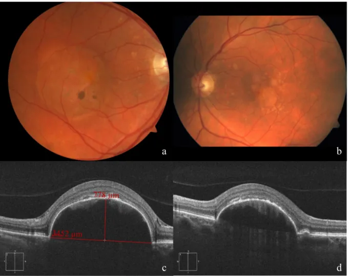
On follow-up, the lesion size further increased. In Decem- ber 2016, the DPED reached a diameter of 3,452 µm and a height of 778 µm (see Figure 1). Inserting those values into the scatter plots used by Balaratnasingam et al. [4], we estimated the risk for DPED collapse and deterioration of function to be very high. In addition, the intraretinal hyperreflective material heralded possible imminent outer retinal atrophy.
Figure 1: Fundus appearance and morphological evaluation of both eyes 4 weeks before operation of the right eye.
Fundus photograph of the right (a) and left (b) eye, showing the drusenoid pigment epithelial detachment. Cross sections of SD-OCT images of the right eye (c), vertical scan with indications for height and diameter of the DPED, and the left eye (d).
Since we have extensive experience with autologous RPE- choroid graft surgery to treat complicated neovascular AMD and DPED in dry AMD is thought to be the result of alterations in the RPE layer and Bruch’s membrane [2], [5], the possibility of translocation surgery was suggested as an option for the worst eye. After careful deliberation between the medical and surgical retina team and after extensive consenting of the patient, the surgery was planned.
The right eye was operated in February 2017 by MV. The standard RPE-choroid graft procedure, as previously reported for neovascular AMD, was carried out under general anesthesia [6]. Of note, opening up the PED re- vealed a clear gelatinous substance without any signs of CNV. The material could not be grasped with forceps but could easily be removed with the vitreous cutter. The graft procedure was otherwise unremarkable. No problems were encountered during oil removal seven weeks later.
VA remained 0.5. Eight months after surgery SD-OCT images revealed a small subretinal fluffy bordered hyper- reflective lesion with intraretinal cysts on the nasal border of the RPE-choroid graft, suggestive of a type 2 CNV, which was confirmed by FA. After treatment with intravit- real Aflibercept (Eylea), the lesion became sharply bordered with a few persisting intraretinal cysts on OCT.
During further follow-up, the DPED in the left eye was found to develop similar characteristics predicting a col- lapse in the near future. Considering the CNV complication in the operated eye and the absence of long-term follow- up data of RPE-choroid graft for this indication, we decided to abstain from surgery for the left eye, despite insistence of the patient.
At the last visit in July 2018, VA in the right eye was 0.6.
The RPE-choroid transplant in the right eye appeared healthy on funduscopic examination and OCTA confirmed perfusion. The CNV on the nasal side of the graft showed no signs of activity. As expected for the left eye, the DPED collapsed and evolved into atrophy (see Figure 2). VA in the left eye decreased to 0.35.
Discussion
DPEDs are a frequent finding in dry AMD and their natural history is characterized by a high rate of progression to geographic atrophy [1]. The larger size of the DPED and the appearance of intraretinal hyperreflective lesions have been shown to be predictive of progression to atrophy [2], [7].
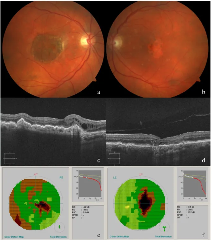
Figure 2: Fundus appearance and morphological and functional evaluation of both eyes at the last consultation.
Fundus photograph of the right eye (a), showing the RPE-choroid graft, and left eye (b), showing macular atrophy. Horizontal cross sections of SD-OCT images of the right (c) and left (d) eye. Octopus visual field of the right (e) and left (f) eye.
To date, no therapeutic options exist that can stabilize or improve VA in patients with DPED.
We hypothesized that replacing the ‘diseased’ RPE-Bruch membrane-choriocapillaris complex by an RPE-choroid graft could rescue the overlying photoreceptors.
It is important to keep in mind that RPE-choroid trans- location surgery remains, in general, a very invasive treatment option that should only be considered in severe cases where other treatment options have failed or are non-existent. Even then, it is essential to weigh the poten- tial benefits very carefully against the risk of postoperative
complications including retinal detachment (with or without proliferative vitreoretinopathy), severe hemor- rhage or graft failure.
When considering RPE-choroid translocation surgery in cases of otherwise untreatable large DPEDs, timing is important. Surgery could be considered when collapse seems unavoidable. A PED diameter greater than 2 disc diameter and the appearance of intraretinal hyperreflect- ive foci may be useful criteria. After all, when the DPED diameter was greater than 2 disc diameter, geographic atrophy occurred in 75% at long term as demonstrated
by Roquet et al. [3]. Furthermore it is known that RPE activation and migration are important precursors to atrophy [8]. Another interesting biomarker for collapse is disruption of the RPE + basal lamina band [4], but we presume that damage to the photoreceptors is already present at that moment and a graft would not be justifi- able anymore.
Another important issue to consider is the lifespan of the graft. Potential long-term survival is suggested by a recent report showing stable VA of 20/32 after 13.5 years, despite several complications [9], [10].
Taking into account the fact that the previously described high-risk characteristics of DPED were clearly present in our patient and were considered predictive of imminent atrophy while the outer retina still appeared healthy on OCT, we considered the timing for surgery to be optimal.
Conclusion
We describe a patient who underwent RPE-choroid graft translocation surgery to treat a DPED on the verge of collapsing. Despite the postoperative development of a CNV at the edge of the graft requiring anti-VEGF injections, functional outcome was favorable compared to the natural history of the fellow eye during the one-and-a-half years of follow-up.
Regarding the results of the RPE-choroid graft transloca- tion surgery in this patient, a prospective, randomized trial appears necessary to evaluate safety and efficacy of such a procedure in DPED patients.
Abbreviations
• RPE: retinal pigment epithelium
• PED: pigment epithelial detachment
• DPED: drusenoid pigment epithelial detachment
• AMD: age-related macular degeneration
• VEGF: vascular endothelial growth factor
• SD-OCT: spectral-domain optical coherence tomo- graphy
• VA: visual acuity
• CNV: choroidal neovascularization
• FA: fluorescein angiography
• OCTA: optical coherence tomography angiography
Notes
Competing interests
The authors declare that they have no competing interests.
Informed consent
The authors declare that the patient‘s consent was re- ceived to use her medical records to write this case re- port.
References
1. Cukras C, Agrón E, Klein ML, Ferris FL 3rd, Chew EY, Gensler G, Wong WT; Age-Related Eye Disease Study Research Group.
Natural history of drusenoid pigment epithelial detachment in age-related macular degeneration: Age-Related Eye Disease Study Report No. 28. Ophthalmology. 2010 Mar;117(3):489-99.
DOI: 10.1016/j.ophtha.2009.12.002
2. Sarks JP, Sarks SH, Killingsworth MC. Evolution of soft drusen in age-related macular degeneration. Eye (Lond). 1994;8(Pt 3):269-83. DOI: 10.1038/eye.1994.57
3. Roquet W, Roudot-Thoraval F, Coscas G, Soubrane G. Clinical features of drusenoid pigment epithelial detachment in age related macular degeneration. Br J Ophthalmol. 2004 May;88(5):638-42. DOI: 10.1136/bjo.2003.017632 4. Balaratnasingam C, Yannuzzi LA, Curcio CA, Morgan WH,
Querques G, Capuano V, Souied E, Jung J, Freund KB.
Associations Between Retinal Pigment Epithelium and Drusen Volume Changes During the Lifecycle of Large Drusenoid Pigment Epithelial Detachments. Invest Ophthalmol Vis Sci. 2016 Oct;57(13):5479-89. DOI: 10.1167/iovs.16-19816 5. Mrejen S, Sarraf D, Mukkamala SK, Freund KB. Multimodal
imaging of pigment epithelial detachment: a guide to evaluation.
Retina (Philadelphia, Pa). 2013 Oct;33(9):1735-62. DOI:
10.1097/IAE.0b013e3182993f66
6. Veckeneer M, Augustinus C, Feron E, Schauwvlieghe PP, Ruys J, Cosemans I, Van Meurs J. OCT angiography documented reperfusion of translocated autologous full thickness RPE-choroid graft for complicated neovascular age-related macular degeneration. Eye (Lond). 2017 Sep;31(9):1274-83. DOI:
10.1038/eye.2017.137
7. Ouyang Y, Heussen FM, Hariri A, Keane PA, Sadda SR. Optical coherence tomography-based observation of the natural history of drusenoid lesion in eyes with dry age-related macular degeneration. Ophthalmology. 2013 Dec;120(12):2656-65. DOI:
10.1016/j.ophtha.2013.05.029
8. Curcio CA, Zanzottera EC, Ach T, Balaratnasingam C, Freund KB.
Activated Retinal Pigment Epithelium, an Optical Coherence Tomography Biomarker for Progression in Age-Related Macular Degeneration. Invest Ophthalmol Vis Sci. 2017 May
1;58(6):BIO211-BIO226. DOI: 10.1167/iovs.17-21872 9. Maaijwee K, Heimann H, Missotten T, Mulder P, Joussen A, van
Meurs J. Retinal pigment epithelium and choroid translocation in patients with exudative age-related macular degeneration:
long-term results. Graefes Arch Clin Exp Ophthalmol. 2007 Nov;245(11):1681-9. DOI: 10.1007/s00417-007-0607-4 10. van Zeeburg EJT, Maaijwee KJM, van Meurs JC. Visual acuity of
20/32, 13.5 years after a retinal pigment epithelium and choroid graft transplantation. Am J Ophthalmol Case Rep. 2018 Jun;10:62-4. DOI: 10.1016/j.ajoc.2018.01.042
Corresponding author:
Dr. Joke Ruys
Department of Ophthalmology, ZNA Middelheim Hospital, Lindendreef 1, 2020 Antwerp, Belgium
joke.ruys@zna.be
Please cite as
Sillen H, Ruys J, Schauwvlieghe PP, Veckeneer M. Autologous full-thickness RPE-choroid graft to treat high-risk drusenoid pigment epithelial detachment without CNV. GMS Ophthalmol Cases.
2020;10:Doc24.
DOI: 10.3205/oc000151, URN: urn:nbn:de:0183-oc0001511
This article is freely available from
https://www.egms.de/en/journals/oc/2020-10/oc000151.shtml Published:2020-04-22
Copyright
©2020 Sillen et al. This is an Open Access article distributed under the terms of the Creative Commons Attribution 4.0 License. See license information at http://creativecommons.org/licenses/by/4.0/.