Spontaneous dislocation of lens bag with acrylic lens after uneventful cataract surgery – unusual complication of cataract surgery
Abstract
Introduction:Spontaneous dislocation of intraocular lens with bag is rare.
Mehul A. Shah
1Shreya M. Shah
1Methods:We report a case of a 56-year-old male who presented with
spontaneous anterior dislocation of an in-the-bag intraocular lens 3 years
Ruchir Mehta
1Prerna Shah
1after manual small incision cataract surgery. He had undergone manual small incision cataract surgery with foldable acrylic intraocular lens implantation, and 18 months after cataract surgery ND: YAG cap-
1 Drashti Netralaya, Dahod, Gujarat, India
sulotomy with uneventful post capsulotomy follow-up. 17 months after capsulotomy, the patient presented with sudden decrease of vision. On anterior segment examination, the intraocular lens with bag was dislo- cated into the anterior chamber.
Result: It was managed with intraocular lens explantation with bag, anterior vitrectomy and sclera fixated intraocular lens.
Conclusion:Spontaneous intraocular lens dislocation with bag is possible after 1.5 years of uneventful surgery which may be managed using dif- ferent techniques.
Keywords:intraocular lens dislocation, Nd: YAG capsulotomy, lens bag dislocation
Introduction
Spontaneous dislocation of an intraocular lens with cap- sular bag after uneventful cataract surgery is rare [1], [2], [3]. It can be predicted in presence of risk factors and be prevented [4], [5], [6], [7]. On the contrary, in the absence of any predictors, it has to be identified and managed promptly. We report a case of spontaneous in-the-bag anterior dislocation of an intraocular lens 36 months after cataract surgery.
Case presentation
A 56-year-old man presented with a 15-day history of sudden, painless blurring of vision in his right eye.
Previous ophthalmic history included an uneventful MSICS (manual small incision cataract surgery) and successful extraction of a mature cataract with a foldable hydrophilic acrylic intraocular lens in the right eye 36 months before presentation.
At 1 month follow-up the patient had a visual acuity of 20/20 with –0.25 DS.
At 18 months after the surgery, the best corrected visual acuity (BCVA) decreased to 20/40 and in the slit lamp examination, a visually significant posterior capsular opacification was found with no other significant ocular finding. Nd: YAG capsulotomy was performed and BCVA
of 20/20 achieved at an uneventful follow-up 10 days post procedure.
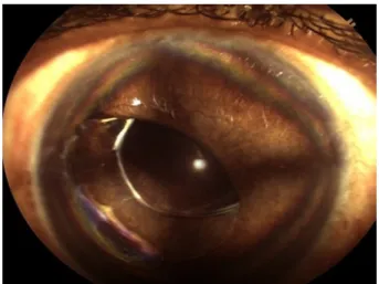
17 months after capsulotomy the patient presented with painless diminution of vision. BCVA was 20/60. Slit lamp examination showed anterior in-the-bag intraocular lens dislocation (Figure 1). Intraocular pressure was 15 mm Hg. There were no abnormal findings in the fundus on examination by indirect ophthalmoscopy with 20D lens.
Figure 1: Clinical photograph. Status post mydriasis: anterior dislocation of intraocular lens. One haptic and optic partially
in anterior chamber
1/4 GMS Ophthalmology Cases 2015, Vol. 5, ISSN 2193-1496
Case Report
OPEN ACCESS
It was managed with intraocular lens explantation with bag, anterior vitrectomy and scleral fixated intraocular lens using scleral flap and four point technique.
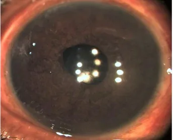
Third day follow-up was uneventful with visual acuity of 20/30 and one month follow-up with a best corrected visual acuity of 20/20 (Figure 2).
Figure 2: Post-operative initial. Day 3 post surgery: status after scleral fixated intraocular lens
Figure 3 shows the result a month after surgery. End result was uncomplicated pseudophakia at end of 6 weeks.
Figure 3: Post-operative late. 1 month post-surgery: status after scleral fixated intraocular lens
Discussion
Late intraocular lens dislocation is defined as occurring 3 months or later after cataract surgery. Late intraocular lens dislocation has been reported with increasing fre- quency in recent years but the pseudophakic community continues to increase in size due to longer life spans. The increased incidence of reported dislocation may be due to large numbers of people undergoing cataract extraction
[3], [4], [5]. The rate of posterior chamber intraocular lens dislocation ranges between 0.2% and 2.8% [6].
Lorente et al. found a mean duration of approximately 8 years ± 2 years between cataract extraction and in- traocular lens dislocation [1]. Our case presented at 3 years after surgery which is similar to most findings in literature.
In-the-bag intraocular lens dislocation may be due to pa- tient or surgeon factors.
The surgical and post-surgical factors include the type of surgery, type of intraocular lens used, rate of capsular contraction, undetected small capsular tears or zonulysis, and post-operative Nd: YAG capsulotomy.
Various types of surgeries are performed for cataract ex- traction. Intraocular lens dislocations have been reported for all types of cataract surgeries at varying rates. In the Rochester Epidemiology Intraocular Lens Project for cataract cohort and in-the-bag intraocular lens dislocation cohort, no significant difference was found between rates of dislocation for phacoemulsification and extra capsular cataract extraction surgeries [5]. Our case was following small incision cataract surgery and to our knowledge no reports of rates of dislocation have been made for the same.
The intraocular lens material and design may also have an effect on intraocular lens position and subsequent dislocation. Pueringer et al. found PMMA lenses to be more commonly dislocated [5]. Davis et al. found 33 in- traocular lenses of silicone out of 86 dislocations [2].
Hwang et al. postulated in their case report that the in- traocular lens they implanted was a sharp three piece intraocular lens with a polymethyl methacrylate (PMMA) haptic [3]. Aqueous movement caused by external pres- sure widened a pre-existing unnoticed break in the cap- sular bag causing intraocular lens dislocation [3]. In our case, the acrylic lens had dislocated with bag without any pre-existing opening in posterior capsule.
Contraction of the capsular opening occurs in all postoper- ative patients and is common with continuous curvilinear capsulorhexis [6], [7], [8]. Anterior capsular phimosis can lead to an imbalance between the centrifugal and centri- petal forces of the capsulorhexis margin, which may exert continuous centrifugal forces on the zonules causing their rupture.
Capsule contraction syndrome plays a role in the devel- opment of in-the-bag intraocular lens dislocation. In the presence of solid zonular support, significant intraocular lens displacement is unlikely. However, compromised zonules can become vulnerable to centripetal forces and rupture. The risk for asymmetrical capsule shrinkage in- creases in eyes with zonular dehiscence or weakness as a result of the centripetal forces exerted by the anterior capsule rim [6].
Cases of intraocular lens posterior luxation with the cap- sular bag have been reported. Luxation in these cases was spontaneous and explained by excessive centripetal force on the zonules after a strong capsule contraction syndrome [9], [10], [11].
2/4 GMS Ophthalmology Cases 2015, Vol. 5, ISSN 2193-1496
Shah et al.: Spontaneous dislocation of lens bag with acrylic lens ...
Intraocular lens material could affect the rate of capsular shrinkage. Single-piece polymethylmethacrylate (PMMA) lenses are thought to be better than three-piece lenses and acrylic intraocular lenses are associated with less fibrosis [11]. In a case series of 45 eyes it was observed that with bag lens dislocations were more common in three piece acrylic intraocular lenses. In our case, capsule fibrosis had probably weakened the zonules.
Nd: YAG capsulotomy may also contribute to zonular weakness.
Framme et al. [12] reported a case of zonulolysis and PMMA intraocular lens dislocation within the intact cap- sular bag six months after Nd: YAG laser capsulotomy. It was suggested that zonulolysis was possibly triggered by contraction of the capsular bag after the capsulotomy.
The factors responsible could be anterior capsular con- traction and shrinkage [11].
Gimbel et al. [6] concluded that the impact of laser energy may have put an additional burden on already comprom- ised zonules and was the triggering event for the sublux- ation. Furthermore, the need for capsulotomy is an indi- cator of significant cell proliferation and of increased capsular bag weight. Lorente et al. [1] had reported about 3 eyes which had undergone Nd: YAG capsulotomy before presenting with spontaneous dislocations. Our patient underwent Nd: YAG capsulotomy 1 year after surgery and it could have been a triggering factor for the dislocation.
The patient factors most commonly reported are pseudoexfoliation [1], [2], others being high axial length [7], uveitis [10], pars planitis, diabetes mellitus, trauma, previous vitrectomy surgery, increased age, eye rubbing habits [3] as in atopic dermatitis, myotonic dystrophy. In our case there was no recorded history of other predis- posing factors, such as pseudoexfoliation, preoperative zonulysis, or other trauma.
Özkan et al. [13], and Gatzioufas et al. [14] reported an- terior dislocation of posterior chamber intraocular lens in patients with pseudoexfoliation syndrome. Fazel et al.
reported similar findings in a patient with blunt trauma [15]. Por and Chee reported anterior intraocular lens dislocation after haptic disinsertion in two cases [9].
However their age group was young (14 years and 22 years) and one of them had a predisposing factor (previous vitrectomy). Similarly Chaudhary et al. reported anterior dislocation of intraocular lens in one eye and posterior dislocation in the other in a 41-year-old with marfanoid features [8]. So, unlike most reports where dislocations were posterior and had predisposing factors, our patient experienced in-the-bag intraocular lens dislo- cation into the anterior chamber in the absence of any known predisposing factors. We can only postulate primary or secondary zonular weakness to be the likely mechanism for this occurrence.
Surgical management usually involves repositioning, ex- plantation with or without replacement and rarely obser- vation [1]. Surgery type and urgency to perform the sur- gery depend on the surgeon’s preferences and specialty and the clinical features of the individual case, including type of IOL, presence of a Capsular Tension Ring, stage
and site of IOL dislocation, and coexisting ocular patho- logy.
Although repositioning and suturing is the management of choice [1], [6] replacement was our choice since the IOL was dislocated with the bag and a zonular dehiscence of more than 180 degrees was observed.
In a review of all pertinent literature, the American Academy of Ophthalmology concluded that there is insuf- ficient evidence to support the superiority of scleral or iris sutured posterior chamber IOLs over open-loop anteri- or chamber IOLs [1], [6]. Since our experience with sclera fixated posterior chamber IOL has been good and the patient had no contraindications, the patient was man- aged with the same.
Conclusion
In conclusion, late in-the-bag intraocular lens dislocation is a potential late complication of cataract surgery in which an in-the-bag intraocular lens is used and is more likely to happen in certain predisposed eyes. It is impor- tant to identify such factors. In the event of dislocation, especially anterior ones, the prognosis after explantation and replacement is good.
Notes
Competing interests
The authors declare that they have no competing in- terests.
References
1. Lorente R, de Rojas V, Vazquez de Parga P, Moreno C, Landaluce ML, Domínguez R, Lorente B. Management of late spontaneous in-the-bag intraocular lens dislocation: Retrospective analysis of 45 cases. J Cataract Refract Surg. 2010 Aug;36(8):1270-82.
DOI: 10.1016/j.jcrs.2010.01.035
2. Davis D, Brubaker J, Espandar L, Stringham J, Crandall A, Werner L, Mamalis N. Late in-the-bag spontaneous intraocular lens dislocation: evaluation of 86 consecutive cases. Ophthalmology.
2009 Apr;116(4):664-70. DOI: 10.1016/j.ophtha.2008.11.018 3. Hwang HB, Yim HB, Kim HS. Anterior dislocation of an empty
capsular bag in a pseudophakic eye: A rare case report. Indian J Ophthalmol. 2015 Mar;63(3):280-2. DOI: 10.4103/0301- 4738.156939
4. Zech JC, Tanniére P, Denis P, Trepsat C. Posterior chamber intraocular lens dislocation with the bag. J Cataract Refract Surg.
1999 Aug;25(8):1168-9. DOI: 10.1016/S0886-3350(99)00124- 8
5. Pueringer SL, Hodge DO, Erie JC. Risk of late intraocular lens dislocation after cataract surgery, 1980-2009: a population- based study. Am J Ophthalmol. 2011 Oct;152(4):618-23. DOI:
10.1016/j.ajo.2011.03.009
6. Gimbel HV, Condon GP, Kohnen T, Olson RJ, Halkiadakis I. Late in-the-bag intraocular lens dislocation: incidence, prevention, and management. J Cataract Refract Surg. 2005
Nov;31(11):2193-204. DOI: 10.1016/j.jcrs.2005.06.053
3/4 GMS Ophthalmology Cases 2015, Vol. 5, ISSN 2193-1496
Shah et al.: Spontaneous dislocation of lens bag with acrylic lens ...
7. Fernández-Buenaga R, Alio JL, Pérez-Ardoy AL, Larrosa-Quesada A, Pinilla-Cortés L, Barraquer R, Alio JL 2nd,Muñoz-Negrete FJ.
Late in-the-bag intraocular lens dislocation requiring explantation:
risk factors and outcomes. Eye (Lond). 2013 Jul;27(7):795-801.
DOI: 10.1038/eye.2013.95
8. Choudhary A, Sahni J, Kaye SB. Late spontaneous anterior dislocation of an intraocular lens (IOL) with the capsular bag.
Eye (Lond). 2005 Jan;19(1):101-2. DOI:
10.1038/sj.eye.6701419
9. Por YM, Chee SP. Spontaneous disinsertion of a multipiece foldable acrylic intraocular lens haptic 3 and 12 months after implantation. J Cataract Refract Surg. 2004 May;30(5):1139- 42. DOI: 10.1016/j.jcrs.2003.08.033
10. Tao LW, Hall A. In-bag dislocation of intraocular lens in patients with uveitis: a case series. J Ophthalmic Inflamm Infect.
2015;5:10. DOI: 10.1186/s12348-015-0036-1
11. Davison JA. Capsule contraction syndrome. J Cataract Refract Surg. 1993 Sep;19(5):582-9. DOI: 10.1016/S0886- 3350(13)80004-1
12. Framme C, Hoerauf H, Roider J, Laqua H. Delayed intraocular lens dislocation after neodymium:YAG capsulotomy. J Cataract Refract Surg. 1998 Nov;24(11):1541-3. DOI: 10.1016/S0886- 3350(98)80182-X
13. Özkan B, Altintas Ö, Yüksel N, Çaglar Y. Spontaneous Late Anterior Dislocation of Intraocular Lens within the Capsular Bag in Pseudoexfoliation Syndrome. Glokom-Katarakt. 2008;3:268- 9.
14. Gatzioufas Z, Kopsidas K, Gyongyossy B, Moschos M, Seitz B.
Late-onset anterior dislocation of a posterior chamber intraocular lens in a patient with pseudoexfoliation syndrome. Case Rep Ophthalmol. 2011;2(1):1-4. DOI: 10.1159/000323861
15. Fazel F, Kianersi F, Rezaei L. In the bag intraocular lens dislocation in anterior chamber along with intact superior zonules following a blunt trauma. Am J Case Rep. 2010;11:201-4.
Corresponding author:
Mehul A. Shah, MD
Drashti Netralaya, Nr. GIDC, Chakalia Road, Dahod-389151, Gujarat, India, Phone:
00-91-2673-645364, Fax: 00-91-2673-221232 omtrust@rediffmail.com
Please cite as
Shah MA, Shah SM, Mehta R, Shah Prerna. Spontaneous dislocation of lens bag with acrylic lens after uneventful cataract surgery – unusual complication of cataract surgery . GMS Ophthalmol Cases.
2015;5:Doc11.
DOI: 10.3205/oc000033, URN: urn:nbn:de:0183-oc0000337
This article is freely available from
http://www.egms.de/en/journals/oc/2015-5/oc000033.shtml Published:2015-11-02
Copyright
©2015 Shah et al. This is an Open Access article distributed under the terms of the Creative Commons Attribution 4.0 License. See license information at http://creativecommons.org/licenses/by/4.0/.
4/4 GMS Ophthalmology Cases 2015, Vol. 5, ISSN 2193-1496
Shah et al.: Spontaneous dislocation of lens bag with acrylic lens ...