Antibody masked cytokines as new approach in targeted tumor therapy
DISSERTATION ZUR ERLANGUNG DES DOKTORGRADES DER NATURWISSENSCHAFTEN (DR. RER. NAT.) DER FAKULTÄT CHEMIE UND PHARMAZIE
DER UNIVERSITÄT REGENSBURG
vorgelegt von
Martin Matthias Kuen aus
Villach, Österreich
im Jahr 2015
von Frau Prof. Dr. Daniela Männel angefertigt.
Promotionsgesuch eingereicht am: 30. September 2015 Datum der mündlichen Prüfung: 05. August 2016
Prüfungsausschuss: Prof. Dr. Jens Schlossmann (Vorsitzender) Prof. Dr. Daniela Männel (Erstgutachter) Prof. Dr. Jörg Heilmann (Zweitgutachter) PD Dr. Miriam Breunig (Drittprüfer)
Prof. Dr. Harald Wajant (externer Gutachter)
For my Dad
Eidesstattliche Erklärung
(1) Ich erkläre hiermit an Eides statt, dass ich die vorliegende Arbeit ohne unzulässige Hilfe Dritter und ohne Benutzung anderer als der angegebenen Hilfsmittel angefertigt habe; die aus anderen Quellen direkt oder indirekt übernommenen Daten und Konzepte sind unter Angabe des Literaturzitats gekennzeichnet.
(2) Bei der Auswahl und Auswertung folgenden Materials haben mir die nachstehend aufgeführten Personen in der jeweils beschriebenen Weise entgeltlich/unentgeltlich
geholfen:
im Text vermerkt
(3) Weitere Personen waren an der inhaltlich-materiellen Herstellung der vorliegenden Arbeit nicht beteiligt. Insbesondere habe ich hierfür nicht die entgeltliche Hilfe eines
Promotionsberaters oder anderer Personen in Anspruch genommen. Niemand hat von mir weder unmittelbar noch mittelbar geldwerte Leistungen für Arbeiten erhalten, die im Zusammenhang mit dem Inhalt der vorgelegten Dissertation stehen.
(4) Die Arbeit wurde bisher weder im In- noch im Ausland in gleicher oder ähnlicher Form einer anderen Prüfungsbehörde vorgelegt.
_______________________________________________
Kuen Martin, Ort, Datum
Clause of confidentiality
This is to certify that the doctoral thesis delivered in July 2015 by Mr Martin Matthias Kuen entitled
"Antibody masked cytokines as new approach in targeted tumor therapy" will NOT be published for a period of 2 years after the oral exam, as confirmed by the University in the work contract between the Roche Diagnostics GmbH and Prof. Dr. Jörg Heilmann.
______________________________________________
place, date
Acknowledgements
I would like to use this opportunity to express my special appreciation and thanks to my scientific advisor Professor Dr. Daniela Männel. I would like to thank her for all the good advices and willingness to share her scientific expertise with me. I am deeply grateful for the outstanding support.
I wish to express my sincere thanks to Dr. Edgar Voss for being my scientific advisor and supervisor.
The good advices in research topics as well as on career issues have been invaluable and I appreciate every conversation and critical discussion. Due to this support I feel well prepared for upcoming challenges in my future career.
My special thanks go to Dr. Manfred Schwaiger for giving me the opportunity to work and further investigate topics of the project proposal regarding interferon fusion proteins. I am glad and thankful for all the support, for being a mentor to me and facilitating my personal development in a competitive scientific environment.
I would also like to thank Professor Dr. Jörg Heilmann, Professor Dr. Achim Göpferich and PD Dr. Miriam Breunig for serving as my committee members and proofreading of my thesis. I am very thankful for providing me with the opportunity to do my dissertation at the Faculty of Chemistry and Pharmacy.
In addition I express my warm thanks to the members of our lab, Guido Werner, Ingrid Munk and Christian Clemens for supporting me and giving me advices whenever I asked for. It was a pleasure for me to be part of this high-performing team.
My special thanks go to the people which are the most important value in my life. I want to thank my parents for their outstanding support during every step of my education and keeping me grounded all the time. Words cannot explain how much you mean to me. At the end I want to thank my beloved girlfriend for being my support all the time.
Table of content
1. Introduction ... 1
1.1 History of Interferon ... 1
1.2 Half a century in interferon research ... 3
1.3 Classification of interferon ... 6
1.4 The interferon receptor family and downstream signalling ... 8
1.5 Interferon in oncology practice... 12
1.6 Interferon treatment in multiple myeloma ... 15
1.7 Limitations of interferon in cancer therapy ... 18
1.8 Description of the proposed therapeutic attempt in interferon based cancer therapy ... 20
1.9 Proposed mode of action of the interferon fusion protein ... 23
2. Scientific objectives ... 27
3. Material ... 29
3.1 Equipment ... 29
3.2 Chemicals and reagents ... 30
3.3 Cytokines ... 30
3.4 Consumable supplies ... 31
3.5 Kits ... 33
3.6 Detection antibody ... 33
3.7 Enzymes ... 33
3.8 Buffers ... 34
3.9 Media and supplements ... 34
3.10 Eukaryotic cell lines ... 35
3.11 Applied computer software ... 35
4. Methods ... 36
4.1 Antibody design ... 36
4.1.1 Knobs-into-holes ... 36
4.1.2 CrossMab ... 38
4.1.3 HY-RF mutation ... 41
4.2 Cloning of expression plasmids ... 42
4.3 Eukaryotic cell transfection and expression of fusion proteins ... 43
4.4 Protein purification ... 44
4.4.1 Protein A chromatography... 44
4.4.2 Size exclusion chromatography ... 44
4.5 Cell viability assay ... 45
4.6 Plasma stability ... 46
4.7 Surface plasmon resonance (SPR – Biacore) ... 47
4.8 Confocal microscopy ... 49
5. Results ... 51
5.1 Screening for interferon responsive cell lines ... 51
5.2 Anti-proliferative effect of Type I interferons... 54
5.3 Improvement of pharmacodynamics parameters of interferon ... 56
5.3.1 Combined mutations for maximum IFNAR1 affinity ... 59
5.4 Masking of interferon ... 63
5.5 Effects of a bivalent non-masking anti-interferon antibody ... 70
5.6 Screening for tumor specific proteases ... 72
5.6.1 Peptide linkers specific for matrix metalloproteinases ... 72
5.6.2 Peptide linkers selectively cleaved by matriptase ... 74
5.6.3 Dual specific tandem linker ... 76
5.7 Manufacturing and evaluation of the interferon fusion protein lead molecule ... 78
5.7.1 Purification of the bispecific interferon fusion protein ... 79
5.7.2 Functionality of the CD138 targeting moiety ... 81
5.7.3 Pharmacologic potency of the interferon fusion protein ... 83
6. Discussion ... 87
6.1 Clinical indications for an interferon fusion protein ... 88
6.2 Improvement of the pharmacological potency of IFN ... 90
6.3 Masking and temporary inhibition of interferon ... 93
6.4 Protease cleavable peptide linkers ... 97
6.5 Experiences and findings during manufacturing of a bispecific antibody cytokine fusion protein ... 102
6.6 Functional characterization of the proposed interferon fusion protein ... 103
7 Outlook and perspectives ... 106
References ... 1
List of figures
Figure 1: Discovery of interferon mediated viral resistance... 2
Figure 2: Major milestones and discoveries in fifty years of interferon research ... 6
Figure 3: Interferon receptors and associated downstream signaling cascades ... 10
Figure 4: Alternative interferon pathway besides classical JAK-STAT signaling ... 11
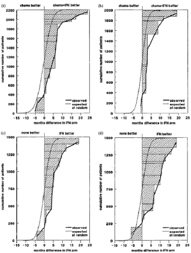
Figure 5: Meta-analysis of thirty clinical trials comparing interferon mono- and combination therapy ... 18
Figure 6: Side-effect profile of interferon determined during clinical investigations ... 19
Figure 7: Binding of interferon to masking antibody ... 21
Figure 8: Stabilized binding of interferon to a masking antibody via a protease cleavable linker ... 21
Figure 9: Summary of the proposed bispecific interferon fusion protein ... 22
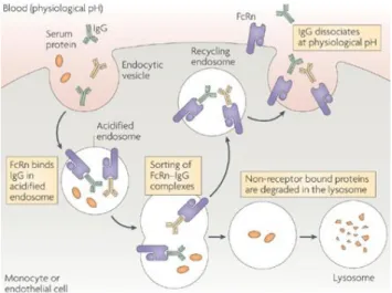
Figure 10: Neonatal Fc-receptor-mediated IgG recycling mechanism ... 23
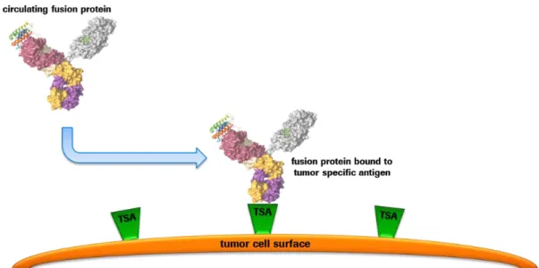
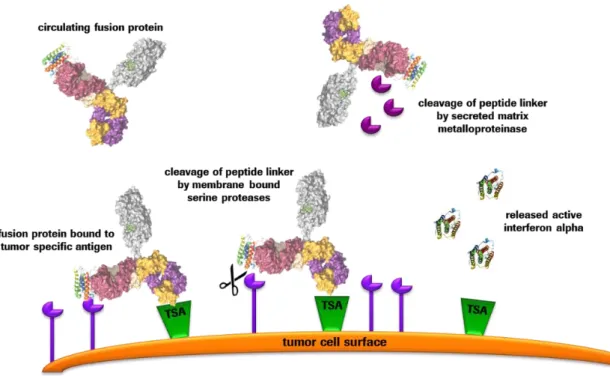
Figure 11: Binding and enrichment of fusion proteins on the surface of tumor cells ... 24
Figure 12: Protease-mediated cleavage of peptide linkers triggering interferon release ... 25
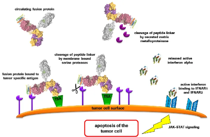
Figure 13: Interferon signaling pathway induces apoptosis induction in tumor cells ... 26
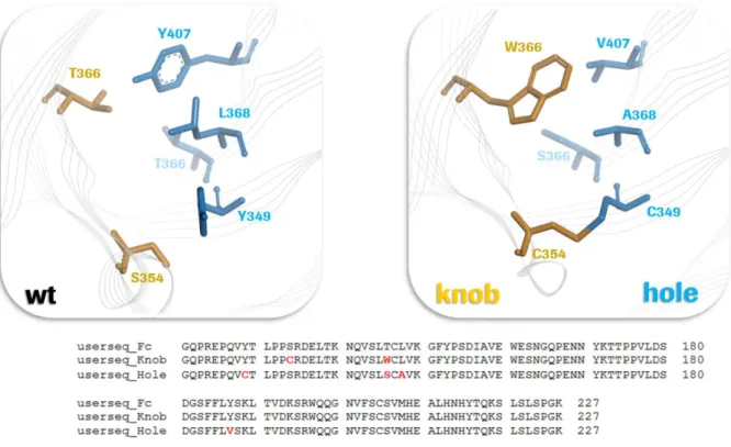
Figure 14: "knobs into holes" modification to enforce heavy chain heterodimerization ... 37
Figure 15: Crossing over of antibody domains ... 39
Figure 16: CrossMab technology used in the interferon fusion protein ... 40
Figure 17: Charge reversal of heavy and light chains ... 41
Figure 18: Modification of protein-A binding capacity ... 42
Figure 19: Seeding and treatment scheme for cell viability assays ... 46
Figure 20: Surface plasmon resonance measurement principle ... 48
Figure 21: Example and explanation of a SPR senorgram ... 48
Figure 22: Scheme of the light pathway used in confocal microscopy ... 50
Figure 23: Interferon-induced apoptosis in different cancer cell lines ... 52
Figure 24: Interferon-induced cell death in multiple myeloma cell lines ... 53
Figure 25: Efficacy screening of human IFN alpha subtypes ... 55
Figure 26: Receptor affinity-mediated increase of anti-proliferative effects ... 56
Figure 27: IFN efficacy study in MM patient-derived bone marrow ... 57
Figure 28: Affinity maturation within the IFN/IFNAR2 interaction domain ... 58
Figure 29: Stepwise improvement of the pharmacodynamic efficacy of interferon alpha ... 59
Figure 30: Potency enhancing mutations in IFN alpha 2a compared to IFN alpha H2 ... 61
Figure 31: Outstanding IFN potency compared to standard of care treatment ... 62
Figure 32: Masking and temporary inactivation of interferon ... 65
Figure 33: Adaptation of antibody masking intensity ... 66
Figure 34: Antibody-induced inactivation of IFN alpha subtypes ... 68
Figure 35: CDR burnishing of the anti IFN alpha 2a antibody 9F3 ... 69
Figure 36: IFN receptor clustering to explain improved anti-proliferative efficacy ... 71
Figure 37: Matrix metalloproteinase cleavable peptide linker ... 73
Figure 38: Membrane bound serine-protease cleavable peptide linker ... 75
Figure 39: Tandem linker with synergistic turnover rate... 76
Figure 40: Plasma stability of protease cleavable linkers ... 77
Figure 41: CD138 (Syndecan-1) expression in multiple myeloma cell lines ... 79
Figure 42: Manufacturing and purification of the bispecific interferon fusion protein ... 80
Figure 43: SDS gel electrophoresis of the IFN antibody conjugate ... 81
Figure 44: Confocal microscopy of CD138 targeted IFN fusion proteins ... 82
Figure 45: Proof of concept evaluation in multiple myeloma ... 83
Figure 46: CD138 targeted IFN in several MM cell lines and ovarian cancer ... 85
List of tables Table 1: Nomenclature of natural occurring IFN-alpha subtypes ... 7
Table 2: IC 50 values of IFN point mutations ... 58
Table 3: Potency of IFN alpha 2a mutations ... 60
Table 4: IC50 values of IFN alpha H2 mutants ... 61
Table 5: Potency of cytostatic compounds ... 63
Table 6: IC50 values of fusion proteins containing different peptide linkers ... 77
Table 7: Evaluated IC50 values of proof of concept fusion proteins ... 84
Table 8: Similar shifts of IC50 values in different cell lines ... 85
Abbreviations
(G4S)6 glutamine serine peptide
ATP adenosine triphosphate
BSA bovine serum albumin
CD138 cluster of differentiation 138
CDK cyclin-dependent kinase
CDRs complementarity determining regions
CH1 – CH3 constant region heavy chain 1 - 3
CHR complete hematologic remission
CL constant region light chain
CML chronic myelogenous leukemia
CMV cytomegalovirus
CR complete remission
DNA deoxyribonucleic acid
ECM extra cellular matrix
EDC 1-ethyl-3(3-dimethylaminopropyl) carbodiimide
hydrochloride
FBN-III fibronectin domain type iii
FcRn neonatal fc receptor
FDA food and drug administration
FRET förster resonance energy transfer
GAS interferon gamma activated sites
GTPase guanosine triphosphate hydrolase
HAI-1 hepatocyte growth factor activator inhibitor-1
HC heavy chain
HCL hairy cell leukemia
hCRs helical cytokine receptors
HCV hepatitis c virus
HEK293F cells human embryonic kidney cells
His histidine
IC50 inhibitory concentration 50%
IFN interferon
IFNAR1 interferon alpha receptor unit 1
IFNAR2 interferon alpha receptor unit 2
IFNGR1 interferon gamma receptor unit 1
IFNGR2 interferon gamma receptor unit 2
IFNα interferon alpha
IFNβ interferon beta
IFNγ interferon gamma
IgG1 immunoglobulin g type i
IL-2 interleukin-2
ILP isolated limb perfusion
INFAR interferon alpha receptor
IRF9 interferon regulatory factor 9
ISG interferon stimulated genes
ISGF3 interferon stimulated gene factor 3
ISRE interferon stimulated response elements
JAK janus activated kinase
KiH knobs-into-holes
KS kaposi’s sarcoma
LC light chain
MgCl2 magnesium chloride
MHC-Class I major histocompatibility complex class i
MIP macrophage inflammatory protein
MM multiple myeloma
MMP matrix metallo proteinase
MTD maximum tolerable dose
NaCl sodium chloride
NHS n-hydroxysuccinimide
NSCLC non-small cell lung cancer
PBS phosphate buffered saline
PBST phosphate buffered saline with tween 20
PR partial remission
RANKL receptor activator of nuclear factor-κb
RNA ribonucleic acid
SPR surface plasmon resonance
STAT signal transducers and activators of transcription
TBST tris-buffered saline containing tween 20
TNF tumor necrosis factor
TSA tumor specific antigen
VEGF® vascular endothelial growth factor (receptor)
VH variable region heavy chain
VL variable region light chain
Abstract
Cytokine-based cancer therapies are commonly applied treatment strategies in oncology. A prominent representative of this potent class of anti-tumor agents is interferon alpha. Interferon is well known for its anti-viral and anti-proliferative activities and a potent therapeutic agent in the treatment of multiple myeloma, chronic myeloid leukemia (CML), hairy cell leukemia (HCL) and malignant melanoma. Unfortunately, the application of interferon is always accompanied by severe adverse side effects and shows a strong toxicological profile. In order to overcome these obstacles and generate a more suitable therapeutic agent, a bispecific interferon-antibody fusion protein was designed. By introducing multiple point mutations, the pharmacodynamic potency of the cytokine was significantly improved compared to wild type interferon by increasing its affinity to the receptor IFNAR1. In order to prevent undesired interactions of the fusion protein during circulation in the periphery and consequently decrease toxic side effects, a masking antibody was connected by a linker sequence which led to a temporary neutralization of interferon. Local reactivation of the cytokine at the tumor site was initiated by the efficient cleavage of the dual MMP-9- and matriptase- cleavable linker peptide between masking antibody and interferon with suitable turnover rates. In addition, enrichment of the interferon fusion protein at the tumor site was enforced by using one arm of the antibody for targeting the fusion protein to the multiple myeloma-associated antigen CD138, as successfully confirmed by confocal microscopy. The potent anti-proliferative activity of the cytokine in combination with a striking interferon masking effect by the linked antibody were shown in several cell lines originating from different oncologic indications. The promising approach of temporary inhibition of the cytokine in combination with tumor-selective reactivation of its pharmacodynamic potency holds the potential to overcome current limitations in cytokine-based treatment strategies and makes a broad clinical application of this extraordinary class of biomolecules in oncology feasible.
1
1. Introduction
1.1 History of Interferon
The story of success of interferon begins early back in 1957 when the scientists Alick Isaacs from the National Institute of Medical Research in London and Jean Lindenmann from the Swiss Academy of Medical Science launched a collaboration to investigate the interference of heat- inactivated influenza viruses on the infectiousness of intact viruses on chorio-allantoic membranes of chicken embryos (Isaacs & Lindenmann, 1957).
Prior to the above mentioned project, Lindenmann worked on red blood cells covered with heat- inactivated virus particles and the processes taking place by challenging those cells with active viruses. To answer the question why the virus particles, which are fixed on the surface of the cells, are able to induce interactions between treated and non-treated cells, Lindenmann decided to cooperate with Alick Isaacs. Due to the experience of Isaacs in the field of electron microscopy both scientists planned to visualize the events taking place between the infected and non- infected cells via a microscope. Unfortunately the experimental set-up turned out to be very challenging and the gained optical data could not be interpreted due to difficulties in discriminating between virus-loaded and empty cells (Pitha, 2007).
In a new attempt the scientists used the Melbourne influenza A strain which was heat-inactivated at 56°C for one hour and following to that, inoculated pieces of chorio-allantoic membranes with the pre-treated virus at 37°C for 24 hours. Following the incubation step, the supernatant was replaced and membranes were washed several times with buffer to remove remaining virus particles from the surface. Afterwards the membranes were transferred into fresh growth medium and challenged with intact and non-modified influenca viruses. To evaluate the reproduction of viruses inside the cells, a haemagglutinin titration series was performed. From this experiment it was seen, that membranes which were pre-treated with heat-inactivated viruses, show a strong resistance against infectious viruses from the same strain. To exclude possible interference of the experiment by remaining virus particles from the pre-treatment step, a new test set-up was carried out (Isaacs & Lindenmann, 1957).
In the final experiment chorio-allantoic membranes were treated with heat-inactivated influenza viruses overnight. Following the incubation, supernatants of the treated membranes were
2
collected and transferred into test tubes containing fresh membranes which have never been in direct contact with any virus particle. After incubation of the chorio-allantoic membranes in the medium derived from the previous performed virus particle treatment, infectious influenza A viruses were added to challenge the membranes. On the next day the viral replication rate inside the cells of each membrane was determined and it was observed, that the fresh membranes which have never been in direct contact with viral particles before, showed a surprisingly high and significant resistance against the attack of intact influenza viruses mediated by an inhibition of viral replication. A schematic drawing of this experiment can be found in figure 1 (Pitha, 2007).
Figure 1: Discovery of interferon mediated viral resistance
From their experiments, Isaacs and Lindenmann concluded that there must be a kind of interfering agent which mediates viral resistance between treated and non-treated cells without getting into close proximity. Due to this kind of “interference” they named this unknown agent
“interferon”. At that moment both scientists had no idea that they had discovered the first cytokine and what tremendous impact this discovery would have on anti-viral and oncological indications in future times (Pitha, 2007).
Jean Lindenmann presented the outcome of the virus induced interference between cells and the discovered mediating agent the first time at a meeting of the Swiss Society for Microbiology in Interlaken in 1957. This was the first time the cytokine interferon became apparent in scientific community and the kick-off for further research in the field of cytokines was initiated (Pitha, 2007).
Supernatants from chorio-allantoic membranes, pre-treated with heat-inactivated viruses, were transferred to fresh membranes followed by challenging them with intact influenza A viruses.
Mediation of viral resistance by an unknown agent contained in the transferred supernatant was determined. (Isaacs & Lindenmann, 1957)
3
1.2 Half a century in interferon research
Over the past fifty years the understanding of interferon-induced molecular mechanisms inside mammalian cells and the clinical application of the cytokine led to major break-troughs in anti- viral therapies and the continuous battle against cancer. It quickly became clear that this paracrine and autocrine signaling molecule reveals the potential to revolutionize treatment strategies in infectious diseases and oncological malignancies (Borden et al., 2007).
However, it took a long way from the discovery of interferon in 1957 to its characterization, the understanding of the mode of action and the clinical application of the cytokine. Before promises made at its discovery were able to be kept, more than a decade elapsed until new technologies in molecular biology raised the opportunities to investigate this cytokine in more detail. In the first years of interferon research scientists mainly tried to answer the question about how synthesis of the molecule is induced and the effects that are later on triggered in vivo. In the early 60s it was already shown that interferon is able to mediate viral resistance in mice and its expression is not limited to viral infections, but rather can be initialized by other stimulants like microbial products of bacteria, protozoa and virus particles (Borden et al., 2007).
One of the main observations in the first years of interferon research was the discovery of its anti- proliferative activity on tumor cells, shown in several mouse models. In 1969, the group of Ion Gresser inoculated Balb/c and C57/B16 mice with 2000 – 3000 tumor cells and determined the overall survival rate of untreated mice compared to animals inoculated with interferon. The experiment showed only 3.7% of the untreated mouse population survived more than 22 days. In contrast to that, the treatment of mice with interferon led to an increased survival rate of 98%
within the first three weeks and an overall survival of 15% after 60 days. None of the surviving mice showed any signs of remaining tumor mass and all of them stayed progression free (Gresser, Bourali, Lévy, Fontaine-Brouty-Boyé, & Thomas, 1969).
These promising results from early in vivo studies enforced the expectations and efforts to apply interferon therapy also for human beings. Already in 1977 the anti-proliferative effects of interferon on human derived tumor cells were able to be confirmed in non-small cell lung cancer (NSCLC) cell lines in vitro. In this first set of experiments, the tumor cells were treated with different subtypes of interferon in mono-therapy or combination with chemotherapeutic agents to determine the most effective treatment strategies for following clinical studies (Fujie et al., 2011).
4
Between 1957 and the late 1970s, the application and study of this cytokine was limited by the low amounts and low purities of material which was isolated from cell cultures stimulated with viral particles. This limitation came to an end in 1980, when striking developments in molecular biology provided the opportunity to produce interferon in a recombinant way. One of the early attempts to produce the cytokine in a recombinant way was performed by the group of Shigekazu Nagata at the institute of molecular biology at the University of Zürich. The scientists isolated the 12S fraction of poly-A RNA, which was coding for interferon, from interferon producing human leukocytes. After back-translation of the RNA by reverse transcriptase, the generated cDNA was cloned in a pBR322 bacterial vector plasmid. As an expression host the E.coli strain HB101 was chosen and transfected with plasmids containing the DNA sequence coding for interferon.
Resulting to these efforts, a bacterial clone was identified which produced a polypeptide with an interferon-like biological activity in a range of 10.000 IU/g cell mass. In further analyses the previously gained inserts in E.coli DNA were isolated and hybridized with poly-A RNA coding for interferon to verify the correct incorporation of the IFN coding gene and to link the observed biological effects to the expression and secretion of interferon (Nagata et al., 1980).
The new acquired possibility to produce interferon in high amounts and with a suitable grade of purity enforced the research in the field of cytokines. From this point of time it was possible to generate sufficient material for animal studies and first clinical trials in humans were launched.
Only one year after the initial production of recombinant interferon, first investigations in cancer patients and multiple sclerosis patients were launched with significant treatment effects. In the following years different subtypes of interferon were identified and the mode of action of this class of cytokines was investigated in more detail. From the beginning to the middle of the 80s, more and more intracellular pathways, which are activated by interferon, became clear and first interferon stimulated genes (ISG) were reported (Borden et al., 2007).
One of the major break-troughs in the history of interferon was the approval of recombinant interferon alpha 2a for clinical application in hairy cell leukemia patients in 1986. This first approval of a cytokine in cancer therapy has led to enormous efforts in pharmaceutical industry to produce high amounts of this new miracle drug. Due to extensive marketing strategies and first beneficial results from the clinic, people became aware of the new drug and a growing need for interferon aroused (Eckart, 2002).
This vital momentum pushed interferon research to a new level and more and more clinical applications of this molecule were investigated in further detail. Due to positive clinical study results and the enforced marketing of interferon by the industry, an interferon hype was initiated
5
and this new miracle drug was published at the cover page of prestigious newspapers like The New York Times or the German Spiegel. Interferon was considered as the most potent drug against cancer and clinical trials for several oncological indications were launched. At this time in 1990, the Federal Food and Drug Administration (FDA) approved the application of interferon also for anti-viral therapies against Hepatitis C (HCV). Because of the duel functionality of the cytokine against viral invaders and malignant cell proliferation, researchers started to study the exact mode of action of interferon to answer the question about how the pleiotropic effects of this molecule are triggered and which pathways are activated in anti-viral and anti-proliferative action (Borden et al., 2007).
The decade from 1990 to the year 2000 was marked by the investigation of interferon signaling pathways and the identification of the main signal transducing molecules Janus activated kinase (JAK) and the Signal Transducers and Activators of Transcription (STAT). In addition to that, the three dimensional structure of interferon in complex with both receptor units was determined via crystal structure analysis. Following the identification of the intracellular signaling, it became clear that the pleiotropic mode of action of interferon cannot be explained by a single activated downstream cascade. New insights in the mode of action of interferon were disclosed in the end of the 90s by the characterization of hundreds of interferon stimulated genes (ISGs). Due to the activation of only one main downstream signaling pathway by interferon, the pleiotropic action of the cytokine can be possibly traced back to a different transcription profile of the newly discovered interferon-stimulated genes (Borden et al., 2007).
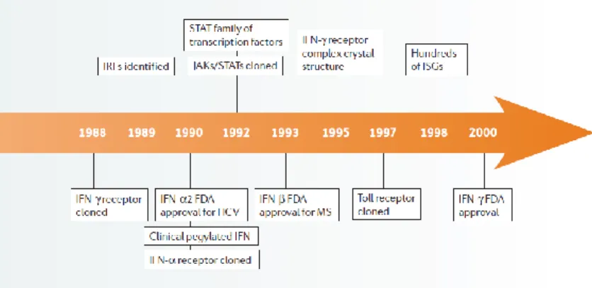
The following illustration summarizes major milestones in interferon history and gives a brief overview about the historical development and scientific efforts in the characterization and clinical application of this miracle drug.
6
Figure 2: Major milestones and discoveries in fifty years of interferon research
In the past 50 years of interferon research, more than 100.000 scientific papers were published but there are still a lot of open questions which have to be addressed. From the first observation of anti-viral activity of the cytokine to the characterization of a genetic transcription profile, it took a lot of time, effort and money, but further investigations are necessary to identify the exact mode of action, to control its side effect profile and to broaden the clinical application of this cytokine. Perhaps the second half of the interferon century will bring up new technologies to gain deeper insights into the field of interferon research and help to answer questions which have been open for more than fifty years and are still open today.
1.3 Classification of interferon
Since the discovery of interferon by Isaacs and Lindenmann, it became clear that there must be several different kinds of this cytokine which all belong to the same family of interferons. At the early beginning of interferon research, newly found subtypes were categorized according to the cell type they originated from. At these times IFN-α was known as leukocyte interferon and IFN-β as fibroblast IFN. Nowadays natural occurring interferon subtypes are named by their coding gene and the corresponding protein sequence (Bekisz, Schmeisser, Hernandez, Goldman, & Zoon, 2004).
The cytokine family of interferons (IFNs) is divided into two major classes, Type I and Type II interferons. The class of Type I interferon consists of seven different subtypes of this cytokine family: IFN-α, IFN-β, IFN-ω, IFN-κ, IFN-ε, IFN-δ and IFN-τ. Most prominent representatives of this
The above shown image summarizes major milestones in interferon research. Starting with the discovery of the cytokine in 1957, early investigations and characterizations of interferon until the end of the 70s and major break-troughs and clinical studies using IFN from 1980 up to 2000. (Borden et al., 2007)
7
Type I interferons are IFN-α and IFN-β according to their extensive biological relevance and clinical trials performed with both agents. IFN-τ was only found in ruminants and giraffes but could not be found in human beings. Although it shows biological activity if administered to the human system. [Bekisz 2004] Similar to that, IFN-δ occurs only in pigs and is produced by trophoblasts, which form the outer layer of a blastocyst. In contrast to IFN-τ, IFN-δ does not show any activity in humans (Lefevre, Guillomot, D'Andrea, Battegay, & La Bonnardiere, 1998).
IFN-κ, produced by keratinocytes, and the less well characterized IFN-ε display only limited anti- viral and anti-proliferative activity and seem to be less important in respect to biological functions. From the functional point of view IFN-α with its thirteen subtypes and the single type IFN-β are the major mediators of viral resistance and suppressors of malignant proliferation inside the Type I interferon family. IFN-ω builds up the third potent anti-viral member of this cytokine family and is secreted by viral infected leukocytes. Interferon alpha (IFN-α) consists of thirteen different family members which display a close structural homogeneity. The genetic information for these several subtypes is located in the same region on chromosome 9 in the eukaryotic genome. All types of IFN-α consist of 166 amino acids with interferon alpha 2a being the exception with only 165 due to a deletion of a single amino acid at position 44. These different types of IFN-α show a strong sequential similarity in their peptide code in the range of 75 – 99%.
The three dimensional structure of those cytokines consist of 6 alpha-helices and structural integrity is ensured by two disulfide bridges (Bekisz et al., 2004).
The following table summarizes the nomenclature of the thirteen members of the IFN-α family and their corresponding genes.
IFN-α protein subtype Corresponding IFN-α gene
A 2a
2 2b
B2 8
C 10
D (Val114) 1
F 21
G 5
H2 14
I 17
J1 7
K 6
4a 4a
4b 4b
WA 16
1 (Ala114) 1
Table 1: Nomenclature of natural occurring IFN-alpha subtypes
Summary of natural occurring interferon alpha subtypes and corresponding nomenclature according their protein and gene name. (Pestka & Meager, 1997)
8
Compared to the complex classification of Typ I interferons, the family of Type II interferons only consist of a single member termed IFN-γ. IFN-γ is mainly secreted by T-lymphocytes after activation by antigen binding. This Type II interferon owns a significant immune-modulatory capacity and evolves strong anti-viral and anti-tumor activity by the recruitment of macrophages and increased expression of antigen presenting MHC-Class I molecules (Pestka, Krause, & Walter, 2004).
Since 2002 an additional class of interferon-like molecules is known. The members of this family are classified as a new type of interferon because they also possess anti-viral activity and show similarities in the downstream signaling via the Jak-STAT pathway. This so called Type III interferon family contains three different interferon lambdas termed IFN-λ1, IFN-λ2 and IFN-λ3.
These three cytokines are also known as IL-29, IL-28A and IL-28B. The genetic information for these types of interferon λ is located at chromosome 19 in contrast to genes for Type I and Type II interferons which are positioned on chromosome 9 (Kotenko et al., 2003).
The previously mentioned interferon subclasses are distinguished from each other not only by their amino acid sequence and the cell type of origin, furthermore they can also bind to different interferon receptors with varying affinities. In case of IFN-α subtypes, the functional unit of the molecule acts in a monomeric fashion whereas IFN-β binds to its receptor in a dimer-like fashion.
In contrast to INF-α, IFN-γ builds up a tetrameric structure to bind to its receptor and induce signal transduction. A brief overview about interferon receptors and the intra cellular signaling cascade will be given in the following part of the thesis (Pestka et al., 1983).
1.4 The interferon receptor family and downstream signalling
Each of the previously described interferon types uses its own cell surface receptor complex which is composed out of multiple chains. These three different kinds of cytokine receptors belong to the group of class II helical cytokine receptors (hCRs) and share similar structures and basic elements. The extracellular domain of interferon receptors is build up by tandem domains with a length of up to 100 amino acids. These extracellular regions contain two type III fibronectin domains (FBN-III) which are similar to the constant domain of an antibody. An exception in the number of extracellular domains is represented by the receptor unit 1 (IFNAR1) of the Type I interferon receptor complex which is composed of four tandem domains. The genes coding for Type I and Type II interferon receptors are located close to each other at chromosome 21 and
9
seem to be evolutionary conserved which indicates a possible functional connection between the receptors and the triggered downstream signaling cascade (de Weerd & Nguyen, 2012).
Despite the heterologous group of Type I interferons and the differences in their peptide sequence, all members of this class signal through the same receptor complex of IFNAR1 and IFNAR2. These two receptor units differ significantly in their affinity for Type I interferons. IFNAR 2 shows high affinity binding to IFN-α or IFN-β in a nM range whereas IFNAR 1 binds in a low affinity mode at µM concentrations. The common form of IFNAR2 is termed IFNAR2c which is composed of an intact intracellular domain connected to the two extracellular tandem domains via a long transmembrane region. IFNAR2c is inevitable for complete signal transduction to induce anti-viral and anti-proliferative activity of interferon. In addition, there are two known splice variants of IFNAR2. IFNAR2b consists of a short transmembrane region without an intracellular domain while IFNAR2a is the soluble form of the receptor lacking the transmembrane and the intracellular domain of IRNAR2c (de Weerd & Nguyen, 2012).
Fully intracellular signaling in case of Type I interferons is engaged after binding of interferon to the receptor unit 2 (IFNAR2) followed by the formation of a ternary complex with IFNAR1. This formation of the IFNAR-complex is common for all members of the Type I interferon family but until these days it is not fully understood how the different biological activity of interferons is mediated via binding to the same receptor units. It was shown that the various binding affinities of the different interferons to their receptors, the stability of the ternary IFNAR complex and the total amount of receptors expressed on the cell surface can influence the expression profile of interferon-stimulated genes (ISGs) and the triggered biological effects (de Weerd & Nguyen, 2012; E. Kalie, Jaitin, Podoplelova, Piehler, & Schreiber, 2008).
The intracellular signal transduction of the IFNAR complex is mediated by the Janus activated kinase 1 (JAK1) located at the intracellular domain of IFNAR2 as well as tyrosine kinase 2 (Tyk2) coupled to IFNAR1. After formation of the ternary complex, JAK1 becomes activated via auto- phosphorylation and triggers the recruitment of STAT (signal transducers and activators of transcription) molecules. Phosphorylation of the receptor-associated kinases induces the activation of the classical Jak/STAT pathway mediated by the phosphorylation of the signal transducers STAT1 and STAT2 which allows STAT homo- or heterodimerization. In case of Type I interferon signaling, the heterodimer composed of phosphorylated STAT1 and STAT2 forms a complex with IRF9 (interferon regulatory factor 9) to build up a transcription factor. This STAT1- STAT2-IRF9 complex, also known as ISGF3 (IFN-stimulated gene factor 3), then translocates into the nucleus and initiates the transcription of interferon-stimulated genes (ISGs) via binding to the
10
upstream positioned interferon stimulated response elements (ISRE) which act as promoters for ISGs (Platanias, 2005).
In contrast to different Type I interferons, which bind to one common receptor complex, the only Type II interferon IFN- γ interacts with its own cell surface receptors. This Type II receptor complex is composed of two subunits termed IFNGR1 and IFNGR2. Similar to the Type I interferon receptors, the intracellular domains of the receptor are associated with kinases to induce signal transduction. JAK 1 is located at the IFNGR1 subunit while IFNGR2 is associated with the JAK2.
Both kinases trigger the phosphorylation of STAT1 molecules which then form homodimers before being translocated into the nucleus. Once in the nucleus, these STAT1 dimers bind to IFN-γ activated sites (GAS) in the genome and induce transcription of certain interferon stimulated genes. The formation of STAT 1 homodimers is not limited to Type II interferon receptor signaling.
Phosphorylated STAT 1 molecules activated by Type I interferon signaling can also dimerize and lead to a crosstalk between Type I and Type II signal transduction (Platanias, 2005).
Figure 3: Interferon receptors and associated downstream signaling cascades Visualization of the downstream signaling cascade of Type I and Type II interferons and their corresponding receptors. Activation of receptor-coupled kinases (JAK1, TYK2), triggers phosphorylation and activation of signal transduction proteins (STAT1 and STAT2). In case of Type I signaling, STAT1 and STAT2 dimerize and form a transcription complex in combination with IRF9. Type II signaling is mediated by phosphorylated STAT1 homodimers. After translocation of the transcription complex to the nucleus, transcription of interferon stimulated genes is initiated. (Platanias, 2005)
11
In addition to the main downstream signaling of interferons via the Jak/STAT pathway an alternative signaling pathway was identified with possible roles in the pleiotropic biological functions of this class of cytokines (shown in figure 4). In this alternative pathway, activated tyrosine kinase 2, interacting with the intracellular subunit of IFNAR1, triggers the phosphorylation of an adaptor protein termed CRKL. Due to this transient tyrosine phosphorylation of CRKL, the C3G-Rap1 signaling pathway becomes activated, leading to the recruitment of the small GTPase Rap 1. Rap 1 is associated with different functions in cell proliferation, differentiation and cell adhesion. It was shown that this small protein is able to mediate direct suppression of cell growth but in some circumstances can even promote proliferation of cells. Due to this opposed biological functions, it is believed that the mode of action of Rap 1 depends on the kind of stimuli triggering recruitment of C3G-Rap1 signaling. The activation of this alternative signaling cascade was also shown in the treatment of human cell lines by the Type II interferon IFN-γ which indicates a possible significant biological role in the anti-proliferative action of this class of cytokines (Ahmad, Alsayed, Druker, & Platanias, 1997;
Alsayed et al., 2000).
Figure 4: Alternative interferon pathway besides classical JAK-STAT signaling Alternative signaling cascade of Type I interferons mediated via Crkl protein.
Phosphorylation of CRKL triggers the signal transduction via C3G and the activation of RAP1 which strongly interacts in cell cycle control mechanisms. (Platanias, 2005)
12
1.5 Interferon in oncology practice
Although the discovery of interferon has been early back in 1957, it took more than twenty years to produce this class of cytokines in a recombinant way to obtain sufficient amounts of material for performing clinical investigations. First clinical studies were conducted with crude formulations of cell lysates containing less than 1% interferon. The establishment of cloning procedures in the 1980s raised the opportunity to express large quantities of interferon with high grade purity and enabled the evaluation of the cytostatic, immune-modulating and antiviral activity of interferon in vivo. Interferon-based treatment strategies in oncologic and infectious diseases always deal with balancing the pharmacological effect of the cytokine and the prevention of adverse side effects. Due to significant unwanted responses, IFN therapies narrow the optimal treatment ability for physicians and side effects strongly influence patient’s quality of life. In oncology research, therefore understanding of the biological mode of action, the choice of suitable clinical indications and the management of the side effect profile and toxicities of Interferon play an important role to exploit the full potential of this class of cytokines (Jonasch &
Haluska, 2001).
Clinical indications for interferon alpha in oncology comprises several hematological cancers and solid tumors, including hairy cell leukemia (HCL), chronic myelogenous leukemia (CML), follicular lymphoma, multiple myeloma (MM), AIDS related Kaposi’s sarcoma (KS) and malignant melanoma. In addition to the application of interferons in oncology, IFNs play a major role in anti- viral therapy such as chronic hepatitis B and chronic hepatitis C (Rote Liste® 2014). The following section summarizes the main clinical oncological indications with regard to interferon-induced side effects.
The majority of interferons used in the clinic are nowadays produced in recombinant form by expression in eukaryotic cells, followed downstream purification methods ensuring very high purity. For clinical application, prominent interferon preparations are Roferon-A™ (IFN-α2a, Roche) and Intron-A™ (IFN-α2b, Schering-Plough Corporation). Both types of interferon alphas are highly similar in their amino acid sequence and differ just in one position. Besides these two interferon alphas, other interferon subtypes like IFN-β1a (Avonex™, Biogen Inc), an interferon beta type with a serine to cysteine alteration and IFN-β1b (Betaseron™, Berlex), which is expressed in an Escherichia coli strain, found their way into the clinic. As an alternative medication to Rofern-A™ and Intron-A™ in chronic hepatitis C treatment, the IFN alfacon-1 (Infergen™, Amgen), an artificial synthesized interferon, based on the sequence comparison of
13
natural occurring IFN alphas, is approved. The only Type II interferon product on the market (IFN- γ, Actimmune™, Genentech Inc) was approved by the U.S. Food and Drug Administration (FDA) for its immune-modulatory function in the treatment of chronic granulomatous disease (Jonasch
& Haluska, 2001).
Interferon alpha therapy in hairy cell leukemia (HCL) was approved in 1986, justified by a multicenter study of 64 patients performed by Harvey M. Golomb et al. in the previous years. In this study, patients were treated with interferon alpha 2b (Intron-A™) subcutaneously at a dosage of 2 x 10⁶ U/m2, three times a week. The outcome of the study displayed complete remission (CR) in three patients (5%), 45 patients (70%) showed up partial response (PR) and nine patients (14%) a minor response. Only three of the 45 patients had no beneficial effect despite interferon treatment. After treatment duration of five month, hemoglobin levels and granulocyte counts re- approached to physiological levels. The evaluation of bone marrow biopsies showed a significant decrease in the median hairy-cell index by half and serious viral infections during treatment were prevented by interferon. Due to promising first responses of interferon treatment, IFN-α2a and IFN-α2b are still applied in HCL therapy with treatment durations usually over two years.
Nevertheless later studies showed that patients relapse after therapy and alternative treatment strategies with 2-deoxycoformycin and cladribine in hairy cell leukemia seem to be more beneficial (Golomb et al., 1986; Jonasch & Haluska, 2001).
Hematologic malignancies in particular show a high treatment potential for interferon administration compared to solid tumors. One of these hematological indications represents chronic myelogenous leukemia (CML). Leading clinical evaluations of interferon in CML treatment were performed by the group of Talpaz et al. in the early 1980s. They evaluated the influence of interferon alpha 2a (Roferon-A™) on 17 patients suffering from CML, which were positive for the Philadelphia-Chromosome [translocation between chromosome 9 and 22, t(9;22)(q34;q11)]. In this study, interferon alpha 2a was administered daily intramuscularly at a dosage of 5 x 10⁶ U/m2 body surface. Out of 17 patients, 14 showed a response to IFN-α2a treatment. 13 patients displayed a complete hematologic remission (CR) and just one patient a partial remission (PR).
The analysis of cytogenetic tests unveiled the suppression of Philadelphia-Chromosome positive cells in 25% of the patients responding to IFN treatment. Besides the promising outcome of the study, physicians had to deal with side effects triggered by interferon which led to treatment interruptions in two patients during the interferon therapy for 9 – 15 month (Talpaz et al., 1986).
A comprehensive study on the anti-proliferative activity of interferon in CML was conducted by the Italian Cooperative Study Group on Chronic Myeloid Leukemia in 1994. To investigate the
14
potency of interferon compared to conventional chemotherapy, 322 CML patients were treated either with IFN-α2a (Roferon-A™) or a standard chemotherapeutic agent, hydroxyurea or busulfan. In the interferon treated patient population (218 patients), a karyotypic reaction was detectable in 30% compared to only 5% of patients treated with standard chemotherapy (104 patients). The duration from the chronic phase of CML to a more severe blastic phase was significantly prolonged in the interferon treated group (72 months in the IFN-group, 45 month in the chemotherapy group). In addition to the decelerated disease progression, the overall survival of patients in the IFN-group was 21% higher than in the standard therapy group over a period of six years. Many other studies at that time confirmed the beneficial effect of interferon monotherapy compared to cytotoxic agents used in chemotherapeutic treatment of CML (Leukemia, 1994).
During 1980s another effective compound against CML was investigated termed cytarabine (ara- C). Ara-C was shown to efficiently suppress proliferation of malignant myeloid cells, when given via continuous infusion at low doses. The group of Kantarjian et al. performed a study on 140 patients positive for Philadelphia-Chromosome in early chronic CML. Interferon was administered subcutaneously in a daily dose of 5 x 10⁶ U/m2 body surface, in combination with a low dose infusion of ara-C at 10 mg/day. Due to the combination of interferon and ara-C a complete hematologic remission (CHR) was achieved in 92% of the treated patient population with a cytogentic response in 74% of responding patients. Despite the promising results of the combination therapy, several adverse events were reported. A significant part of the patients suffered from fatigue accompanied by weight loss, muscle ache, diarrhea and neurologic disorders. According to the outcome of the study, the combination of interferon with low-dose ara-C in the treatment of early-stage CML seemed to be beneficial due to the reduction of the applied interferon doses to a minimal but still effective level and the subsequent prevention of severe side effects (Kantarjian et al., 1999).
Beside the main clinical indications of interferon in malignant hematologic diseases, several kinds of solid tumors can be treated efficiently. One example is the application of interferon in combination with other cytotoxic agents in the treatment of malignant melanoma. For growth inhibition of this aggressive and highly metastatic cancer type, many combination studies including interferon therapies were conducted. In early attempts IFN-alpha was combined with another cytokine, namely interleukin-2 (IL-2). Unfortunately the combination of both immune- modulating agents did not show an additional anti-tumor effect. Other clinical trials in malignant melanoma combining interferon with conventional chemotherapeutics like cisplatin, dacarbacin
15
and vinblastine showed an increase in treatment efficacy which reached significant levels but were limited due to severe toxicities occurring by overstimulation of the immune system. A more beneficial application of interferon in malignant melanoma therapy was shown in clinical trials with an adjuvant setting, by using IFN as a follow-up therapy after initial chemotherapy. In one of these trials, published by Kirkwood et al.in 1994, 287 patients were treated with INFα2b (Intron- A™) following surgical removal of the tumor mass. In the first month after surgery the maximum tolerable dose of interferon 20 x 106 U/m2 per day was administered intravenously. Following the initial high dose treatment, 10 x 106 U/m2 INFα2b were given subcutaneously three times a week over a period of 48 weeks. Summarizing the data generated during this study, performed by the Eastern Cooperative Oncology Group, the relapse-free interval of patients after surgery was prolonged from 1 to 1.7 years due to adjuvant interferon therapy. The overall survival of high risk patients was significantly increased from 2.8 to 3.8 years. The outcome of this study confirmed the potency of interferon in fighting against solid tumors and confirmed interferon as the first molecule in this indication which showed a tremendous impact on the relapse-free period and overall survival rate in malignant melanoma patients classified with high-risk for a relapsing disease (Jonasch & Haluska, 2001; Kirkwood et al., 1996).
Interferon based cancer therapy shows significant effects in many different indications such as hematologic malignancies and solid tumors, e.g. malignant melanoma. Besides the promising activity in chronic myelogenous leukemia (CML) and hairy cell leukemia (HCL) therapy, this class of cytokines shows the potential to efficiently inhibit proliferation of another type of malignant cells of the lymphatic system. Multiple myeloma is one of the currently approved clinical indications (Rote Liste® 2014) for IFN-alpha and provides the capability for further investigations in interferon research due to a high unmet medical need in this field of oncology. According to this, the biology of multiple myeloma and possible interventions in the pathological development of this kind of lymphatic cancer will be explained in more detail in the following section.
1.6 Interferon treatment in multiple myeloma
Multiple myeloma accounts for 13% of all hematologic cancers and is characterized by an uncontrolled clonal expansion of plasma cells in the humoral immune system. The relatively low incidence rate of 5.6 cases in 100.000 persons, with a medium diagnose age of 70 years, should not disguise the fact that multiple myeloma is an incurable type of cancer and demonstrates a
16
high medical need for effective treatment. Current therapies comprise cytostatic and cytotoxic agents like the proteasome inhibitor bortezomib, the anti-angiogenic and anti-proliferative compounds thalidomide and its derivate lenalidomide. The application of thalidomide (formerly known by the trade name Contergan™) and its derivative lenalidomide in multiple myeloma is one of the last indication these compounds are still applied after severe side effects on newborn children had been detected in times when thalidomide was sold as an over the counter drug for its mild sedative activity. The combination of autologous stem cell transplantation and the earlier mentioned cytotoxic agents increased the overall survival rate of patients suffering from multiple myeloma (30% likelihood for a 10-year survival) (Palumbo & Anderson, 2011).
This kind of hematologic malignancy derives from misguided plasma cells originating from pre- germinal B-cells. Due to a genetic malfunction in terminal differentiation of B-lymphocytes into plasma cells, these cells gain a malignant phenotype. In more than half of the multiple myeloma cells, an incorporation of oncogenes in the genetic locus of the immunoglobulin heavy chain gene on chromosome 14 can be found. Accompanied by the uncontrolled proliferation of a single plasma cell clone, the hematologic system gets flooded with enormous amounts of a type of pathological immunoglobulin which is termed monoclonal gammopathy. In this first stage of the disease, called MGUS (monoclonal gammopathy of undetermined significance), gammopathy can lead to a dysfunction of kidneys by clocking them with large amounts of monoclonal antibodies.
Later on during disease progression, further genetic alterations like numerous genetic translocations trigger the activation of known cancer forcing genes like MYC, NRAS and KRAS which are involved in cell cycle control and enable increased proliferation rates. Simultaneously proteins which limit uncontrolled cell cycling like cyclin-dependent kinases inhibitors (CDKN2A and CDKN2C) become silenced. Based on the characterization and progress of genetic alterations, multiple myeloma can be subdivided into three stages, depending on the degree of severity and accompanying symptoms (Palumbo & Anderson, 2011; Smith & Yong, 2013).
In late stage multiple myeloma, large amounts of malignant plasma cells enrich in the bone marrow of the limbs, hip, spinal cord and skull with dramatic consequences. After settling of malignant cells in the bone marrow, myeloma plasma cells bind to bone marrow stromal cells and stimulate the overexpression and release of cytokines like tumor necrosis factor (TNF), macrophage inflammatory protein (MIP) and receptor activator of nuclear factor-κB ligand (RANKL) into the microenvironment. The overstimulation of cells by this cytokine-cocktail triggers bone resorption by transformation of osteoblasts into lytic osteoclasts. As a consequence of the destruction of bones, calcium from the bone marrow is released into the periphery in high