• Keine Ergebnisse gefunden

Microglia as therapeutic targets in retinal degeneration: role of translocator protein (18 κDa) (TSPO) and minocycline

N/A
N/A
Protected

Academic year: 2021

Aktie "Microglia as therapeutic targets in retinal degeneration: role of translocator protein (18 κDa) (TSPO) and minocycline"

Copied!
120
0
0

Wird geladen.... (Jetzt Volltext ansehen)

Volltext

(1)

Microglia as therapeutic targets in retinal degeneration: role of translocator protein (18 κ Da) (TSPO) and minocycline

Inaugural Dissertation zur

Erlangung des Doktorgrades Dr. nat. med.

der Medizinischen Fakultät und

der Mathematisch-Naturwissenschaftlichen Fakultät der Universität zu Köln

vorgelegt von Rebecca Scholz

aus Neuss

Köln, 2016

(2)

Berichterstatter/Berichterstatterin: Prof. Dr. Rudolf Wiesner Prof. Dr. Ines Neundorf

Tag der letzten mündlichen Prüfung: 12.07.2016

(3)

1

Table of contents

Table of contents 1

1 Summary 3

2 Zusammenfassung 5

3 Introduction 7

3.1 Structure and function of the retina 7

3.2 Inherited retinal degenerative diseases 9

3.2.1 Light damage as retinal degeneration model 10

3.3 Microglia 11

3.3.1 Microglial reactivity markers 11

3.3.2 Origin and self-renewal of microglia cells 12 3.3.3 Microglial function in the developing and adult retina 13

3.3.4 Microglia in the aging retina 16

3.3.5 Microglia reactivity in retinal degenerative diseases 17 3.4 Modulation of microglial cells as a therapeutic strategy 19

3.4.1 Endogenous factors: AMWAP, micro RNAs and Interferon- 20 3.4.2 Natural compounds: Luteolin, Curcumin, Crocin and DHA 21

3.4.3 Minocycline 22

3.4.4 Translocator protein (18κDa) (TSPO) and its ligands 23

3.5 Aims of the thesis 28

4 Results 29

4.1 Translocator protein (18 kDa) (TSPO) is expressed in reactive retinal microglia and modulates microglial inflammation and phagocytosis 29 4.2 Targeting translocator protein (18 kDa) (TSPO) dampens pro-inflammatory microglia

reactivity in the retina and protects from degeneration 31

4.3 Minocycline counter-regulates pro-inflammatory microglia responses in the retina and

protects from degeneration 33

5 Discussion 36

5.1 TSPO-reactivity marker and target for therapy 36

5.1.1 TSPO as a microglial reactivity marker in retinal degeneration 36

5.1.2 Understanding TSPO induction in microglial cells 37

(4)

Table of contents

2

5.1.3 Transcriptional regulation of TSPO 38

5.1.4 Immune modulation and neuroprotection by TSPO ligand XBD173 38 5.1.5 TSPO - future challenges and perspectives 42

5.2 Minocycline in retinal degeneration 42

5.2.1 Immune modulation and retinal degeneration rescue by Minocycline 43 5.2.2 Minocycline – future challenges and perspectives 45

5.3 Conclusion 46

References 47

Publications 62

Lebenslauf 116

Danksagung 117

Erklärung 118

(5)

3

1 Summary

Microglia are the resident immune cells of the central nervous system (CNS) and play an important role in innate immune defense as well as tissue homeostasis. Chronic microglial reactivity, microgliosis, is a general hallmark of inflammatory and degenerative diseases that affect the CNS, including the retina. There is increasing evidence that chronic microgliosis is more than just a bystander effect, but rather actively contributes to progression of degeneration through processes such as toxic nitric oxide (NO) production and even phagocytosis of stressed but viable photoreceptors. Therefore immunmodulation of microglia presents a possible therapeutic strategy for retinal degenerations. Notably, the expression of the mitochondrial translocator protein 18 ( κ Da) (TSPO) is highly elevated in reactive microglia as seen in several neuroinflammatory diseases such as Alzheimer’s disease, Parkinson’s disease and multiple sclerosis. Therefore it is used as a gliosis biomarker in the brain. Moreover TSPO ligands show potent effects in resolving neuroinflammatory brain disorders. However, TSPO expression in the eye had not been investigated before. Further, it was unknown whether TSPO ligands’ potent immunomodulatory effects could be used to treat retinal degenerations.

To fill this gap, the study aimed to analyze whether TSPO is also a potential biomarker for degenerative processes in the retina. Moreover the thesis attempted to determine whether a specific TSPO ligand, XBD173, might modulate microglial reactivity and is a potent therapeutic, to treat retinal degenerative diseases.

The findings revealed that TSPO is strongly upregulated in microglial cells of

retinoschisin-deficient (RS1

-/y

) mice, a model of inherited retinal degeneration and in

a murine light damage model. A co-localization of TSPO and microglia was

furthermore detectable in human retinal sections, indicating a potential role for TSPO

as a biomarker for retinal degenerations. In vitro assays showed that the TSPO

ligand XBD173 effectively inhibited features of microglial activation such as

morphological transformation into reactive phagocytes and enhanced expression of

pro-inflammatory cytokines. XBD173 also reduced microglial migration and

proliferation and reduced their neurotoxic potential on photoreceptor cells. In two

independent mouse models of light-induced retinal degeneration, the treatment with

XBD173 reduced accumulation of amoeboid, reactive microglia in the outer retina

(6)

Summary

4 and attenuated degenerative processes, indicated by a nearly preserved photoreceptor layer.

A further question addressed in this thesis was whether minocycline, an antibiotic with additional anti-inflammatory properties is able to reduce microglial neurotoxicity and to protect the retina from degeneration.

Minocycline administration dampened microglial pro-inflammatory gene expression, NO production and neurotoxicity on photoreceptors. Interestingly, in addition to its immunomodulatory effect, minocycline also increased the viability of photoreceptors in a direct manner. In the light damage model, minocycline administration counter- acted microglial activation and blocked retinal degeneration.

Taken together these results identified TSPO as a biomarker for microglial reactivity and as therapeutic target in the retina. Targeting TSPO with XBD173 was able to reverse microglial reactivity and could prevent degenerative processes in the retina.

In addition, the study showed that the antibiotic minocycline effectively counter-

regulates microgliosis and light-induced retinal degeneration. Considering that

microgliosis is a major contributing factor for retinal degenerative disorders, this

thesis supports the concept of a microglia-directed therapy to treat retinal

degeneration.

(7)

5

2 Zusammenfassung

Mikrogliazellen sind die Gewebsmakrophagen des zentralen Nervensystems (ZNS).

Sie spielen eine wichtige Rolle bei der Immunabwehr sowie der Aufrechterhaltung der Gewebshomöostase. Chronische Mikrogliaaktivierung, Mikrogliose, gilt als generelles Kennzeichen von degenerativen und entzündlichen Erkrankungen des ZNS, inklusive der Retina. Eine zunehmende Anzahl von Studien belegt, dass persistierende Mikrogliareaktivität nicht nur eine Folgeerscheinung degenerativer Erkrankungen, sondern aktiv am Voranschreiten solcher Prozesse beteiligt sein kann, beispielsweise durch Produktion reaktiver Sauerstoffspezies oder sogar Phagozytose gestresster, aber lebender Photorezeptoren. Mikroglia-gerichtete Therapien könnten daher ein Behandlungskonzept für retinale Degenerationen darstellen. Bei neurodegenerativen Erkrankungen wie Alzheimer, Parkinson oder Multipler Sklerose wurde festgestellt, dass reaktive Mikrogliazellen eine erhöhte Expression des mitochondrialen Translokatorproteins (18κDa) (TSPO) aufweisen.

Deshalb wird TSPO als Gliosemarker im Gehirn verwendet. Spezifische TSPO- Liganden zeigten außerdem Wirksamkeit bei der Behandlung von neuroinflammatorischen Erkrankungen des ZNS. Ob TSPO auch im Auge exprimiert wird und ob TSPO-Liganden möglicherweise zur Therapie von retinalen Degenerationen geeignet sind, ist bisher noch nicht untersucht worden.

Neben der Überprüfung, ob TSPO auch als Biomarker für degenerative Veränderungen der Netzhaut verwendet werden kann, wurde in dieser Arbeit untersucht, inwiefern der spezifische TSPO-Ligand, XBD173, Mikroglia Reaktivität modulieren kann und sich zur Therapie von retinalen Degenerationen eignet.

Sowohl Untersuchungen an Retinoschisin-defizienten Mäusen, einem Modell für

erbliche Netzhautdegeneration, als auch in einem Licht-Schadensmodell zeigten eine

Hochregulation von TSPO in Mikrogliazellen. Eine Kolokalisation von TSPO und

Mikrogliazellen war außerdem in humanen Retinapräparaten erkennbar und

verdeutlicht die potentielle Eignung als Biomarker für retinale Erkrankungen. In vitro

Analysen zeigten, dass XBD173 Zeichen von Mikrogliareaktivität wie erhöhte

Expression inflammatorischer Marker, Migration, Proliferation und die

morphologische Transition zu reaktiven Phagozyten reduziert sowie das

neurotoxische Potential von Mikrogliazellen verringert. In zwei unabhängigen

murinen Modellen für Licht-induzierte Degeneration, reduzierte die Behandlung mit

(8)

Zusammenfassung

6 XBD173 die Akkumulation amoeboider, reaktiver Mikrogliazellen in der äußeren Netzhaut und schützte die Retina vor degenerativen Prozessen, erkennbar durch den Erhalt der Photorezeptorschicht.

Ein weiteres Ziel dieser Arbeit war zu untersuchen, ob Minocyclin, ein Antibiotikum mit zusätzlichen anti-inflammatorischen Eigenschaften, Mikroglia Reaktivität modulieren und die Retina vor degenerativen Prozessen schützen kann.

Stimulation von Mikrogliazellen mit Minocycline verringerte die Expression von Entzündungsmarkern, die Produktion von toxischem Stickstoffmonoxid und die Neurotoxizität von Mikrogliazellen gegenüber Photorezeptoren. Interessanterweise war zusätzlich zu der immunmodulativen Wirkung war auch ein direkter protektiver Effekt auf Photorezeptoren festzustellen. Behandlung mit Minocyclin bei Licht- induzierter Degeneration wirkte pro-inflammatorischer Mikroglia-Aktivierung entgegen und schützte die Retina vor Degeneration.

Die Ergebnisse zeigen, dass sich TSPO als Biomarker für Mikroglia-Reaktivität und

therapeutisches Target bei retinalen Degenerationen eignet. TSPO gerichtete

Therapie mit XBD173 reduzierte Mikroglia-Reaktivität und verhinderte degenerative

Prozesse in der Retina. Zusätzlich zeigt die vorliegende Arbeit, dass das Antibiotikum

Minocyclin, Mikrogliose entgegenwirkt und die Retina vor Licht-induzierter

Degeneration schützt. Unter der Annahme, dass chronische Mikrogliose maßgeblich

den Verlauf von degenerativen Prozessen beeinflussen kann, stellen Mikroglia-

gerichtete Therapieansätze ein erfolgsversprechendes Konzept zur Behandlung von

retinalen Degenerationen dar.

(9)

7

3 Introduction

3.1 Structure and function of the retina

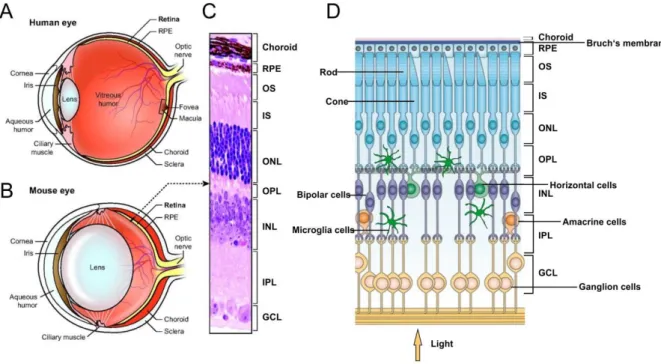
The gross morphological structure of the human eye consists of three different layers (shown as sagittal section of the adult human eye Fig. 1, A). The external layer consists of the sclera and the cornea. The intermediate layer is generally further divided into an anterior part, which includes iris and ciliary body and a posterior part, the choroid. The retina belonging to the CNS is designated as inner layer. The chambers that occur between these layers are the anterior chamber (between iris and cornea) and the vitreous humor (between lens and retina). Both are filled with differently viscous fluids (Kolb, 2005).

Figure 1: Schematic overview of the mammalian eye and its retinal structure. (A) Cross section of the human eye. The retina is located at the posterior part of the eye and is loosely stocked to the retinal pigment epithelium (RPE) (B) Cross section of the murine eye. (C) H&E stained murine retinal cross section showing the different retinal layers. (D) Schematic overview of the different retinal layers, including respective cell types. (GCL) ganglion cell layer, (IPL) inner plexiform layer, (INL) inner nuclear layer, (OPL) outer plexiform layer, (ONL) outer nuclear layer, (IS) inner segments, (OS) outer segments (A-C) modified from (Veleri et al., 2015) D modified from (Kimbrel and Lanza, 2015)

During embryogenesis, the retina derives from the neural tube (Lamb et al., 2007).

As sensory part of the eye, the retina absorbs light and converts it into electrical signals that are transferred to the brain and are decoded to gain visual information.

The retina consists of different neuronal cell types that roughly divide the retina into

(10)

Introduction

8 three cellular layers, the ganglion cell layer (GCL), the inner nuclear layer (INL) and the outer nuclear layer (ONL) which are separated from each other by two synaptic layers, the inner plexiform layer (IPL) and the outer plexiform layer (OPL)) (Fig. 1 C, D). The retinal pigment epithelium (RPE) lies juxtaposed to the apical side of the photoreceptors and is essential to support the retina (Sung and Chuang, 2010).

Beside the absorption of light the RPE phagocytoses sheded membrane discs of the photoreceptors that are damaged due to photo-oxidative stress. This is a daily occurring process that leads to a complete membrane disc renewal in about 10-15 days. Furthermore the RPE is co-responsible for the recycling of rhodopsin after bleaching, a process called visual cycle and supports the retina with nutrients (Travis et al., 2007). Additionally the RPE is part of the blood-retina barrier (BRB) and controls the transport of molecules between choroidal blood flow and retina (Campbell and Humphries, 2012, Strauss, 2005, Young, 1967).

After passing the different retinal layers light is absorbed by the photoreceptors that

are located at the outer side of the retina. In the human retina, photoreceptors can be

divided into two subtypes: rods (~120 million cells), which are responsible for dim

light vision and cones (~6 million cells), which mediate color vision under brighter

light conditions (Bommas-Ebert et al., 2006). Dependent of its spectral sensitivity

three cone types with different opsins can be distinguished in primates: L- (long,

564nm), M- (medium, 533 nm) and S- (short, 437nm) wavelength cones (Bowmaker

and Dartnall, 1980). In the human retina, a certain spot called fovea (~1,5 mm

2

)

consists only of cones and has the highest visual acuity. The area (~3 mm

2

) around

the fovea, recognizable by its yellow screening pigments (zeaxanthin and lutein), is

named macula. (Veleri et al., 2015, Morrow et al., 1998, Balashov and Bernstein,

1998). The absorption of a photon by the chromophore 11-cis retinal within the

photoreceptors, leads to isomerization to all-trans retinal and initiates the process of

phototransduction, which is a cascade of biochemical events resulting in closure of

ion channels, photoreceptor hyperpolarization and signal transmission to second

order neurons, such as bipolar, horizontal and amacrine cells that form the inner

nuclear layer (INL) (Travis et al., 2007, Sung and Chuang, 2010, Yau and Hardie,

2009). Neurons of the INL are interconnected to each other, which allows reciprocal

excitation or inhibition and serves as contrast intensification (Bommas-Ebert et al.,

2006). After processing of the signal by the different cell types of the INL, the signal is

forwarded to the ganglion cells, whose axons form the optic nerve and transmit the

(11)

9 converted light signals to the brain (Sung and Chuang, 2010). Furthermore three glial cell types are found in the retina: Müller cells, microglia and astrocytes (Bringmann et al., 2006). Microglia are the immune cells of the eye which are under homeostatic conditions located in the plexiform layers (Kettenmann et al., 2011). Müller cells span the entire retina and are important for the maintenance of retinal homeostasis (Bringmann et al., 2006).

Although the murine eye is structurally very similar to the human eye, some differences remain: the lens of the mouse eye is much bigger in proportion to the rest of the eye (Fig. 1, B) (Veleri et al., 2015). Furthermore mice, as nocturnal animals, rely predominantly on rod-mediated vision and in contrast to humans possess only two different types of cones, M- and S-cones (dichromatic vision) and no macula or fovea (Morrow et al., 1998).

3.2 Inherited retinal degenerative diseases

Loss of visual function means a severe loss of life quality for the affected individuals.

Major causes for blindness are neurodegenerative retinal diseases that lead to dysfunction or death of photoreceptors (Veleri et al., 2015). In general, retinal degenerations can be divided into monogenic or multifactorial (complex) disorders (Berger et al., 2010). A relatively common form of monogenic retinal degeneration affecting 1 in 4000 individuals, is retinitis pigmentosa (RP) (Ferrari et al., 2011).

Within the group of multifactorial retinopathies, age-related macular degeneration (AMD) is the leading cause of blindness in elderly populations in developed countries (The Eye Diseases Prevalence Research Group, 2004) (Coleman et al., 2008).

Genetic susceptibility factors such as variants in complement factor H (CFH) and age-related maculopathy susceptibility 2 (ARMS2) genes, risk factors such as smoking, diet and higher age are associated with the development of this late-onset disease (Swaroop et al., 2009, Cooke Bailey et al., 2013, Jager et al., 2008).

Improvement of molecular techniques such as next generation sequencing and large scale genotyping allowed the identification of many disease-causing genes.

Nowadays more than 250 causal genes are known for this heterogeneous group of

retinal diseases (RetNet:https://sph.uth.edu/retnet/). Despite this knowledge, there is

a need to develop novel therapeutic strategies for retinal degenerations.

(12)

Introduction

10 Because of limitations to study disease mechanisms in patients, mouse models provide a valuable tool to analyze disease pathology and to validate novel therapeutic strategies (Veleri et al., 2015). Despite the fact that the mouse eye is not completely identical to the human eye, mouse models allow to study important features of retinal degeneration such as immune activation or photoreceptor apoptosis. Several genetic as well as experimental models are available including those that mimick the damaging features of bright light. These models vary basically in the onset and severity of degeneration and discriminate underlying causes such as defects in cilia or RPE function, phototransduction and synaptic transmission (Veleri et al., 2015, Wenzel et al., 2005, Morrow et al., 1998). A common feature of retinal degenerative diseases, which is also detectable in most mouse models, is microglial activation. There is now ample evidence that chronic microglial activation is more than a bystander effect of such pathologies, but rather actively contributes to progression of retinal degeneration (Karlstetter et al., 2015, Chen and Xu, 2015, Gupta et al., 2003). For instance, it was reported that reactive microglial cells are able to produce cytotoxic oxygen radicals such as NO and may even phagocytose stressed, but viable photoreceptors (Zhao et al., 2015, Yang et al., 2007a). For the therapeutic intervention studies in this thesis, we have chosen the acute white light- induced degeneration paradigm, which models both disease characteristics, the fast degeneration of photoreceptors and activation of microglial cells (Zhang et al., 2004) (Joly et al., 2009, Ng and Streilein, 2001).

3.2.1 Light damage as retinal degeneration model

Light-induced retinal degeneration represents an inducible model to study

mechanisms of neuronal cell death and to test immune-directed treatment strategies

simultaneously. Besides that, light is also a promoting factor in human retinal

degenerations and is therefore not just an artificial stimulus (Cruickshanks et al.,

1993, Swaroop et al., 2009, Wenzel et al., 2005). The advantage over genetically

modified mouse strains is that the start of degeneration can be well defined. It has a

relatively synchronized wave of cell death and progresses relatively fast. These

features allow to better analyze biochemical processes leading to photoreceptor

degeneration and proper timing of intervention approaches (Grimm and Reme,

2013). Different light exposure settings can be distinguished: short-term exposure to

(13)

11 bright white light, long-term exposure to low levels of white light and exposure to monochromatic light of a specific wavelength (Grimm and Reme, 2013). The damaging potential of light critically depends on its wavelength. Short wavelength blue light has a higher damaging potential than long-wavelength green light. This is due to a process called photo-reversal, the regeneration of rhodopsin from bleaching intermediates that results in a higher absorption of photons in a certain time span (Grimm et al., 2001). In addition, there are also indications that the different light- damage setups vary in mechanisms of cell death, and caspases involved in cell death induction are differentially expressed (Wenzel et al., 2005). Responsible for the induction of light-induced degeneration is the excessive and ongoing bleaching of rhodopsin. This leads to the production and accumulation of retinoid metabolites and other toxic byproducts that initiate a cascade of death signals, subsequently resulting in photoreceptor cell death (Wenzel et al., 2005). Crucial for this process is a functional visual cycle as RPE 65 deficiency, an enzyme involved in the rhodopsin recycling process, protects from light-induced degeneration (Grimm et al., 2000).

Furthermore, the RPE65

Leu450Met

variation, in which leucine is substituted by methionine, slows down the visual cycle and thereby decreases the light damage susceptibility (Wenzel et al., 2001b). Other factors that can influence light damage susceptibility are increased levels of corticosteroids, which suppress AP-1-induced cell death and circadian dependent effects (Wenzel et al., 2001a, Organisciak et al., 2000).

3.3 Microglia

3.3.1 Microglial reactivity markers

Microglia cells represent the resident immune cells of the CNS, including the retina

(Kettenmann et al., 2011). They were first described by the Spanish histopathologist

Pio Del Rio Hortega back in the 1920s. The invention of the silver carbonate staining

procedures enabled Hortega, to identify and distinguish microglia from neurons,

astrocytes and oligodendendroglia by morphological criteria (Rio-Hortega, 1939,

Perez-Cerda et al., 2015). Nowadays, several macrophage-related surface and

intracellular markers are known and used to study the role of microglia in health and

disease. These markers include F4/80 ( -glucan receptor dectin-1), CD68

(14)

Introduction

12 (macrosialin), CD11b/CD18 (also called complement factor 3 (CR3), Mac1) and ionized calcium-binding adapter molecule 1 (iba1), which is currently the most reliable and widely used marker in humans and mice (Hume et al., 1983, Xu et al., 2007, Ito et al., 1998, Karlstetter et al., 2015). Other known markers are CD16, CD169 (siglec-1), CD 206 and MHC II. A few of these markers were shown to increase with microglial reactivity. This was observed for iba1 and CD11b. As microglial reactivity is often associated with neurodegeneration, the identification of activity markers is important as it complements the known morphological criteria of ramified versus amoeboid cell shape. Unfortunately, none of the introduced markers is exclusively expressed on microglia so that resident microglia can be hardly separated from CNS-recruited monocytes (Kettenmann et al., 2011). Another method to specifically label microglia is the use of genetically modified animals that express enhanced green fluorescent protein (EGFP) under the control of a microglia/macrophage specific gene promoter factor such as CX3CR1 (fraktalkine receptor) (Xu et al., 2007, Kezic and McMenamin, 2008).

3.3.2 Origin and self-renewal of microglia cells

The origin and renewal of microglial cells has been discussed controversially over the

last twenty years. Adoptive transfer experiments with bone marrow (BM) cells from

EGFP+ transgenic donors transferred into full body-irradiated adult wild-type mice,

initially indicated that microglial cells originate and renew from BM precursors,

because EGFP+ BM cells were detectable in the retina of wildtype recipients a few

weeks after transfer (Xu et al., 2007, Kezic and McMenamin, 2008). But criticism was

raised that whole body irradiation and the transfusion of BM do not reflect normal

physiological conditions, because they may lead to the breakdown of the blood-brain-

barrier (BBB) and the blood-retina-barrier (BRB) and bring progenitors into the

circulation which would normally rest in the BM (Li et al., 2004, Ajami et al., 2007). In

contrast to adoptive transfer studies, parabiosis experiments, in which the blood-flow

of wildtype and GFP+ transgenic mice were connected, showed that cells from the

circulation are not recruited to the retina, neither in healthy nor in disease conditions

(Ajami et al., 2007). This and other findings strongly suggest that microglial cells are

not recruited from the circulation under physiological conditions and that observed

microglial replacement in older studies was very likely caused by the experimental

(15)

13 procedure of irradiation and bone marrow transplantation (Mildner et al., 2007, Ransohoff, 2007, Kierdorf et al., 2013b, Alliot et al., 1999). Furthermore, different fate mapping approaches independently reveal that microglia cells are ontogenetically distinguishable from hematopoietic cells and derive from the yolk-sac during early embryonic development (Ginhoux et al., 2010, Schulz et al., 2012, Kierdorf et al., 2013a).

3.3.3 Microglial function in the developing and adult retina

During retinal development, before the complex structure of the retina is fully differentiated, a burst of developmental apoptosis can be observed in neurons located in the GCL and in the IPL (Hume et al., 1983). The phenomenon of developmental apoptosis is detectable in the entire CNS and different studies proof that microglia cells actively contribute to that process (Bessis et al., 2007). Microglia cells seem to orchestrate apoptosis by the production of several molecules, like tumor necrosis factor- � (TNF- �) , nerve growth factor (NGF) and nitric oxide (NO).

However, they are also important for phagocytosis of occurring cellular debris (Sedel et al., 2004, Frade and Barde, 1998, Chao et al., 1992, Hume et al., 1983). Although the underlying reasons of developmental apoptosis are not completely understood, the chronology showing developmental apoptosis preceding synaptogenesis, indicates a possible correlation between both processes (Bessis et al., 2007). This is further sustained by studies that propose microglia cell involvement in synaptogenesis. The deletion of the microglial specific protein KARAP/DAP 12 as well as the blockage of complement receptor 3 CR3/C3 signaling negatively influenced synaptic function and the process of synaptic pruning and also reduced the connectivity between neurons (Roumier et al., 2004, Schafer et al., 2012). These studies implicate that microglia can control neuronal function by orchestrating synaptogenesis and apoptosis, starting early during retinal development.

In the healthy adult retina microglia cells are mainly located in the plexiform layers,

where they build up a not overlapping meshwork that spans the entire retina (Lee et

al., 2008). Under physiological conditions microglia have a ramified morphology with

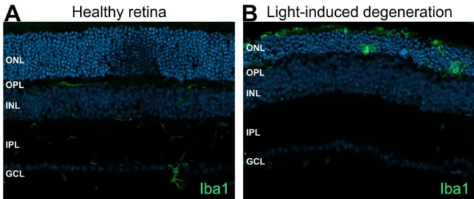
a small somata and wide ranging, fine processes (Figure 2, A). Normal microglial

appearance is often denoted as “resting state”, but their processes are highly motile

and permanently scan their microenvironment to clear it from metabolic products or

(16)

Introduction

14 tissue debris and to detect pathological changes. They also directly contact other cells such as astrocytes, neurons and blood vessels. Furthermore, microglial protrusions undergo a permanent reconstruction process. The permanent monitoring and the expression of several surface receptors, such as purino-and fractalkine receptors, receptors for complement factors, immunoglobulins, adhesion molecules and chemokines enable microglia to sense and react quickly to potential danger signals (Nimmerjahn et al., 2005, Karlstetter et al., 2015, Langmann, 2007, Kettenmann et al., 2011). Such activation goes along with an extensive change in morphology, gene expression and function (Figure 2, B) (Kettenmann et al., 2011).

Figure 2: Microglial retinal location and appearance during health and disease. (A) In the healthy retina Iba1 positive ramified microglial cells are located in the plexiform layers. (B) After exposure to bright white light microglial cells migrate to the degenerating outer retina and the amoeboid shape points towards a much more activated state. (ONL) outer nuclear layer, (OPL) outer plexiform layer, (INL) inner nuclear layer, (IPL) inner plexiform layer, (GCL) ganglion cell layer.

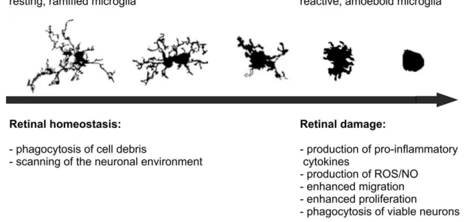
In general reactive microglia cells have a more amoeboid shape, with less branches

and thicker processes. But there are many different intermediate forms described,

ranging from highly ramified to more or less round cells completely lacking processes

(Figure 3) (Karperien et al., 2013). In addition to these morphological changes,

microglia cells can become motile and migrate into damaged areas following

chemotactic signals from neurons, astrocytes and endothelial cells.

(17)

15

Figure 3: Microglial morphological plasticity. Under homeostatic conditions microglial cells have a small cell body and scan their environment with their long-branched protrusions. Microglia are able to sense subtle degenerative changes and respond quickly. Activation of microglial goes a long with a morphological transformation from a ramified to a much more amoeboid shape. Drawings modified from Karperien et al. 2013.

The phenotypical changes are accompanied by several functional changes.

Enhanced phagocytic activity is observed, release of several neurotropic factors, chemokines or molecules like NO, upregulation of intracellular cytokines, expression of several surface markers, which are important for cell-cell interaction and enhanced proliferation (Hanisch and Kettenmann, 2007, Kettenmann et al., 2011).

Microglial cells are located in a highly specialized tissue, where immune reactions including chronic activation and recruitment of other immune cells such as T-cells would have detrimental effects. So there are many control mechanisms available to surveil microglial reactivity. These mechanisms constitute to the immune privilege of the eye and the CNS to prevent overwhelming and damaging immune reactions (Carson et al., 2006, Zhou and Caspi, 2010). Several retinal cell types are essentially involved in the control of microglial reactivity either by direct interaction or by the production of different soluble factors. For instance, healthy neurons express CD200 (OX2), a membrane glycoprotein that represents an inhibitory signal for microglia.

CD200 deficiency or the missing interaction of CD200 and its receptor leads to an

activation of microglial cells and promotes the susceptibility to autoimmune and

degenerative pathologies of the CNS in animal models (Hoek et al., 2000, Wang et

(18)

Introduction

16 al., 2007, Chitnis et al., 2007, Dick, 2003). Fractalkine (CX3CL1) is a further regulatory molecule produced by neurons and endothelial cells. In the CNS only microglia cells express the respective receptor for fractalkine and its deficiency can increase microglial neurotoxicity in models for Parkinson’s disease, amyotrophic lateral sclerosis (ALS) and Alzheimer’s disease (Cardona et al., 2006, Cook et al., 2001, Fuhrmann et al., 2010). In addition to these regulatory molecules, healthy neurons have a sugar-coated exterior surface, designated as glycocalyx which is covered with sialic acids caps. Sialic acids can be recognized by microglial siglec receptors. This interaction signals the healthy state of neurons and suppresses microglial activation (Karlstetter and Langmann, 2014, Linnartz and Neumann, 2013).

The retinal pigment epithelium (RPE) is also involved in modulation of microglial reactivity. The RPE is source of several cytokines in the eye including transforming growth factor- (TGF- ) (Tanihara et al., 1993). Microarray analysis of murine primary microglia pre-stimulated with INF- and TNF - α and then with TGF - revealed, that TGF- can downregulate the expression of pro -inflammatory genes involved in chemotaxis, migration, apoptosis and host response to infection (Paglinawan et al., 2003). Thus, microglial reactivity is a complex process that is fine- tuned by the quality of the activating stimulus and the interaction with other retinal cells.

3.3.4 Microglia in the aging retina

In the entire CNS, including the retina, aging-related changes in microglial cells can

be observed in healthy conditions and in responses to injury. Comparative studies

between young and aged healthy mice revealed that microglia undergo phenotypic

changes with increasing age. Resting microglia from aged mice compared to young

ones appear smaller and stretched out fewer branched protrusions that were less

motile (Damani et al., 2011). Furthermore the number and density of microglial cells

increase with age. Of note is that the mosaic like arrangement of microglia in the

retina became more irregular and the territory monitored by a single cells is smaller

(Tremblay et al., 2012). Microglia cells which are in the young and healthy retina

confied to the plexiform layers are also detected in the outer part in aged retinas

(Wong, 2013, Ma et al., 2009).

(19)

17 The phenotypic changes occur along with altered microglial functions. Markers such as MHCII and pro-inflammatory cytokines like IL-6, TNF α - and IL-1 are upregulated with age, whereas cytokines associated with attenuation of microglial cells such as IL-4, IL-10 and TGF- are generally downregulated. A further aging effect is that receptor interactions involved in suppression of microglial reactivity, such as CD200- CD200r and CX3CL1-CX3CR1 interactions, are significantly weaker with age (Matt and Johnson, 2016). These age-related changes promote inadequate microglial behavior also in response to injuries. Investigations in a laser -induced retinal injury model revealed that process motility as well as migration capacity was slowed down in aged microglia. Although the capability to respond to pathological stimuli was reduced, once activated the cells failed to transform back into a “resting” state (Damani et al., 2011). Since aging is a risk factor and microglia can contribute to degenerative diseases of the CNS, age-dependent microglial alterations like reduction of neuroprotective function, increase of neurotoxicity and inadequate responses to injury, are possible drivers for pathological processes (Flanary et al., 2007).

3.3.5 Microglia reactivity in retinal degenerative diseases

Several studies revealed an association between enhanced microglial reactivity and

neurodegeneration. It is still discussed whether microglial reactivity is just the

consequence of pathological alterations or if microglia cells are actively involved in

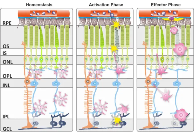
the progression of degenerative diseases (Figure 4) (Karlstetter et al., 2015, Masuda

and Prinz, 2016, Zhao et al., 2015, Giulian, 1999). An increasing number of studies

indicate that microglial activation is an early event and precedes or accompanies

degeneration of photoreceptors in various retinal degeneration models (Zhang et al.,

2004, Zeiss and Johnson, 2004, Gehrig et al., 2007, Ebert et al., 2009, Zhao et al.,

2015). For instance, an association between microglial proliferation and ongoing

degenerative processes was detectable in the Pde6b

rd1

mice, an inherited model of

retinal degeneration (Zeiss and Johnson, 2004) . Also in the RS1h knockout mouse

that shows several features of X-linked juvenile retinoschisis (XLRS), microarray

analysis revealed an upregulation of microglial-related genes starting at postnatal

day p11, before any occurrence of photoreceptor apoptosis (Gehrig et al., 2007,

Ebert et al., 2009).

(20)

Introduction

18

Figure 4: Schematic representation of microglial action during retinal degenerations. (A) Under homeostatic conditions ramified microglia mainly populate the plexiform layers and keep up tissue maintenance by phagocytoses of cell debris, secretion of neurotrophic factors and scan their environment in regard of pathological alterations. (B) Several factors signaling damage or abnormal function are sensed by microglia and lead to their activation. (C) Microglia and in case of breakdown of the blood-retina barrier, recruited blood-derived precursors migrate to the lesion sites, where they transform into amoeboid reactive phagocytes. These effector cells may be protective or detrimental depending on their immunological phenotype and the local cytokine milieu.

RPE, retinal pigment epithelium; ONL, outer nuclear layer; OPL, outer plexiform layer; INL, inner nuclear layer;

IPL, inner plexiform layer; GCL, ganglion cell layer (modified from (Karlstetter et al., 2010a)

Furthermore analysis of human retinal sections from patients with AMD, late-onset

retinal degeneration (L-ORD) or retinitis pigmentosa (RP) revealed an accumulation

of amoeboid shaped microglia in regions of ongoing photoreceptor death. Of note is

that these microglia cells contained rhodopsin-positive cytoplasmatic inclusions,

indicating that they phagocytosed photoreceptor cell debris and are possibly also

involved in cell death of adjacent cells by production of neurotoxic factors (Gupta et

al., 2003). Despite the fact that microglia cells basically exert several functions to

keep-up tissue homeostasis and support neurons e.g. by production of different

neurotrophic factors, many different studies revealed that microglia cells that lose

their autoregulatory capacity can actively participate in the initiation and perturbation

of retinal degenerative diseases. (Harada et al., 2002, Karlstetter et al., 2015). In line

(21)

19 with this, in vitro studies that compared the neurotoxic potential of activated versus non-activated supernatant of microglia and Müller cells, revealed that activated microglia cells induce much more photoreceptor cell death than conditioned medium from non-activated microglia or Müller cells. These findings suggest that soluble factors produced by reactive microglia such as NO, pro-inflammatory cytokines, complement factors or proteinases may contribute to induction of cell death during degenerative processes (Roque et al., 1999).

Which factors are involved in microglial activation is an open field of research. It was reported that stimulation with Toll-like receptor (TLR) ligands disturb the ability of microglial cells to distinguish between dead and alive, resulting in phagocytosis of viable neurons (Brown and Neher, 2014). The phagocytosis of stressed, but still viable neurons is designated as “phagoptosis” or pri mary phagocytosis. Phagoptosis generally mediates turnover of different cells like neutrophils or erythrocytes and is one form of cell death; however experiments in models of brain inflammation indicate that this process also contributes to neuron loss during degeneration. An important factor for phagoptosis induction seems to be the expression of ‘eat me’ signals, like the reversible expression of phosphatidylserine (PS) or the loss of ‘don’t eat me’

signals, as blocking of phagocytic signaling in microglia prevents neuron loss without inhibiting inflammation (Brown and Neher, 2012). In line with that, Zhao et al. showed that microglial phagocytosis of living photoreceptors is also involved in cell death in the CX3CR1

GFP/+

/rd10 retinal degeneration model that mimics features of retinitis pigmentosa. Depletion of microglial cells as well as blockage of microglial phagocytosis in the same model attenuated photoreceptor degeneration, suggesting reactive microglia as key players of cell death during degeneration (Zhao et al., 2015).

3.4 Modulation of microglial cells as a therapeutic strategy

The observation that microglia cells can be actively involved in retinal degeneration

brings them into focus as a potential therapeutic target. Although causative genetic

defects and contributing factors can vary tremendously, different pathologies share

the common feature of innate immune activation. Microglial reactivity and

inflammation is a general hallmark of neurodegenerative diseases. Therefore,

(22)

Introduction

20 modulation of microglial cells could be a good therapeutic strategy to diminish degenerative processes in different diseases. Several compounds including natural occurring substances, synthetic pharmaceuticals as well as endogenous factors are investigated with regard to their immune modulatory competence.

3.4.1 Endogenous factors: AMWAP, micro RNAs and Interferon- β

After activation, microglia must be able to return back into a normal ramified scanning state. As loss of auto-regulatory function can have detrimental effects, microglia possess endogenous control mechanisms. Activated microglia/macrophe whey acidic protein (AMWAP) is produced by microglial cells and acts in a paracrine fashion.

Overexpression of AMWAP as well as treatment of microglia cells with recombinant AMWAP reduced reactivity and limited their neurotoxic potential. Thus, AMWAP is an efficient endogenous counter-regulator of pro-inflammatory activity and a possibly therapeutic modulator of microglial reactivity (Karlstetter et al., 2010b, Aslanidis et al., 2015).

Further endogenous molecules that are able to modulate microglial cells are MicoRNAs (miRNAs). miRNAs are small non-coding RNA molecules that are involved in the regulation of diverse physiological processes including functions of the immune system. Different miRNAs were identified to control microglial reactivity, proposing epigenetic regulation by miRNAs as a potential therapeutic application (Cardoso et al., 2016). For example, overexpression of the microglial specific miR- 124 in a model of experimental autoimmune encephalomyelitis (EAE) promoted the formation of a resting microglial phenotype, suppressed microglial activation and clearly ameliorated disease symptoms (Ponomarev et al., 2011).

Interferon- (IFN- ) is a body own cytokine, but is also therapeutically used for the

treatment of multiple sclerosis (MS) (Waubant et al., 2003, Kasper and Reder, 2014,

Teige et al., 2003). Data from our research group recently revealed an important role

for INF- in a laser -induced mouse model of choroidal neovascularization (CNV),

which mimics several features of age-related macular degeneration (AMD). Complete

absence of IFNAR or specific deletion in microglia enhanced lesion size and

microglial reactivity, whereas treatment with INF- reduced CNV and microgliosis

(Lückoff et al., 2016).

(23)

21

3.4.2 Natural compounds: Luteolin, Curcumin, Crocin and DHA

Different naturally occurring substances can modulate microglial reactivity and reduce their neurotoxic potential. Luteolin is a plant derived flavone that displays anti- oxidative and anti-inflammatory capacity in micromolar concentrations (Seelinger et al., 2008). In vitro studies with LPS-pre-activated BV-2 microglial cells revealed that Luteolin can reduce pro-inflammatory and pro-apoptotic gene expression as well as production of NO; whereas markers associated with enhanced phagocytosis, ramification, chemotaxis and anti-oxidative pathways were clearly upregulated, resulting in less neurotoxicity on photoreceptor cells (Dirscherl et al., 2010).

Similar findings were reported for Curcumin, a spice often found in curry powder (Oyagbemi et al., 2009). Transcriptome analysis of resting and activated microglia showed that treatment with curcumin potently downregulates transcripts that are associated with microglial activation. Furthermore, supernatant from curcumin challenged microglia were less toxic to photoreceptors cells compared to controls (Karlstetter et al., 2011).

A further plant-derived compound with microglial modulative capacity is crocin, a carotenoid that can be found in stigmas of safran or fruits of gardenia. Crocin was shown to reduce LPS, interferon- and amyloid- induced microglial NO production and cell death ex vivo in hippocampal slice cultures as well as in vivo in light induced retinal degeneration and even improved retinal function measured by electroretinograms (ERG)(Nam et al., 2010, Yamauchi et al., 2011).

Another naturally occurring substance that also influences microglial reactivity is

docosahexaenoic acid (DHA), which can be found in fish oil and some seeds. DHA is

derived from an essential polyunsaturated fatty acid (PUFA) that must be obtained

from dietary sources. DHA is an important structural fatty acid in brain and retina and

is responsible for its integrity and functionality (Singh, 2005). Levels of DHA were

shown to decrease during different forms of retinal degeneration, including

retinoschisin deficient mice, a model of inherited retinal degeneration (Schaefer et al.,

1995, Ebert et al., 2009). Dietary intervention with DHA alone or combined with

curcumin reduced microgliosis and enhanced photoreceptor survival in retinoschisin

deficient mice as well as in a model of neuronal lipofuscinosis (NCL) (Ebert et al.,

2009, Mirza et al., 2013).

(24)

Introduction

22

3.4.3 Minocycline

Minocycline is a semisynthetic tetracycline which is in use against a wide range of infectious diseases caused by gram-positive and gram-negative bacteria for at least 30 years. The ability to bind bacterial 30s ribosomal subunit results in blockage of protein synthesis and is mainly responsible for minocycline’s an ti-biotic effect. Beside the bacteriostatic action minocycline exerts several other biological activities, including anti-inflammatory and anti-apoptotic effects, neuroprotective capacity as well as inhibition of proteolysis, angiogenesis and tumor metastasis. In various disease models reaching from inflammatory diseases (dermatitis, atherosclerosis), autoimmune disorders (inflammatory bowel disease, rheumathoid arthritis) and neurodegenerative disorders (Parkinson’s disease, Alzheimer’s disease, Multiple s clerosis, amyotrophic lateral sclerosis, spinal cord injury, Huntington’s disease and Ischemic injury) beneficial effects were observed. Accountable for the broad application possibilities is at least partially the rapid absorption, long half-life, good bioavailability and lipophilic properties that allow overcoming of the blood-brain barrier (Garrido-Mesa et al., 2013, Du et al., 2001, Wu et al., 2002b, Biscaro et al., 2012, Brundula et al., 2002, Nikodemova et al., 2010, Wang et al., 2003, Wu et al., 2002a).

In the eye several studies reported minocycline to act immune modulatory and neuroprotective. Glaucoma is a disease caused by degeneration of retinal ganglion cell axons. In DBA/2J mice, a chronic glaucoma model, microglial activation and migration into the optic nerve head are thought to be involved in disease progression.

Treatment with minocycline was able to calm down microglial reactivity and to

improve axonal integrity and transport. However, minocycline failed to reduce Müller

cell and astrocyte gliosis as well as high intraocular pressure (IOP) (Bosco et al.,

2008). Furthermore, administration of minocycline successfully preserved outer

retinal structure and improved retinal function in a model of green light-induced

degeneration that mimics features of dry AMD. Of note is that protection was

accompanied by a reduced number of CD11b+ cells in the outer retina (Zhang et al.,

2004). Additionally, different studies independently revealed that minocycline was

able to reduce microgliosis and preserved retinal structure and function in different

models for retinitis pigmentosa. For instance, Yang et al reported that minocycline

treatment reduced iNOS expression in the retina and the authors suggested that

(25)

23 minocycline’s neuroprotective effect could be at least partially mediated by blockage of microglial NO production (Yang et al., 2007a). Peng et al identified microglial activation to be an early event in the rd 10 model of RP and suggests minocycline to act neuroprotective through an anti-inflammatory and anti-apoptotic mechanism.

Interestingly they also observed that microglia, dysregulated by Cx3cr1 deficiency, results in enhanced degeneration in rd 10 mice, suggesting microglial cells to be critically involved in disease progression (Peng et al., 2014). In contrast, others also proposed a microglia independent mechanism (Hughes et al., 2004). Of not is also minocycline’s beneficial effect in the treatment of diabetic retinopathy. Diabetic retinopathy is characterized by intra-retinal vascular leakage and neuronal degeneration and there is increasing evidence that inflammatory processes including activation of microglial cells are involved. In a streptozotocin (STZ) induced model of diabetic retinopathy minocycline administration successfully suppressed the production of cytotoxins produced by reactive microglial cells and reduced neuronal apoptosis (Krady et al., 2005). In line with that, minocycline has already been tested in a phase I/II clinical trial for the treatment of diabetic macular edema (DME). In patients treatment with minocycline improved visual function and decreased vascular leakage, although retinal thickness was reported to decrease (Cukras et al., 2012) It is still not fully clear which mechanisms are mainly responsible for the observed immune modulatory and neuroprotective capacity of minocycline. Proposed mechanisms include the modulation of microglial reactivity (Wang et al., 2005, Henry et al., 2008, Zhang et al., 2004), mitochondrial stabilization, interference with caspase dependent and independent cell death pathways (Chen et al., 2000, Wang et al., 2003, Wang et al., 2004), inhibition of p38 MAPKinase phosphorylation (Matsui et al., 2010, Hua et al., 2005), blockage of matrix metalloproteases (MMP) activity (Brundula et al., 2002, Park et al., 2011, Koistinaho et al., 2005, Xiao et al., 2012) and anti-oxidative as well as direct radical scavenging activity (Kraus et al., 2005) (Plane et al., 2010).

3.4.4 Translocator protein (18 κ Da) (TSPO) and its ligands

Translocator protein (18 κ Da) (TSPO) until 2006 also known as peripheral-type

benzodiazepine receptor (PBR) is a transmembrane protein in the outer

mitochondrial membrane (Papadopoulos et al., 2006, Anholt et al., 1986). TSPO is

(26)

Introduction

24 an evolutionary well conserved gene and its expression is detectable in different organs like kidney, heart, testis, ovary and brain but highest expression levels were found in steroid-synthesizing tissue such as adrenal glands (Gavish et al., 1999).

Within the brain TSPO is expressed in reactive astrocytes (Kuhlmann and Guilarte, 2000, Maeda et al., 2007) and in microglial cells (Casellas et al., 2002, Gavish et al., 1999, Park et al., 1996). In addition, detection has also been reported for some neuronal cell types for example in neurons of the olfactory bulb (Anholt et al., 1984), in neuro- and glioblastoma cell lines (Decaudin et al., 2002), rat dorsal root sensory neurons (Karchewski et al., 2004) and in cultures of rat cortical astrocytes (Jayakumar et al., 2002).

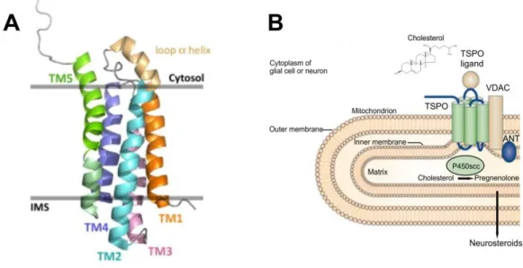
Both older hydropathy profile analysis (Joseph-Liauzun et al., 1998) as well as recently published nuclear-magnetic resonance (NMR) analysis (Jaremko et al., 2014) show that TSPO has a five transmembrane α -helix structure (Figure 5, A).

Figure 5: TSPO structure and mitochondrial localization. (A) Structure of TSPO in the outer mitochondrial membrane (side view) showing the five a-helix transmembrane structure (TM1 – 5). IMS, intermembrane space.

(B) TSPO is localized in the outer mitochondrial membrane where it forms a complex with the voltage-dependent anion channel (VDAC) and the adenine nucleotide transporter (ANT). TSPO mediates the transition of cholesterol from the inner to the outer mitochondrial membrane. Cholesterin-monooxygenase P450 scc converts cholesterol into pregnenolone, a precursor for several neurosteroids. A is modified from (Selvaraj and Stocco, 2015), B is modified from (Rupprecht et al., 2010), Cholesterol structure modified from (Palmer, 2004)

Protein interaction studies proposed that TSPO forms a complex with other proteins

of the inner and outer mitochondrial membrane such as the voltage-dependent anion

channel (VDAC) and the adenine nucleotide transporter (ANT) (Garnier et al., 1994,

(27)

25 Veenman et al., 2008). TSPO, as part of this complex reaching from the inner to the outer mitochondrial membrane, is thought to mediate the transport of lipophilic substances such as cholesterol across the intermembrane space (McEnery et al., 1992, Papadopoulos et al., 2006). At the inner mitochondrial membrane the cholesterol side-chain cleaving enzyme cytochrome P450 (P450 scc) converts cholesterol to pregnenolone, a precursor for several neurosteroids (Figure 5, B) (Veenman et al., 2007, Papadopoulos et al., 2007). TSPO is thought to be involved in this crucial a rate-limiting step in steroidogenesis (Lacapere and Papadopoulos, 2003, Papadopoulos et al., 2006, Papadopoulos et al., 2007). In line with this role, TSPO ligands were reported to stimulate production of steroid hormones and neurosteroids in steroidogenic cells and isolated mitochondria (Mukhin et al., 1989, Papadopoulos et al., 1990). Additionally, knocking out TSPO or blocking TSPO with antisense oligodeoxynucleotides (ODNs) suppresses steroid production in Leydig tumor cells (Papadopoulos et al., 1997a, Hauet et al., 2005). Other described functions for TSPO include mitochondrial respiration (Hirsch et al., 1989, Gavish et al., 1999), formation of the mitochondrial permeability transition pore (MPTP) (Zamzami and Kroemer, 2001), cell proliferation (Corsi et al., 2008) and apoptosis (Veenman et al., 2008). Investigations with new established CRISPR/Cas9-mediated TSPO knock-out steroidogenic MA-10 Leydig cells, furthermore propose a role for TSPO in regulation of mitochondrial energy metabolism by modulation of fatty acid oxidation (FAO) (Tu et al., 2016). Of note is that many of the functions were detected in experiments using TSPO ligands.

Two separate studies have however shown that a global TSPO knockout is not lethal as was previosly thought, with the absence of TSPO having no impact on steroidogenesis. This raised a lot of doubt and controversy to the functions ascribed to TSPO (Papadopoulos et al., 1997b, Tu et al., 2014a, Banati et al., 2014).

Stimulation of TSPO-knockout in MA-10 mouse Leydig tumor cells with TSPO ligand

PK11195 also revealed that enhancement of steroid production is TSPO independent

(Tu et al., 2014b). Leading to further speculation that some of the observed effects of

TSPO ligands on steroidogenesis are just side effects and not TSPO specific

(Selvaraj and Stocco, 2015). There is also conflicting evidence on the role of TSPO

as regulator of the mitochondrial permeability transition pore (MPTP), a function

associated with cell death induction. More recently published data propose that the

(28)

Introduction

26 complex of TSPO, VDAC and ANT is not involved in this process (Baines et al., 2007, Kokoszka et al., 2004, Selvaraj and Stocco, 2015).

Independent of the discussion about TSPO function several studies show a strong upregulation of TSPO during different neuropathologies (Rupprecht et al., 2010). In humans, TSPO upregulation is observed in neurological disorders like Alzheimer’s disease (Edison et al., 2008, Yasuno et al., 2008), Multiple sclerosis (Banati et al., 2000, Versijpt et al., 2005) , Parkinson’s disease (Gerhard et al., 2006) Amyotrophic lateral sclerosis (Turner et al., 2004), dementia (Cagnin et al., 2004), and stroke (Gerhard et al., 2005). This is why TSPO serves as a biomarker for brain damage and neurodegeneration. Radio labeled TSPO-ligands are used to detect TSPO expression by positon emission tomography (PET) or single photon emission computed tomography (SPECT), allowing to localize damaged areas or active disease processes (Vivash and O'Brien, 2016, Chauveau et al., 2008, Chen and Guilarte, 2008). However, human studies with healthy volunteers revealed variations in the binding affinity of TSPO ligands, so that results of TSPO density analysis have to be interpreted carefully (Owen et al., 2011b).

Besides their use as diagnostic tools to visualize TSPO expression, TSPO ligands have been shown to be potent in resolving neuroinflammation and neurodegeneration in various disease models (Rupprecht et al., 2010). Endogenous ligands for TSPO include cholesterol (Li et al., 2001), porphyrins (Verma et al., 1987), diazem-binding inhibitor (DBI) and its proteolytic products: octadecaneuropeptide (ODN) and triakontatetraneuropeptide (TTN) (Costa and Guidotti, 1991, Mocchetti and Santi, 1991). The production of DBI-derived ligands is upregulated in response to peripheral nerve injury, suggesting a role for TSPO and its endogenous ligands in nerve regeneration (Lacor et al., 1999). In the retina, DBI-derived ligands produced by astrocytes and Müller cells, were shown to negatively regulate microglial reactivity, by suppression of reactive-oxygen species (ROS) production, TNF- α expression and blockage of microglial proliferation. This suggests DBI-TSPO signaling in the retina to be important for macroglia-microglial interaction and essential for returning activated microglial into a quiescent state after an inflammatory response (Wang et al., 2014).

Synthetic TSPO ligands were primarily developed as neuroimaging agents.

Nevertheless classical TSPO ligands such as PK11195 or Ro-5 4864 as well as more

recently developed ligands have been reported to exert anti-inflammatory (Ryu et al.,

(29)

27 2005) , neuroprotective (Bordet et al., 2007, Veiga et al., 2005), immune suppressive (Daugherty et al., 2013), regenerative (Girard et al., 2012) and anti-anxiety activity (Rupprecht et al., 2009, Rupprecht et al., 2010). For example, treatment with PK 11195 reduced pro-inflammatory cytokine production, microglial reactivity and neuronal death in a model of quinolinic acid-induced brain lesion (Ryu et al., 2005).

Similarly administration of Ro-5 4864 prevented neuron loss in two independent models of brain damage (Veiga et al., 2005, Soustiel et al., 2008). Another TSPO ligand, Etifoxine was shown to be protective in an experimental murine model for multiple sclerosis (MS). Etifoxine improved recovery from MS symptoms was accompanied by clearly reduced immune cell infiltration in the spinal cord and improved oligodendroglial regeneration (Daugherty et al., 2013). In an experimental model for amytrophic lateral sclerosis (ALS), characterized by death of cortical and spinal cord motor neurons, treatment with the TSPO ligand olesoxime (TRO 19622) reduced neuronal cell death and promoted regeneration (Bordet et al., 2007).

Furthermore in psychiatric disorders, the TSPO ligand, XBD173 (AC-5216, Emapunil), diminished experimentally induced panic attacks in rodents and exerted antipanic capacity in human volunteers (Rupprecht et al., 2009).

However, how the different TSPO ligands exert neuroprotection and other beneficial

effects remains largely unresolved. Possible mechanisms include the modulation of

microglial cells, hence glial cells upregulate TSPO in response to pathological

alterations and TSPO ligands were shown to reduce microgliosis (Rupprecht et al.,

2010). In addition, beneficial effects of TSPO ligands have been associated with

changes in neurosteroid production (Giatti et al., 2009, Barron et al., 2013, Selvaraj

and Stocco, 2015). However, considering recent findings which challenge TSPO’s

role in steroidogenesis further approaches are needed to elucidate the exact function

of TSPO and how TSPO ligands work to mediate therapeutic effects.

(30)

Introduction

28

3.5 Aims of the thesis

There is increasing evidence that microglial reactivity actively contributes to the development and perturbation of retinal degenerations. Observations in different brain pathologies revealed furthermore an upregulation of TSPO in activated microglial cells. Moreover ligands for TSPO were effective in resolving degenerative CNS pathologies. Thus, attenuation of chronic microglial activation via TSPO ligands might represent a promising approach to diminish degenerative processes in the retina.

Therefore the study aimed to elucidate the role of TSPO as potential biomarker for retinal microgliosis and possible therapeutic target for the modulation of microglial cells. A comprehensive in vitro approach, testing key microglial functions, was performed to determine the capacity of TSPO ligand, XBD173 to interfere with microglial reactivity and resultant neurotoxicity in vitro. A further question addressed in this thesis was whether XBD173 might modulate microglial reactivity in vivo and thereby reduce light induced retinal degeneration.

A further goal of the thesis was to determine whether minocycline, an antibiotic with

additional anti-inflammatory properties is able to reduce microglial neurotoxicity and

to protect the retina from degeneration. Therefore a model of bright white light-

induced retinal degeneration, mimicking features of inherited degenerations such as

microgliosis and death of photoreceptors, was used to assess the immunomodulatory

and neuroprotective effects of minocycline during retinal degeneration.

(31)

29

4 Results

4.1 Translocator protein (18 kDa) (TSPO) is expressed in reactive retinal microglia and modulates microglial inflammation and phagocytosis

Marcus Karlstetter, Caroline Nothdurfter, Alexander Aslanidis, Katharina Moeller, Felicitas Horn, Rebecca Scholz, Harald Neumann, Bernhard H F Weber, Rainer Rupprecht* and Thomas Langmann*

Background: The translocator protein (18 kDa) (TSPO) is a mitochondrial protein expressed on reactive glial cells and a biomarker for gliosis in the brain. TSPO ligands have been shown to reduce neuroinflammation in several mouse models of neurodegeneration. Here, we analyzed TSPO expression in mouse and human retinal microglia and studied the effects of the TSPO ligand XBD173 on microglial functions.

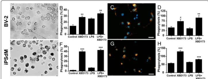
Methods: TSPO protein analyses were performed in retinoschisin-deficient mouse retinas and human retinas. Lipopolysaccharide (LPS)-challenged BV-2 microglial cells were treated with XBD173 and TSPO shRNAs in vitro and pro-inflammatory markers were determined by qRT-PCR. The migration potential of microglia was determined with wound healing assays and the proliferation was studied with Fluorescence Activated Cell Sorting (FACS) analysis. Microglial neurotoxicity was estimated by nitrite measurement and quantification of caspase 3/7 levels in 661 W photoreceptors cultured in the presence of microglia-conditioned medium. The effects of XBD173 on filopodia formation and phagocytosis were analyzed in BV-2 cells and human induced pluripotent stem (iPS) cell-derived microglia (iPSdM). The morphology of microglia was quantified in mouse retinal explants treated with XBD173.

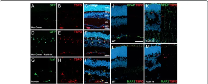
Results: TSPO was strongly up-regulated in microglial cells of the dystrophic mouse

retina and also co-localized with microglia in human retinas. Constitutive TSPO

expression was high in the early postnatal Day 3 mouse retina and declined to low

levels in the adult tissue. TSPO mRNA and protein were also strongly induced in

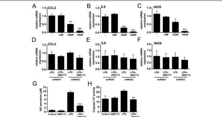
LPS-challenged BV-2 microglia while the TSPO ligand XBD173 efficiently

suppressed transcription of the pro-inflammatory marker genes chemokine (C-C

motif) ligand 2 (CCL2), interleukin 6 (IL6) and inducible nitric oxide (NO)-synthase

Abbildung

Figure  1:  Schematic  overview  of  the  mammalian  eye  and  its  retinal  structure
Figure 2: Microglial retinal location and appearance during health and disease. (A) In the healthy retina Iba1  positive  ramified  microglial  cells  are  located  in  the  plexiform  layers
Figure 3: Microglial morphological plasticity. Under homeostatic conditions microglial cells have a small cell  body  and  scan  their  environment  with  their  long-branched  protrusions
Figure  4:  Schematic  representation  of  microglial  action  during  retinal  degenerations
+7

Referenzen

ÄHNLICHE DOKUMENTE

Previous studies have shown that microglia can promote tumor growth and invasion through various molecular and cellular mechanisms, one of which includes MT1-MMP

On the other hand, during tumor growth or therapies, different endogenous TLR ligands could be released by dying cancer cells or necrotic tissue to activate TLRs, this moderate

elegans counterparts in terms of sequence (let-7) or highly similar in terms of domain architecture (lin-28 and lin- 41), they are even interconnected in a conserved pathway

Even though the cell ultrastructure was preserved differently depending on the fixation method used, we were able to confirm our previous

Materials and Methods: Stem cell marker expression of CD133, CD44, C-KIT, CD34, ITGA6, OCT4, DDX5 and MELK in canine prostate carcinomas and prostate cyst cell lines were

Microglial cells expressing GFP (green) in the deep retinal layer, vessels are stained with lectin (red). bar =

Based on the quantitative analysis of the spatial growth distribution, and the specific transcript accumulation of FpXET1 in young elongating tissue, it is proposed that FpXET1 is

Besides the contribution of mast cells to allergic inflammation [132], they also seem to be involved in non-allergic inflammatory diseases of the gut, since in the intestine of