Cicatricial keratoconjunctivitis associated with lichen planus
Abstract
Purpose:To describe a case of cicatricial keratoconjunctivitis associated with lichen planus.
Susan Huang
1Prabjot Channa
1Methods:Case report.
Results:To our knowledge, this is the sixth reported case of cicatricial
keratoconjunctivitis associated with lichen planus. A 73-year-old woman 1 Department of Ophthalmology, Albert had persistent cicatricial keratoconjunctivitis. Histopathologic studies
of the buccal mucosa biopsy specimen revealed lichen planus. Einstein College of Medicine, Montefiore Medical Center, Bronx, NY, USA
Conclusion:Lichen planus is a possible cause of cicatricial keratocon- junctivitis. Topical cyclosporine may stabilize the ocular surface, and additional systemic immunosuppression may be needed in severe cases.
A correct diagnosis through biopsy is essential to start aggressive anti- inflammatory treatment to avoid vision loss.
Keywords:lichen planus, cicatricial keratoconjunctivitis, keratitis, cyclosporine
Introduction
Lichen planus (LP) is an autoimmune, inflammatory con- dition of unknown etiology that affects the skin and mu- cous membranes. Most commonly, the mouth and gen- italia mucous membranes are involved [1]. Conjunctival involvement is rare, and may become indistinguishable from other forms of cicatricial conjunctivitis due to severe scarring that results. Even more rarely reported is corneal involvement. We report a case of lichen planus keratocon- junctivitis.
Case report
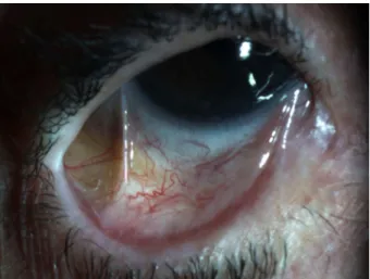
A 73-year-old Hispanic woman with a history of left eye enucleation secondary to endophthalmitis, history of dacryocystitis, and primary open-angle glaucoma presented with persistent right eye conjunctivitis and epithelial defect. On ophthalmic examination, her visual acuity was 20/50 in the right eye. The left eye had pros- thesis. Intraocular pressure was within normal limits. The right eyelid showed thickened, telangectatic lid margins, with upper trichiasis and distichiasis. The semilunar fold was keratinized. There was subepithelial fibrosis of the right superior palpebral conjunctiva (Figure 1) and symblepharon in the inferior medial palpebral conjunctiva (Figure 2). The bulbar conjunctiva had notable amount of conjunctival injection. The cornea had superior pannus with diffuse severe punctuate epithelial erosions.
Figure 1: Subepithelial fibrosis of the right superior palpebral conjunctiva
Figure 2: Foreshortening and symblepharon of the right lower palpebral conjunctiva
1/3 GMS Ophthalmology Cases 2015, Vol. 5, ISSN 2193-1496
Case Report
OPEN ACCESS
Flat-topped polygonal papules were noted on the patient’s forearm. The patient was referred to Oral Surgery to per- form buccal mucosal biopsy given the suspicion of the chronic conjunctivitis. A punch biopsy of the buccal mu- cosa was performed with histopathology confirming lichen planus, which was IgG-, IgA-, and C3-negative.
The patient was started on 0.05% cyclosporine eyedrop twice daily, very frequent lubrication with Artificial Tears, and Genteal Gel at bedtime. Later, 0.1% fluoromethalone acetate twice daily was added. Throughout the fourteen years that the patient was followed in our clinic, she had frequent epilation for her trichiasis. During this time course, she presented with several episodes of corneal abrasion and two episodes of corneal ulcer, which healed with topical antibiotic treatment. The cicatricial conjunctivi- tis remained grossly stable without further significant progression and with her best-corrected visual acuity minimally changed at 20/70 in her right eye. The option to start oral cyclosporine was discussed with the patient, but the patient deferred starting the medication due to the potential medication side effects and the very slow progression of her disease.
Comment
Lichen planus is an autoimmune papulosquamous dis- ease that affects the skin and mucous membranes. The etiology of LP is unclear, but is thought to be likely from T-cell mediated immunological response to an induced antigenic change in the basal membrane zone of mucosa or skin, triggering apoptosis of epithelial cells [1].
The cutaneous form of LP presents with a preference for the anterior aspect of the wrists and ankles and is char- acterized by violaceous, flat-topped polygonal papules with a superficial network of fine white lines [1]. Mucous membranes involvement may be associated with the cu- taneous form and is characterized as reticular, erosive whitish macules, most typically on the buccal mucosa, lips, and genitalia. This form of LP is usually self-limiting, with spontaneous remission after one to two years.
Whereas the second form of isolated mucosal LP may follow a chronic, unremitting course. Conjunctival involve- ment is rare, and usually presents as a chronic cicatricial conjunctivitis or keratoconjunctivitis [2]. Ocular involve- ment usually presents with other clinical manifestations of LP, but isolated conjunctival involvement has also been reported [3], [4], [5].
Histopathologic analysis of tissue samples, whether from conjunctiva or another body site, helps differentiate from other cicatricial conjunctivitis causes, such as mucous membrane pemphigoid, pemphigus vulgaris, Stevens- Johnson syndrome, graft-versus-host disease, linear IgA disease, and atopic keratoconjunctivitis, since they may be indistinguishable from clinical presentation alone [6], [7]. A definitive diagnosis is imperative because improper treatment would lead to progressive subepithelial fibrosis, trichiasis, entropion, corneal opacification, and keratocon- junctivitis sicca from chronic, persistent inflammation
leading to severe vision loss [8]. 8.3% of pseudopemphi- goid disorders with conjunctival involvement are actually caused by LP [6].
Lichen planus-associated keratitis has even more of a limited collection of published reports than conjunctival involvement, with only 5 previous reports to our know- ledge based on a PubMed search of the English literature [5], [8], [9], [10], [11]. It is unclear whether corneal in- volvement is a direct effect of lichen planus, secondary to the abnormal tear film from conjunctival cicatricial changes, or a combination of both [9]. In our patient, tri- chiasis and ocular surface toxicity from chronic use of glaucoma eye drops may have also played a role.
First-line treatment includes topical corticosteroid and cyclosporine. Aggressive use of preservative-free artificial tears is also important. If there is not a good response from topical treatments, systemic corticosteroids and immunosuppresants, such as cyclosporine, azathioprine, or mycophenolate mofetil can be used [3], [8], [12], [13], [14]. For severe disease state, amniotic membrane transplantation can be used [9].
Thus, our case reflects the need for accurate diagnosis through biopsy to distinguish this disease entity from other forms of cicatricial conjunctivitis or keratoconjuncti- vitis. A correct diagnosis is vital to start an aggressive anti-inflammatory treatment to avoid vision loss.
Notes
Patient consent
The patient gave informed consent for submission of this report to this journal.
Competing interests
The authors declare that they have no competing in- terests.
References
1. Sugerman PB, Savage NW, Walsh LJ, Zhao ZZ, Zhou XJ, Khan A, Seymour GJ, Bigby M. The pathogenesis of oral lichen planus.
Crit Rev Oral Biol Med. 2002;13(4):350-65. DOI:
10.1177/154411130201300405
2. Weisenthal RW, Streeten BW, Levin DL. Lichen planus. In: Mannis MJ, Macsai MS, Huntley AC, editors. Eye and skin disease.
Philadelphia: Lippincott-Raven Publishers; 1996. p. 328-32.
3. Thorne JE, Jabs DA, Nikolskaia OV, Mimouni D, Anhalt GJ, Nousari HC. Lichen planus and cicatrizing conjunctivitis: characterization of five cases. Am J Ophthalmol. 2003;136(2):239-43. DOI:
10.1016/S0002-9394(03)00147-8
4. Rozas Muñoz E, Martínez-Escala ME, Juanpere N, Armentia J, Pujol RM, Herrero-González JE. Isolated conjunctival lichen planus: a diagnostic challenge. Arch Dermatol. 2011 Apr;147(4):465-7. DOI: 10.1001/archdermatol.2011.68
2/3 GMS Ophthalmology Cases 2015, Vol. 5, ISSN 2193-1496
Huang et al.: Cicatricial keratoconjunctivitis associated with lichen ...
5. Brewer JD, Ekdawi NS, Torgerson RR, Camilleri MJ, Bruce AJ, Rogers RS 3rd, Maguire LJ. Lichen planus and cicatricial conjunctivitis: disease course and response to therapy of 11 patients. J Eur Acad Dermatol Venereol. 2011 Jan;25(1):100-4.
DOI: 10.1111/j.1468-3083.2010.03693.x
6. Thorne JE, Anhalt GJ, Jabs DA. Mucous membrane pemphigoid and pseudopemphigoid. Ophthalmology. 2004 Jan;111(1):45- 52. DOI: 10.1016/j.ophtha.2003.03.001
7. Camisa C, Meisler DM. Immunobullous diseases with ocular involvement. Dermatol Clin. 1992 Jul;10(3):555-70.
8. Hutnik CM, Probst LE, Burt WL, Hooper PL, Tokarewicz AC, Heathcote JG. Progressive, refractory keratoconjunctivitis associated with lichen planus. Can J Ophthalmol. 1995 Jun;30(4):211-4.
9. Rhee MK, Mootha VV. Bilateral keratoconjunctivitis associated with lichen planus. Cornea. 2004 Jan;23(1):100-5. DOI:
10.1097/00003226-200401000-00019
10. Luhr AF. Lichen planus of the conjunctiva. Am J Ophthalmol.
1924;7(6):456-7. DOI: 10.1016/S0002-9394(24)91142-1 11. Goldsmith J. Deep keratitis associated with atypical lichen planus;
report of a case. Arch Ophthal. 1948 Aug;40(2):138-46. DOI:
10.1001/archopht.1948.00900030143004
12. Neumann R, Dutt CJ, Foster CS. Immunohistopathologic features and therapy of conjunctival lichen planus. Am J Ophthalmol.
1993 Apr 15;115(4):494-500. DOI: 10.1016/S0002- 9394(14)74452-6
13. Pakravan M, Klesert TR, Akpek EK. Isolated lichen planus of the conjunctiva. Br J Ophthalmol. 2006 Oct;90(10):1325-6. DOI:
10.1136/bjo.2006.096263
14. McNab AA. Lacrimal canalicular obstruction in lichen planus.
Orbit. 1998 Sep;17(3):201-2. DOI: 10.1076/orbi.17.3.201.2744
Corresponding author:
Susan Huang, MD
Montefiore Medical Center, Department of Ophthalmology, 3332 Rochambeau Avenue, 3rd floor, Room 306, Bronx, NY 10467, USA, Phone: (510) 828-6562
Susan.huang16@gmail.com
Please cite as
Huang S, Channa P. Cicatricial keratoconjunctivitis associated with lichen planus. GMS Ophthalmol Cases. 2015;5:Doc08.
DOI: 10.3205/oc000030, URN: urn:nbn:de:0183-oc0000305
This article is freely available from
http://www.egms.de/en/journals/oc/2015-5/oc000030.shtml Published:2015-10-07
Copyright
©2015 Huang et al. This is an Open Access article distributed under the terms of the Creative Commons Attribution 4.0 License. See license information at http://creativecommons.org/licenses/by/4.0/.
3/3 GMS Ophthalmology Cases 2015, Vol. 5, ISSN 2193-1496
Huang et al.: Cicatricial keratoconjunctivitis associated with lichen ...