DNA barcoding of the German green supralittoral zone indicates the distribution and phenotypic plasticity of Blidingia species and reveals Blidingia cornuta sp. nov.
Sophie Steinhagen,1,2 Luisa Düsedau1 & Florian Weinberger1
1 GEOMAR Helmholtz Centre for Ocean Research Kiel, Marine Ecology Department, Düsternbrooker Weg 20, 24105 Kiel, Germany 2 Department of Marine Sciences-Tjärnö, University of Gothenburg, 452 96 Strömstad, Sweden
Address for correspondence:Sophie Steinhagen, sophie.steinhagen@gu.se DOI https://doi.org/10.1002/tax.12445
Abstract In temperate and subarctic regions of the Northern Hemisphere, green algae of the genusBlidingiaare a substantial and environment-shaping component of the upper and mid-supralittoral zones. However, taxonomic knowledge on these important green algae is still sparse. In the present study, the molecular diversity and distribution ofBlidingiaspecies in the German State of Schleswig-Holstein was examined for the first time, including Baltic Sea and Wadden Sea coasts and the off-shore island of Helgo- land (Heligoland). In total, three entities were delimited by DNA barcoding, and their respective distributions were verified (in decreasing order of abundance:Blidingia marginata,Blidingia cornutasp. nov. andBlidingia minima). Our molecular data revealed strong taxonomic discrepancies with historical species concepts, which were mainly based on morphological and ontogenetic char- acters. Using a combination of molecular, morphological and ontogenetic approaches, we were able to disentangle previous mis- identifications ofB.minimaand demonstrate that the distribution ofB.minimais more restricted than expected within the examined area.Blidingia minima, the type of the genus nameBlidingia, is epitypified within this study by material collected at the type locality Helgoland. In contrast withB.minima,B.marginatashows a higher phenotypic plasticity and is more widely distributed in the study area than previously assumed. The third entity,Blidingia cornutasp. nov., is clearly delimited from other describedBlidingiaspecies, due to unique characters in its ontogenetic development and morphology as well as by itstufAandrbcLsequences.
Keywords Baltic Sea; barcoding; Helgoland; Heligoland; phylogeography; ontogeny;tufA
Supporting Information may be found online in the Supporting Information section at the end of the article.
■INTRODUCTION
Green algae of the genusBlidingiaKylin are a substantial component of the upper and mid-supralittoral zones of the Northern Hemisphere, where they can be found as dense mats on various natural and artificial surfaces and additionally as epi- phytes on other macrophytobenthic organisms.Blidingiaspp.
can withstand extreme conditions such as periodic and sus- tained desiccation, heat waves or frost periods with snow cover, and marine flooding as well as extended freshwater immersion.
Thus, representatives of the genusBlidingiaoften indicate the transition between the marine or estuarine zone and the terres- trial zone.Blidingiaspecies shape these particular environments and give them a unique and important ecological character by providing habitats for small invertebrates. However, in-depth understanding of the genetic species diversity within the genus and hence about geographic species distribution is still sparse. The recognition ofBlidingiaspp. is largely based on
morphological characters of mature thalli and ontogenetic stages, while molecular knowledge is limited.
Kylin (1949) proposed the new genusBlidingiaforEn- teromorpha minimaNägeli ex Kütz., based on observations made by Bliding (1938). One of the main characteristics that distinguishesBlidingiafrom the closely related and morpho- logically similar genusEnteromorphaLink (nowadaysUlvaL.) is its small cells, with a diameter less than 10μm (Kylin, 1949).
Both genera include species with monostromatic, tubular thalli; however, their ontogenetic development was also found to differ significantly (Kylin, 1949). The motile swarmers of Blidingiahave no eyespot, in contrast to the motile swarmers ofUlva. The settled Blidingiaswarmer grows into an elon- gated tube that incorporates the intracellular spore contents, and the empty spore sleeve is separated by a transverse mem- brane. A prostrate disc develops, and from the expanded cen- tre of this partly bi-layered disc, a monostromatic tube begins to emerge (Bliding, 1938; Kylin, 1949). While several studies
Article history:Received: 29 Apr 2020 | returned for (first) revision: 12 Aug 2020 | (last) revision received: 5 Nov 2020 | accepted: 10 Nov 2020 | published online: 27 Jan 2021 |Associate Editor:John Marinus Huisman | © 2021 The Authors.
TAXON published by John Wiley & Sons Ltd on behalf of International Association for Plant Taxonomy.
This is an open access article under the terms of the Creative Commons Attribution License, which permits use, distribution and reproduction in any medium, provided the original work is properly cited.
supported these ontogenetic findings (Dangeard, 1961; Gayral, 1967), others emphasized variations in the ontogenetic deve- lopment between differentBlidingia species (Kornmann &
Sahling, 1978; Tatewaki & Iima, 1984; Iima, 1989), particularly during spore sleeve formation (Bliding, 1963; Kornmann &
Sahling, 1978). Bliding (1963) stated,“the germinating tube of the swarmer is mostly divided in a basal empty-cell and an upper cell containing all the cytoplasm”, suggesting that differences in spore sleeve formation have been observed.
Detailed observations of early ontogenetic developmental pat- terns on spore sleeve formation were made by Kornmann
& Sahling (1978), who focused on the differentBlidingiaspe- cies from Helgoland (Heligoland). Whereas half of the obser- ved entities (B.minima(Nägeli ex Kütz.) Kylin,B.chadefaudii (Feldmann) Bliding) cut off an empty spore sleeve (called embryospore), this pattern was not observed forB.subsalsa (Kjellm.) Kornmann & Sahling ex Scagel & al. andB.mar- ginata(J.Agardh) P.J.L.Dang. ex Bliding. Thus, it was sug- gested that early ontogenetic stages were suitable criteria for species delimitation (Kornmann & Sahling, 1978).
Three different ontogenetic developmental patterns of prostrate discs were observed in studies focused on the sexual reproduction and early development ofBlidinga minimafrom Japan (Tatewaki & Iima, 1984; Iima, 1989). The authors distin- guished compact discs with short cells (D-type), open discs with longer cells (F-type) and an intermediate disc type (M- type) (Iima, 1989). Notably, European specimens that exhib- ited thallus discs similar to the F-type were assigned to a newly differentiated species, namelyB.chadefaudii(Bliding, 1938; Chadefaud, 1957; Kornmann & Sahling, 1978). How- ever, due to the interfertility of specimens forming the different disc-types identified by Iima (1989), it was suggested that B.chadefaudiishould be considered as a variant ofB.minima (Woolcott & al., 2000). Those authors undertook a molecular reassessment of the taxonomic affiliation of individuals ex- hibiting different disc-morphotypes based on nuclear rDNA ITS sequences. By showing that the allegedly ontogenetic cri- teria that were used for species delimitation encompass an integrated continuum of variation, together with the fact that molecular analysis and interbreeding-experiments did not de- monstrate a species boundary between JapaneseB.minimaand B.chadefaudii, the authors concluded that the two taxa are conspecific. Thus, the observations of Woolcott & al. (2000) blur the boundary betweenB.minimaandB.chadefaudiiand underline that morphological characters within most Ulvales are highly variable and that molecular sequencing is needed to delimit species boundaries.
Currently, sixBlidingia species are accepted taxonomi- cally: B. chadefaudii, B.dawsonii (Hollenb. & I.A.Abbott) S.C.Lindstr. & al.,B.marginata,B.minima,B.subsalsa,B.tuber- culosa(P.J.L.Dang.) Benhissoune & al. (Guiry & Guiry, 2020).
However, molecular knowledge of these species is sparse and most have primarily been distinguished by morphological and ontogenetic traits.
The present study aimed to examine the diversity ofBlidin- giaspecies in the northern German State of Schleswig-Holstein.
Schleswig-Holstein’s coastline is of limited length, but is struc- turally very diverse, including a south-east section of the fully marine North Sea and a south-west section of the brackish Bal- tic Sea, as well as various estuaries and lagoons. The area also includes the North Sea island of Helgoland, the focus of phyco- logical research in the mid-19th century (Reinke, 1889) and among the best-studied seaweed habitats in Europe (Bartsch &
Kuhlenkamp, 2000). Long-term observations of the benthic flora of Helgoland have shown that the establishment of artificial substrata resulted in an increased abundance ofBlidingiaspp.
(Bartsch & Kuhlenkamp, 2000). Subsequent to C.W. Nägeli collecting the holotype ofB.minimaon Helgoland around the mid-19th century (historically:Enteromorpha minima, suppl.
Fig. S1), the first recordings of otherBlidingiaspecies for the island were made by Wollny (1881). More recently, Kornmann
& Sahling (1978) documented theBlidingiaspecies of Helgo- land in a detailed synopsis, focusing on their developmental dif- ferences. The study emphasized the presence of fourBlidingia species (B.chadefaudii,B.marginata,B.minima,B.subsalsa).
The authors observed several–and often vague–micro-morpho- logical differences between the four species, but also one com- monly shared macro-morphological character: All species were characterized as unbranched tubes (Kornmann & Sahling, 1978).
According to species inventories, only B. minima and B.marginatahave been recorded from Schleswig-Holstein’s mainland coasts (Schories & al., 2009). However, due to the aforementioned taxonomic difficulties, historical records of Blidingiaspecies require confirmation, and the first molecular assessment of the distribution of Ulvales and Ulotrichales in the region (Steinhagen & al., 2019a,b) has indicated that the present distribution of Blidingia spp. may not accord with recent inventories. However, there has not been a molecular characterization of the northern EuropeanBlidingia species or a review of their allegedly significant identification criteria undertaken until now.
A crucial point for molecular studies using DNA barcod- ing is always the choice of suitable marker genes that are on the one hand side variable enough to delimit species but also stable within the respective species group. A marker gene that has been widely used within molecular studies of green algae is the coding gene for the ribulose-bisphosphate car- boxylase large subunitrbcL(Saunders & Kucera, 2010). Later, the plastid encodedtufAgene was promoted as the most suit- able marker for delimitation of green algae, providing the high- est amplification success and the largest barcode gaps for green macroalgae (Saunders & Kucera, 2010). Genetic data- bases such as GenBank include numerous sequences for both markers. Reference sequences, however, need to be selected with caution, as type specimens of Ulvales have rarely been successfully sequenced.
By combiningtufAandrbcLsequencing with morpholog- ical and ontogenetic observations, we have assessed the diver- sity ofBlidingiaspecies within Schleswig-Holstein and have recognized an undescribed new species that is relatively abun- dant in the area. In addition, our study highlights past difficul- ties in distinguishingBlidingiaspecies.
■MATERIALS AND METHODS
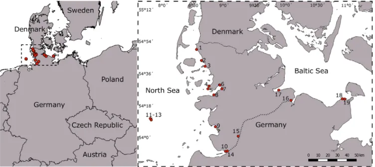
Field collection and sample preparation.—Sites along the North Sea and Baltic Sea coasts of Schleswig-Holstein– including the island of Helgoland–were repeatedly visited in the years 2014–2017 (see also Steinhagen & al., 2019a). In 2016, sampling also covered the heavily trafficked Kiel Canal, which connects both sea areas (Fig. 1, sites 14–16, see also Steinhagen & al., 2019b). Upper littoral and supralittoral zones were checked for macroalgal growth with a focus on freshwater inflows (e.g., drainages, river inflows, beach showers). Several sites were re-visited in the years 2018 and 2019, to verify the presence of populations and obtain material for cultivation (Table 1). Altogether,Blidingiaspp. were observed at 19 of the visited sites (Fig. 1; Table 1).
Specimens were collected, placed into sealed plastic bags, and stored on ice until further processing in the lab. Most sam- ples were preserved as herbarium vouchers (see Table 1) and lodged in the Natural History Museum Denmark, Copenha- gen (C). A subsample was divided, with part of it stored at 4C or−20C for subsequent morphological observation and part of it stored in a microreaction tube at−80C for genomic DNA extraction. Light microscopy observations were under- taken, and microscopic documentation was carried out on the remaining part, using a digital camera (Nikon DS-Vil) attached to a light microscope (Nikon Eclipse TS 100). Salin- ity was measured using a WTW portable conductivity meter (Xylem Analytics, Weilheim, Germany).
In addition to the field collected samples, herbarium spec- imens in the Herbarium of the Helgoland Biological Station of the Alfred Wegener Institute (BRM) were included in our ana- lyses (barcode numbers: BRM007967 and BRM008079; see also Table 1). Several additional herbarium specimens were
investigated, including the type specimen ofB.minima(Natu- ralis Biodiversity Center, Leiden, Netherlands [L], barcode L 0054691; suppl. Fig. S1). However, they yielded insufficient amounts of DNA due to the vouchers being pre-treated with fixation solutions, or the amplification of marker genes was impossible due to impurities of the herbarium vouchers (dia- toms, multi-algal vouchers, etc.). Thus, these herbarium vou- chers were excluded from the molecular analyses of this study.
DNA extraction, amplification and sequencing. — Genomic DNA was isolated from the lyophilized algal tissue with an Invisorb Spin Plant Mini Kit (Stratec, Birkenfeld, Ger- many) following the manufacturer’s protocol. Extracted DNA was stored at−80C and used for amplification of therbcL andtufAgenes. PCR amplifications of therbcLgene used the primer pairs rbcLstart and R750, as well as F650 and rbcLend (Shimada & al., 2003). The PCR reactions were performed as follows: 94C for 1 min; 35 cycles at 94C for 30 s, at 56.3C for 30 s, and at 72C for 1 min; and a final extension step at 72C for 7 min. PCR amplification of thetufAgene followed the detailed description of Steinhagen & al. (2019a). PCR prod- ucts were purified using the QUIAquick PCR Purification Kit (Quiagen, Hilden, Germany). Subsequent sequencing of the purified amplicons was provided by GATC Biotech (Konstanz, Germany). Forward and reverse sequence reads were assem- bled to produce contigs in Sequencher (v.4.1.4, GeneCodes, Ann Arbor, Michigan, U.S.A.), and a multiple sequence align- ment was constructed for each gene region using MAFFT v.7.402 (Katoh & al., 2002) (for Alignments, see supplemen- tary Appendices S1 and S2). Sequences obtained in this study are publicly available in GenBank (for accession numbers, see Table 1).
Phylogenetic analyses.—RbcLandtufAsequences were analysed in separate datasets. Newly generated sequences
Fig. 1.Sites of theBlidingiasamples in northern Germany processed in this study. Overview map about northern Germany with numbered sampling sites at the Wadden Sea (no. 1–10), on Helgoland (no. 11–13), within the Kiel Canal (no. 14–16) and in the Baltic Sea (no. 17–19).
Table1.ListofBlidingiasamplescollectedandgeneticallyassessedin2014–2017innorthernGermany.Additionally,theherbarium(BRM)specimensincludedinthisstudyarelisted. SpeciesSampleIDCountryRegionLat/LongStation Fig.1DateCollectorVoucher
tufA accession no.
rbcL accession no. Blidingia marginataS_129Germany:Schleswig- Holstein,SchluettsielWaddenSeaN54.6813333 E8.7544167330Jul2014S.SteinhagenC-A-99366MH538542– Blidingia marginataS_147_AGermany:Schleswig- Holstein,PellwormWaddenSeaN54.49882 E8.8087431Jul2014S.SteinhagenC-A-99378MH475464MN258044 Blidingia marginataS_147_BGermany:Schleswig- Holstein,SchobuellWaddenSeaN54.5078167 E8.9955667631Jul2014S.SteinhagenC-A-99379MH538543– Blidingia marginataS_156Germany:Schleswig- Holstein,WoehrdenWaddenSeaN54.1173167 E8.9359333905Aug2014S.SteinhagenC-A-99382MH538548– Blidingia marginataS_327Germany:Schleswig- Holstein,HeiligenhafenBalticSeaN54.3765333 E10.98006671925Aug2014S.Steinhagen–MH538549– Blidingia marginataS_474Germany:Schleswig- Holstein,SchluettsielWaddenSeaN54.68435 E8.75385312Sep2014S.SteinhagenC-A-99458MH538546– Blidingia marginataS_577Germany:Schleswig- Holstein,Brunsbuettel estuary
WaddenSeaN53.889 E9.1011331014Apr2015S.Steinhagen–MH475465MN258045 Blidingia marginataS_661Germany:Schleswig- Holstein,NordstrandWaddenSeaN54.4707167 E8.8068333521Apr2015S.Steinhagen–MH538544– Blidingia marginataS_708Germany:HelgolandHelgolandN54.1780333 E7.88871671123Apr2015S.Steinhagen–MH538545– Blidingia marginataS_737Germany:HelgolandHelgolandN54.1825 E7.89061671224Apr2015S.SteinhagenC-A-99417MH538547– Blidingia marginataS_911Germany:Schleswig- Holstein,AschauBalticSeaN54.4608 E9.926651726Aug2017S.Steinhagen–MN258036MN258046 Blidingia marginataS_930Germany:Schleswig- Holstein,SchobuellWaddenSeaN54.50782 E8.995567727Aug2017S.SteinhagenC-A-99681MN258037MN258047 Blidingia marginataS_941Germany:Schleswig- Holstein,FinkhaushalligWaddenSeaN54.41558 E8.903633829Aug2017S.Steinhagen–MN258038MN258048 Blidingia marginataS_944Germany:Schleswig- Holstein,FinkhaushalligWaddenSeaN54.41558 E8.903633829Aug2017S.Steinhagen–MN258039– Blidingia sp.1–Germany:Schleswig- Holstein,KielCanalKielCanalN54.031933 E9.30016715May2016S.Steinhagen–MG797655– Blidingia sp.1–Germany:Schleswig- Holstein,KielCanalKielCanalN54.369100 E10.12491716May2016S.Steinhagen–MG797656– Blidingia sp.1–Germany:Schleswig- Holstein,KielCanalKielCanalN53.888778 E9.11656714May2016S.Steinhagen–MG797657– Blidingia sp.1S_21Germany:HelgolandHelgolandN54.1825 E7.8906171223Jul2014S.SteinhagenC-A-99667MH538693– (Continues)
Table1.Continued. SpeciesSampleIDCountryRegionLat/LongStation Fig.1DateCollectorVoucher
tufA accession no.
rbcL accession no. Blidingia sp.1S_93Germany:Schleswig- Holstein,AschauWaddenSeaN54.4608 E9.926651724Jul2014S.Steinhagen–MH538691– Blidingia sp.1S_179Germany:Schleswig- Holstein,Brunsbuettel estuary
WaddenSeaN53.889 E9.1011331006Aug2014S.SteinhagenC-A-99682MH475459MN258049 Blidingia sp.1S_578Germany:Schleswig- Holstein,Brunsbuettel estuary
WaddenSeaN53.888778 E9.1165671414May2015S.Steinhagen–MN258043– Blidingia sp.1S_622Germany:Schleswig- Holstein,HeiligenhafenBalticSeaN54.3787167 E10.955451816Apr2015S.Steinhagen–MH538692– Blidingia sp.1S_813Germany:Schleswig- Holstein,Friedrich- Wilhelm-Luebke-Koog
WaddenSeaN54.83735 E8.6122124Jul2017S.SteinhagenC-A-99677MH475458MN258050 Blidingia sp.1S_815Germany:Schleswig- Holstein,FinkhaushalligWaddenSeaN54.41558 E8.903633824Jul2017S.SteinhagenC-A-99678MH475457MN258051 Blidingia sp.1S_818Germany:Schleswig- Holstein,HusumWaddenSeaN54.47113 E9.027917724Jul2017S.SteinhagenC-A-99679MH475456– Blidingia sp.1S_828Germany:Schleswig- Holstein,SchobuellWaddenSeaN54.50782 E8.995567624Jul2017S.SteinhagenC-A-99680MH475455MN258052 Blidingia sp.2Hel_59Germany:HelgolandHelgoland–ca.11–1325Jul1977P.-H.SahlingBRM008079MT076212– Blidingia sp.2S_1Germany:HelgolandHelgolandN54.18367 E7.8886331322Jul2014S.SteinhagenC-A-99660MH475461– Blidingia sp.2S_34Germany:HelgolandHelgolandN54.18367 E7.8886331323Jul2014S.SteinhagenC-A-99668MH475460MN258053 Blidingia sp.2S_39Germany:HelgolandHelgolandN54.1825 E7.8906171223Jul2014S.SteinhagenC-A-99671MH475462MN258054 Blidingia sp.2S_124Germany:Schleswig- Holstein,DagebuellWaddenSeaN54.73007 E8.689167230Jul2014S.SteinhagenC-A-99663MH475463MN258055 Blidingia sp.2S_949Germany:Schleswig- Holstein,NordstrandWaddenSeaN54.4707167 E8.8068333529Aug2017S.Steinhagen–MN258040– Blidingia sp.2S_951Germany:Schleswig- Holstein,NordstrandWaddenSeaN54.4707167 E8.8068333529Aug2017S.Steinhagen–MN258041MN258056 Blidingia sp.2S_950Germany:Schleswig- Holstein,NordstrandWaddenSeaN54.4707167 E8.8068333529Aug2017S.Steinhagen–MN258042– Ulva intestinalisHel_27Germany:HelgolandHelgoland–ca.11–1329Jul1997I.BartschBRM007967MT076211– Specimensusedinphylogeneticanalysisareindicatedbyaccessionnumbersinbold;however,allindividualswereincludedinthecalculationofintra-andinterspecificgeneticdivergencevalues.
were aligned with reference sequences downloaded from GenBank and used for further phylogenetic analysis. Particu- larly, sequences of specimens of the generaUlva L.,Korn- mannia(Kjellm.) Bliding andMonostromaThuret were inclu- ded to assess relationships with closely related taxa, whereas sequences ofProtomonostroma undulatum(Wittr.) K.L.Vinogr.
(rbcL: HQ603387;tufA: HQ619275, MH475501) were cho- sen as outgroups. Preference for reference sequence selection was given to peer-reviewed sequences. The models that best fit our data were found under the Akaike information criterion by employing MrModeltest v.2.2. (Nylander, 2004). For both datasets, the optimal substitution model was determined and found to be GTR+Γ+I. Maximum likelihood (ML) analyses were then carried out using RAxML v.8 (Stamatakis, 2014), employing the chosen substitution model with 1000 boot- strap replicates for each alignment.
Cultivation.—Complete thalli of selected matureBlidin- giaspecimens were washed thoroughly and repeatedly with sterile seawater (salinity of the respective collection site) to remove dirt and adhering impurities and were isolated into cultures. Clean thalli were transferred into polystyrene 24- well plates and were incubated in sterile artificial seawater adjusted to the salinity of the respective sites at 15C under a photon flux density of 40–70μmol m−2s−1and a 17 : 7 h light : dark photo regime. To prevent the growth of diatoms, 1 mg l−1 GeO2 was added. The thalli were examined daily for sporulation events. After sporulation had taken place, adult thalli were removed from the wells, and spore development was observed with an inverted microscope (Nikon Eclipse TS 100) and photographed (Nikon DS-Vil).
■RESULTS
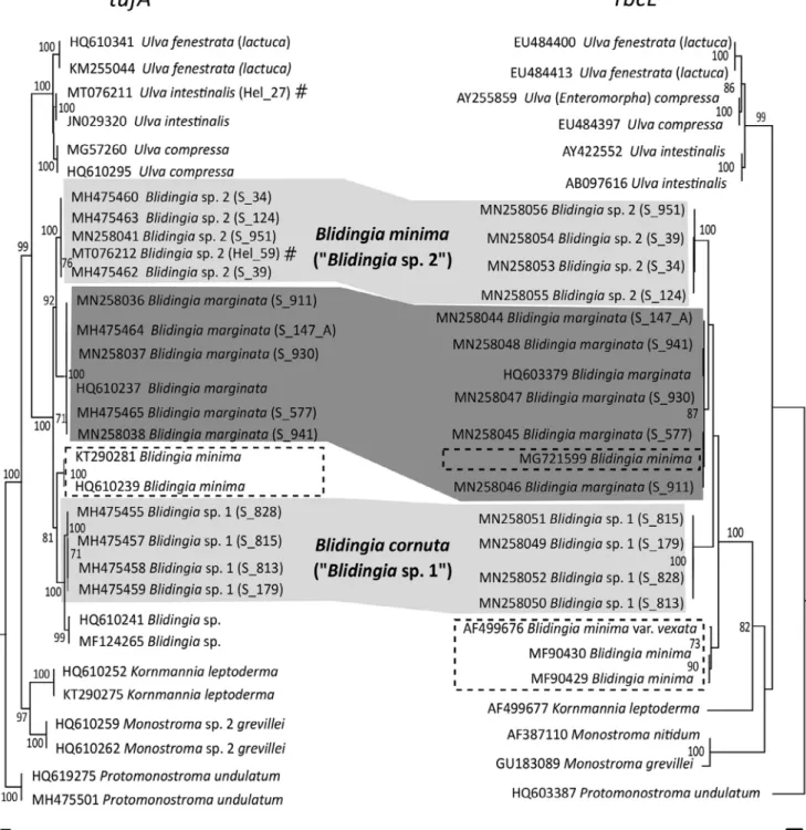
Phylogeny.—The phylogenetic analyses performed on datasets of therbcLandtufAmarkers resulted in comparable and nearly identical results, and almost equivalent evolution- ary relationships of both investigated marker genes were dis- covered (Fig. 2). TherbcLalignment consisted of a total of 695 positions (suppl. Appendix S1), whereas the tufAgene dataset was 771 basepairs long (suppl. Appendix S2).
The node separating the genus Blidingia from closely related and outgroup taxa received full bootstrap support for both marker genes and unequivocably confirmed the taxo- nomic position ofBlidingiaas a separate genus. Whereas the tufAdataset revealsUlvaas sister clade toBlidingia(bootstrap support: 99), therbcLphylogram displays a reference sequence ofKornmannia leptoderma(AF499677) as the closest relative ofBlidingiabeforeUlva. Both analyses resolve the German Blidingia samples from the examined area in three clades (Blidingia marginata,Blidingiasp. 1,Blidingiasp. 2) with high (>85) to full bootstrap support (Fig. 2). The clades delimiting B. marginata showed low intraspecific genetic variability (tufA: 0%–0.3%;rbcL: 0%–0.2%) (see also Table 2) and could be resolved with reference sequences (rbcL: HQ603379, Canada;tufA: HQ610237, Canada). A reference sequence that
seemed to be incorrectly assigned toB.minima (MG721599) clustered within the clade representingB.marginata.
The clusters representing Blidingia sp. 1 and Blidingia sp. 2 could not be matched to any GenBank sequence entries.
However, both clades are clearly separated from otherBlidingia species and receive full bootstrap support (Fig. 2).Blidingia sp. 1 finds its next relative in a well-delimited cluster of un- identifiedBlidingiaspecimens from North America (HQ610241, MF124265) (genetic dissimilarity ofBlidingiasp. 1 andBli- dingiasp.:tufA4.8%;rbcLn.a.), whereas the next closest rel- ative ofBlidingiasp. 2 isB.marginata(genetic dissimilarity ofB.marginataandBlidingiasp. 2:tufA8.6%–9.4%;rbcL 3.2%–3.7%) (see also Table 2).
The phylograms of both thetufA(KT290281, HQ610239) andrbcLgenes (AF499676, MF90430, MF90429) include clades of downloaded reference sequences from GenBank that were supposed to representBlidingia minima(Fig. 2).
However, the topology of these clades and thus their place- ment within the trees is not congruent. One of the specimens assigned to the clade representingB.minimawithin thetufA phylogram originates from a site 30 km to the east in the neighbouring German State of Mecklenburg-Vorpommern, Wohlenberg, which was observed by us in a previous study (Steinhagen & al., 2019a).
Notably, GenBank entries of therbcLandtufAgenes of Blidingia minima showed significant differences in their nucleotide reads (indicating different species are combined in GenBank under this species name). Since most of the uploaded sequences contain several ambiguous bases and are rather short (250–500 bp), we decided to exclude them from the displayed results. However, within this study, we were not able to validate a genotype representing any of the genotypes associated withB.minimain GenBank, not even at the type locality of B. minima on Helgoland. Intra- and interspecific divergence values of the here investigatedBlidin- giaentities are summarized and listed in Table 2.
Both herbarium specimens from Helgoland (Table 1) showed no agreement between their previous identification based on morphological traits and their molecular identifica- tion by DNA barcoding. Within our phylogenetic analysis of thetufAgene, voucher BRM008079 (morphologically identi- fied asBlidingia minima, GenBank accession no.: MT076212) clustered within the clade representingBlidingiasp. 2, and voucher BRM007967 (morphological identityB.marginata, GenBank accession no.: MT076211) was assigned to the clade ofUlva intestinalis(Fig. 2).
Morphological characterization, habitat and distribu- tion.—In the following section theBlidingiaspecies present in the study area of northern Germany are described in more detail. Macro- and micromorphological observations includ- ing respective ontogenetic features, as well as ecological and distribution information, are presented for each species:
Blidingia marginata (J.Agardh) P.J.L.Dang. ex Bliding in Opera Bot. 8(3) [Crit. Surv. Eur. Taxa Ulvales 1]: 32.
1963 ≡ Enteromorpha marginata J.Agardh., Alg. Mar.
Medit.: 16. 1842 ≡ Enteromorpha nana var. marginata (J.Agardh) V.J.Chapm. in J. Linn. Soc. London, Bot. 55:
416. 1956–Lectotype: FRANCE. Nice, Alpes-Maritimes, France, 1842 (LD Herb. Agardh No. 14161).
Habitat,seasonality and distribution.–Blidingia margi- natais the most commonBlidingiaspecies within the study
area and has the widest distribution. It is abundant on Baltic Sea and Wadden Sea coasts and at Helgoland (Table 1). This species can be found in remote as well as anthropogenically strongly impacted habitats (see also Steinhagen & al., 2019a,b) and inhabits fully marine and brackish water ecosystems.
However,B.marginatawas seldomly observed in water bodies
Fig. 2.Comparative maximum likelihood phylograms oftufAandrbcLsequences from taxa ofBlidingiafrom northern Germany. The grey-shaded boxes indicate clades that were present in the study area and connects the respective clades of species in both phylogenetic analyses for direct com- parison. Dashed boxes indicate the ambiguous use of the historic construct of“Blidingia minima”and highlight reference sequences identified as such. Numbers at nodes indicate bootstrap values. Poorly supported nodes (<70% bootstrap support) are not labelled. Branch lengths are propor- tional to sequence divergence. # symbol marks herbarium samples (see also Table 1).
below 5 PSU, and it was not detected in the Kiel Canal. As is typical for the genus,B.marginatawas observed growing as dense turf mats in the upper and middle intertidal zone (Fig. 3A–F). Adults ofB.marginatacan be found through- out the year, but their peak abundance was observed during early and mid-summer (May–July). In late summer and fall (August–October), dense stands began to bleach and popu- lations shrank in size, so that in winter and spring only a few smaller individuals were encountered. Individuals were always found attached to the substratum and only observed detached after extreme weather events. Especially in the North Sea areas,B.marginatawas the most abundant alga in the intertidal zone. It occupied a variety of natural and artificial hard substrata (stones, cobble, breakwaters, wooden piles, etc.) and was often found growing epiphytically on other macrophytes (e.g.,Fucusspp.) and higher plants (Phragmi- tessp.) in the intertidal (Fig. 3B,C). The same distribution patterns were encountered in the Baltic Sea. Here, however, B.marginatawas found in mixed stands withUlva intestina- lis, and in some cases, young individuals ofU.intestinalis were difficult to distinguish fromB.marginataby morpho- logical characters alone. Individuals ofB.marginatacould resist strong UV-radiation and desiccation in summer as well as snow cover and long frost periods in winter (Fig. 3F).
Morphology.–Individuals ofBlidingia marginataexhib- ited all the morphological features described for this species (Bliding, 1963), but also some differences. The long (1–12 cm) and narrow thallus was tubular and usually formed dense mats (Fig. 3D–F). Broader thalli were often wrinkled and twisted and resembledUlva intestinalis(Fig. 3G–I). Contrary to the observations of Bliding (1963), who stated that specimens ofB.marginataexhibited rare small proliferations, 80% of the individuals of the investigated material had microscopi- cally visible branches or branchlet-like appendages (Fig. 3H).
These branchlets were often uni- or biseriate with a single api- cal cell. Macroscopic branching in the middle or apical thallus
parts was rarely observed, whereas some specimens had mac- roscopic branches in the rhizoidal zone. In young, narrow thalli, the cells were arranged in distinct rows (Fig. 3J,K); how- ever, mature or broader thalli exhibited only short cell rows or no cell organization. Cells were quadratic to rectangular, some- times of amorphous shape, 4–10μm long and 2–9μm wide in surface view. The chloroplast filled the whole cell with one central pyrenoid (Fig. 3J,K).
Ontogeny.–As also described by Kornmann & Sahling (1978), the quadriflagellate spore settled on the substratum and immediately developed a germination tube without cutting off an empty cell (Fig. 4A,B). The first mitotic cell divisions resulted in the formation of a prostrate disc (Fig. 4C,D), which then in most of the cases became 2-layered (Fig. 4E). The tubus typically started to develop from lateral initial cells and was rarely observed to develop form the disc’s centre.
Blidingiasp. 1
Habitat and distribution. – Specimens of this species were also found on Baltic Sea and Wadden Sea coasts and on Helgoland (Table 1). However, dense, turf-like populations ofBlidingiasp. 1 were not as frequent and abundant as those ofB.marginata, and they were more clearly restricted to the upper supralittoral zone. Notably, most of the observed popula- tions ofBlidingiasp. 1 grew in the direct vicinity of freshwater inflows (drain pipes, beach showers, stream run-off, etc.;
Fig. 5A). As a consequence–and in contrast withB.margi- nataandBlidingiasp. 2–specimens ofBlidingiasp. 1 were rarely found desiccated during the summer months, despite their location in the upper supralittoral. When freshwater in- flows were more located towards the medio- or infralittoral, Ulva intestinalis was the prevailing species, andBlidingia sp. 1 was absent. The species was also absent from more inland freshwater inflows that had no direct connection to the sea.Blidingiasp. 1 was present throughout all seasons;
however, it was more common during July to August.
Table 2.Overview on intra- and interspecific divergence values of theBlidingiaentities investigated within this study.
Interaction tufA[% difference] rbcL[% difference]
Intraspecific Blidingia marginata 0–0.3 0–0.2
Blidingiasp. 1 0–0.6 0
Blidingiasp. 2 0–0.2 0–0.2
Interspecific Blidingia marginata:Blidingiasp. 2 8.6–9.4 3.2–3.7
Blidingia marginata:Blidingiasp. 1 15.6–17.9 3.2–5.6
Blidingiasp. 1 :Blidingiasp. 2 11.8–13.1 6.4–6.8
Blidingia marginata:“Blidingia minima” 13.7–15.4 4.1–4.4
Blidingiasp. 1 :“Blidingia minima” 10.7–12.1 5.8–6.0
Blidingiasp. 2 :“Blidingia minima” 10–11.1 4.7–5.7
Blidingiasp. 1 :Blidingiasp. (MF124265, HQ610241) 4.8 n.a.
Values for interspecific comparison of“Blidingia minima”were calculated on the respective historically mis-identified clades represented in Fig. 2 (dashed boxtufA;rbcLlower dashed box). Therefore, they are framed by double quotation marks within this table.
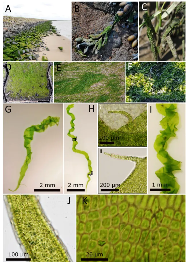
Fig. 3.Morphology ofBlidingia marginataspecimens from Germany. Population ofB.marginatagrowing on a breakwater (A), epiphytic onFucus vesiculosus(B) andPhragmites(C), on cobbles (D) and on sand (E). As a species of the upper intertidal zone, it can manage frost periods (F). Indi- viduals mostly exhibit no macromorphological branches (G&H) but often have microscopic branchlets (H i&ii) and can be of cork-screw-like morphology (I). Cells usually exhibit one central pyrenoid and are arranged in longitudinal rows (J&K).—Photos by S. Steinhagen.
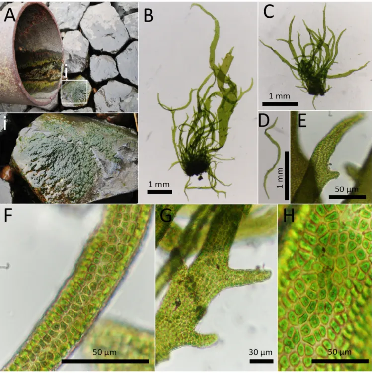
Morphology.–The morphology ofBlidingiasp. 1 shows similarities with the description of Bliding’s (1963) “Bli- dingia minimavar.ramifera”(not validly published, type not indicated), which was later“raised to species rank”by Gar- bary & Barkhouse (1987; as“Blidingia ramifera”).“Blidingia ramifera” is currently regarded as a synonym of B. mar- ginata (Guiry & Guiry, 2019). However, there are distinct differences:
The small, tubular and branched thalli were compact and reached 1–10 mm in length (rarely taller) (Fig. 5B,E) whereas thalli of“Blidingia ramifera”were distinctly larger (Bliding, 1963; Garbary & Barkhouse, 1987). The width of the thallus increased as it proceeded from the rhizoidal zone to the tip (0.08–0.8 mm wide). Branches were antler shaped, uni- to multiseriate and present across the whole thallus (Fig. 5B,C).
Unbranched individuals were rarely encountered. In addition to the clearly formed branches, spine-like microscopic append- ages (10–60μm) were frequently found across the whole thal- lus (Fig. 5E,G). Branches and microscopic appendices were blunt-ended. Thalli grew in tufts that formed dense stands, and several individuals were connected by their rhizoidal zones. Cells formed clear, longitudinal rows (Fig. 5F) that
sometimes blurred in broader thallus areas of the apical region (Fig. 5G), whereas no distinct cell arrangement was observed in mature individuals of“B.ramifera”(Bliding, 1963; Garbary
& Barkhouse, 1987). Individuals with unordered cell arrange- ments were observed infrequently. The cells were quadratic, rectangular, often polygonal with blunt to rounded corners and 3–8μm long and 2–8μm broad. The chloroplast filled the cell or was rarely parietal, with 1 (rarely 2) central pyre- noid(s) (Fig. 5F,H).
Ontogeny.–After the quadriflagellate spore had settled, a germination-tube began to form (Fig. 4F). The cell content migrated through the germination-tube and formed a germi- nating cell, detaching the initial spore sleeve and in most cases also parts of the germination tube (Fig. 4G,H). By mitotic cell divisions, a monostromatic, relatively open disc developed, and its cells were not as dense as in other entities (Fig. 4I,J).
After becoming distromatic, the disc bulged out, and an erect tube formed.
Molecular analysis. – The sequence divergence of the clade representing Blidingia sp. 1 from other species within the genus, in combination with morphological dif- ferences, indicates that Blidingia sp. 1 is genetically and
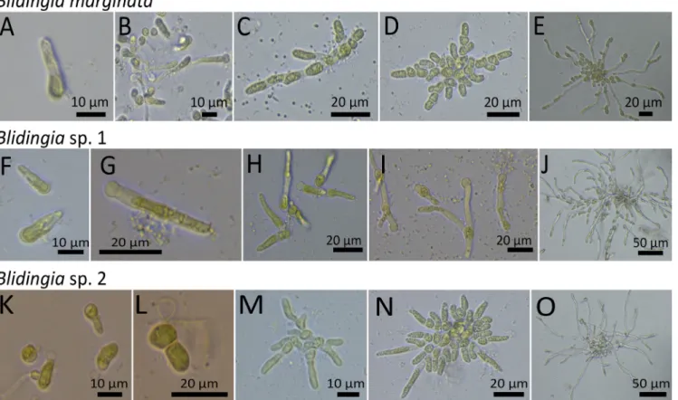
Fig. 4.Ontogenetic development of GermanBlidingiaspp. After the spore ofB.marginatahad settled, a germination-tube began to form (A). No cutting-off of an empty cell was observed (B). A prostrate disc began to form (C–E) with densly packed cells in its centre (E). The early development ofBlidingiasp. 1 started with the formation of a germination-tube after the spore had settled (F). The spores cell content migrated through the ger- mination tube and formed a germinating cell while detaching the empty initial spore sleeve (G&H). Open discs were formed (I&J). The settled spore ofBlidingiasp. 2 formed a small germination tube (K) that then divided into an empty initial cell and a cell containing the cell content (L).
A monostromatic disc with dense cell arrangement formed (M) that in its centre became distromatic (N&O).—Photos by L. Düsedau.
morphologically distinct from other previously described species within the genus Blidingia. We here describe this new species asBlidingia cornuta:
Blidingia cornuta S.Steinhagen & F.Weinberger, sp. nov. – Holotype: GERMANY. Brunsbüttel harbour, Schleswig-
Holstein, N 53.889 E 9.101133, 6 Aug 2014, S.Stein- hagen S_179(C barcode C-A-99682).
Figures 4F–J & 5A–H.
Species description.–Thalli tubular, light to dark green, compressed, bearing antler-like uni- and multiseriate branches across the whole thallus (rarely unbranched), with rounded
Fig. 5.Morphology of the holotype ofBlidingia cornutasp. nov. (“Blidingiasp. 1”). (A) Type locality ofB.cornutaat the Elbe estuary in Bruns- büttel, Germany. The thalli are growing on a stone (i) directly underneath a water drainage pipe permanently releasing freshwater. The individuals exhibited small, tubular and antler-like branched thalli (B&C), and also microscopic, blunt-ended, spine-like appendages were frequently observed across the whole blade (D&E). The cells formed clear, longitudinal rows (F) that sometimes blurred in broader thallus areas of the apical region (G). The cells were quadratic, rectangular, often polygonal with blunt to rounded corners, and the chloroplast filled the cell (rarely parietal) and con- tained 1 (rarely 2) central pyrenoid(s) (F&H).—Photos by S. Steinhagen.
intact branch apices, attached by rhizoids to substratum, 1–10 mm (mean ± 6 mm; rarely >1 cm) long, 0.08–0.8 mm (mean ± 0.3 mm; rarely >1 mm) broad. Main axis increasing in breadth from 10–35μm (rhizoidal zone) to 0.3 mm (middle and apical thallus). Middle thallus often with spine-like appendices (20–35μm in length), these appendices shorter than real branches. Cells in surface view in clear longitudi- nal rows in basal and middle thallus parts, short longitudi- nal rows in apical regions, square, rectangular or polygonal with blunt to round corners, most commonly 3–8μm long and 2–8μm broad. The chloroplast is cell-filling (rarely pari- etal covering most of the cell wall) and cells containing 1 (rarely 2) central pyrenoid(s). Reproduction by quadrifla- gellate spores. From the spore a germination-tube arises, and the cell contents migrate to a germinating cell, detaching the initial spore sleeve and germination-tube. By mitotic cell divi- sions a monostromatic disc develops, and after becoming dis- tromatic, the disc bulges out and an erect tube is formed.
Etymology.– The species epithet cornuta (“horned”in Latin) refers to the antler-like morphology of branches.
GenBank accessions.–MH475459 represents the sequence of the tufAmarker gene, and MN258049 is the respective rbcLsequence.
Type locality.–Brunsbüttel harbour, Schleswig-Holstein, Germany (N 53.889 E 9.101133). Thalli were growing as dense turf in the high intertidal zone directly under a drainage with constant freshwater seepage from land on a bulkhead (Fig. 5A). The site is part of the Elbe estuary, close to the out- let of the Kiel Canal.
Other selected specimens examined (paratypes). – Aschau, Schleswig-Holstein, Germany (N 54.4608; E 9.92665), 24 Jul 2014,S.Steinhagen S_93, GenBank MH 538691 (tufA), Baltic Sea; Heiligenhafen, Schleswig-Hol- stein, Germany (N 54.3787167; E 10.95545), 16 Apr 2015, S. Steinhagen S_622, GenBank MH538692 (tufA), Bal- tic Sea; Helgoland, Germany (N 54.1825; E 7890617), 23 Jul 2014, S. Steinhagen S_21, GenBank MH538693 (tufA), North Sea; Schobuell, Schleswig-Holstein, Germany (N 54.50782; E 8.995567), 24 Jul 2017, S. Steinhagen S_828, GenBank MH475455 (tufA) and MN258052 (rbcL), Wadden Sea; Husum, Schleswig-Holstein, Germany (N 54.47113; E 9.027917), 24 Jul 2017,S.Steinhagen S_818, GenBank MH475456 (tufA), Wadden Sea; Finkhaushallig, Schleswig-Holstein, Germany (N 54.41558; E 8.903633), 24 Jul 2017,S.Steinhagen S_815, GenBank MH475457 (tufA) and MN258051 (rbcL), Wadden Sea; Friedrich-Wilhelm- Luebke-Koog, Schleswig-Holstein, Germany (N 54.83735; E 8.6122), 24 Jul 2017, S. Steinhagen S_813, GenBank MH475458 (tufA) and MN258050 (rbcL), Wadden Sea;
Brunsbuettel estuary, Schleswig-Holstein, Germany (N 53.893567; E 9.141733), 14 May 2015, S. Steinhagen S_578, GenBank MN258043 (tufA), Wadden Sea/river Elbe.
Blidingiasp. 2
Habitat and distribution.–This entity was only observed on Helgoland and in the northeastern Wadden Sea (Table 1) and
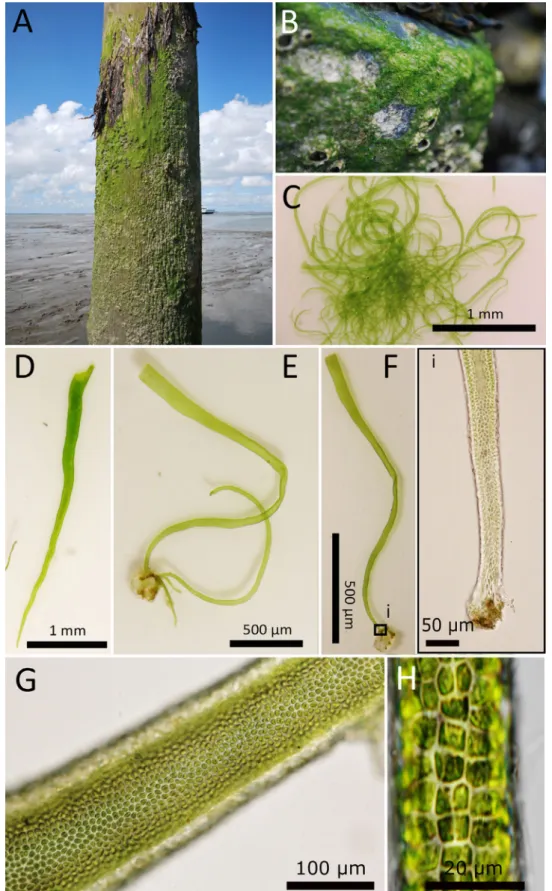
was not present in the Baltic Sea. It inhabited the upper supra- littoral zone and was found growing as turfs, but more often it was observed as small patches on stones, concrete, wooden piles or other hard substrates (Fig. 6A,B). When growing epiphytic on macrophytobenthic species (e.g., Fucus spp.), Blidingia sp. 2 did not cover the host likeB.marginata. Instead, single individuals were found to be scattered across the host plants.
Morphology.–The morphology ofBlidingiasp. 2 shows strong similarities with the description of Kützing’s (1849) Enteromorpha minima, which was later raised to the type ofBli- dingia(asB.minima) by Kylin (1949). Striking morphological congruities amongBlidingiasp. 2 andB.chadefaudiifrom Hel- goland (Kornmann & Sahling, 1978) are also obvious.
The thalli ofBlidingiasp. 2 were mostly only few milli- metres long (rarely taller than 1 cm) and 50–300μm wide (sin- gle individuals had broader thalli up to 700μm) (Fig. 6C–E).
No branches in the middle or apical thallus parts were observed, however the base sometimes exhibited branches (Fig. 6D,E).
Thalli were most often compressed, but inflated individuals were also present. Whereas cells form clear and distinct longitu- dinal rows in the basal thallus parts (Fig. 6F), the arrangement of cells is less organised in the middle and apical thallus parts (Fig. 6G). Cells were of various shapes, quadratic to polygonal with rounded corners, 4–8μm long and 4–6μm wide in surface view. No thickened cell walls, nor any lamellar internal struc- tures were observed. The chloroplast was parietal or filled the cell, with one central pyrenoid (Fig. 6H).
Ontogeny.–The main ontogenetic patterns discovered in this study agree with the developmental descriptions ofBlidin- gia chadefaudii(rather thanB.minima) made by Kornmann
& Sahling (1978). From the attached quadriflagellate spore, a small germination tube was formed (Fig. 4K). The tube divided into an empty initial cell and an upper cell containing the cell contents (Fig. 4L). A monostromatic disc with a dense cell arrangement was formed (Fig. 4M,N), which became dis- tromatic in its centre (Fig. 4N,O) and gave rise to an erect tube.
Molecular analysis. – Our molecular analysis demon- strates thatBlidingiasp. 2 is distinct from otherBlidingiaspe- cies represented in GenBank. However, as described above, morphological (Kützing, 1849; Kylin, 1949; Kornmann & Sah- ling, 1978) as well as ontogenetic (Kornmann & Sahling, 1978) patterns are in accordance with previously described entities.
■DISCUSSION
We here provide a strongly revised picture of the diversity, distribution and morphological variability ofBlidingiaspp. in Schleswig-Holstein, Germany, a study area that includes coas- tal sections of two important ecosystems in northern Europe, the North and Baltic Seas (Fig. 1). In total, 30 populations ofBlidingiaspp. at 19 sites were analysed to cover the full diversity in the study area. However, whereas older studies and recent species inventories mention fourBlidingiaspecies as abundantly present along the coastlines of Schleswig-
Fig. 6.Morphology ofBlidingiasp. 2. Typical habitat ofBlidingiasp. 2 at the German peninsula Nordstrand (A&B). Individuals were found grow- ing in patches and turfs on wooden piles (A) and stones of breakwaters (B). The thalli were unbranched in the middle and apical thallus region (C&D); however, branching in the basal parts was observed infrequently (E). Cells of the basal part proceeded in clear and distinct longitudinal rows (F), but the arrangement of cells is disturbed in the middle and apical thallus parts, and no clear structure was observed (G). Cells were quadratic to polygonal with rounded corners, and the chloroplast was found to be parietal or cell filling and one central pyrenoid was observed (H).—Photos by S. Steinhagen.
Holstein (Kornmann & Sahling, 1978; Schories & al., 2009), we only discovered three. Our findings suggest that this dis- crepancy results from the exclusive use of morphological traits and ontogenetic developmental characters as identification cri- teria in the past. These criteria were probably invalid in part, due to morphological variability within species. That morpho- logical identification criteria for Blidingiaspecies are not in concomitance with more clear-cut molecular identification cri- teria should be discussed in detail.
Of the four Blidingia species listed for Schleswig- Holstein, onlyB.marginatawas frequently observed.Blidin- gia marginatawas found to be the dominant species within the intertidal zone, and it was frequently observed at Baltic Sea and Wadden Sea coasts and on Helgoland. The species is represented by a well-delimited clade with low intraspecific variation (0%–0.3% tufA and 0%–0.2% rbcL) within both provided phylogenetic trees (Fig. 2) and was well resolved to the species level by including several peer-reviewed refe- rence sequences (GenBank accessions HQ610237tufAand HQ603379 rbcL). Its closest relative was Blidingia sp. 2, and both entities had a sequence dissimilarity of 8.6%–9.4%
within thetufAgene and 3.2%–3.7% in theirrbcLsequences (Table 2). However, the combination of morphological and molecular techniques revealed thatB.marginata exhibited a larger morphological variability than expected (Fig. 3).
Individuals ofBlidingia marginataexhibited all the mor- phological features described for this species (Bliding, 1963), and our ontogenetic observations (Fig. 4) are in accordance with previous findings (Bliding, 1963; Kornmann & Sahling, 1978). The tubular thalli where either thin and elongated or had a corkscrew-like morphology, and their cells were arranged in clear longitudinal rows. It should be noted that mature thalli are morphologically similar to smallUlva intes- tinalisand could easily be confused with that species. Corre- spondingly, a herbarium sample from Helgoland that was in concordance with the morphological characters described for B.marginata(Biological Station Helgoland Herbarium BRM007967) was identified asU.intestinalisby DNA bar- coding (Fig. 2).
Contrary to the observations of Bliding (1963), who stated that specimens exhibited rare small proliferations, more than 80% of the individuals of the examined material had microscopic branches or branchlet-like appendages (Fig. 3H).
Bliding (1963) assigned specimens with branchlet-like struc- tures toBlidingia marginatasubsp.subsalsa(Kjellm.) Bliding.
Later, Scagel & al. (1989) raisedB.marginatasubsp.subsalsa to species level, as already suggested by Kornmann & Sah- ling (1978). Our observations indicate thatB.marginata can exhibit various morphologies, including those assigned to B. subsalsa; however, such morphological differences were not reflected by molecular differences of the marker genestufA or rbcL, and no delimitations of the different morphotypes within our phylogenetic analyses were observed (Fig. 2).
Kornmann & Sahling (1978) observed that cultivated swarmers, released by material identified asBlidingia subsalsa, did not grow into the“naturally looking wildtype forms”of
B. subsalsa and that their first-generation offspring were macro-morphologically rather identical with individuals of B.marginata. Based on our results we conclude that the mor- phological spectrum of B. marginata is broader than pre- viously expected, as it includes the morphologies assigned toB.subsalsa.
Based on literature (Kornmann & Sahling, 1978; Pankow, 1990; Schories & al., 2009),Blidingia minimawas expected to have the widest distribution in northern Germany. However, our study revealed that this species is restricted to Helgoland and some other North Sea locations. Past records, in particular those from Baltic Sea locations, may represent misidentifica- tions due to flawed historic species concepts.
This view is strongly supported by our analysis of avail- able barcoding sequences. Screening of databases such as GenBank for sequences ofBlidingia minimaprovides several hits fortufAandrbcLsequences of various length. However, within our phylogenetic analyses (Fig. 2), different sequences from around the globe and identified asB.minimafell in sev- eral well delimited clusters (Fig. 2, dashed boxes) that are not necessarily closely related or even cluster withB.marginata.
This unequivocally confirms that the recent species concept ofB.minimais ambiguous, combines genetically distinct enti- ties, and needs clarification.
Even though sampling sites were chosen in a way that dis- tances between the sites did not exceed 25 km (see also Stein- hagen & al., 2019a,b), no genotypes in accordance with any GenBank entries forBlidingia minimawere observed in the investigated area. One of the reference sequences allegedly representingB.minima(KT290281) originates from Wohlen- berg, 30 km to the east in the neighbouring German State of Mecklenburg-Vorpommern and was observed in a previous study. However, sequences from the type locality ofB.minima, Helgoland–several hundred kilometres away from Wohlen- berg –, were so far missing, which gains importance when we consider that this island was extensively studied within our survey (Fig. 1). A frequently found entity on Helgoland that could not be resolved to species level due to the absence of any similar GenBank entries wasBlidingiasp. 2 (Fig. 2).
Blidingiasp. 2 delimits in a unique clade and is the next clos- est relative toB.marginata(Fig. 2, Table 2).
In addition to the above-mentioned molecular differences ofBlidingiasp. 2 andB.marginata, distinctive morphological delimitations of the two entities were also observed.Blidingia sp. 2 differed from specimens ofB.marginata(Fig. 3) (Bli- ding, 1963; Kornmann & Sahling, 1978) in generally exhibit- ing smaller thalli and being mostly unbranched (Fig. 6). Only few individuals exhibited macroscopic branching in the basal thallus parts, and no microscopic branches were observed (Fig. 6). Concurrently, the morphological features of adultBli- dingiasp. 2 (Fig. 6) showed high similarity with both the type description ofB.minima(Kützing, 1849; Kylin, 1949) and traits described for B. chadefaudii (Kornmann & Sahling, 1978) from Helgoland.
Based on the results obtained within our study we conclude: (1) The type locality of Blidingia minima (as