zur Erlangung der Doktorwürde der Naturwissenschaftlich-Mathematischen Gesamtfakultät der
Ruprecht-Karls-Universität Heidelberg
Molecular biology of the entomopathogenic fungus Beauveria bassiana:
Insect-cuticle degrading enzymes and
Development of a new selection marker for fungal transformation
vorgelegt von
Hong Wan
Gutachter:
Prof. Dr. Hans Ulrich Schairer P.D. Dr. rer. nat. Dorothea Keßler
submitted to the
Combined Faculties for the Natural Sciences and for Mathematics of the Ruperto-Carola University of Heidelberg, Germany
for the degree of Doctor of Natural Sciences
Presented by
Diplom-Biology: Hong Wan
Born in Tianjin, People’s Republic of China Heidelberg
January 2003 Oral examination:
Insect-cuticle degrading enzymes and
Development of a new selection marker for fungal transformation
Referees: Prof. Dr. Hans Ulrich Schairer P.D. Dr. rer. nat. Dorothea Keßler
First of all I would like to express my sincere gratitude to Prof. Hans Ulrich Schairer for giving me the opportunity to study the fascinating field of microbiology and for his supervision throughout my PhD years.
Thanks also goes to all my colleagues, especially to Andreas Leclerque for introducing me to the field of invertebrate pathology, to Wulf Plaga and Ana Milosevic for useful suggestions concerning protein purification experiments and to Diana Hofmann and Susanne Müller for warmhearted advice during the thesis writing process.
I am grateful to Dr. Gisbert Zimmermann (Biologische Bundesanstalt für Land- und ForstwirtschaftInstitut für biologischen Pflanzenschutz) for providing the beetles during the whole time and to all the people of ZMBH who helped me during my thesis work.
Finally, I am thankful for the support and attention my family and my former supervisors in China have given me throughout the past and will, without doubt, give me in the coming years.
I. General Introduction
1.1. Presently used bio-insecticides 1
1.1.1. Entomopathogenic viruses 1
1.1.2. Entomopathogenic bacteria 2
1.1.3. Entomopathogenic nematodes 2
1.1.4. Entomopathogenic fungi 3
1.2. The entomopathogenic deuteromycete B. bassiana 5
1.2.1. The life cycle of B. bassiana 6
1.2.2. The infection process 7
1.2.2.1 Adhesion and germination of conidia 8
1.2.2.2. Formation of an infection structure 8
1.2.2.3. Penetration of the cuticle 9
1.2.2.4. Production of toxins 9
1.2.3. Host defense system 10
1.3. Aspects of using entomopathogenic fungi as bio-control agents 11
1.4. Genetic engineering of entomopathogenic fungi 12
1.5. The aims of this thesis 13
II. Characterization of insect cuticle-degrading enzymes from B. bassiana
2.1. Introduction 14
2.1.1. The mechanism of cuticle degradation by endoprotease 14 2.1.2. The function of phospholipases in cuticle penetration 15
2.1.3. General remarks 16
2.1.4. Experimental plan 17
2.2. Results 18
2.2.1. Cloning of the genes encoding PR2 and PLB from B. bassiana 252 18
2.2.2. Sequence analysis of the pr2 genes 19
2.2.3. Sequence analysis of the plb genes 21 2.2.4. Functional studies of two enzymes involved in the infectious process 25
2.2.4.1. Heterologous expression of the fragments encoding
antigenic determinants of PR1 and PLB2 25
2.2.4.2. Purification of the fusion proteins and raising of antisera 26 2.2.4.3. Insect cuticle as an inducer of PR1 and PLB2 production 27
2.3. Discussion 30
2.3.1. Production of proteases by B. bassiana 252 30
2.3.2. Trypsin-like serine proteases 31
2.3.3. Expression of phospholipase B in B. bassiana 252 32 2.3.4. Function of cuticle-degrading enzymes during fungal infection processes 33
III. Development of a new dominant selection marker, sorR, for fungal transformation
3.1. Introduction 35
3.1.1. Auxotrophic selection markers 36
3.1.1.1. General introduction of commonly used auxotrophic selection markers 36 3.1.1.2. Auxotrophic selection markers used for counterselection 38 3.1.1.3. Characters of auxotrophic selection markers 38
3.1.2. Dominant selection markers 39
3.1.2.1. General introduction of commonly used dominant selection markers 39
3.1.2.2. Characters of dominant selection markers 42
3.1.3. Utilization limits of the current available selection markers 42 3.1.4. sorR, a new dominant selection marker for fungal transformation 45 3.1.4.1 Basic characterization and biosynthesis of soraphen A 45 3.1.4.2. Antimicrobial spectrum and the mechanism of action of soraphen A 46
3.1.4.3. Acetyl-CoA Carboxylases 47
3.1.4.4. The strategy of utilizing sorR selection marker for fungal transformation 49
3.1.5. The focus of this part of work 50
3.2. Results 51 3.2.1. The sensitivity of B. bassiana 252 to Soraphen A 51
3.2.2. Construction of B. bassiana 252 genomic library 52
3.2.3. Isolation of the accB1 gene from B. bassiana 252 53
3.2.4. Sequence analysis of B. bassiana accB1 gene 54
3.2.4.1. Amino acid sequence homology 54
3.2.4.2. Transcription initiation site of accB1 gene 55
3.2.4.3. Introns in the accB1 gene 57
3.2.4.4. The upstream region of accB1 57
3.2.4.5. The downstream region of accB1 57
3.2.5. Construction of transformation vectors for identification of the core
promoter sequence of the B. bassiana accB1 gene 62
3.2.6. Expression of E. coli ACC in Pichia pastoris and effects on
the resistance to soraphen A 63
3.2.6.1. The sensitivity of P. pastoris GS115 to soraphen A 64 3.2.6.2. Construction of the E. coli ACC expression vector 65 3.2.6.3. Expression of E. coli ACC in P. pastoris GS115 67 3.2.6.4. Chromosomal DNA analysis of putative SorR transformants 68 3.2.7. Development of a stepwise transformation procedure for sorR marker 71 3.2.7.1. Construction of the expression vector pPIC3.5K-BCCP 71 3.2.7.2. Expression of E. coli accC gene in strain HWA3-2 71 3.2.7.3. Soraphen A sensitivity assay of the transformants HWA3pG 73 3.2.7.4. Chromosomal DNA analysis of SorR transformants 74
3.3. Discussion 80
3.3.1. Sequence of the gene encoding acetyl-CoA carboxylase in B. bassiana 80 3.3.2. Expression of sorR conferred soraphen A resistance in P. pastoris 82
IV. Materials and Methods
4.1. Materials 86
4.1.1. Chemicals and consumables 86
4.1.2. Radioisotope 86
4.1.3. Antibodies 86
4.1.4. Enzymes and kits 87
4.1.5. Plasmids 88
4.1.6. E. coli strains 88
4.1.7. Fungus and yeast strains 88
4.1.8. Antibiotic stock solutions 89
4.2. Methods 89
4.2.1. Microbiologic techniques 89
4.2.1.1. Growth of E. coli 89
4.2.1.2. Electroporation of E. coli 90
4.2.1.3. Growth of B. bassiana 90
4.2.1.4. Media transfer of B. bassiana culture in order to
achieve C/N derepression 91
4.2.1.5. Sensitivity assay of B. bassiana to soraphen A 92
4.2.1.6. Growth of P. pastoris 92
4.2.1.7. Sensitivity assay of P. pastoris to soraphen A 92
4.2.1.8. Transformation of P. pastoris 93
4.2.1.9. Preparation of comminuted cuticles from adult colorado potato beetle 95
4.2.2. DNA techniques 95
4.2.2.1. Isolation of high molecular chromosomal DNA from B. bassiana 95 4.2.2.2. DNA fraction by partial cleavage with Mbo I 96 4.2.2.3. Construction of phage genomic library of B. bassiana 97
4.2.2.4. Phage library screening 98
4.2.2.5. Subcloning of try1, try2, plb1 and plb2 genes from the phage library 99
4.2.2.6. Isolation of genomic DNA from P. pastoris 99
4.2.2.7. Southern hybridisation 100
4.2.2.8. Construction of the transformation vector pTGT-PaccB1 101
4.2.2.9. Primers used for construction of pAO815-ACC4 102 4.2.2.10. Recovery of DNA fragment from crystal violet agarose gel 102
4.2.2.11. DNA sequencing 103
4.2.3. RNA techniques 103
4.2.3.1. Isolation of RNA from B. bassiana 103
4.2.3.2. RT-PCR 104
4.2.3.3. Primer extension 105
4.2.3.4. 5’RACE 106
4.2.4. Protein methods 107
4.2.4.1. Protein preparation from B. bassiana mycelium 107 4.2.4.2. Protein preparation from supernatant of B. bassiana culture 108
4.2.4.3. Construction of pQE-PR1 and pQE-PLB2 108
4.2.4.4. Expression of the fusion protein 109
4.2.4.5. Large scale protein preparation and affinity purification from E. coli 110
4.2.4.6. Immunoblot analysis 110
4.2.5. Raising antisera 111
4.2.6. Soft ware and computational analysis 112
V. References 113
VI. Summary 126
VII. Appendixes 128
Appendix1: Papers and patent application arising from this work 128
Appendix2: Abbreviations 129
Appendix3: Sequences 132
I. General Introduction
Insecticide resistance and the demand for reduced chemical inputs in agriculture have provided an impetus to the development of alternative forms of pest control.
Biological control offers an attractive alternative or supplement to the use of chemical pesticides. Microbial biological control agents are naturally occurring organisms and perceived as being less damaging to the environment. Furthermore, their generally complex mode of action makes it unlikely that resistance could be developed to a bio-pesticide. Biological pest control agents includes viruses, bacteria, fungi, and nematodes. The use of microorganisms as selective pesticides has had some notable successes.
1.1. Presently used bio-insecticides 1.1.1. Entomopathogenic viruses
The insect pathogenic viruses belong to of the family Baculoviridae (BV). BV includes two genera (Nucleoplyhedrovirus, NPV and Granulovirus, GV) of arthropod-specific pathogens and the majority of the baculoviruses are infectious only for insect species within the order Lepidoptera, with no adverse effect on members of other orders. In addition, most of the baculoviruses exhibit a very narrow, mostly single-species, host range. Target specificity makes them good candidates for use in integrated pest management systems (Romanowski, 2002).
Up-to-date, the most successful case of using baculoviruses as bio-control was the exploitation of the multinucleocapsid nucleopolyhedrovirus of Anticarsia gemmatalis (AgMNPV) for the control of the velvetbean caterpillar in soybean in Brazil. In the season 2001/2002, the treated area with AgMNPV (tradename Coopervirus®) was over 1,550,000 ha (more than 11% of the soybean cultivated area in the country) and the use of the AgMNPV in Brazil has generated substantial economical, ecological, and social benefits (Moscardi et al., 2002). In addition, the multinucleocapsid nucleopolyhedrovirus of Spodoptere frugiperda (SfMNPV), which is the principal pest of maize and sorghum, has been developed as a bioinsecticide in Mexico and Central America (Williams, 2002); a single nucleocapside nucleopolyhedrovirus of Helicoverpa armigera (HaSNPV) has been developed as a commercial pesticide for
the control of the cotton bollworm in China. The genome of the HaSNPV has been entirely sequenced and several genetically modified HaSNPVs have been constructed and tested in the laboratory and in the field (Sun et al., 2002). However, the ingestion of a lethal dose by the target insect and the length of time required to kill or prevent the damage of the crop are the major limitations for their extensive usage in agronomic systems.
1.1.2. Entomopathogenic bacteria
The best example of bacterial insecticides that have been investigated in detail belong to the genus Bacillus. Four species have been used as insecticides commercially: B. thuringiensis, B. popilliae, B. moritai and B. sphaericus.
B. thuringiensis, the most successful biological control agent produced entomocidal toxins: the α- and β-exotoxins and δ-endotoxin. The latter, also called insecticidal crystal protein (ICP), is synthesized during sporulation, and accounts for the commercial value of B. thuringiensis as biological pest control agent (Zhong, 2000).
B. thuringiensis var. israelensis (Bti) was first discovered in 1976 and has proven to be highly pathogenic for many aquatic Diptera. Bti has been successfully utilized for control of mosquitoes and black flies worldwide. Additionally, Bti has an excellent safety record and resistance has not been found probably due to its complex mode of action involving synergistic interaction between up to four proteins. Furthermore, several toxin genes from B. thuringiensis have been used in transgenic crops such as cotton, corn and soybean (Khachatourians, 1986).
1.1.3. Entomopathogenic nematodes
Entomopathogenic nematodes of the families Steinernematidae and Heterorhabditidae together with their symbiotic bacteria, Xenorhabdus and Photorhabdus, respectively, represent a unique biological control agent. The nematode vectors the entomopathogenic bacteria within their gut into the insect host, where the bacteria multiply and produce a wide range of toxins and hydrolytic exoenzymes that are responsible for the death and bioconversion of the insect larvae into a nutrient soup, that is ideal for nematode growth and reproduction. The nematodes reproduce until the nutrient supply becomes limiting at which time they develop into infective juveniles, which are recolonized by the symbiotic bacterium (Forst and Clarke, 2002). The nematode-bacterium complex kills insects rapidly (48hr). they do not form intimate, highly adapted, host-parasite relationship that
could be found in the majority of other parasites used for biological control and therefore can exploit a wide host range of insect species. The target insects of EPN include the black vine weevil in nurseries and landscape, cranberry girdler in cranberry bogs, Diaprepes root weevil in citrus, fungus gnats in nurseries, sciarid flies in mushrooms, Western flower thrips in the greenhouses, hunting billbugs in turfgrass, white grubs in turfgrass, and mole crickets in turfgrass. The proper nematode species must be used against the target insect as all nematode species are not effective against a given target insect (Stock, 2002).
EPNs and their associated symbiotic bacteria have been considered to be a save approach to pest control. They have been proved to be not harmful to humans and other vertebrates. EPNs can be easily mass-produced using conventional fermentation technology, are exempt from registration requirements in many countries and therefore several formulations of EPNs are available on the market for the control of soil and cryptic pests in North America, Europe, Asia, and Australia.
1.1.4. Entomopathogenic fungi
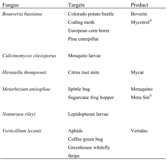
Entomopathogenic fungi were among the first organisms to be used for the biological control of pests. More than 700 species of fungi from around 90 genera are pathogenic to insects. Most are found within the deuteromycetes and entomophthorales. some insect-pathogenic fungi have restricted host ranges, for example, Aschersonia aleyrodis infects only scale insects and whiteflies, while other fungal species have a wide host range, with individual isolates being more specific, for example, Metarhizium anisopliae and Beauveria bassiana(Fig. 1.1). The commercial produced entomopathogenic fungi and their targeted hosts are shown in Table 1.1.
Fungal species such as M. anisopliae and B. bassiana are well characterized in respect to pathogenicity to several insects and they have been used as agents for the biological control of agriculture pests worldwide. In Colombia, about 11 companies offer at least 16 products based on the entomopathogenic fungi B. bassiana. These products are used not only in the coffee crop but also in other crops such as cabbage, corn, bean, tomato, potato. They are also used to treat public disease vectors (e.g., flies and mosquitoes) (Florez, 2002).
Fig. 1.1. Biological control of insect pest (alfalfa weevil). Left and Center, Adult insect killed by Beauveria bassiana. Right, Healthy adult insect (control).
Table 1.1. Entomopathogenic fungi in commercial and experimental production (adapted from Khachatourians, 1986)
Fungus Targets Product
Beauveria bassiana
Culicinomyces clavisporus Hirsutella thompsonii Metarhizium anisopliae
Nomuraea rileyi Verticillium lecanii
Colorado potato beetle Coding moth
European corn borer Pine caterpillar Mosquito larvae Citrus rust mite
Spittle bug
Sugarcane frog hopper Lepidopteran larvae
Aphids
Coffee green bug Greenhouse whitefly thrips
Boverin Mycotrol®
Mycar
Metaquino Meta-Sin®
Vertalec
1.2. The entomopathogenic deuteromycete B. bassiana
The filamentous fungus Beauveria bassiana belongs to a class of insect pathogenic deuteromycete (imperfect fungus). The different Beauveria strains are highly adapted to particular host insects. A broad range of B. bassiana species have been isolated from a variety of insects worldwide that are of medical or agricultural significance.
An interesting feature of Beauveria is the high host specificity of many isolates.
Hosts of medical importance include vectors for agents of tropical infectious diseases such as the tsetse fly Glossina morsitans morsitans, the sand fly Phlebotomus that transmits Leishmania, and the bugs of the genera Triatoma and Rhodnius, the vectors of Chagas’ disease. Hosts of agricultural significance include the Colorado potato beetle, the codling moth and several genera of termites. Furthermore, the high level of persistence in the host population and in the environment provides long-term effects of the entomopathogenic fungi on pest suppresson.
In China, B. bassiana is applied against the European corn borer Ostrinia mubilalis, pine caterpillars Dendrolimus spp., and green leafhoppers Nephotettix spp.. In the Soviet Union, B. bassiana is produced under the trade name Boverin for control of the Colorado potato beetle Leptinotarsa decemlineata and the codling moth Laspeyresia pomonella.
1.2.1. The life cycle of B. bassiana
B. bassiana has a dimorphic mode of growth. In the absence of the specific insect host Beauveria passes through an asexual vegetative life cycle that includes germination, filamentous growth and the formation of sympoduloconidia (Fig. 1.2B).
In the presence of its host insect, Beauveria switches to the pathogenic life cycle.
The conidiospores germinate on the surface of the cuticle and the germinated hyphal tubes penetrate the insect’s integument directly. When having penetrated the cuticle, the fungus alters its growth morphology to a yeast-like phase and produces hyphal bodies, which circulate in the haemolymph and proliferate by budding (Fig. 1.2A).
Following the death of the host, fungal growth reverts back to the typical hyphal form (the saprotrophic stage). The ability to convert to the yeast-like phase may be a prerequisite for pathogenicity.
Fig. 1.2. Dimorphic growth mode of B. bassiana. (A) yeast.like parasitic phase when infecting susceptible species. (B) Saprobic phase shows filamentous hypha.
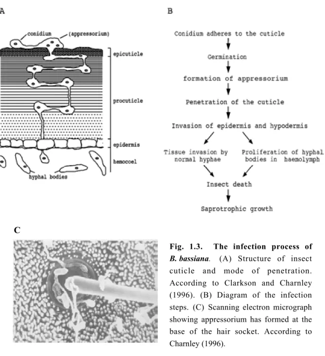
1.2.2. The infection process
In contrast to bacteria and viruses that pass through the gut wall from contaminated food, fungi have an unique mode of infection. They reach the hamocoel through the cuticle or possibly through the mouth parts. Ingested fungal spores do not germinate in the gut and are voided in the faeces. The death of the insect results from a combination of factors: mechanical damage resulting from tissue invasion, depletion of nutrient resources and toxicosis (Fig. 1.3).
C
Fig. 1.3. The infection process of B. bassiana. (A) Structure of insect cuticle and mode of penetration.
According to Clarkson and Charnley (1996). (B) Diagram of the infection steps. (C) Scanning electron micrograph showing appressorium has formed at the base of the hair socket. According to Charnley (1996).
1.2.2.1. Adhesion and germination of conidia
Attachment of a fungal spore to the cuticle surface of a susceptible host represents the initial event in the establishment of mycosis. For most entomopathogenic fungi host location is a random event and attachment is a passive process with the aid of wind or water. It was found that dry spores of B. bassiana possess an outer layer composed of interwoven fascicles of hydrophobic rodlets. This rodlet layer appears to be unique to the conidial stage and has not been detected on the vegetative cells.
The adhesion of dry spores to the cuticle was suggested to be due to non-specific hydrophobic forces exerted by the rodlets (Boucias et al., 1988). In addition, lectins, a kind of carbohydrate binding glycoproteins, have been detected on the conidial surface of B. bassiana. It was also suggested that lectins could be involved in binding between conidia and the insect cuticle. The exact mechanisms responsible for the interaction between fungal spores and the cuticle remain to be determined (Latge, 1988).
After the pathogen reaches and adheres to the host surface, it proceeds with rapid germination and growth which are profoundly influenced by the availability of nutrients, oxygen, water, as well as pH, and temperature, and by the effects of toxic host-surface compound. Generally, fungi with a broad host range germinate in culture in response to a wide range of nonspecific carbon and nitrogen sources.
Entomopathogenic fungi with restricted host range appear to have more specific requirements for germination (St Leger, 1989a).
1.2.2.2. Formation of an infection structure
Entomopathogenic fungi invade their hosts by direct penetration of the host cuticle.
The cuticle has two layers, the outer epicuticle and the procuticle. The epicuticle is a very complex thin structure that lacks chitin but contains phenol-stabilized proteins and is covered by a waxy layer containing fatty acids, lipids and sterols (Hackmann, 1984). The procuticle forms the majority of the cuticle and contains chitin fibrils embedded into a protein matrix together with lipids and quinones (Neville, 1984).
Protein may account for up to 70% of the cuticle. In many areas of the cuticle the chitin is organized helically giving rise to a laminate structure.
In common with many entomopathogenic fungi, B. bassiana conidia germinate on the host surface and differentiate an infection structure termed appressorium. The
appressorium represent an adaptation for concentrating physical and chemical energy over a very small area so that ingress may be achieved efficiently (Fig. 1.3C). Thus, formation of the appressorium plays a pivotal role in establishing a pathogenic interaction with the host. Appressorium formation may be influenced by host surface topography and biochemical investigations indicate the involvement of the intracellular second messengers Ca2+ and cyclic AMP (cAMP) in appressorium formation (St Leger et al., 1991).
1.2.2.3. Penetration of the cuticle
Pathogenic fungi need to penetrate through the cuticle into the insect body to obtain nutrients for their growth and reproduction. Entry into the host involves both enzymic degradation and mechanical pressure as evidenced by the physical separation of lamellae by penetrated hyphae. A range of extracellular enzymes that can degrade the major components of insect cuticle, including chitinases, lipases, esterases and at least four different classes of proteases, have been suggested to function during the fungi pathogenesis. The production of cuticle-degrading enzymes by M. anisopliae during infection structure formation on Calliphor vomitoria and Manduca sexta has been investigated by biochemical and histochemical analysis both in vivo and in vitro. Among the first enzymes produced on the cuticle are endoproteases (termed PR1 and PR2) and aminopeptidases, coincident with the formation of appressoria. N-Acetylglucosaminidase is produced at a slow rate as compared to the proteolytic enzymes. Chitinase and lipase activities were not detected (St Leger et al., 1989b ). Although the complex structure of the insect cuticle suggests that penetration would require the synergistic action of several different enzymes, much of the attention has focused on the cuticle-active endoprotease as a key factor in the process.
1.2.2.4. Production of toxins
There is considerable circumstantial evidence from deuteromycete pathogens for the involvement of fungal toxins in host death. The action of cytotoxins is suggested by cellular disruption prior to hyphae penetration. Behavioural symptoms such as partial or general paralysis, sluggishness and decreased irritability in mycosed insects are consistent with the action of neuromuscular toxins (Charnley, 1984). B. bassiana and M. anisopliae produced significant amounts of toxic compounds within their hosts.
For example, the toxins Beauvericin, Beauverolides, Bassianolide and Isarolides
have been isolated from B. bassiana infected hosts (Hamill, 1969; Elsworth, 1977);
toxins Destruxins (DTXs) and Cytochalasins have been isolated from M. anisopliae infected hosts. The toxins have shown to have diverse effects on various insect tissues. DTX depolarizes the lepidopteran muscle membrane by activating calcium channels. In addition, function of insect hemocytes can be inhibited by DTX (Bradfisch, 1990). Presumably, there are still many toxins that remain to be isolated from parasized insects and except DTXs, their relevance to patogenicity remains to be established.
1.2.3. Host defense systems
In order to prevent invasion by fungi, insects have evolved various defense mechanisms. The defensive arsenal of insects contains both passive structural barries, such as the cuticle, and a cascade of active responses to pathogens that gain access to the hemocoel. This active responses include melanization, cellular reactions, humoral reaction to recognize the non-self pathogen, and production of protease inhibitors.
Melanization: the oxidation of phenolic compounds to dihydroxyphenylalanine, typified by the production of brown or black melanic pigments, is a common feature of the response of many insects to fungal infection. Melanin may partially shield cuticle from enzymatic attack or may be toxic to fungi. However, such protection is incomplete. The investigation from St Leger (1988) indicates that melanization is primarily an effective defence against weak or slow growing pathogens, but is ineffective against more virulent fungi.
Cellular reactions: once the cuticle and epidermis have been breached the invading fungus is faced with the defence systems of the hemolymph. The responses to mycopathogens within the haemocoel include phagocytosis, encapsulation and nodulation. However, the effect on fungal elements is uncertain. With the arbitrary injection method, Bidochka (1987) found that haemocytes of the migratory grasshopper, M. sanguinipes, encapsulate viable conidia of B. bassiana, however they fail to suppress conidial germination within the nodule. It was suggested that the production of toxins and extracellular proteases by B. bassiana could trigger the evadation of encapsulation.
Humoral reactions: in response to fungal challenge, insects elicit an acquired humoral “immunity” to subsequent infection. Recognition of “non-self” is critical to the initiation of the hemocytic defense reaction and this selective response in insects depends on a specific chemical recognition on part of the hemocytes. Serum and hemocyte cell membrane-bound lectins have been found in many insects (Mello, 1999). They could play a role in immune defense reactions since they agglutinate pathogens as well as fungi (Mello, 1999). Thus, insect serum agglutinin may function as opsonic mediating the enhanced attachment of granulocytes to the hyphal bodies (Pendland et al., 1988).
Production of protease inhibitors: host-produced protease inhibitors, which inhibit cuticle-degrading enzyme activities of pathogens, may contribute to insect defence systems. Such compounds have been isolated from the serum of Anticarsia gemmatalis larvae which were resistant to infection by Nomuraea rileyi (Boucias et al., 1987).
1.3. Aspects of using entomopathogenic fungi as bio-control agents The advantages of using fungi as insecticides are:
(1) Their high degree of specificity for pest control. Fungi can be used to control harmful insect pests without affecting beneficial insect predators and non- harmful parasites.
(2) The absence of effects on mammals and thus the reduction of the hazards normally encountered with insecticide applications, such as pollution of the environment.
(3) The lack of problems caused to insect resistance and prolonged pest control.
(4) A high potential for further development by biotechnological research.
(5) High persistence in the environment provide long-term effects of entomopathogenic fungi on pest suppression.
However, there are also a number of constraints on the use of fungi as insecticides:
(1) 2-3 weeks are required to kill the insects whereas chemical insecticides may need only 2-3 hours.
(2) Application needs to coincide with high relative humidity, low pest numbers and a fungicide free period.
(3) Due to the high specificity additional control agents are needed for other pests.
(4) Their production is relatively expensive and the short shelf life of spores necessitates cold storage.
(5) The persistence and efficacy of entomopathogenic fungi in the host population varies among different insects species, thus insect-specific application techniques need to be optimised to retain long-term impacts.
(6) A potential risk to immunodepressive people
1.4. Genetic engineering of entomopathogenic fungi
A more widespread use of fungi for bio-control will depend on improvements of wild-type strains by combining characteristics of different strains and mutants.. Two type of improvements may be considered: (i) improvement the efficacy of the insecticide, e.g., by reducing the dose necessary to kill the insects, by reducing the time to kill the pest or decreasing crop damage caused by the pest by reducing the feeding time; (ii) expanding the host range. Essential for the development of a hypervirulent strain is a complete understanding of the remarkable pathology of fungi infections.
Molecular biology provides the necessary tools for dissecting the mechanisms of pathogenesis and in the longer term for producing recombinant organisms with new characteristics. Initial development towards these goals has occurred with M. anisophliae and to a much lesser extent with B. bassiana (Hegedus, 1991).
Genetic transformation systems, which are an essential part of modern fungal research, and is necessary for the experimental manipulation of virulence genes in vitro and in vivo, have been established (Goettel and St Leger, 1990). The success of utilizing these procedures depend on the availability of selectable transformation markers. transformation techniques have been used to isolate specific pathogenic genes, investigate virulence determinants of M. anisophliae, and to produce a strain with enhanced virulence. Unravelling the molecular mechanisms of fungal pathogenesis in insects will provide the basis for the genetic engineering of entomopathogenic fungi.
1.5. The aims of this thesis
As described above, the entomopathogenic fungus B. bassiana is attracting increased attention as a potential tool for biological control of insect pests. Understanding the mechanisms of fungal pathogenesis in insects will provide a rational basis for strain selection and improvement. The research of this thesis is undertaken with a B. bassiana strain, ARSEF 252, which is highly pathogenic for the Colorado potato beetle, Leptinotarsa decemlineata. The aim of this thesis is to evaluate the contribution of individual enzymes to pathogenesis of B. bassiana252. In addition, in order to help in the manipulation of pathogenic genes in entomopathogens or even to introduce heterologous pathogenic genes designed to improve their potential for biocontrol, we also interested in optimizing the transformation procedures which is suitable for B. bassiana252. Therefore, a new dominant selection marker for fungal transformation is developed. The outcome of these two independent studies will be presented and discussed separately in two sections.
II. Characterization of insect cuticle-degrading enzymes from B. bassiana
2.1. Introduction
2.1.1. The mechanism of cuticle degradation by endoprotease
Metarhizium anisopliae is by far the best studied entomopathogenic fungus and several virulence factors involved in the disease process have been identified (Clarkson and Charnley, 1996). A subtilisin-like protease, termed PR1 has been cloned and characterized (St Leger et al., 1992). PR1 is synthesized as a large precursor containing an 18-amino acid signal peptide and an 89-amino acid propeptide. The mature protein (28.6 kDa) contains 281 amino acid residues. The sequence shows considerable similarities with other enzymes of the subtilisin subclass of serine endoproteases. In particular, the serine, histidine, and aspartate residues that comprise the active site of these proteases are conserved in PR1. PR1 possesses a broad primary specificity for amino acids with a hydrophobic side group at the second carbon atom (e.g., phenylalanine, methionine, and alanine) but also possesses a secondary specificity for extended hydrophobic peptide chains with the active site recognizing at least five subsite residues. This relative nonspecificity accounts for its activity against a range of proteins.
By immunogold electron microscopy, it was shown that PR1 is secreted by the appressorium and penetrating hyphea within the cuticle. For M. anisopliae, PR1 appears to be a pathogenicity determinant by virtue of its ability to extensively degrade the cuticle and its production at high levels by the pathogen in situ during infection (St Leger et al., 1987). Furthermore, addition of multiple copies of pr1 under the control of a constitutive promoter increases the virulence of the transformants (St Leger et al., 1996). The mechanism of cuticle degradation by PR1 in M. anisopliae was suggested as follows: (1) PR1 is adsorbed to the cuticle via nonspecific electrostatic forces; (2) the active site comes into contact with any part of the accessible cuticle protein chains and under appropriate conditions, e.g., temperature, splits susceptible peptide bonds thus relaesing cuticle proteins; (3) solubilized proteins are further degraded until a chain length of around 5 is obtained (St Leger et al., 1987). The progression of knowledge regarding the major pathogen protease, PR1, followed a course that may serve as a model for research on other entomopathogenic fungi. Furthermore, the characterization of PR1 also aided in
unravelling additional factors that contribute to pathogenicity. In addition to PR1, another endoprotease, trypsin-like protease (PR2), has also been characterized from M. anisopliae, but the role of PR2 is not clear (St Leger, 1996).
2.1.2. The function of phospholipases in cuticle penetration
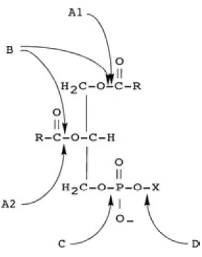
Since lipids represent major chemical constituents of the insect cuticle, enzymes capable of hydrolyzing these compounds, such as phospholipases, could be expected to be involved in the cuticle disruption processes that occur during host invasion.
Phospholipases are a heterogeneous group of enzymes that are able to hydrolyse one or more ester linkages in glycerophospholipids. The action of phospholipases can result in the destabilization of membranes, cell lysis and release of lipid second messengers (Ghannoum, 2000). These enzymes are categorized according to the location of the ester link that is cleaved (Fig. 2.1). Although phospholipase B (PLB) refers to an enzyme that can remove both sn-1 and sn-2 fatty acids, this enzyme also has lysophospholipase-transacylase activity.
Fig. 2.1. Sites of action of phospholipases. A1, A2, B, C and D indicate cleavage sites of the corresponding phospholipases (PLA1, PLA2, PLB, PLC and PLD).
Extracellular phospholipases have been implicated as pathogenicity factors for bacteria, rickettsiae and protozoa, The type of phospholipase involved in virulence varies with the organism. For example, C. perfringens (Alape-Giron, 2000) secretes a phospholipase C (PLC), whereas T. gondii secretes a phospholipase A (PLA). The importance of these enzymes, especially PLB, for virulence has so far only been verified in medically important fungi. PLB was secreted by different clinically important fungal species such as Candida albicans (Mukherjee, 2001), Aspergillus fumigatus (Burch,1996) and Cryptococcus neoformans (Cox, 2001). The role of PLB in the pathogenicity of entomopathogenic fungi remains to be determined, even in the best-studied species M. anisopliae.
2.1.3. General remarks
The great diversity of fungi, and the many routes by which they have achieved their success as pathogens, render it imprudent to consider M. anisopliae or any other fungi as an overall model for pathogenicity. The general validity of the “protease concept” remains to be proved. Nevertheless, given that the proteinaceous cuticle is a barrier to all entomopathogenic fungi, the involvement of proteases in the infection process is likely to be ubiquitous.
There is a surprising degree of species and strain variability observed in the production of insect cuticle-degrading enzymes. In M. anisopliae, variability was not limited to the quantitative levels of enzymes produced, but extended to the patterns of enzyme expression on different growth media. Fungal strain variability in cuticle- degrading enzyme production may be directly related to variability in virulence. It is also possible that these enzymes may be involved in host range determination.
2.1.4. Experimental plan:
So far there have been no detailed investigations of the enzymes involved in the infection processes of B. bassiana. Because of the strong evidence supporting the role of proteases (PR1 and PR2) and phospholipase B in fungal pathogens and the correlation observed in M. anisopliae and C. albicans. An initial characterization of these potential virulence determinants in B. bassiana was undertaken. The following experimental work was performed:
• Cloning of the genes encoding PR2 and PLB from B. bassiana.
• Sequence analysis of the genes encoding PR2 and PLB.
• Partial recombinant expression and the development of antisera against PR1 and PLB.
• Examination of the expression of PR1 and PLB in cuticle cultures.
2.2. Results
2.2.1. Cloning of the genes encoding PR2 and PLB from B. bassiana 252
Initial experiments by A. Leclerque indicated the presence of two pr2 homologues and two plb homologues in B. bassiana. Degenerate primers complementary to conserved regions within fungal pr2 and plb genes were used for PCR amplification from the B. bassiana genome. Two amplicons of different size from each gene were cloned and sequenced. Their homology to other fungal pr2 and plb genes was confirmed by alignments with the related genes. The results indicated that there are two isotypes of the pr2 gene (designated try1 and try2) and two isotypes of the plb gene (designated plb1 and plb2) in B. bassiana (A. Leclerque, unpublished).
Plasmids containing the pr2 and plb amplicons were obtained from A. Leclerque and the cloned fragments were amplified by PCR. The PCR products were used as probes for screening a phage genomic DNA library of B. bassiana in order to isolate phage clones harbouring the full length of try1, try2 genes and plb1, plb2 genes, respectively. A total of 20,000 plaques were screened at high stringency and 11 try1 clones, 4 try2 clones, 3 plb1 clones and 4 plb2 clones were identified. One phage clone of each gene was further purified and designated TA4, TB3, PA1 and PB3.
Fig. 2.2. Restriction pattern of phage clones.
1 µg of DNA from the phage clones TA4, TB3, PA1 and PB3 was digested with Sal I, EcoR I, Xba I and BamH I, respectively, and separated on an 0.8% agarose gel. The presence of the respective fungal genes try1, try2, plb1 and plb2 in the restricted fragments was proved by Southern Blot analysis. Positive bands are indicated by arrowheads. M, 1 kb DNA ladder.
Sal I restricted fragments from the clone TA4, EcoR I restricted fragments from the clone TB3, Xba I restricted fragments from the clone PA1, and BamH I restricted fragments from the clone PB3 were subcloned into a pUC18 vector and sequenced.
Thefragments selected for sequencing are indicated in Fig. 2.2. In addition, fragment/
fragments obtained by Not I restriction from all four clones was/ were cubcloned into pBSSKII+ vector to acquire sequence data derived from overlapping clones to ensure that the subcloned DNA fragments were contiguous.
2.2.2. Sequence analysis of the pr2 genes
Nucleotide sequence and deduced amino acid sequence of the pr2 genes
The try1 gene has an ORF of 813bp which is interrupted by three introns. The intron locations were inferred by disruption of consensus trypsin sequence and supported by the presence of consensus 5’ and 3’ splice sites which are characteristic of fungal introns (Gurr et al., 1987). The protein encoded by the try1 gene comprises 271 amino acid residues (28.1 kDa). The try2 gene has an ORF of 738bp and no obvious intron splice sites were found. The encoded protein contains 245 amino acid residues (25.8 kDa).
A signal-peptide cleavage site was predicted for both TRY1 and TRY2 peptide sequences according to the “(-3, -1) rule” of Von Heijne (1986), indicating that the encoded enzymes are preproenzymes which are subjected to post-translational cleavage.
Homology comparison and secondary structure of the pr2 genes
The predicted peptide sequences of try1 and try2 show 22.4% identity. Furthermore, they contain all of the most conserved amino acid positions common to other trypsin-like proteins, and possess a putative histidine, asparagines and serine catalytic triad that is indicative of a serine protease (Fig. 2.3).
Fig. 2.3. Amino acid sequence alignment of the proteins encoded by try1 and try2 from B. bassiana. Putative His-Asp-Ser catalytical triad is shaded.
Database searches using the BLASTP programe showed that both sequences have high homology with other serine proteases of the trypsin subclass found in the database as indicated by phylogenetic analysis (Fig. 2.4). Interestingly, the two trypsin-like genes are related to two different genes. The deduced amino acid sequence of try1 showed high homology to the trypsin-like proteases from fungi, e.g., 41% identity to Trichoderma harzianum; 41% identity to Metarhizium anisopliae; 37% identity to Phaeosphaeria nodorum; and 36% identity to Fusarium oxysporum. In contrast, the deduced amino acid sequence of try2 showed the highest homology to the trypsin-like proteases from insects, e.g., 41% identity to that of Drosophila melanogaster; 39% identity to Anopheles darlingi; 39% identity to Stomoxys calcitrans; 37% identity to Aedes aegypti; and 37% identity to Anopheles gambiae.
Fig. 2.4. Phylogenetic tree of trypsins inferred from amino acid sequence alignments. A phylogenetic tree was constructed for trypsin-like genes using the Multiple Sequence Alignment program of the lasergene package.
2.2.3. Sequence analysis of the plb genes
Nucleotide sequence and deduced amino acid sequence of the plb genes
The ORF of the plb1 gene was found to be 1980 bp in size encoding a 660-amino- acid protein with a predicted molecular mass of 70.9 kDa. The plb2 gene has an open reading frame of 2007 bp, encoding a predicted protein of 669 amino acid residues with a molecular mass of 72.5 kDa. The presence of two introns in both the plb1 and the plb2 gene was confirmed by RT-PCR with primers flanking the splice sites.
Several potential N-glycosylation sites, indicated by a Asn-X-Ser/Thr motif, were identified in the predicted protein sequence of both PLB1 and PLB2. Furthermore, three amino acid residues essential for the catalytic function of lipolytic enzymes were found in both PLB1 (138Arg, 177Ser, 429Asp) and PLB2 (143Arg, 182Ser,
436Asp) in accordance with the following motifs: SGGGXRA(M/L), GLSG(G/S) and D(S/G)G(E/L)XXXN.
Homology comparison and secondary structure of the putative PLBs
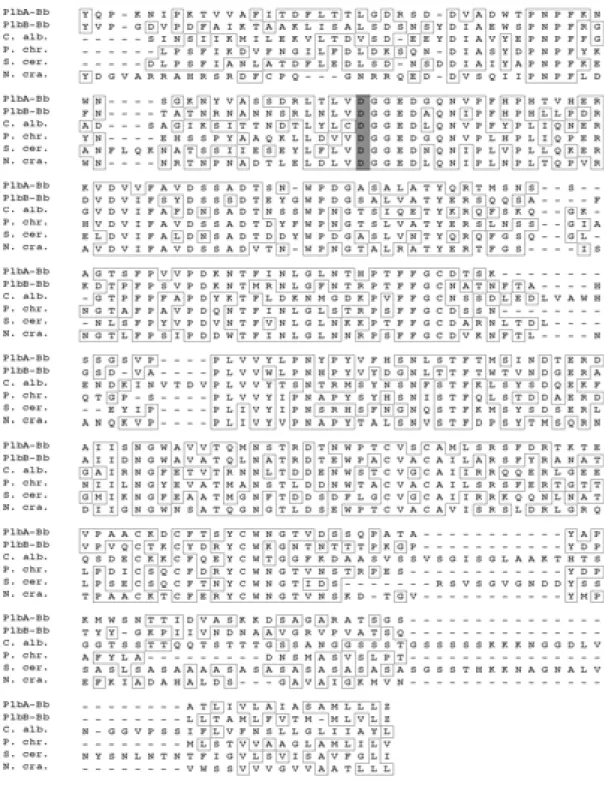
The deduced amino acid sequence of PLB1 was highly homologous to that of PLB2 (57% identity). Comparison of the putative B. bassiana PLBs (PLB1 and PLB2) with other proteins in the database (using the BLASTP program) revealed high homology to known fungal PLBs from S. cerevisiae (40% and 38%), P. chrysogenum (46% and 43%), N. crassa (44% and 43%), C. albicans (38% and 35%), S. pombe (34% and 29%) (Fig. 2.5).
Unlike the PLBs of the nonpathogenic fungi, S. cerevisiae and S. pombe, B. bassiana PLB1 and PLB2 were characterized by the absence of a hydrophobic COOH terminus which may signal for the addition of a glycosylphophatidylinositol (GPI) anchor (Lu, 1995).
Fig. 2.5. Sequence alignment of fungal PLBs. The amino acid sequences predicted by B. bassiana PLB1 and PLB2 were aligned with PLBs from C. albicans (C. alb.; gb/
AAF08980.1), P. chrysogenum (P. chr.; WISS-PROT: P39457), S. cerevisiae (S.cer.; ref/
NP_013721.1), N. crassa (N.cra.; gb/ AAC03052.1), and S. pombe (S. Pom.; ref/
NP_593196.1). Identical amino acids are boxed. Shaded areas indicate residues of putative importance for catalysis.
2.2.4. Functional studies of two enzymes involved in the infectious process
2.2.4.1. Heterologous expression of the fragments encoding antigenic determinants of PR1 and PLB2
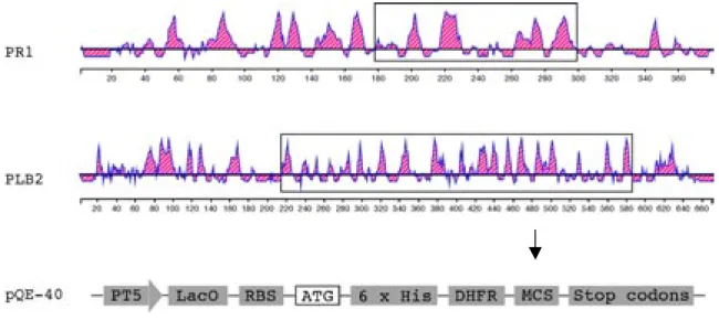
In order to investigate the significance of cuticle-degrading enzymes and their formation in the course of the infectious process, antisera against peptides derived from PR1 and PLB2 were raised. Part of the cDNAs encoding PR1 (obtained from A. Leclerque) and PLB2, which display high antigenicity were chosen as antigens, and cloned in frame into the multiple cloning sites of the vector pQE40 generating the plasmids pQE-PR1 and pQE-PLB2 (Fig. 2.6). The gene encoding Dehydrofolate reductase (DHFR) and a hexahistidine tag, harboured by pQE40, was located at the 5’ end of the cloning site. The hybrid gene encodes the corresponding fusion protein.
The expression of the fusion protein is under the control of the T5 promoter that is fused to the lacZ operon. The generated fusion proteins were termed DHFR-PR1 and DHFR-PLB2, respectively.
Fig. 2.6. Cloning the DNA fragments encoding the PR1 and PLB2 antigen in the pQE-40 expression vector. The antigenicity of the PR1 and PLB2 amino acid sequences was obtained using the Lasergene program package. Domains with high antigenicity correspond to shaded areas above the baseline. DNA sequence encoding the regions with strong antigenicity (boxed) were selected for bacterial expression using the vector pQE40.
2.2.4.2. Purification of the fusion proteins and raising of antisera
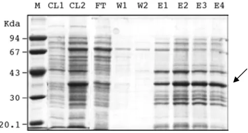
The plasmids pQE-PR1 and pQE-PLB2 were transformed into the E. coli strain SG13009 (Qiagen). A helper plasmid pREP4 contained in the recipient strain encodes the lacZ repressor, thus the expression of the fusion protein was regulated by the addition of IPTG which can induce the lacZ promoter. The fusion protein which was present mainly in a soluble form was purified under denaturing conditions on a Ni-NTA agarose column (Fig. 2.7). The fusion proteins of about 39kDa for DHFR- PR1 and 70kDa for DHFR-PLB2 were visible on Coomassie-stained SDS-PAGE gels and their identity was confirmed by Western blot analysis using an anti-DHFR antiserum. The proteins were excised from the gel and used to raise antisera in rabbits.
Fig. 2.7. Purification of DHFR-PR1 using a Ni-NTA agarose column under denaturing conditions. Fractions were visualized by Coomassie blue staining after separation on a 12.5% SDS-PAGE gel. M, protein marker; CL1, cleared lysate of noninduced control cells; CL2, cleared lysate of IPTG (1 mM) induced cells; FT, flow-through; W1-W2, first and second washing fractions; E1-E4, serial eluates. The arrow indicates the PR1 fusion protein band.
2.2.4.3. Insect cuticle as an inducer of PR1 and PLB2 production
Previous results from M. anisopliae have shown that production of PR1 is transcriptionally modulated by carbon catabolite and nitrogen metabolite repression.
Further induction is obtained in poor media by the addition of insect cuticles.
Therefore, two kinds of media were prepared: (1) Cuticle medium (Cu), 0.8%(w/v) of pulverized insect cuticle was added in basal salt medium as the sole carbon and nitrogen source; (2) Complete Medium (CM) containing sucrose and nitrate as the carbon and nitrogen source.
B. bassiana was grown for 12 hours in complete medium and cuticle medium following the medium transfer steps introduced in Materials and Methods. Gene expression and enzyme synthesis was detected by RT-PCR and Western blot analysis, respectively.
Production of PR1 by B. bassiana
From the Coomassie-stained gel (Fig. 2.8A) it can be seen that the total array of protein production in B. bassiana has been altered in cuticle medium as compared to that in the complete medium. A band corresponding to the predicted size of PR1 was clearly detected in the mycelium fraction as well as in the extracellular protein fraction when B. bassiana was grown in cuticle medium (Fig. 2.8B). In contrast, PR1 could not be detected extracellularly and intracellularly when the cells were grown in the complete medium. Synthesis of PR1 occurs rapidly upon cuticle induction. The protein can be detected in cells and the supernatant of a culture inoculated in cuticle medium after four hours (data not shown).
Corresponding results at the transcriptional level were obtained by RT-PCR analysis. As shown in Fig. 2.8C, pr1 mRNA was highly expressed in B. bassiana grown in cuticle medium. Formation of pr1 mRNA could not be detected after growth in complete medium.
C
Fig. Fig. 2.8. PR1 production by B. bassiana. Mycelium was grown in complete medium (CM) and cuticle medium (Cu) for 12 hours. The proteins from mycelium (P) and supernatants (S) were extracted and separated on a 12.5% SDS- PAGE gel followed by: (A) Coomassie analysis for estimation of protein production;(B) Immunoblot analysis using the antisera against PR1. Arrowhead indicates the PR1 band. M, protein marker. Panel (C):
Transcription of pr1 mRNA detected by RT- PCR analysis. Total RNA was isolated from mycelium harvested from different growth media. The RT-PCR reactions were carried out using the same amounts of RNA. M, DNA marker VI (Roche).
Production of PLB2 by B. bassiana
By immunoblot analysis using the PLB2 antisera, a polypeptide fragment that corresponds in size to PLB2 (70 kDa) was detected in B. bassiana culture grown in complete medium and cuticle medium, respectively. The highest amount of PLB2 was produced by cells grown in cuticle medium (Fig. 2.9A). Secretion of PLB2 to the culture medium was not detected under the same experimental conditions.
The transcription of the plb2 gene was detected by RT-PCR analysis. As shown in Fig. 2.9B, plb2 mRNA was expressed in the cells independent of growth conditions.
A B
Fig. 2.9. (A) PLB2 production by B. bassiana. Mycelium was grown in cuticle medium (Cu) or complete medium (CM) and the proteins from mycelium (P) and supernatants (S) were extracted and separated on a 10% SDS-PAGE gel followed immunoblot analysis with anti-PLB2 antisera., arrow indicates the PLB2 band. M, protein marker. (B) plb2 mRNA transcription detected by RT-PCR. Total RNA was isolated from mycelium harvested from different growth medium. The same amounts of RNA was applied for each RT-PCR reactions. M, DNA marker VI (Roche).
2.3. Discussion
2.3.1. Production of proteases by B. bassiana 252
Insect pathogenic fungi produce an array of enzymes capable of degrading protein, chitin, and lipid components of the insect cuticle (St Leger et al., 1986a). However, a convincing role in pathogenesis has been established only for the serine protease PR1. The activity of PR1 is a prerequisite for successful penetration of the host by M. anisopliae ( St Leger et al., 1989b).
From the cDNA sequence and protein structure, it has been shown that PR1 resembles the powerful serine endoprotease proteinase K, but PR1 is far more effective than proteinase K in degrading insect cuticle, indicative of pathogenic specialization. The higher activity of PR1 could come from the positively charged residues (His17, Arg18 and Arg20) located on the surface of the protease PR1, that are absent from Proteinase K. The presence of this charged domain might allow electrostatic interactions with negatively charged groups in the insect cuticle (St Leger, 1992).
The study of PR1 from M. anisopliae has proven that PR1 expression is regulated by both carbon catabolite and nitrogen metabolite repression and is specifically induced by cuticular proteins after carbon and nitrogen starvation. The level of PR1 was induced tenfold within 24h of contact with cuticle and the regulation was found to be at the transcriptional level (Paterson et al., 1994). Data from this work show that although mycelium growing in complete medium containing carbon and nitrogen sources can synthesize different proteins, the production of PR1 is simultaneously repressed by high levels of nutrients (e.g., sucrose). Under these conditions expression of PR1 was not detectable at both transcriptional and translational level.
In B. bassiana, the production of PR1 seems to be regulated in a similar way as in M. anisopliae. PR1 was found to be exclusively expressed under the cuticle-induced conditions, as judged by immunoblot analysis using an anti-PR1 antiserum.
A protein band migrating slightly slower than PR1 was detected by Western Blot analysis, but its identity is not determined. Several possibilities can be considered: it may arise from protein degradation, crossreaction of the antibody possibly related to the infectious process as suggested by St Leger (1989) or posttranslational modification.
2.3.2. Trypsin-like serine proteases
Another major endoprotease produced in vitro on insect cuticle is the trypsin-like serine protease PR2. The expression of this enzyme has been detected in M. anisopliae. The PR2 protease has been purified and its regulation has been investigated in both M. anisopliae and B. bassiana (Cole, 1993 and St Leger, 1987).
But biochemical characterization of PR2 has only been performed in M. anisopliae.
Two isoforms of PR2 have been identified from M. anisopliae, however, the unique, specific functions of PR2 in pathogenesis has not yet been elucidated. St Leger (1996) determined the location of PR2, using immunogold labeling, to be at the M. anisopliae cell wall during growth through insect cuticles. This indicates that the PR2 proteins might have a role in degrading extracellular proteins as a complement to PR1 and other enzymes. PR1 is produced as a pro-enzyme, requiring processing before it is active. Thus, PR2 could also play a role in the activation of PR1.
Cloning data from this work showed that B. bassiana possesses two genes encoding PR2-like serine proteases of the trypsin family. The sequences of these two genes, designated try1 and try2, have been determined. Interestingly, the predicted amino acid sequence of try1 showed highest homology to the trypsin-like proteases from fungi, whereas the predicted TRY2 protein showed highest homology to the trypsin- like proteases from insects. Considering the experimental conditions and previous results obtained under the same conditions, the possibility of contamination was ruled out. Similar observations have been made regarding trypsin-like proteases from the bacterium Streptomyces griseus which showed close homology to mammalian trypsin-like proteases. It was hypothesized that a gene transfer might have been taken place from a mammal to a bacterium (Olafson, 1975). However, this suggestion could later be ruled out using phylogenetic analysis of sequence data accumulated from different organisms (Rypniewshi, 1994). The structure similarity of TRY2 protease from B. bassiana to insects enzymes might allow the fungal cells to evade host “non-self” recognition and thus might represents one of the important virulence determinants.
Like PR1, the trypsin-like proteases PR2 is also controlled by multiple regulatory mechanisms which include carbon and nitrogen metabolite repression/ derepression as well as induction by a range of proteins. Based on the model system M. anisopliae, St Leger (1988) found that the regulation of PR1 and PR2 is not identical; although PR1 was not detected under carbon/nitrogen repression
conditions, low levels of PR2 were produced. It was found that the soluble protein bovine serum albumin (BSA) represses production of PR1, and at the same time stimulates synthesis of PR2 in poor medium. In addition, PR2 production could be more tightly regulated by nitrogen than carbon availability (Paterson, 1993). This type of protein regulation is not uncommon. There are examples of fungal proteases which are induced by any protein substrate, as observed in N. Crassa (Drucker, 1975) and Candida spp. (Ross, 1990). These proteases are produced when the fungus is starved for either carbon, nitrogen or sulphur sources. Some fungal proteases appear to be regulated by derepression alone, for example, in Schizophyllum commune (Willick et al., 1984) and many Aspergillus species (Hanzi et al., 1993).
2.3.3. Expression of phospholipase B in B. bassiana 252
Two PLB-encoding genes were cloned from B. bassiana and their sequences were determined. They displayed 57% identity at the amino acid level. Three PLB- encoding genes in S. cerevisiae (Witt et al., 1984) and two PLB homologues in C.
albicans (Takahashi et al., 1991) have been isolated. The multiple PLB homologues from these fungi display high homology to each other, but the significance of this genetic redundancy is unclear.
Potential N-glycosylation sites were identified in both PLB1 and PLB2. All characterized fungal phospholipases have been glycosylated. Chen et al., (2000) found that deglycosylation of the protein resulted in almost total loss of enzyme activity. This indicates that N-linked carbohydrates are important for the catalytic function of the protein. The immuno blot assay in this work with an antibody against PLB2 produced a broad band corresponding to a mass of around 70 kDa. The slightly heterogeneous migration of PLB2 upon SDS/PAGE separation has also been observed in C. neoformans (Chen et al., 2000) and other fungal PLBs (Oishi et al.,1999). It is supposedly due to heterogeneity of the carbohydrate moiety linked to the enzyme.
A hydrophobic carboxy-terminal region was identified in PLBs from non-pathogenic fungi, such as S. cerevisiae, S. pombe, P. notatum (Caro et al., 1997; Gerber et al., 1992). These COOH-terminal regions have been hypothesized to contain conserved sequence motif that is a signal for the addition of a glycosylphosphatidylinositol (GPI) anchor. Proteins modified with a GPI anchor may be transiently tethered to the
plasma membrane or ultimately cross-linked to the insoluble glucan component of the cell wall (Lu et al., 1995). Release of proteins associated with the plasma membrane would require the action of a GPI-specific phospholipase. Therefore, the GPI anchor may serve to regulate the release of the enzyme to the surroundings. The absence of a GPI anchor in B. bassiana PLBs that is also observed in the C. albicans PLBs, could cause the PLBs from these organisms to be secreted directly thereby enhance the virulence of pathogenic fungi.
The high amounts of PLB2 synthesised from B. bassiana grown on insect cuticle suggests that this enzyme is likely to be involved in the early steps of host invasion.
Unlike PR1, production of PLB was not tightly regulated by carbon/ nitrogen repression mechanisms. The transcription of PLB mRNA was detected by RT-PCR in both mycelial samples harvested from rich medium and cuticle-induced medium.
Although PLB2 synthesis is highest in the mycelium grown in cuticle-induced medium, the enzyme is also detected in cultures grown on rich medium. Some unspecific bands were detected by immunoblot analysis and this unspecific banding maybe because the synthesized antisera was not pure enough.
2.3.4. Function of cuticle-degrading enzymes during fungal infection processes
The high-level production of proteases during infection process implies an important function for these enzymes. Aside from the proteolytic degradation of the cuticular barrier, other possible roles include the utilization of host proteins for nutrition, the destruction of antifungal proteins of the host, and the release of amino acids for amine production which could elevate the pH to produce better growth conditions.
Other effects may be indirect, for example, the proteolytic activation of toxin precursors.
The results obtained from this work are consistent with the idea that a major function of the extracellular proteases is to make nutrients available from the cuticle. The pathogenic process involving enzyme production and penetration of host cuticle occurs only when it is necessary to establish a nutritional relationship with the host.
It is probable that the synthesis of proteolytic enzymes and phospholipases occurring in cultures containing host cuticle may also occur when the pathogens are infecting living insects.
A highly reliable method to categorically demonstrate the involvement of a protein in pathogenicity is through genetically engineered null mutants. The multiplicity of cuticle-degrading enzymes provides a major challenge with respect to establish which particular enzyme has which function in adapting to a new environment or in pathogenicity. The exact role of individual cuticle-degrading enzymes in pathogenicity could be assessed by disruption of multiple genes in combination with site-directed mutagenesis experiments. An increased understanding of these virulence factors could ultimately lead to the generation of genetically engineered strains more suited for insect pest control.
III. Development of a new dominant selection marker, sorR, for fungal transformation
3.1. Introduction
The filamentous fungi are a diverse group of eukaryotic microorganisms with a number of properties which make them important both scientifically and economically. The economic importance can be illustrated by the large variety of products that are made by filamentous fungi, such as organic acids (e.g. citric acid), antibiotics (e.g. penicillin and cephalosporin) and numerous industrial enzymes (e.g.
glucoamylase, cellulase, alpha-amylase, proteases, lipases, etc.). Filamentous fungi are also used as food products (e.g. cheese, mushrooms), food additives (e.g. the meat extender “Quorn”) and condiments (e.g. soy sauce). Furthermore, entomopathogenic fungi have been widely used for the biological control of either insect pests or plant pathogenic fungi in agriculture and forestry.
A severe, negative economic influence of filamentous fungi is their detrimental effect on crop yield. Plant pathogenic fungi cause devastating crop losses all over the world. In the medical field, fungal diseases have become increasingly important.
Candida albicans and some fungal pathogens, although normally existing as a harmless commensal, can cause significant morbidity and mortality in patients immunodepressive due to AIDS, organ transplantation, or chemotherapy.
In addition to their economic importance, genetic and biochemical studies of filamentous fungi have contributed to the development of concepts about biosynthetic pathways and gene-enzymes relationships. The “ one-gene-one- enzyme” hypothesis was based on work done with the filamentous fungus Neurospora crassa (Beadle et al., 1941). Filamentous fungi have several interesting biological properties such as a complex life cycle, cell differentiation, highly regulated metabolic pathways and efficient secretion of proteins and they offer many of the complexities of a eukaryote with the ease of manipulation of a microorganism which make them attractive as a model for basic biological research. (Timberlake and Marshall, 1989)
The importance of fungi in medicine, industry, agriculture, and science has led to intensive investigation for many years with the aim of understanding and controlling
their detrimental and beneficial activities. Genetic engineering of filamentous fungi provides the ultimate tools for this aim in novel ways. Genetic transformation of fungi started from the experiments with S. cerevisiae (Hinnen,1978), A. nidulans (Yelton,1984) and N. crassa (Case,1980) which are the traditional favourites of the fungal geneticist. A great deal of information was accumulated concerning the nature of transformation events and selection procedures. Shortly thereafter, representatives from all major fungal classes have been shown to be amenable to transformation.
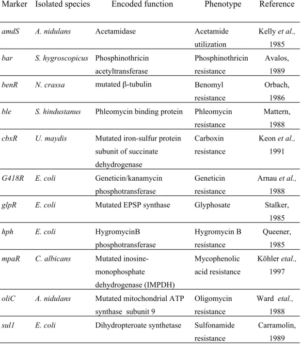
The application of recombinant DNA techniques and the development of transformation systems for filamentous fungi initially was hampered by the lack of useful selectable markers and/or the availability of the appropriate recipient. This was overcome by the isolation and application of homologous and heterologous selection markers and the development of selection systems (Goosen,1980). Two types of selection marker have been used: (1) auxotrophic selection markers, (2) dominant selection markers.
3.1.1. Auxotrophic selection markers
Transformation using auxotrophic selection markers depends on the availability of auxotrophic mutants. The wild-type gene, in plasmid-borne form, complements corresponding auxotrophic mutations of both homologous and heterologous recipients and therefore provides a selectable marker. Several genes have been used as auxotrophic selection markers for transformation studies, e.g., N. crassa pyr-4 gene, Aspergillus nidulans amdS, trpC, pyrG, niaD,sC and argB genes. Such transformation systems have been developed for a number of filamentous fungi, such as, N.crassa (Buxton and Radford, 1984), A. nidulans (Johnstone, 1985) and Penicillium chrysogenum (Sanchez , 1987). An overview of commonly used auxotrophic markers is given in table 3.1.
3.1.1.1. General introduction of commonly used auxotrophic selection marker
For P.chrysogenum, relief of an auxotrophic requirement for tryptophan has been achieved by transformation with the wild-type trpC gene that, similarly to the N.crassa trp-1 gene (Schechtman and Yanofsky, 1983) or the A. nidulans (Yelton et al., 1983) and A. niger (Kos,1985) trpC genes, encodes a trifunctional polypeptide
that contains the GAT, IGPS and PRAI activities of the tryptophan biosynthetic pathway (Sanchez, 1986).
The pyrG gene has been used successfully as a selectable marker in filamentous fungi. It encodes the orotidine-5-monophosphate decarboxylase enzyme which is required for the synthesis of uracil, providing the precursor of the three pyrimidine trinucleotides as well as for the synthesis of di- and polysaccharides. The cell with wild-type pyr gene is uridine prototroph but unable to grow in the presence of 5- fluoroorotic acid (5-FOA) which can be converted to 5-fluorouracil, a toxic compound to the cells whereas the pyr(-) strain confers resistance against 5-FOA.
pyrG homologs have been identified in a number of other species as well, including A. nidulans (pyrG) (Oakley and Rinehart,1987), A. niger (pyrG) (Wilson &
Carmona,1988), Candida albicans (ura3) (Losberger and Ernst,1989), and Schizosaccharomyces pombe (ura4) (Grimm and Kohli,1988).
The niaD gene encodes nitrate reductase which is a key enzyme involved in the first step of nitrate assimilation. The niaD gene is required for growth when nitrate is the sole nitrogen source. The niaD(+) strain is nitrate prototroph and sensitive to chlorate whereas the niaD (-) strain is resistant to chlorate. The chlorate is not itself toxic, but is rendered toxic by conversion to chlorite as a result of the catalytic action of nitrate reductase.
The sC gene, which encodes an ATP sulfurylase that catalyses the activation of inorganic sulfate to produce adenosine-5’-phosphosulfate (APS), has been developed as a selectable marker for the transformation of A. fumigatus (De Lucas and Dominguez, 2001) and A. niger (Buxton and Gwynne, 1985). In filamentous fungi, expression of the sC gene is required for the assimilation of sulfate as a source of sulfur. sC (-) mutants are unable to utilize sulfate as sulfur source. Instead they require reduced sulfur (e.g., methionine) as sulfur source. In addition, they are resistant to selenate, a metabolite that resembles sulfate but becomes toxic after reduction inside the cell.