Adhesive polymer films for the prevention of postsurgical adhesions
Dissertation zur Erlangung des Doktorgrades der Naturwissenschaften (Dr. rer. nat.)
der Fakultät für Chemie und Pharmazie der Universität Regensburg
vorgelegt von Eva Esser aus Kleinenbroich
Januar 2016
This work was conducted from July 2010 until October 2012 at the Department of Pharmaceutical Technology of the University of Regensburg and from November 2012 until June 2014 at the Department for Functional Materials in Medicine and Dentistry of the University Hospital of Würzburg under the supervision of Prof. Dr. Achim Göpferich and Prof.
Dr. Jürgen Groll.
Promotionsgesuch eingereicht am: 18.01.2016 Datum der mündlichen Prüfung: 17.02.2016
Prüfungsausschuss: Prof. Dr. Sigurd Elz (Vorsitzender)
Prof. Dr. Achim Göpferich (Erstgutachter) Dr. Jörg Teßmar (Zweitgutachter)
Prof. Dr. Jörg Heilmann (Drittprüfer)
i Contents
1. Barrier films for adhesion prevention: What can we learn from wound dressings ... 1
1.1 Introduction ... 2
1.2 Differences of the two application sites ... 4
1.3 Structure and function of skin and peritoneum ... 4
1.4 Differences in wound healing ... 4
1.5 Different Requirements on wound dressings and barriers for adhesion prevention 6 1.6 Wound dressings and barrier films prepared from polymers ... 12
1.7 Summary and conclusions ... 15
2. Aim of this work ... 17
3. Alginate films cross-linked with different methods ... 21
3.1 Introduction ... 22
3.2 Film preparation techniques ... 24
3.3 Optimizing the mechanical properties ... 29
3.4 Erosion and swelling of the alginate films ... 51
3.5 Biological evaluations of alginate films ... 60
3.6 Summary and Conclusion ... 65
4. Optimization of alginate film preparation with predefined calcium amounts ... 67
4.1 Pretests before film preparation with “optimized” calcium amount ... 69
4.2 Film preparation ... 75
4.3 Preparation with optimized film preparation technique ... 77
4.4 Mechanical evaluation ... 78
4.5 Erosion in different buffers ... 89
4.6 Cytotoxicity tests of the prepared alginate films ... 96
4.7 Discussion ... 97
4.8 Summary and Conclusion ... 98
ii
5. Drug release from thin polymer films ... 101
5.1 Introduction ... 102
5.2 Drug loading of thin polymer films ... 104
5.3 Release studies ... 112
5.4 Water uptake of the different polymer films ... 114
5.5 Biological activity of released drug tested via microbiological testing ... 114
5.6 Results and Discussion ... 118
5.7 Summary and Conclusion ... 122
6. Thiolated hyaluronic acid ... 125
6.1 Introduction ... 126
6.2 Synthesis of thiolated hyaluronic acid ... 127
6.3 Characterization of thiolated hyaluronic acid ... 130
6.4 Film preparation ... 138
6.5 Mechanical evaluation ... 141
6.6 Film swelling ... 146
6.7 Summary and Conclusion ... 148
7. Multilayer ... 151
7.1 Introduction ... 152
7.2 Materials and methods ... 153
7.3 Preparation of Multilayers using different cohesion promoters ... 153
7.4 Preparation of Multilayers using increased surface roughness132 ... 165
7.5 Results ... 169
7.6 Discussion ... 175
7.7 Conclusion ... 175
8. Summary and Outlook ... 177
8.1 Summary ... 178
iii
8.2 Conclusion and Outlook ... 184
9. Zusammenfassung und Ausblick ... 185
9.1 Zusammenfassung ... 185
9.2 Schlussfolgerung und Ausblick ... 192
10. References ... 193
11. Danksagung ... 203
iv
v Abbreviations and Symbols
% percent
° degree
°C degree Celsius
µg microgram
µl microliter
µm micrometer
ANOVA analysis of variance
ASTM American Society for Testing and Materials
C12H10Ca3O14 4 H2O calcium citrate tetrahydrate
CA contact angle
CaCl2 calcium chloride
CaCO3 calcium carbonate
CaHPO4 calcium phosphate
CaSO4 calcium sulfate
cm centimeter
CO2 carbon dioxide
D2O deuterium oxide
Da Dalton
DCM dichloromethane
DIAD diisopropyl azodicarboxylate
DMEM Dulbecco’s modified eagle’s medium
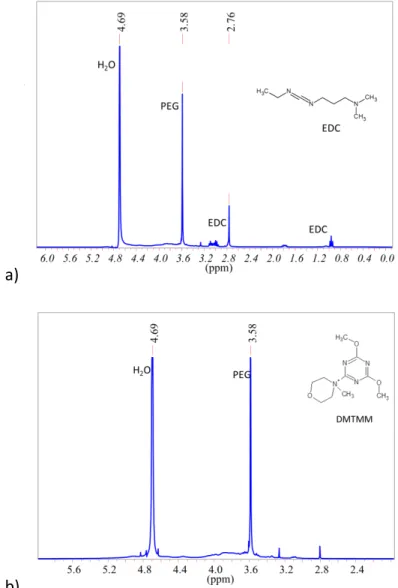
DMTMM 4-(4.6-Dimethoxy-1,3,5-triazin-2-yl)-4-
methylmorpholinium chloride
DTP Dithiopropionic acid dihydrazide
DTT 1,4-Dithiothreitol
EDC N-(3-Dimethylaminopropyl)-N’-
ethylcarbodiimide hydrochloride
EDTA ethylenediaminetetraacetic acid
EGTA ethylene glycol tetraacetic acid
EMEM Eagle`s minimal essential medium
e-modulus elastic modulus
eq equivalent
vi
FCS fetal calf serum
g gram
G` Elastic modulus
G`` Viscous modulus
G-block Guluronic acid block
GDL gluconic acid δ-lactone
GPC gel permeation chromatography
h hour
H2O2/ HOOH hydrogen peroxide
H2SO4 sulfuric acid
HA hyaluronic acid
HA-SH thiolated hyaluronic acid
HEPES 4-(2-hydroxyethyl)-1-
piperazineethanesulfonic acid
High G alginate alginate with high amount of guluronic acid
I2 iodine
ITC isothermal titration calorimeter
kDa kilo Dalton
kg kilogram
KI potassium iodide
kPa kilopascal
l liter
Low G alginate alginate with low amount of guluronic acid
M molar
m mass
MDa mega Dalton
mg milligram
MgSO4 magnesium sulfate
min minute
ml milliliter
mm millimeter
mM (mmol) millimolar
Mn number average
vii
mol molar
MPa megapascal
MTT-Assay 3-(4,5-dimethylthiazol-2-yl)-2,5-
diphenyltetrazolium assay
MW molecular weight
Mw weight average molecular weight
Mw/ Mn polydispersity index
N Newton
NaCl sodium chloride
NaOH sodium hydroxide
nm nanometer
NMR nuclear magnetic resonance
Pa pascal
PBS phosphate buffered saline
PEG poly (ethylene glycol)
pH negative logarithmic value of the hydrogen
ion concentration
PLA Poly (L-lactide-co-D,L-lactide; 70:30)
PLA PEG PLA
(PLA35kDa-PEG10kDa-PLA35kDa)
lipophilic triblock copolymer of poly(lactic acid) and poly(ethylene glycol)
PPh3 triphenlyphosphine
rpm revolutions per minute
SD Substitution degree
SDS sodium dodecyl sulfate
sec second
SEM scanning electron microscopy
THF tetrahydrofuran
Tris Tris(hydroxymethyl)aminomethane
UV-Vis ultraviolet-visible
W0 dry weight
WST Water-soluble Tetrazolium salt
wt wet weight
1
1. Barrier films for adhesion prevention: What can we learn from wound dressings
This chapter gives an overview of polymers that can be used as wound dressings and are therefore good candidates for the preparation of films for the prevention of peritoneal adhesions.
2 1.1 Introduction
In the modern wound care natural and synthetic polymers are often used for wound dressings due to specific beneficial characteristics, like the ability to regulate a moisture imbalance by removing wound exudate and maintaining a moist wound environment or the ability to donate moisture 1,2. Advanced non-adherent hydrogel dressings can be furthermore removed pain free without causing damage to the injured tissue 2, which promotes the desired wound healing and enhances the restoration of the skin defect. To prevent peritoneal adhesions after surgery similarly designed barrier films made of different degradable and non-degradable polymers already showed excellent clinical results due to an appropriate separation of the damaged tissues during the wound-healing period.
Accordingly, a closer look onto the characteristics of polymers, which are used for effective wound dressings, should allow the identification of new candidates for the preparation of alternative films for adhesion prevention and moreover improve currently used systems for this application.
In order to appropriately compare the two applications and learn from each other, the differences in the healing of skin defects and defects of the peritoneum and the resulting polymer requirements, which are used for the preparation of the medical devices, will be highlighted in more detail.
Modern wound dressings for the treatment of acute and chronic external wounds are made of polymers, which fulfill many different clinical functions, like for example the uptake of wound exudate or the maintenance of a humid wound environment to enhance the healing process. The established polymers for this application, like for example alginate and hyaluronic acid, already showed many benefits in the treatment of skin defects. But also for the prevention of peritoneal adhesions hyaluronic acid is already used in the application form of a gel or a film, because the separation of the traumatized tissues is currently considered to be the most effective way to prevent the formation of adhesions.
For both types of wounds biomaterial characteristics like biocompatibility, biodegradability, non-immunogenicity, hemostatic activity, as well as the absence of toxic, carcinogenic or thrombogenic activities are essential to fulfill the criteria of suitable devices to treat patients
3,4. Accordingly many polymers have already been used for both applications, for wound
3
dressings, as well as for films for adhesion prevention, with different success and a different focus of their application.
The research of George D. Winter in 1962 firstly demonstrated the beneficial effect of a moist environment on the wound healing process, because wounds that are exposed to air form a scab that retards epithelization. Wounds, covered with a polyethylene film to keep them moist do not form a scab and the epithelization is more rapid 5. Since this time, many wound dressings made of different hydrogel forming polymers, which create a moist environment for healing, were invented and successfully brought to the clinic. Besides a moist environment these hydrogels have many more beneficial advantages. They are non- adherent on the wound and therefore can be removed from the wound without causing additional pain and further damage to the newly grown skin epithelium 6. On the other hand, exuding wounds need a wound dressing that is able to incorporate large amounts of wound exudate, which can be accomplished by wound dressings made of alginate 7, a natural polymer that is known to absorb large amounts of liquid resulting in the formation of stable hydrogels. Another quite similar polysaccharide, which is used for wound dressings due to its ability to reduce scar formation and alternatively already applied for adhesion prevention as a biodegradable and biocompatible hydrogel, is hyaluronic acid, which only differs slightly in structure and functional groups (Figure 1).
a) b)
Figure 1: a) alginic acid, b) hyaluronic acid
To prevent the formation of peritoneal adhesions, a reduction respectively inhibition of scar formation is also highly desirable. Therefore hyaluronic acid seems to be an advantageous polymer for the preparation of films for adhesion prophylaxis. In the peritoneal cavity the effective uptake of wound exudates or bacteria by a polymer like alginate should not be expected to be that helpful. However, alginate also showed promising results in the
4
prevention of peritoneal adhesions due to some other beneficial characteristics like its mucoadhesive properties 8.
1.2 Differences of the two application sites
To identify the essential prerequisites for the two application sites, differences in structure and function should be highlighted and compared in more detail. Furthermore, it is also necessary to understand the differences in two wound healing processes as well as the resulting requirements for wound dressings and adhesion barriers.
1.3 Structure and function of skin and peritoneum
The largest and most important organ of the human body is the skin. It protects the human body from microbes and chemical agents. Furthermore it helps to regulate the body temperature. The skin consists of three different layers the epidermis, the dermis and the hypodermis. The epidermis is the outer layer of the skin. It provides physical protection and works as a waterproof barrier. The dermis, as well as the hypodermis consists of connective tissue consisting of collagen and glycosaminoglycans. Furthermore the dermis contains sweat glands and hair follicles and the hypodermis fat 9.
The peritoneum is a protective membrane, too. Unlike the skin, its assignment is not to protect the human body from environmental influences, but to cover the organs and allow them to move upon each other without friction 10. It consists of a sheet of connective tissue and a single layer of mesothelial cells that are anchored to the basement membrane 11,12. Both the skin as well as the peritoneum can like any other tissue or organ lose their functionality by the formation of scar tissue after trauma. Furthermore injured peritoneum can form peritoneal adhesions. Therefore it is of utmost importance to enhance the wound healing and reduce the scar formation to retain their functionality.
1.4 Differences in wound healing
The wound healing of a skin defect differs from that of a peritoneal defect. The healing of a skin defect can be divided into the following stages. In the first stage, called the immediate, the hemostasis occurs. The initiation of the coagulation cascade leads to the formation of a fibrin clot and platelet aggregation. In the second stage, the inflammation takes place with
5
the activation of the complement cascade, which comes along with the invasion of neutrophils and macrophages. In the third stage, the proliferation, the formation of granulation tissue occurs. Fibronectin, hyaluronan and collagen are produced by migrated fibroblasts and the wound is closed by epithelization. In case of skin defects, the epidermalization takes place from the borders of the wound until the wound is closed. The final stage is the remodeling stage that leads to the scar maturation. In this stage fibronectin and hyaluronan are broken down and replaced by collagen. In open wounds the wound healing is finished with a contraction step 1,13–15.
In contrast to the skin, the entire surface of a peritoneal wound becomes epithelialized simultaneously and not gradually from the borders like in skin defects. When new mesothelial cells are attracted to the site of injury by chemotactic messengers that are released by blood clots, platelets or leukocytes, they form multiple individual islands on the peritoneal defect. From these islands they divide until the surface of the injury is completely covered by a new layer of mesothelial cells 16. Therefore, the time needed for the healing process is more or less the same for smaller and larger peritoneal defects, since no cell migration is necessary and cell division is initiated from multiple healing sites. In contrast skin defects typically need longer healing times depending on the size and depths of the wound.
During the peritoneal healing injured parts of the peritoneum can be connected due to the formation of stable fibrin bands, which is enhanced due to the immediate contact of the insured tissues. In normal peritoneal healing these fibrin bands are subsequently resolved by fibrinolysis and the natural tissue organization is restored. If the bands are not effectively resolved due to impaired fibrinolytic activity, these weak fibrous bridges can be later infiltrated with fibroblasts, followed by vascularization and collagen deposition, which ultimately results in the formation of persisting adhesions. As a consequence different side effects can occur like pain, small-bowel obstruction, secondary infertility and furthermore 16–
18.
6
Figure 2: Wound healing of skin and peritoneum
Both healing processes of skin and peritoneum have much in common. They can result in the formation of scared tissue that ultimately lost its functionality and causes discomfort or even pain for the patient. For the application of a wound dressing as well as for a film for adhesion prevention, the aim should be to reduce the scar formation and moreover the formation of adhesive bonds in case of the peritoneum.
1.5 Different Requirements on wound dressings and barriers for adhesion prevention Wound dressings as well as barrier films should obviously be non-toxic, non-immunogenic and biocompatible. The also essential preparation of sterile wound dressings and barrier films comes along with the difficulties that most natural and synthetic polymers partially degrade or chemically suffer during autoclaving or irradiation procedures, resulting in altered device properties as well as undesirable breakdown products. Therefore, it is also essential to find a way to prepare sterile products or at least to be able to sterilize them as gentle as possible after preparation of the device.
Skin defect
Haemostasis Fibrin clot formaqon
Inflammaqon Neutrophil invasion Macrophage invasion
Angiogenesis Epithelizaqon Fibroblast recruitment
Remodeling Collagen synthesis
Collagenolysis
Contracqon aser healing of open
wounds
Peritoneal trauma
Hameostasis
Inflammaqon Neutrophil invasion Macrophage invasion
Formaqon of a fibrin matrix
Fibrinolysis
Normal healing
Formaqon of a fibrin matrix
Infiltraqon of fibroblasts
Vascularizaqon
Adhesion persisqng
7
The most significant requirements for the wound dressings depend on the actual type of wound that has to be covered. For an appropriate wound healing an optimal environment is necessary, which may be different for every type of wound. Most wounds need a moist environment. This is naturally given in the peritoneal cavity and moreover should be effectively protected from bacteria, which could cause inflammation and result in further complications of the wound healing process. In order to keep a moist environment, a dry or desiccated skin defect should be wetted and kept in this state by the wound dressing.
Whereas a skin defect, that produces large amounts of wound exudate should be ideally covered with a wound dressing that is able to take up the exudate and provide a surrounding that stabilizes the healing skin and prevents the elimination of the newly grown epithelium
1,19.
For the peritoneal application the main task of the barrier film is consequently not the protection of the wound from environmental influences and the maintenance of the moist environment, but to effectively separate the damaged tissue long enough to prevent the undesired formation of peritoneal adhesions. However, for both applications it is beneficial, if the surgeon is able to cut the device into the desired size and shape in order to effectively cover different defects. For the application with the peritoneum it is also desirable, if the device can be implanted via laparoscopic intervention and is still flexible enough to cover uneven surfaces of the small intestine or other organs within the patient.
An external wound dressing is most commonly fixed via the adhesive parts of the wound dressing composite itself or via additionally applied external fixing materials like bandages or adhesive tape. An intestinal adhesion barrier on the other hand can only be fixed with the help of sutures or staples, which in consequence can disadvantageously lead to the formation of adhesions and therefore should be most effectively avoided by the preparation of devices or materials, which stick to the treated tissue on their own without causing undesired connections or bands, which later result in the formation of new connections of the adjacent tissue sites. Even more important than the fixation is the removal of both types of wound dressings. This should generally be possible without causing further damage to the wounded skin or peritoneum. For a skin defect, the wound dressing can easily be removed from the wound, if it does not stick to it. Therefore devices are beneficial, which maintain a thin layer of liquid between the tissue and the device, ultimately resulting in an easy removal
8
from the skin. In the peritoneal cavity the removal of the anti-adhesive barrier film has obviously to be done by a second surgical intervention, this again may cause new adhesion sites. To avoid this, peritoneal barrier films should be biodegradable and consequently be removed from the peritoneal cavity by either enzymatic or hydrolytic degradation or just by simple dissolution.
1.5.1 Purity
Polymers, which are used for wound dressings, as well as adhesion barriers, should generally be non-toxic and non-immunogenic, since they both get in close contact with the patient’s tissue and consequently with individual cells, as well as the patient’s immune system 20. As adhesion barriers the polymers are applied parenterally and therefore have to be of even higher purity than the polymers that are used for wound dressings, where the polymer is usually not resorbed and only remains on the outer skin of the patient until it is removed by the doctor or just falls off. Before the preparation of any barrier film or wound cover, it should be additionally considered, that a subsequent sterilization can have dramatic effects on natural macromolecules, as well as synthetic polymers 4. Therefore it is always important to know, if the used polymer can be sterilized before film preparation or if the final product has to be sterilized, obviously without altering its properties or molecular structure. To remove toxic chemicals that have been applied for the synthesis of the polymer like solvents, cross-linkers or unreacted monomers, the obtained polymer should always be sufficiently purified with the help of solvent washing or dialysis 21 before it can be used for the preparation of wound dressings or barrier films. The efficient purification is even more important for natural polymers from plants or bacteria, like alginate 22 and hyaluronic acid, which have to be intensively purified before parenteral application to remove for example residues that remain from the leaves of the algae.
1.5.2 Degradability
Regarding the degradation of polymer devices at the application site, several different pathways are discussed, which all ultimately have to lead to water-soluble breakdown products that can be metabolized or eliminated from the body. Based on the chemical structure of the device two main steps of the degradation have to be distinguished. The first applies to the erosion of a device due to the separation or dissolution of the whole polymer
9
chains that are only loosely fixed by entanglements or weak ionic interactions. A second process involves the breakdown of few chemical bonds, if cross-linked networks of water- soluble polymers are degraded, or even the breakage of the whole polymer chain into monomers, if water insoluble polymers are applied.
Accordingly, devices made of water insoluble polymers, like polyesters or polyanhydrides, can undergo surface erosion or bulk erosion, depending on the diffusion speed of water into the polymer device, this ultimately determines the kinetics of polymer breakdown 23. The degradation of devices made of biodegradable polymers can furthermore involve the hydrolytic or an enzymatic cleavage of the polymer chain, which may also lead to different kinetics of polymer erosion 24. Hydrolytically degradable polymers, which carry functional groups, like esters or anhydrides, only degrade based on the amount of water and steric effects of neighboring groups 24. On the opposite, enzymatically degradable polymers, like collagen, albumin, fibrin or others are cleaved by specific enzymes, which further enhance the polymer’s elimination based on their local amount and activity 24.
The overall degradation of polymers can be largely influenced by secondary modifications of the polymer chains or additional side functionalities. Alginate itself is a hydrophilic polymer, but in a humid environment the alginate chains do not degrade efficiently and devices made of alginate only undergo erosion based on the speed of dissolution of the loose chains. With the help of partial oxidation using sodium periodate, aldehyde groups can be introduced, which render the polymer more susceptible to hydrolysis and oxidation 25. When a wound dressing made of a hydrogel is removed from the wound area, polymer parts may also still stick to the wound, but they can be subsequently washed away during a necessary cleaning of the wound area. Nevertheless it might still be possible, that during the healing process small amounts of the hydrogel have been incorporated in the granulation tissue and will be later removed by biodegradation or can be incorporated in the newly formed tissues, as for example in case of hyaluronic acid hydrogels.
If a peritoneal barrier device will not be removed from the body by a second surgery, the used polymers have to be fully biodegradable or at least the prepared device should completely erode into fragments that can be eliminated from the body. Biodegradable polymers that are used for all kinds of parenteral applications can be eliminated unaltered or metabolized into endogenous compounds, which can be metabolized or also eliminated 20.
10
For example barrier films that are prepared of polylactic acid like Surgiwrap® degrade via hydrolytic degradation ultimately into bioresorbable monomers like lactic acid enantiomers or glycolic acid that can be inserted in biochemical pathways 4.
To prolong the persistence time of very hydrophilic polymers, like alginate or hyaluronic acid, which are dissolved very fast in the humid environment of the peritoneum and washed away from the application side, the polymers usually have to be chemically or physically cross-linked in order to provide a sufficient long persistence time at the application site.
Nevertheless these cross-linked polymer networks should still degrade after the peritoneal healing is ended. For example barrier films made of alginate that have been physically cross- linked with divalent cations like calcium degrade due to a slow exchange of the divalent cations by monovalent cations like sodium 4.
If the released polymer chains can be finally withdrawn from the body, also depends on the hydrodynamic radius of the free polymer chains. Molecules with a size of about 3-4 nm like for example albumin (3.6 nm) and larger would stay in the blood system instead of being withdrawn by renal filtration 26. Accordingly larger water soluble polymers always have to contain certain cleavage sites, where the molecules can be broken down in order to be eliminated from the patient.
1.5.3 Adhesion
Most wound dressings firmly adhere to the skin because they are surrounded by sticky surfaces covered with glue or they can be fixed with the help of bandages or adhesive tape.
Barrier films in the peritoneum cannot be fixed at their application site that easy. They have to be sutured to the tissue or be fixed with staples if they do not stick to the tissue themselves.
The use of mucoadhesive polymers here would allow an application without further fixation and the risk of adhesion formation.
Mucoadhesion can be defined as the ability to adhere to the mucus gel layer 27. Mucoadhesive materials can be described as hydrophilic macromolecules that contain numerous hydrogen bond forming groups. The mechanism of mucoadhesion can be divided into two stages. The contact, where the wetting takes place and the consolidation state where the adhesive interactions are established 28. Chitosan, sodium alginate and cellulose
11
derivatives belong to the so called “wet” adhesives. They can be activated by moistening due to capillary forces and adhere to many surfaces in a wet state. But these polymers can also overhydrate and form a slippery gel, which in consequence detaches from the application site 28. Different mucoadhesive interactions can be achieved by the formation of differently strong bonds like ionic bonds, covalent bonds, hydrogen bonds, van-der-Waals bonds or hydrophobic bonds 27,29. To improve the mucoadhesive properties of polymers like chitosan or alginate thiol groups can be introduced into the polymer chains, which later stabilize the adhesive by forming covalent disulfide bridges with components of the mucus. This can be achieved with the help of polymer analogue modification by coupling small bifunctional molecules like cysteine, thioglycolic acid or cysteamine to the existing polymer chain 27,28,30. Due to its non-adhesive properties to skin defects, wound dressings prepared from alginate always have to be fixed with secondary dressings 3. Regarding barrier devices, alginate is a good candidate due to its mucoadhesive properties, because it can adhere, due to the formation of hydrogen bonds, to the traumatized tissue without further fixation 8. Therefore this polymer can be quite advantageous for both applications.
1.5.4 Uptake of wound exudates and bacteria
The uptake of wound exudates and bacteria is mainly beneficial for external wound dressings, because after incorporation and uptake of the bacteria the wound dressing can be exchanged with a fresh one 6.
In the peritoneal cavity, a polymer film that is capable to incorporate wound exudates like a wound dressing would also incorporate the surrounding peritoneal fluid until it is soaked completely. Furthermore, the wound exudate that is formed by a peritoneal defect commonly is anyway diluted with peritoneal fluid and washed away from the wound site. An efficient uptake of bacteria into a peritoneal adhesion barrier would furthermore be undesired, because they consequently cannot be removed from the peritoneal cavity. On the contrary, the bacteria would finally be released from the barrier again upon its ultimate degradation in the body.
12
1.6 Wound dressings and barrier films prepared from polymers
Many different wound dressings and peritoneal barrier films based on different natural and synthetic polymers are already available on the market and in clinical use.
One external wound dressing, Intracell®, for example is based on maltodextrin and is mainly available as powder for exuding wounds or formulated as gel containing 1 % ascorbic acid for drier wounds lacking the necessary liquid to bind the dry powder 31. Usually when the powder comes in contact with the wound exudate it immediately forms a permeable, hydrophilic film that provides a moist environment to cover the wound. As an internal barrier for the prevention of peritoneal adhesions maltodextrin powder on the other hand would dissolve much too fast in the constantly produced peritoneal liquids and be eliminated from the wound area before the wound healing is finally completed. For an easy accessible skin defect the application as a dry powder that sticks to the wound surface and forms a gel with the uptake of wound exudate, is very promising and moreover allows adjusting the size of the protective hydrogel to the size of the defect. However for a peritoneal application, especially using the laparoscopic route of entry, a powder is obviously not applicable due to many technical reasons. Consequently a homogeneous application like on a skin defect is not possible, this is why dry polymer powders are not considered as alternative coatings for peritoneal wounds.
Further available and already marketed wound dressings are prepared from another polysaccharide, namely alginate, which is cross-linked and stabilized with bivalent calcium ions, like for example Algicell®, Algisite® or Curasorb® 1. All these products are still highly absorptive and soft, which is beneficial for the effective treatment of exuding skin defects.
Alginate dressings furthermore improve blood clotting and increase epithelization and the formation of granulation tissue, which finally enhances the closure of the wounds 19,31. Other described properties, like alginate’s erosion and hemostatic activity 1,31, could also be very beneficial for the preparation of peritoneal barrier films, however the most important material property of alginate being a good candidate for the preparation of barrier films are its mucoadhesive properties 8. For the preparation of degradable barrier films, which only persist for several weeks and will be withdrawn from the wound after wound healing, alginate would also be ideal due to its reversible cross-linking possibility. With a defined amount of bivalent cations the cross-linking extent and therefore the resulting erosion time
13
of a prepared film can be easily adjusted and adapted to the speed of restoration of the peritoneum. Furthermore different techniques are already established to draw very thin and homogenous alginate films, which would provide the basis to improve the handling within the patient as well as the subsequent erosion time 32.
Other hydrocolloid dressings, like Comfeel®, Tegasorb® or Hydrocoll®, are made of carboxymethylcellulose, gelatin or pectins and also form soft gels by absorbing wound exudates. The fact, that hydrocolloid dressings adhere to moist as well as dry sites 19, make them not only promising as wound dressings but also for the preparation of barrier films.
Studies moreover have shown that dressings prepared of carboxymethylcellulose could encapsulate large numbers of the bacteria Pseudomonas aeruginosa and Staphylococcus aureus 19, which both are bacteria known from infections of dermal wounds. After the uptake of bacteria, the wound dressing can be obviously exchanged and the bacteria can therefore be dislodged from the wound side. The uptake of bacteria into a peritoneal barrier film that degrades after several weeks in contrary is quite inadequate, since the bacteria would be released from the film again during the occurring erosion. Nevertheless carboxymethylcellulose is available as highly cross-linked Seprafilm® or Sepragel® for the preparation of barrier films in combination with hyaluronic acid as second polymer component. Beneficial for its use as barrier film is the finally occurring enzymatic degradation of the natural polymers, that takes place in the peritoneal cavity in about two to four weeks 18,33. Hyaluronic acid itself is used as sodium salt or ferric salt in Sepracoat® and Intergel® as highly viscous liquid or partially cross-linked hydrogel. Due to the hydrophilicity and high water solubility elimination half-lifes of these devices are about 26 hours and 51 hours, respectively 33. To enhance the overall persistence time at the application site thiolated hyaluronic acid was already chemically cross-linked with polyethylene glycol methacrylate following a michael addition reaction, in order to prepare an injectable, in situ forming hybrid hydrogel wound dressing with an enhanced dissolution stability 34. Here, the ability of an easy chemical modification of hyaluronic acid to obtain thiolated hyaluronic acid derivatives with the help of carbodiimide chemistry is very promising. Accordingly thiolated hyaluronic acids can be cross-linked via stable but cleavable disulfide bonds to prolong the persistence time in the peritoneal cavity as barrier film without creating permanent implants. The crosslinking extend of the prepared films can be adjusted via the chosen substitution degree and consequently be used to adjust degradation times 35. For the
14
preparation of thin films (100 µm), a solution of thiolated hyaluronic acid was dissolved in phosphate-buffered saline and poured into a petri dish after the adjustment of the pH. After drying the film was finally oxidized by immersion in H2O2 to further chemically link the individual polymer chains.
Even fully synthetic polymers are used to prepare in situ cross-linkable barrier devices, like SprayGel®, which is based on a hydrogel composed of branched polyethylene glycol NHS esters and PEG amines, which contain degradable bonds in order to allow elimination of the individual polyethylene glycol units within a degradation time of about 6 days 33. The here applied PEG active esters however cross-link too fast in order to prepare evenly thick hydrogel films, which would be necessary for a prefabricated barrier system in form of a thin film.
Polyurethan foams or films like Lyofoam® or Tegaderm® 36,37 can also function as semipermeable wound dressing to cover hydrated alginate dressings. They allow the skin to breath because they are permeable for water and oxygen. Opsite™ a thin film made of polyurethane is semi-permeable, too and easily conforms to irregular contours like knees and does not need additional taping 19, its ability to adapt to uneven wound surfaces would also be quite beneficial for the use as a barrier film, too. However due to their non- degradability all these urethane based devices cannot be applied internally without being removed during a second surgical intervention.
Besides its non-degradability Gore-Tex Surgical Membrane is used as anti-adhesion barrier in pericardial surgery to replace the pericardium 38–40. One advantage of Gore-Tex surgical membrane is that no adhesions are formed due to its very low porosity 40. Furthermore the Gore-Tex Surgical membrane is antiadhesive to cells due to its hydrophobic character (see manufacturer information). The disadvantage of this membrane is that it has to be removed from the body with another surgery. Gore-Tex can also be used for a permanent gastrostomy tube as support conduit in open gastrostomy 41.
In order to combine the beneficial properties of different polymer compounds multilayered (multifunctional) devices have also been investigated, which furthermore enhance the possibilities to design devices for more sophisticated applications.
15
For initial clinical research a bilayered construct was prepared from silk and gelatin, two quite different proteins 42. It consists of a non-adhesive layer prepared of wax-coated silk fibroin woven fabric to provide mechanical stability and a bioactive layer to enhance wound healing, which is a sponge made of sericin and glutaraldehyde-crosslinked silk fibroin/gelatin
42. For this advanced wound dressing it is obviously beneficial when the dressing does not stick to the wound and can be removed without causing new damage to the new grown tissue. Fixation of the dressing on the wound must consequently be performed externally.
Another very promising approach for multifunctional devices are antimicrobial dressings that release an antiseptic agent at the wound surface, like Acticoat absorbent® made of calcium alginate releasing ionic silver 43. Wound dressings made of chitosan have also been loaded with the procoagulant polyphosphate and the antimicrobial silver to prepare a hemostatic and antimicrobial wound dressing 44. In current research a barrier prepared from chitosan alginate has been prepared via a new processing technique, electrospinning. In a rat model this device was quite effective in reducing the formation of tissue adhesions 45. Gentamicin sulphate could be released from a hydrogel wound dressing prepared of acacia gum and carbopol, a cross-linked derivative of polyacrylic acid 46. A controlled release of ibuprofen from an alginate based bilayer hydrocolloid film was also achieved by coating an upper layer with ibuprofen and combining it with a drug-free lower layer that functioned as membrane that ultimately controlled the release rate 47.
1.7 Summary and conclusions
From the above comparison of different polymers and essentially different prerequisites for the peritoneum in contrast to the cutaneous application, the most universally applicable polymers for both applications are certainly the degradable and easily hydrogel forming polymers, alginate and hyaluronic acid, which were consequently further investigated in this thesis.
Alginate itself is widely used as wound dressing due to its ability to take up large amounts of wound exudate, furthermore it is soft, hemostatic due to platelet activation 36,48 and biodegradable 1. When it comes in contact with the wound exudate it becomes a viscous gel, which would also be beneficial for internal applications. Another advantage is the adjustability to different types of wounds and the fact, that it can be cut and perfectly fitted
16
to the wound’s dimensions 36. The released calcium ions from cross-linked alginate films can furthermore activate platelets 19 and macrophages, which is beneficial for the wound healing process 31. Furthermore alginate dressings are able to bind bacteria and therefore decrease the infection of wounds. In research alginate is used for the preparation of barrier films due to its mucoadhesive properties. For oral formulations alginate of a pharmaceutical grade is sufficient, however, for parenteral applications, like the peritoneal application, it should be of ultrapure grade (NovaMatrix) with low levels of residual endotoxins.
The alternative polymer, hyaluronic acid, is also already available on the market in many different internal and some external wound dressing. Like alginate it fulfills most of the requirements for a peritoneal application. Being part of the natural human tissue and its ability to reduce scar formation makes it very promising to prepare barrier devices that reduce the formation of adhesions and can be withdrawn from or even incorporated in the human body. To prolong its degradation time, films prepared from hyaluronic acid can be cross-linked for example with disulfide cross-linking after preparation of thiolated hyaluronic acid via carbodiimide chemistry or other alternative cross-linking schemes.
17 2. Aim of this work
In this chapter the aim of the thesis is presented. Furthermore, it gives an overview and short summaries of the following chapters.
18
There are currently many different strategies for the prevention of the formation of postsurgical adhesions after peritoneal surgery in clinical application, but also under development. The most effective way to prevent the formation of adhesions is the spatial separation of the affected tissues after surgery. This approach can be done with liquids, gels or films 17, however, films are the most promising way of tissue separation because of their longer retention time in the peritoneum and the fact, that they stay in place, when sutured to the tissue. For the surgeon the applicability of the separation device is very important, since he or she decides based on the ease of application and the performance in different clinical situations which material will be applied. If only the route of application is concerned liquids and gels have the edge over films, because they can be easily applied by rinsing or spraying, which certainly reduces handling issues during application. To date most of the films that are commercially available like SurgiWrap® or Gore Tex® have to be sutured to the tissue in order to secure them at the application site. Hereby especially for the smooth PLA films the suturing step to the tissue could be moreover facilitated, if the films were slightly sticking to the tissue like iron-on patches before they are finally sewed on.
The general aim of this work was to prepare new films for adhesion prevention with several beneficial properties. Using hydrophilic alginate, films could be prepared that are mucoadhesive and have adjustable mechanical properties and erosion time due to physical cross-linking with divalent cations. Films prepared with alternative chemical cross-linking could be prepared by using hyaluronic acid that has previously been modified by introducing thiol groups. Another consequent aim was to improve the handling of PLA films by covering them with an adhesive layer, developed earlier. This adhesive layer should then fulfill several requirements for the application, which includes degradability and subsequent absorption as well as obviously good mucoadhesive properties.
Therefore this thesis deals with the establishment of film preparation methods of polymer films made of alginate and hyaluronic acid and the preparation of multilayers consisting of PLA, PLA-PEG-PLA and alginate. To obtain very thin, homogenously thick films with best suited properties and required degradation time the films were drawn on glass plates, cross- linked with different techniques and further low molecular weight additives like plasticizers were investigated.
19
Chapter 3 deals with the establishment of different cross-linking methods that were performed to investigate the best method for the preparation of thin alginate films with a homogenous distribution of cross-linking calcium 32. Although till now no barrier device prepared of alginate is available on the market, many research groups have worked with it due to its promising properties.
The preparation by casting films into Teflon molds was replaced by the drawing of films on a glass plate, because this led to evenly thick and much larger films. As calcium source the hardly soluble calcium salts calcium citrate and dicalcium phosphate as well as a calcium- EDTA-complex were tested. There dicalcium phosphate showed the best properties, because it could be distributed in the alginate solution more homogenously than calcium citrate and the phosphate did not compete with the guluronic acid blocks for the calcium like EDTA did.
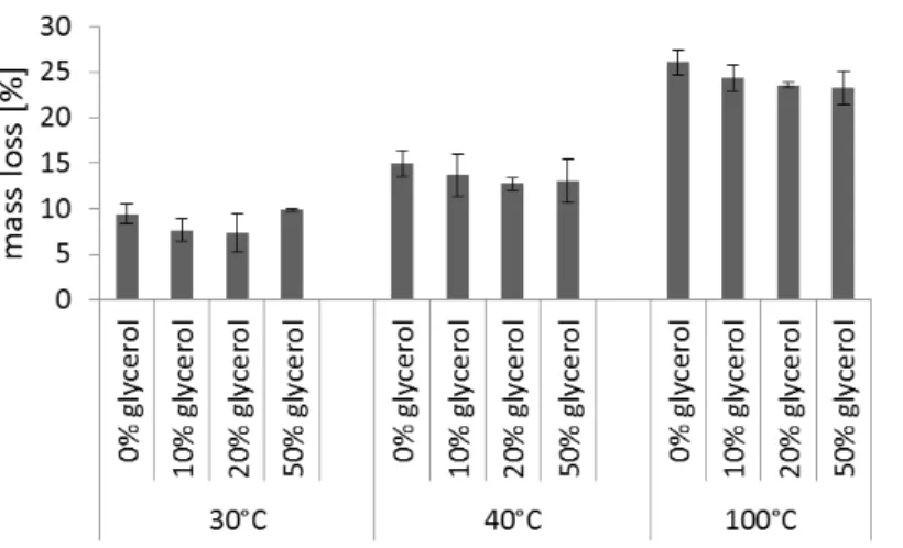
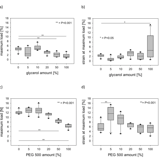
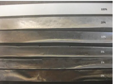
For the acidic release of the calcium, GDL showed the best properties. Only glycerol showed the softening effect that was expected from the different investigated plasticizers. Therefore the best composition for a cross-linked alginate film is comprised of alginate, glycerol, water, dicalcium phosphate and GDL.
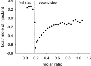
Due to the fact, that different types of alginate with varying amounts of guluronic acid and mannuronic acid can bind more or less calcium the composition should be perfected by finding out how much calcium can be effectively bound by the different alginates. Chapter 4 deals with the assignment to find out, how much calcium is needed to cross-link an alginate film properly and achieve good mechanical properties and erosion time, but also to have binding sites left that are able to bind more calcium if necessary. Furthermore the differences in the cross-linking ability and the resulting properties of alginates with a high guluronic acid content and a low guluronic acid content were investigated. With the help of pretests like ITC measurements, viscosity measurements, rheological tests, turbidity measurements and a defined compression test it was found out how much calcium can be bound by the guluronic acid blocks of the alginates and how much calcium is necessary to cross-link the film properly. The film prepared regarding these results were tested mechanically. Furthermore an erosion test and a cytotoxicity test were performed.
Regarding peritoneal surgery not only the formation of adhesions is a major problem.
Postsurgical infections, which also can enhance the formation of adhesions, are a problem, which can only be solved by sufficient antibiotic treatment. To minimize the side effects of
20
antibiotics, which often come along when they are given systemically, a controlled local application would be beneficial. Therefore the prepared films could be loaded with antibiotics to have a local release after implantation. Chapter 5 deals with the preparation of thin polymer films prepared from alginate, a block copolymer made of PLA-PEG-PLA and PLA.
These films were loaded with the two antibiotics vancomycin hydrochloride and gentamicin sulfate using different methods. Release studies and microbiological testing were performed with the prepared films to test their biological efficacy.
Beside alginate, hyaluronic acid was thought to be a suitable polymer for the preparation of mucoadhesive films. Like alginate, hyaluronic acid is a hydrophilic polymer, which would be dissolved in the humid environment of the peritoneum and therefore has to be cross-linked to increase the stability. Chapter 6 deals with the preparation of films made of a hydrophilic polymer that has not been physically cross-linked by ionic interactions like the alginate films, but chemically cross-linked via disulfide bridges using thiofunctionalization of hyaluronic acid. Carbodiimide chemistry was performed to prepare thiolated hyaluronic acids with different molecular weights and substitution degrees to investigate the impact on cross- linking of prepared films. With the help of oxidizing additives and the adaption of the drying process the cross-linking of the films could be controlled.
After successful preparation of monolayers from alginate and hyaluronic acid, multilayers were prepared to investigate, if a layer of mucoadhesive alginate can improve the properties of a PLA film and also increase the mechanical stability of the hydrophilic polymer films. Due to the fact, that PLA is a lipophilic and alginate a hydrophilic polymer the combination could not be done by just drawing one layer on top of the other. To make the two layers stick to each other additives and surface modifications were tested in Chapter 7. The prepared multilayers were tested mechanically and a mucoadhesion test was performed to investigate, if the alginate layer improves the handling of the PLA film.
21
3. Alginate films cross-linked with different methods
Different cross-linking methods were established to investigate the best method for the preparation of thin alginate films with a homogenous distribution of cross-linking calcium.
Parts of this chapter were published in Eva Esser, Joerg K. V. Tessmar
Preparation of well-defined calcium cross-linked alginate films for the prevention of surgical adhesions
Journal of Biomedical Research Part B: Applied Biomaterials
22 3.1 Introduction
Alginate is a linear block-copolymer produced in the stems or the leaves of marine brown algae and functions as structural component in the plant 49. Furthermore it can also be produced via microbial fermentation 49–53. The 1-4 linked uronic acids are arranged in homopolymeric blocks of β-D-mannuronic acid (M) or α-L-guluronic acid (G) and in MG- blocks containing both uronic acids 49,54. Due to its biocompatibility and biodegradability as well as its non-toxic and non-immunogenic properties 55–57 alginate is widely used as an additive in the food industry as well as in the pharmaceutical industry 49. With the addition of divalent cations like calcium, alginate can be cross-linked, because the guluronic acid blocks can complex bivalent cations and form a so called “egg-box” 58.
Alginates are provided in a broad range of products, therefore they can be used in many different applications. Alginic acid forms different salts with counter ions like sodium, magnesium or potassium. The gelling properties are highly influenced by the ratio of guluronic acid and mannuronic acid in the alginate chains. To prepare alginate films with adjustable mechanical properties and erosion time an adequate cross-linking is necessary.
For this study three different sodium alginates Protanal® LF 10/ 60, its follower Protanal®
LF 10/ 60 FT and Protanal® LF 10/ 60 LS (FMC BioPolymer) 59,60 were used to prepare thin films for the prevention of abdominal adhesions. These alginates are assigned for the preparation of wound healing dressings 61 and therefore ought to be suitable for the preparation of thin alginate films for adhesion prevention.
Its temperature independent 49 ability of forming a gel by a reversible cross-linking of the guluronic acid blocks with the help of bivalent cations like calcium 62 makes alginate a very promising polymer for the preparation of erodible polymer films. When the G-blocks in the alginate chains get in contact with calcium the guluronic acids can complex it and form a junction zone with other G-blocks by forming the so called “egg-box” 58. For the preparation of different products of any shape made from cross-linked alginate many procedures for the internal gelation (Table 1) with a calcium release from a calcium source that has been mixed with the alginate solution and the external gelation (Table 1) with a calcium source that surrounds the prepared alginate product 61 have been established 63.