www.biotechnology-journal.com
Alginate Hydrogels as Scaffolds and Delivery Systems to Repair the Damaged Spinal Cord
Santiago Grijalvo, Manuel Nieto-Díaz, Rodrigo M. Maza, Ramón Eritja, and David Díaz Díaz*
Alginate (ALG) is a lineal hydrophilic polysaccharide present in brown algae cell walls, which turns into a gel state when hydrated. Gelation readily produces a series of three dimensional (3D) architectures like fibers, capillaries, and microspheres, used as biosensors and bio-actuators in a plethora of biomedical applications like drug delivery and wound healing.
Hydrogels have made a great impact on regenerative medicine and tissue engineering because they are able to mimic the mechanical properties of natural tissues due to their high water content. Recent advances in neurosciences have led to promising strategies for repairing and/or regenerating the damaged nervous system. Spinal cord injury (SCI) is particularly challenging, owing to its devastating medical, human, and social consequences. Although effective therapies to repair the damaged spinal cord (SC) are still lacking, multiple pharmacological, genetic, and cell-based therapies are currently under study. In this framework, ALG hydrogels constitute a source of potential tools for the development of implants capable of promoting axonal growth and/or delivering cells or drugs at specific damaged sites, which may result in therapeutic strategies for SCI. In this mini-review, the current state of the art of ALG applications in neural tissues for repairing the damaged spinal cord is discussed.
1. Introduction
The use of hydrogels provides a broad range of possibilities in biomedicine, ranging from cell and gene therapy,[1,2] and drug delivery,[3]to regenerative medicine and tissue engineering applications.[4–7]The success of this technology derives from the ability of hydrogels to retain high levels of water, as much as
Dr. S. Grijalvo, Dr. R. Eritja
Institute for Advanced Chemistry of Catalonia (IQAC, CSIC) Jordi Girona 18–26, E-08034 Barcelona, Spain
The ORCID identification number(s) for the author(s) of this article can be found under https://doi.org/10.1002/biot.201900275
© 2019 The Authors.Biotechnology Journalpublished by WILEY-VCH Verlag GmbH & Co. KGaA, Weinheim. This is an open access article under the terms of the Creative Commons Attribution License, which permits use, distribution and reproduction in any medium, provided the original work is properly cited.
DOI: 10.1002/biot.201900275
over 99%, which favors the entrapment of biological entities and increases their biocompatibility.[8]
Synthetic (e.g., 2-hydroxyethyl methacry- late [HEMA], polyacrylamide [PAA], polyethylene glycol [PEG]) and natural poly- mers (e.g., alginate [ALG], carrageenan, chitosan, hyaluronic acid) have been used in appropriate concentrations to trigger gelation processes by applying chemical or physical cross-linking strategies.[9] These two processes permit the self-assembly of such polymers into hydrophilic networks, giving rise to a plethora of 3D materials con- taining both strong and weak interactions throughout polymer networks.
Although synthetic polymers have played a meaningful role in multiple biomedi- cal applications,[10,11]extensive studies have led to natural polymer-based hydrogels emerging as equally or more suitable ma- terials, due to their low immunogenic- ity, ready biodegradability, and easy large- scale production.[12,13]To this end, specific polysaccharide-based hydrogels are being engineered. They prove to be efficient depots for living cells, small molecules, growth factors, and liposomes, for use in a wide range of biomedical applications including cell transplantation or drug delivery. Hydrogels,[14] in particular those composed of
ALG,[15,16] have recently[17–21] emerged as bridging materials
capable of delivering cells and drugs into specific locations, thus promoting tissue regeneration when implanted in injured tissues.[22]
Dr. S. Grijalvo, Dr. R. Eritja
Networking Centre in Bioengineering, Biomaterials and Nanomedicine (CIBER-BBN)
Jordi Girona 18–26, E-08034 Barcelona, Spain Dr. M. Nieto-Díaz, Dr. R. M. Maza
Molecular Neuroprotection Group
Research Unit, National Hospital for Paraplegics (SESCAM) E-45071 Toledo, Spain
Dr. D. D. Díaz
Institut für Organische Chemie Universität Regensburg,
Universitätsstr. 31, 93053 Regensburg, Germany Dr. D. D. Díaz
Institute of Natural Products and Abrobiology of the CSIC
Avda. Astrofísico Francisco Sánchez 3, E-3826 La Laguna, Tenerife, Spain E-mail: d.diaz.diaz@csic.es
1.1. Spinal Cord Injury
The brain together with the spinal cord (SC) forms the central nervous system (CNS), which integrates information and coordi- nates activity across the entire body. Spinal cord injury (SCI) is a dreadful disorder that affects more than 180 000 people world- wide every year, causing permanent disability—including loss of sensory and motor functions in trunk and limbs, together with the loss of autonomous regulation of breathing, bladder empty- ing, and sexual function.[23]
Traumatic injury to the SC leads to a complex pathophysiol- ogy that affects the neural, vascular, and immune systems and largely determines the functional outcome from the SCI. The trauma damages the membranes of spinal cord cells, mainly in the gray matter, causing cell death, axotomy, and blood ves- sel rupture.[24]The initial damage may consist of rupture of the blood–spinal cord barrier, ionic disbalance, massive excitatory neurotransmitter release, oxidative stress, inflammation, and im- mune response, among others. The molecular and cellular events triggered by such damage induce cell death among neurons and glial cells for weeks after injury, and extend the damage to re- gions far from the injury epicenter.[25,26]The cellular responses lead to the formation of a glial scar that reestablishes the blood–
brain barrier and isolates the damaged region, where cysts usu- ally form. Functional deficits due to SCI are usually permanent because the regenerative response of the injured neurons is very limited,[27,28] and regenerating axons are exposed to inhibitory molecules,[29]glial scars, and cysts that prevent growth across the injury site.[30]
Despite the resources and efforts applied in research to seek an efficient therapy to restore loss of function, only rehabilita- tion and epidural electrical stimulation[31]have proven effective in recovering the functional deficits of SCI. Significant efforts have been made to develop therapeutic strategies aiming to pro- tect spared tissue and replace lost cells, promote axonal growth and myelination, and restore or replace lost neural signaling.
In this context, novel therapeutic treatments involving regener- ative medicine and tissue engineering have emerged as promis- ing strategies focused on promoting axon growth and/or deliv- ering cells or drugs to specific damaged sites for SCI. These approaches rely on using cell transplantation procedures, anti- oxidative or anti-inflammatory molecules, specific growth or neu- rotrophic factors, as well as biomaterial scaffolds. The positive outcomes achieved in vitro and in vivo have facilitated launch- ing various pre-clinical and clinical controlled trials to validate such therapeutic effectiveness, giving rise to promising therapeu- tic strategies for SCI.[32–36]
1.2. Hydrogel-Based Materials in SCI and Neural Regeneration
The rapid development and extensive research sustained by poly- mer science have facilitated the appropriate design and prepara- tion of a large number of scaffolds for tissue engineering appli- cations. Both synthetic and natural materials have demonstrated their usefulness to reconstruct and replace the great majority of damaged tissues.[6,7,37]To do so, preformed materials should fulfill a series of requirements to be considered as optimal im-
Santiago Grijalvoreceived his bachelor’s degree in chemistry from the Autonomous Univer- sity of Madrid in 2000. In 2002, he moved to Barcelona to join Dr. Antonio Delgado’s group to do his Ph.D. focusing on the synthesis of modified sphin- golipid combinatorial libraries at the Institute of Research Chemistry (IIQAB-CSIC). In 2007, he joined Prof. Dr. Ramón Eritja’s research group as post- doctoral researcher at the Institute of Advanced Chemistry (IQAC). He has participated in several research projects fo- cusing on improving the cellular uptake of nucleic acids using either covalent or electrostatic strategies.
David Díaz Díazearned his Ph.D. in chemistry (2002) at the University of La Laguna (Spain). Then, he joined Prof.
Finn’s group as postdoc at The Scripps Research Institute (San Diego, USA). Since 2006, he has held various positions in academia and industry. Cur- rently, he is a Privatdozent at the University of Regensburg (Germany) and a Tenured Sci- entist at the CSIC (Spain). His main research focuses on soft materials.
plants for promoting SC repair and neural tissue engineering.
These include their lack of toxicity and immunogenicity, thus contributing to high biocompatibility.[38,39]Appropriate degrada- tion rates,[40] shear modulus, substrate stiffness, and superficial geometry are important properties that should also be taken into account.[40]
A good number of 3D materials and drug delivery devices in conjunction with injectable hydrogels have been properly de- signed for SCI.[16,39,41–44] Some non-degradable materials com- posed of synthetic polymethacrylates have shown notable success as implants due to their stiffness without causing compression of the SC tissue.[44]This feature makes non-degradable materials favorable to cell encapsulation and to providing the proper 3D networks for bridging neural tissues.[45,46]
Degradable synthetic polymers like PEG and poly-peptide hydrogels,[47–49] among many others,[50–53] have emerged as promising alternatives for reconstructing damaged tissues.[44]
This is mainly due to optimizing several parameters like molec- ular weight, degree of crosslinking, or polymer structures, which tend to favor surface degradation rates and consequently the ab- sence of immune response in the body and nerve compression after the scaffold implantation.[54,55]A selection of other synthetic
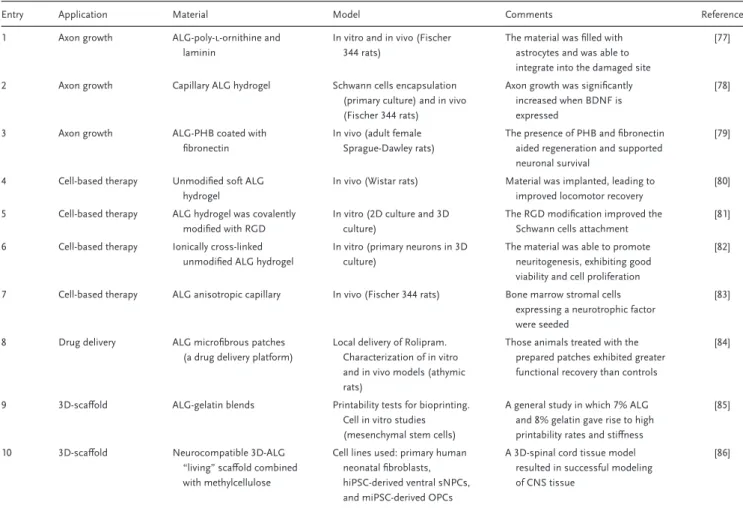
Table 1.Use of alginate (ALG) as a natural hydrogel involved in neural tissue engineering applications such as axon growth, cell-based therapies, drug delivery, and 3D bioprinting.
Entry Application Material Model Comments Reference
1 Axon growth ALG-poly-l-ornithine and
laminin
In vitro and in vivo (Fischer 344 rats)
The material was filled with astrocytes and was able to integrate into the damaged site
[77]
2 Axon growth Capillary ALG hydrogel Schwann cells encapsulation
(primary culture) and in vivo (Fischer 344 rats)
Axon growth was significantly increased when BDNF is expressed
[78]
3 Axon growth ALG-PHB coated with
fibronectin
In vivo (adult female Sprague-Dawley rats)
The presence of PHB and fibronectin aided regeneration and supported neuronal survival
[79]
4 Cell-based therapy Unmodified soft ALG hydrogel
In vivo (Wistar rats) Material was implanted, leading to improved locomotor recovery
[80]
5 Cell-based therapy ALG hydrogel was covalently modified with RGD
In vitro (2D culture and 3D culture)
The RGD modification improved the Schwann cells attachment
[81]
6 Cell-based therapy Ionically cross-linked unmodified ALG hydrogel
In vitro (primary neurons in 3D culture)
The material was able to promote neuritogenesis, exhibiting good viability and cell proliferation
[82]
7 Cell-based therapy ALG anisotropic capillary In vivo (Fischer 344 rats) Bone marrow stromal cells expressing a neurotrophic factor were seeded
[83]
8 Drug delivery ALG microfibrous patches
(a drug delivery platform)
Local delivery of Rolipram.
Characterization of in vitro and in vivo models (athymic rats)
Those animals treated with the prepared patches exhibited greater functional recovery than controls
[84]
9 3D-scaffold ALG-gelatin blends Printability tests for bioprinting.
Cell in vitro studies (mesenchymal stem cells)
A general study in which 7% ALG and 8% gelatin gave rise to high printability rates and stiffness
[85]
10 3D-scaffold Neurocompatible 3D-ALG
“living” scaffold combined with methylcellulose
Cell lines used: primary human neonatal fibroblasts, hiPSC-derived ventral sNPCs, and miPSC-derived OPCs
A 3D-spinal cord tissue model resulted in successful modeling of CNS tissue
[86]
hydrogels used for SC regeneration[56–59]have been listed in Table S1, Supporting Information.
Extensive efforts have been also made to use natural hydrogels in SC repair.[60–67] Natural materials made up of ALG, agarose, cellulose, chitosan or hyaluronic acid, among others, have been used in vitro and in vivo with the aim of promoting cell adhesion, controlled and localized delivery of neurotrophic factors, cell de- livery (including pluripotent or multipotent stem cells), or the formation of filament bridges and scaffolds for neurite regrowth.
[3,12,13,16,21,68]Unlike synthetic polymers, natural implanted mate- rials display high biocompatibility in living tissues. In this way, the cytotoxicity and immune response exhibited by these mate- rials are practically zero.[69]Recent uses of natural hydrogels are listed in Table S1, Supporting Information.
Taking advantage of the characteristics of synthetic and natural hydrogels, a combination of both materials has been proposed for attempting SC regeneration. The idea of using synthetic/natural hydrogels is not novel, as they are employed in other biomedi- cal applications.[70,71]In particular, this strategy involves putting the most common features of both hydrogels into practice: bio- compatibility and biodegradability in the case of natural hydro- gels and the ability to “tune” the physical and mechanical prop- erties of the resultant materials when using synthetic hydrogels.
This combination allows the preparation of functionalized ma- terials to provide protection at the injured site as well as deliver neurotrophic factors and neural stem cells.[72–76]Detailed infor- mation on the progress and uses of these composites is shown in Table S1, Supporting Information.
In this article, we aim to review the current state-of-the-art strategies employed to prepare natural hydrogels for neural tis- sue engineering applications. Particularly, this revision is focused on ALG as an artificial scaffold, analogous to the ECM used in regenerative applications to repair the damaged SC. The possi- bilities of ALG for promoting axon growth, cell-based therapies, drug delivery, or as a 3D-scaffold will be discussed below, and are displayed inTable 1.
2. Alginate Biomaterials for the Repair of the Injured Spinal Cord
ALG is a hydrophilic lineal polysaccharide naturally occurring in the cell walls of brown algae. ALG consists of repeated units of (1-4)-𝛽-d-mannuronic acid and 𝛼-l-guluronic acid building blocks. These residues are found as flexible coils in aqueous so- lution. The presence of divalent cations (e.g., Ca2+, Mg2+, Ba2+,
and many others) is essential for the chelation process amid the two aforementioned building blocks. As a consequence, co- operative ionic inter-chain forces are spontaneously produced, giving rise to ordered 3D structures that cause gelation of the ALG solution.[87] Physical properties of the resultant hydrogels (e.g., mechanical strength, stability or elasticity) can also vary, de- pending on the divalent cation used. This tunable property fa- cilitates engineering different ALG-based materials used in nu- merous minimally invasive biomedical applications, from oral administration to hydrogel injection. Versatility, high biocompat- ibility and lack of toxicity, ease of gelation, and external structural similarity to the extracellular matrix (ECM) of living tissues are key properties of ALG for biomedical applications. However, de- spite ALG being a biocompatible and naturally derived hydrogel, some impurities isolated in commercially available samples have proved to be cytotoxic and mitogenic.[88,89]This might lead to un- wanted responses after cell transplantation, reducing its potential use unless a prior purification of the commercial sample is car- ried out.[88]
ALG has proven to be an efficient biomaterial and a model scaf- fold when working as a bridging material in neural tissue engi- neering, including SCI and peripheral nerve regeneration, as well as in the delivery of cell and growth factor molecules (EGF and bFGF) for SC repair.[90–95]
2.1. 3D-Scaffolds
3D-scaffolds have resulted in a prominent strategy for CNS re- generation, as they can facilitate cell proliferation and provide cellular adhesion onto the new material.[96]Hydrogels may confer additional advantages as cell and molecule delivery vehicles, due to their high water content and good response to several stimuli like pH or temperature. Natural polymers like chitosan, agarose, fibrin, collagen, gellan-gum, Matrigel, and hyaluronic acid have been used as 3D scaffolds to aid in soft-tissue reconstructions of the injured ECM of the SC, through filling cavities arising from the injury.[97–105]To accomplish this, such bio-scaffolds have been successfully engineered. They exhibit tubular-like struc- tures, besides containing some components of the ECM, includ- ing hyaluronic acid[106]and fibronectin.[107]
3D-bioprinting has recently emerged as a potential technique with numerous therapeutic applications, including neural tissue engineering.[108]Alginate has been used in the search of bioinks for 3D-bioprinting applications.[85] On this point, soft ALG hy- drogels have proved to be efficient as a bioink for 3D bioprint- ing experiments to provide an appropriate scaffold to accommo- date neuronal cells. These uses are favored by their biocompati- ble and biodegradable properties in contact with nerve tissues. To achieve this, different viscosities of ALG were tested and mixed with methylcellulose (MC) in a 1:3 volume ratio (ALG:MC) to find the most suitable ink composition for printing.[86]This strat- egy allowed a microenvironment to be created by preparing a 3D SC-based platform containing neuronal and oligodendrocyte progenitor cells. Interestingly, both progenitor cells were able to proliferate precisely, showing the usefulness of this platform in modeling CNS tissue architectures. Besides MC, other addi- tives like fibrin,[109]chitosan, or genipin[110]have been also tested
to produce useful bioinks and 3D-bioscaffolds for neural tissue engineering.[111]
2.2. Axon Growth
In the past century, Aguayo and colleagues demonstrated that SC axons can regenerate if provided with the appropriate substrate.[112,113]Since then, many laboratories have explored the possibilities of biomaterials to build up scaffolds that support and promote axonal growth across the injury region. Biomate- rials should have thin walls with appropriate diameters, capa- ble of promoting axon growth and orientation in the SC.[22,42]
The generation of cross-linked ALG-based capillaries has facil- itated preparing materials with various channel diameters (11, 13, 29, and 89 µm), according to the divalent cation used in vitro (Ba2+, Ca2+, Sr2+, and Zn2+, respectively). In this regard, Pawar and coworkers showed that axon density growth within hydrogel strongly depended on the channel diameter, which ranged from 10 to 100 µm, whereas a decrease in the linear orientation of such axons was obtained with increasing channel diameter.[114]
Not only unmodified anisotropic scaffolds have been used as promising implants to be integrated into the SC without detect- ing any inflammatory responses. Recently, alternative attempts involving the modification of anisotropic ALG capillary surfaces with cationic polymers (poly-l-ornithine, PLO) and laminin have resulted in enhanced axonal growth and cellular adhesion, af- ter 2 weeks of incubation in vitro.[77] These positive responses were replicated in adult rats after implanting this PLO-laminin- ALG hydrogel scaffold, which promoted cell migration and slight axon growth throughout the capillary channels. Notably, the au- thors observed that neurite growth was significantly improved in the presence of cationic peptides when additional astrocytes were first seeded within the material before grafting.
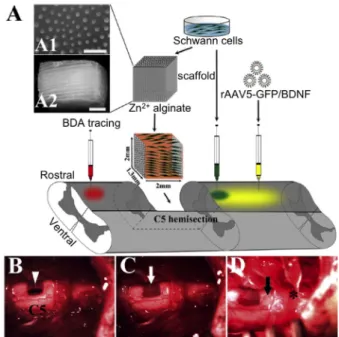
A combined therapy involving the transplantation of ALG cap- illary hydrogels containing Schwann cells followed by the in- jection of a neurotrophic factor (BDNF) as a supplementary growth stimulus facilitated the regeneration of axons at the le- sion site. Injection into the caudal spinal parenchyma led to sig- nificant regeneration, compared to transplants that lacked BDNF (Figure 1).[78] The positive effect promoted by BDNF after SCI was also observed by Novikov et al., when used a biodegrad- able synthetic implant made up of fibers containing poly-𝛽- hydroxybutyrate (PHB).[79] These PHB fibers were coated with neonatal Schwann cells as well as a combination of ALG and fi- bronectin. The resultant 2–3 mm long matrix was evaluated in vivo and surprisingly showed neuronal survival and axonal re- generation after its implantation in the lesion site.
2.3. Cell-Based Therapy
Embryonic, pluripotent, neural, and mesenchymal stem cells as well as Schwann cells, neurons, and other neural cells have been transplanted into specific damaged areas to replace lost neu- ral cells, protect the surviving ones, and facilitate regenerative processes.[31,115]However this strategy, though promising, faces the risk of poor cell survival after transplantation.[116]
Figure 1. Lesion paradigm and experimental procedures. A) Schematic diagram of the experimental design. A 1.5–2 mm long segment of the SC was removed unilaterally at the C5 level before implanting a Schwann cells (SCs)-seeded alginate scaffold. Subsequently, viral vectors (yellow) for the regulatable expression of green fluorescent protein (GFP) (rAAV5- GFP) or BDNF (rAAV5-BDNF) were injected into the caudal SC, ipsilateral to the lesion. SCs (blue) were also injected into the caudal SC in one group.
Biotinylated dextran amine (BDA, red) was injected in the SC rostral to the lesion to trace descending axons 3 or 7 weeks post-lesion. A1) Cross- sectional and A2) longitudinal view of the capillary lumen. B) After a hemi- section lesion (arrowhead,≈2 mm in length) of the rat SC, C) the alginate scaffold loaded with SCs (white arrow) was grafted into the lesion and D) virus (*) or SCs (black arrow) were injected into the SC caudal to the scaf- fold, using a glass capillary. Scale bar: 300 µm in (A1); 500 µm in (A2).
Reproduced with permission[78]. Copyright 2017, Elsevier.
Figure 2. Alginate reduces fibrous scarring. Examples of hematoxylin &
eosin (H&E) stained tissue sections 140 days after SCI of 2 mm size. A) Overview of a cross section of control SC. B) Magnification of the area indicated by the black box showing the fibrotic scar and adjacent SC tissue.
C) Overview of a cross section of SC that received an alginate implant.
D) Magnification of the area indicated in (C) by the black box illustrating the lack of a fibrous scar. Reproduced with permission[80]. Copyright 2018, Springer Nature.
In this regard, ALG has proved to be efficient as a soft hy- drogel material capable of adhering to and protecting neurons when acting as an implant. Furthermore, such ALG implants have also favored functional recovery, particularly in the locomo- tor system after SCI[80](Figure 2), including the outgrowth of ax-
ons in the presence of some divalent cations (e.g., Ca2+, Ba2+, or Sr2+).[117,118]
Unfortunately, poor adhesion properties have also been re- ported when Schwann cells are seeded onto the above-mentioned ALG scaffolds.[81,119]To avoid this limitation and achieve superior adhesiveness, both the preparation of ionically ultrasoft cross- linked ALG hydrogels[82] and modifying the ALG surface with a argininylglycylaspartic acid tripeptide (RGD) peptide[81]led to cell proliferation and facilitated Schwann cell adhesion in 3D cell cultures.
Alginate scaffolds have been also used as an appropriate depot for bone-marrow stromal cells (BMSCs). These cells were embed- ded into a 2 mm-long capillary hydrogel and finally implanted to evaluate the release of neurotrophic factors (BDNF).[83]Data con- firmed ALG was able to integrate into the SC lesion without toxic- ity, favoring axonal regeneration. Encouragingly, survival of BM- SCs and proliferation of Schwann cells and blood vessels were observed when compared to ALG-based biomaterials containing only BMSCs.
Recent studies carried out by Wen et al. showed the feasibil- ity of modifying ALG hydrogels with an integrin ligand (𝛼3𝛽1).
This 3D culture system proved to be efficient in favoring the en- capsulation and differentiation of neural progenitor cells (NPCs) in vitro, after a 3-month incubation.[120] These authors used surface plasmon resonance (SPR) to measure the specific in- teractions between the integrin ligands and the NPCs. This 3D platform also enabled differentiation of both mouse and human NPCs into the distinct neural cell types, after being entrapped within ALG hydrogel.
2.4. Drug Delivery
Available evidence has demonstrated that drugs and bioac- tive molecules, such as nucleotides and proteins, can help re- pair the damaged SC, promoting neurogenesis, plasticity, or regeneration.[121–126] Hydrogels have been employed to deliver many of these molecules to target tissues and cells under precise conditions. For example, interesting strategies have been pro- posed to achieve controlled release of rolipram—a blood-brain barrier permeable neuroprotectant with significant effects on functional recovery after SCI[127]—and increase its therapeutic ef- fects at the action site. In this direction, Downing and cowork- ers prepared series of ALG microfibrous patches for the con- tinuous delivery of rolipram in vivo, achieving good therapeu- tic results (Figure 3).[84] Improvements in the functional recov- ery of motor function after injury were observed (open-field loco- motion, forelimb articular movement, and animal coordination) when small doses of rolipram were released (≈60 µg cm−2) over 12 days through the material. In contrast, when authors used high-doses of rolipram under regular administration conditions, a pronounced decline in animal survival rates (up to 50%) was observed.
3. Conclusions and Future Perspectives
The use of ALG-based hydrogels faces a good number of chal- lenges but also opportunities for tissue repair and regenerative
Figure 3. Implantation of drug-eluting microfibrous patches after SCI. A) Schematic of subdural implantation of drug-eluting microfibrous patches into the injured cord. B) Macroscopic view of lesion site and patch during animal surgery. An asterisk was used to mark location of drug-eluting mi- crofibrous patch. C,D) Gross histology of SC cross section 8 weeks post SCI. H&E staining reveals new tissue formation at the lesion site and patch’s ability to integrate into the surrounding tissues (D). C) Less re- generated control for comparison. Here, arrows point to the implantation site for drug-eluting microfibrous patches. Red dashes outline areas of less tissue formation for comparison. Reproduced with permission[84]. Copy- right 2012, Elsevier.
medicine. The structure, physical properties, and specific func- tions of hydrogels may vary when tuning these materials. Ben- eficial properties such as biocompatibility, long-term stability in vivo, or their suitability to be chemically modified with appro- priate ligands, make these materials promising scaffolds for em- bedding drugs, nerve cells, growth factors, etc.
The use of ALG-based hydrogels in regenerative medicine has grown exponentially in the last decade, achieving significant and promising results as shown by a good number of in vivo models.
However, the translational step from the bench with new designs of materials for the injured CNS in the field of tissue engineering to implementing an effective treatment in humans is still a daunt- ing challenge. Promotion of axon regeneration and neural cell replacement after SCI by means of ALG and other biomaterials has been addressed through a variety of strategies, for instance embedding and subsequent cell release, the use of neurotrophic factors, and small drug molecules.
In a plethora of in vitro and in vivo models, the development of these combined strategies has shown greater therapeutic effec- tiveness than those involving single treatments.[128] The design and development of new biodegradable materials capable of en- trapping stem cells and/or therapeutic drugs could enhance this combinatorial approach, providing maximal functional recovery and the highest chance of success in the clinic. Regrettably, a full understanding of biomaterial properties as well as choosing the right therapeutic combination (e.g., stem cells, neurotrophic fac- tors, or small drugs) requires great investigation in depth, so as to improve the relevant translational research.
In addition to using cell therapy and biomaterials for treat- ing SCI, tissue engineering offers alternative technologies like 3D-bioprinting and microfluidic devices. These are emerging as potential applications for replacing tissues and creating disease
models aimed at developing further new therapies and also un- derstanding more precisely the behavior of the disorder.
Supporting Information
Supporting Information is available from the Wiley Online Library or from the author.
Acknowledgements
M.N.-D. and R.M.M. contributed equally to this work. This re- search was funded by the Spanish Ministerio de Ciencia e Inno- vación (MICINN), grant number CTQ2017-84415-R; CIBER-BBN, grant number CB06_01_0019; the Generalitat de Catalunya, grant number 2017/SGR/114; and the Junta de Comunidades de Castilla La Man- cha, grant number SBPLY/17/180501/000376. The authors thank Guido Vaughan Jones Carter (IPNA-CSIC) for manuscript proofreading.
Conflict of Interest
The authors declare no conflict of interest.
Keywords
alginate, biomaterial, bioprinting, central nervous system, neural tissue engineering, regenerative medicine, spinal cord injury
Received: June 14, 2019 Revised: October 12, 2019 Published online: November 14, 2019
[1] C. Wang, R. R. Varshney, D.-A. Wang,Adv. Drug Delivery Rev.2010, 62, 699.
[2] R. L. Youngblood, N. F. Truong, T. Segura, L. D. Shea,Mol. Ther.
2018,26, 2087.
[3] E. Larrañeta, S. Stewart, M. Ervine, R. Al-Kasasbeh, R. F. Donnelly, J. Funct. Biomater.2018,9, 13.
[4] B. V Slaughter, S. S. Khurshid, O. Z. Fisher, A. Khademhosseini, N.
A. Peppas,Adv. Mater.2009,21, 3307.
[5] J.-H. Lee, H.-W. Kim,J. Tissue Eng.2018,9, 2041731418768285.
[6] E. Jabbari,Gels2019,5, 30.
[7] E. S. Place, J. H. George, C. K. Williams, M. M. Stevens,Chem. Soc.
Rev.2009,38, 1139.
[8] J. Fu, M. In Het Panhuis,J. Mater. Chem. B2019,7, 1523.
[9] J. Maitra, V. K. Shukla,Am. J. Polym. Sci.2014,4, 25.
[10] O. Wichterle, D. Lím,Nature1960,185, 117.
[11] M. C. Hacker, J. Krieghoff, A. G. Mikos, inPrinciples of Regenerative Medicine, 3rd ed., (Eds: A. Atala, R. Lanza, A.G. Mikos, R. Nerem), Academic Press, Boston2019, pp. 559–590.
[12] J. Pushpamalar, A. K. Veeramachineni, C. Owh, X. J. Loh, ChemPlusChem2016,81, 504.
[13] S. Grijalvo, J. Mayr, R. Eritja, D. D. Díaz,Biomater. Sci.2016,4, 555.
[14] R. Y. Tam, T. Fuehrmann, N. Mitrousis, M. S. Shoichet,Neuropsy- chopharmacology2014,39, 169.
[15] L. N. Novikova, A. Mosahebi, M. Wiberg, G. Terenghi, J.-O. Kellerth, L. N. Novikov,J. Biomed. Mater. Res., Part A2006,77A, 242.
[16] G. Perale, F. Rossi, E. Sundstrom, S. Bacchiega, M. Masi, G. Forloni, P. Veglianese,ACS Chem. Neurosci.2011,2, 336.
[17] R. Barbucci, M. Consumi, S. Lamponi, G. Leone,Macromol. Symp.
2003,204, 37.
[18] N. B. Shelke, R. James, C. T. Laurencin, S. G. Kumbar,Polym. Adv.
Technol.2014,25, 448.
[19] J. Radhakrishnan, A. Subramanian, U. M. Krishnan, S. Sethuraman, Biomacromolecules2017,18, 1.
[20] A. Kirschning, N. Dibbert, G. Dräger,Chem. - Eur. J.2018,24, 1231.
[21] T. Coviello, P. Matricardi, C. Marianecci, F. Alhaique,J. Controlled Release2007,119, 5.
[22] Q. Zhang, B. Shi, J. Ding, L. Yan, J. P. Thawani, C. Fu, X. Chen,Acta Biomater.2019,88, 57.
[23] M. Nieto-Díaz, F. Esteban, D. Reigada, T. Muñoz-Galdeano, M.
Yunta, M. Caballero-López, R. Navarro-Ruiz, Á. del Águila, R. Maza, Front. Cell. Neurosci.2014,8, 53.
[24] M. G. Fehlings, A. R. Vaccaro, M. Boakye,Essentials of Spinal Cord Injury: Basic Research to Clinical Practice, Thieme, New York2012.
[25] M. J. Crowe, J. C. Bresnahan, S. L. Shuman, J. N. Masters, M. S.
Beattie,Nat. Med.1997,3, 73.
[26] X. Z. Liu, X. M. Xu, R. Hu, C. Du, S. X. Zhang, J. W. McDonald, H.
X. Dong, Y. J. Wu, G. S. Fan, M. F. Jacquin, C. Y. Hsu, D. W. Choi,J.
Neurosci.1997,17, 5395.
[27] M. Murray, I. Fischer,Neurosci.2001,7, 28.
[28] W. Plunet, B. K. Kwon, W. Tetzlaff,J. Neurosci. Res.2002,68, 1.
[29] M. L. Condic, M. L. Lemons,Neuroreport2002,13, A37.
[30] J. W. Fawcett, R. Asher,Brain Res. Bull.1999,49, 377.
[31] F. B. Wagner, J. B. Mignardot, C. G. Le Goff-Mignardot, R. Demes- maeker, S. Komi, M. Capogrosso, A. Rowald, I. Seáñez, E. Piron- dini, M. Vat, L. A. McCracken, R. Heimgartner, I. Fodor, A. Watrin, P.
Seguin, E. Paoles, K. Van Den Keybus, G. Eberle, B. Schurch, E. Pra- long, F. Becce, J. Prior, N. Buse, R. Buschman, E. Neufeld, N. Kuster, S. Carda, J. Von Zitzewitz, V. Delattre, T. Denison, et al.,Nature2018, 563, 65.
[32] J. D. Steeves, D. Lammertse, A. Curt, J. W. Fawcett, M. H. Tuszynski, J. F. Ditunno, P. H. Ellaway, M. G. Fehlings, J. D. Guest, N. Kleitman, P. F. Barlett, A. R. Blight, V. Dietz, B. H. Dobkin, R. Grossman, D.
Short, M. Nakamura, W. P. Coleman, M. Gaviria, A. Privat,Spinal Cord2007,45, 206.
[33] H. Hasebe, M. Ito, inMetals for Biomedical Devices, 2nd ed. (Ed: M.
Niinomi), Woodhead Publishing Series in Biomaterials, Woodhead Publishing, Cambridge, UK2019, pp. 475–493.
[34] H. Zhao, Q.-L. Sun, L.-J. Duan, Y.-D. Yang, Y.-S. Gao, D.-Y. Zhao, Y.
Xiong, H.-J. Wang, J.-W. Song, K.-T. Yang, X. M. Wang, X. Yu,Eur.
Spine J.2019,28, 1092.
[35] A. D. Levi, K. D. Anderson, D. O. Okonkwo, P. Park, T. N. Bryce, S. N.
Kurpad, B. Aarabi, J. Hsieh, K. Gant,J. Neurotrauma2019,36, 891.
[36] S. Han, W. Yin, X. Li, S. Wu, Y. Cao, J. Tan, Y. Zhao, X. Hou, L. Wang, C. Ren, J. Li, X. Hu, Y. Mao, G. Li, B. Li, H. Zhang, J. Han, B. Chen, Z. Xiao, X. Jiang, J. Dai,J. Neurotrauma2019,36, 2316.
[37] Y. D. Taghipour, N. Asadi, V. R. Hokmabad, A. R. D. Bakhshayesh, N. Asadi, R. Salehi, H. T. Nasrabadi,Curr. Med. Chem.2019,26, https://doi.org/10.2174/0929867326666190711103956.
[38] O. Alluin, C. Wittmann, T. Marqueste, J.-F. Chabas, S. Garcia, M.-N.
Lavaut, D. Guinard, F. Feron, P. Decherchi,Biomaterials2009,30, 363.
[39] Š. Kubinová, Neurochem. Res. 2019, 1. https://doi.org/10.1007/
s11064-019-02808-2. [Epub ahead of print]
[40] D. Shahriari, J. Koffler, D. A. Lynam, M. H. Tuszynski, J. S. Sakamoto, J. Biomed. Mater. Res., Part A2016,104, 611.
[41] M. Tsintou, K. Dalamagkas, A. M. Seifalian,Neural Regener. Res.
2015,10, 726.
[42] K. S. Straley, C. W. P. Foo, S. C. Heilshorn,J. Neurotrauma2010,27, 1.
[43] D. Macaya, M. Spector,Biomed. Mater.2012,7, 012001.
[44] R. C. Assunção-Silva, E. D. Gomes, N. Sousa, N. A. Silva, A. J. Sal- gado,Stem Cells Int.2015,2015, 948040.
[45] Š. Kubinová, D. Horák, A. Hejˇcl, Z. Plichta, J. Kotek, E. Syková,J.
Biomed. Mater. Res., Part A2011,99A, 618.
[46] Z. Cai, Y. Gan, C. Bao, W. Wu, X. Wang, Z. Zhang, Q. Zhou, Q. Lin, Y. Yang, L. Zhu,Adv. Healthcare Mater.2019,8, 1900013.
[47] Z. Hassannejad, S. A. Zadegan, A. R. Vaccaro, V. Rahimi-Movaghar, O. Sabzevari,Injury2019,50, 278.
[48] K. J. Lampe, R. G. Mooney, K. B. Bjugstad, M. J. Mahoney,J. Biomed.
Mater. Res. Part A2010,94A, 1162.
[49] X. Lu, T. H. Perera, A. B. Aria, L. A. S. Callahan,J. Exp. Pharmacol.
2018,10, 37.
[50] L. N. Novikova, J. Pettersson, M. Brohlin, M. Wiberg, L. N. Novikov, Biomaterials2008,29, 1198.
[51] R. T. H. Chan, R. A. Russell, H. Marçal, T. H. Lee, P. J. Holden, L. J.
R. Foster,Biomacromolecules2014,15, 339.
[52] F. Facchiano, E. Fernandez, S. Mancarella, G. Maira, M. Miscusi, D.
D’Arcangelo, G. Cimino-Reale, M. L. Falchetti, M. C. Capogrossi, R.
Pallini,J. Neurosurg.2002,97, 161.
[53] W. Bensaı¨d, J. T. Triffitt, C. Blanchat, K. Oudina, L. Sedel, H. Petite, Biomaterials2003,24, 2497.
[54] V. M. Tysseling-Mattiace, V. Sahni, K. L. Niece, D. Birch, C. Czeisler, M. G. Fehlings, S. I. Stupp, J. A. Kessler,J. Neurosci.2008,28, 3814.
[55] A. Göpferich,Biomaterials1996,17, 103.
[56] Y. Katayama, R. Montenegro, T. Freier, R. Midha, J. S. Belkas, M. S.
Shoichet,Biomaterials2006,27, 505.
[57] A. Hejˇcl, J. Šedý, M. Kapcalová, D. A. Toro, T. Amemori, P. Lesný, K.
Likavˇcanová-Mašínová, E. Krumbholcová, M. Pˇrádný, J. Michálek, M. Burian, M. Hájek, P. Jendelová, E. Skyková,Stem Cells Dev.2010, 19, 1535.
[58] P. A. Ramires, M. A. Miccoli, E. Panzarini, L. Dini, C. Protopapa,J.
Biomed. Mater. Res., Part B2005,72B, 230.
[59] N. Comolli, B. Neuhuber, I. Fischer, A. Lowman,Acta Biomater.
2009,5, 1046.
[60] Z. Wang, J. Nong, R. B. Shultz, Z. Zhang, T. Kim, V. J. Tom, R. K.
Ponnappan, Y. Zhong,Biomaterials2017,112, 62.
[61] H. Gu, Z. Yue, W. S. Leong, B. Nugraha, L. P. Tan,Regener. Med.
2010,5, 245.
[62] M. M. Pakulska, C. H. Tator, M. S. Shoichet,Biomaterials2017,134, 13.
[63] M. Boido, M. Ghibaudi, P. Gentile, E. Favaro, R. Fusaro, C. Tonda- Turo,Sci. Rep.2019,9, 6402.
[64] J. Chedly, S. Soares, A. Montembault, Y. von Boxberg, M. Veron- Ravaille, C. Mouffle, M.-N. Benassy, J. Taxi, L. David, F. Nothias,Bio- materials2017,138, 91.
[65] K. E. Crompton, J. D. Goud, R. V Bellamkonda, T. R. Gengenbach, D. I. Finkelstein, M. K. Horne, J. S. Forsythe,Biomaterials2007,28, 441.
[66] J. Park, E. Lim, S. Back, H. Na, Y. Park, K. Sun,J. Biomed. Mater. Res.
Part A2010,93A, 1091.
[67] R. E. Thompson, J. Pardieck, L. Smith, P. Kenny, L. Crawford, M.
Shoichet, S. Sakiyama-Elbert,Biomaterials2018,162, 208.
[68] Z. Z. Khaing, C. E. Schmidt,Neurosci. Lett.2012,519, 103.
[69] M. S. Shoichet,Macromolecules2010,43, 581.
[70] J. Shang, Z. Shao, X. Chen,Polymer.2008,49, 5520.
[71] K. M. Park, S. Y. Lee, Y. K. Joung, J. S. Na, M. C. Lee, K. D. Park,Acta Biomater.2009,5, 1956.
[72] C. Martínez-Ramos, L. R. Doblado, E. L. Mocholi, A. Alastrue-Agudo, M. S. Petidier, E. Giraldo, M. M. Pradas, V. Moreno-Manzano,J. Tis- sue Eng. Regener. Med.2019,13, 509.
[73] Z. Z. Khaing, N. K. Agrawal, J. H. Park, S. Xin, G. C. Plumton, K. H.
Lee, Y. J. Huang, A. L. Niemerski, C. E. Schmidt, J. W. Grau,J. Mater.
Chem. B2016,4, 7560.
[74] R. Yang, C. Xu, T. Wang, Y. Wang, J. Wang, D. Quan, D. Y. B. Deng, RSC Adv.2017,7, 41098.
[75] G. Perale, C. Giordano, F. Bianco, F. Rossi, M. Tunesi, F. Daniele, F.
Crivelli, M. Matteoli, M. Masi,Int. J. Artif. Organs2011,34, 295.
[76] N. D. Leipzig, R. G. Wylie, H. Kim, M. S. Shoichet,Biomaterials2011, 32, 57.
[77] T. Schackel, P. Kumar, M. Günther, S. Liu, M. Brunner, B. Sandner, R. Puttagunta, R. Müller, N. Weidner, A. Blesch,Tissue Eng., Part A 2019,25, 522.
[78] S. Liu, B. Sandner, T. Schackel, L. Nicholson, A. Chtarto, L. Tenen- baum, R. Puttagunta, R. Müller, N. Weidner, A. Blesch,Acta Bio- mater.2017,60, 167.
[79] L. N. Novikov, L. N. Novikova, A. Mosahebi, M. Wiberg, G. Terenghi, J. O. Kellerth,Biomaterials2002,23, 3369.
[80] K. H. Sitoci-Ficici, M. Matyash, O. Uckermann, R. Galli, E. Leipnitz, R. Later, C. Ikonomidou, M. Gelinsky, G. Schackert, M. Kirsch,Acta Neurochir.2018,160, 449.
[81] L. Ning, Y. Xu, X. Chen, D. J. Schreyer,J. Biomater. Sci., Polym. Ed.
2016,27, 898.
[82] G. Palazzolo, N. Broguiere, O. Cenciarelli, H. Dermutz, M. Zenobi- Wong,Tissue Eng., Part A2015,21, 2177.
[83] M. I. Günther, N. Weidner, R. Müller, A. Blesch,Acta Biomater.2015, 27, 140.
[84] T. L. Downing, A. Wang, Z. Q. Yan, Y. Nout, A. L. Lee, M. S. Beattie, J. C. Bresnahan, D. L. Farmer, S. Li,J. Controlled Release2012,161, 910.
[85] M. Di Giuseppe, N. Law, B. Webb, R. A. Macrae, L. J. Liew, T. B.
Sercombe, R. J. Dilley, B. J. Doyle,J. Mech. Behav. Biomed. Mater.
2018,79, 150.
[86] D. Joung, V. Truong, C. C. Neitzke, S.-Z. Guo, P. J. Walsh, J. R. Monat, F. Meng, S. H. Park, J. R. Dutton, A. M. Parr,Adv. Funct. Mater.2018, 28, 1801850.
[87] K. Y. Lee, D. J. Mooney,Prog. Polym. Sci.2012,37, 106.
[88] H. Nomura, C. H. Tator, M. S. Shoichet,J. Neurotrauma2006,23, 496.
[89] U. Zimmermann, F. Thürmer, A. Jork, M. Weber, S. Mimietz, M. Hill- gärtner, F. Brunnenmeier, H. Zimmermann, I. Westphal, G. Fuhr, U.
Nöth, A. Haase, A. Steinert, C. Hendrich,Ann. N. Y. Acad. Sci.2001, 944, 199.
[90] K. Suzuki, Y. Suzuki, M. Tanihara, K. Ohnishi, T. Hashimoto, K. Endo, Y. Nishimura,J. Biomed. Mater. Res.2000,49, 528.
[91] K. Suzuki, Y. Suzuki, K. Ohnishi, K. Endo, M. Tanihara, Y. Nishimura, NeuroReport2891,1999,10.
[92] I. Grulova, L. Slovinska, J. Blaško, S. Devaux, M. Wisztorski, M.
Salzet, I. Fournier, O. Kryukov, S. Cohen, D. Cizkova,Sci. Rep.2015, 5, 13702.
[93] J. Piantino, J. A. Burdick, D. Goldberg, R. Langer, L. I. Benowitz,Exp.
Neurol.2006,201, 359.
[94] P. Prang, R. Müller, A. Eljaouhari, K. Heckmann, W. Kunz, T. Weber, C. Faber, M. Vroemen, U. Bogdahn, N. Weidner,Biomaterials2006, 27, 3560.
[95] Y. Suzuki, M. Kitaura, S. Wu, K. Kataoka, K. Suzuki, K. Endo, Y.
Nishimura, C. Ide,Neurosci. Lett.2002,318, 121.
[96] R. G. Ellis-Behnke, G. E. Schneider, in (Ed: S. J. Hurst), Humana Press, Totowa, NJ2011, pp. 259–281.
[97] P. J. Johnson, S. R. Parker, S. E. Sakiyama-Elbert,Biotechnol. Bioeng.
2009,104, 1207.
[98] M. M. Pakulska, B. G. Ballios, M. S. Shoichet,Biomed. Mater.2012, 7, 024101.
[99] H. Lee, R. J. McKeon, R. V Bellamkonda,Proc. Natl. Acad. Sci. U. S.
A.2010,107, 3340 LP.
[100] S. Kim, S. K. Nishimoto, J. D. Bumgardner, W. O. Haggard, M. W.
Gaber, Y. Yang,Biomaterials2010,31, 4157.
[101] Y. Kim, J.-M. Caldwell, R. V Bellamkonda,Biomaterials2009,30, 2582.
[102] W. M. Tian, S. P. Hou, J. Ma, C. L. Zhang, Q. Y. Xu, I. S. Lee, H. D.
Li, M. Spector, F. Z. Cui,Tissue Eng.2005,11, 513.
[103] E. C. Tsai, P. D. Dalton, M. S. Shoichet, C. H. Tator,Biomaterials 2006,27, 519.
[104] N. A. Silva, M. J. Cooke, R. Y. Tam, N. Sousa, A. J. Salgado, R. L. Reis, M. S. Shoichet,Biomaterials2012,33, 6345.
[105] T. Freier, R. Montenegro, H. Shan Koh, M. S. Shoichet,Biomaterials 2005,26, 4624.
[106] S. Rochkind, A. Shahar, M. Alon, Z. Nevo,Neurol. Res.2002,24, 355.
[107] G. D. Sterne, R. A. Brown, C. J. Green, G. Terenghi,Eur. J. Neurosci.
1997,9, 1388.
[108] S. Knowlton, S. Anand, T. Shah, S. Tasoglu,Trends Neurosci.2018, 41, 31.
[109] M. Thomas, S. M. Willerth,Front. Bioeng. Biotechnol.2017,5, 69.
[110] I. Y. Kim, S. J. Seo, H. S. Moon, M. K. Yoo, I. Y. Park, B. C. Kim, C. S.
Cho,Biotechnol. Adv.2008,26, 1.
[111] S.-J. Lee, W. Zhu, N. Castro, L. G. Zhang, in (Eds: L. G. Zhang, D. L.
Kaplan), Springer International Publishing, Cham2016, pp. 1–24.
[112] P. M. Richardson, V. M. K. Issa, A. J. Aguayo,J. Neurocytol.1984,13, 165.
[113] S. David, A. J. Aguayo,Science1981,214, 931.
[114] K. Pawar, P. Prang, R. Müller, M. Caioni, U. Bogdahn, W. Kunz, N.
Weidner,Acta Biomater.2015,27, 131.
[115] S. M. Willerth, S. E. Sakiyama-Elbert,Adv. Drug Delivery Rev.2008, 60, 263.
[116] E. A. Stoll,Mol. Cell. Ther.2014,2, 12.
[117] M. Matyash, F. Despang, C. Ikonomidou, M. Gelinsky,Tissue Eng., Part C2014,20, 401.
[118] K. Kataoka, Y. Suzuki, M. Kitada, T. Hashimoto, H. Chou, H. Bai, M.
Ohta, S. Wu, K. Suzuki, C. Ide,Tissue Eng.2004,10, 493.
[119] H. Tabesh, G. Amoabediny, N. S. Nik, M. Heydari, M. Yosefifard, S.
O. R. Siadat, K. Mottaghy,Neurochem. Int.2009,54, 73.
[120] H. Wen, W. Xiao, S. Biswas, Z.-Q. Cong, X.-M. Liu, K. S. Lam, Y.-H.
Liao, W. Deng,ACS Appl. Mater. Interfaces2019,11, 5821.
[121] F. Zufall, G. M. Shepherd, C. J. Barnstable,Curr. Opin. Neurobiol.
1997,7, 404.
[122] J. H. P. Skene,Annu. Rev. Neurosci.1989,12, 127.
[123] P. Lu, H. Yang, L. L. Jones, M. T. Filbin, M. H. Tuszynski,J. Neurosci.
2004,24, 6402 LP.
[124] M. Sandberg, M. Källström, J. Muhr,Nat. Neurosci.2005,8, 995.
[125] J. T. Neary, H. Zimmermann,Trends Neurosci.2009,32, 189.
[126] J. Delic, H. Zimmermann,Purinergic Signalling.2010,6, 417.
[127] E. Nikulina, J. L. Tidwell, H. N. Dai, B. S. Bregman, M. T. Filbin,Proc.
Natl. Acad. Sci. U. S. A.2004,101, 8786.
[128] T. Führmann, P. N. Anandakumaran, M. S. Shoichet,Adv. Healthcare Mater.2017,6, 1601130.