https://doi.org/10.1007/s12104-020-09994-z ARTICLE
Assignment of the Ile, Leu, Val, Met and Ala methyl group resonances of the DEAD‑box RNA helicase DbpA from E. coli
Jan Philip Wurm
1Received: 28 September 2020 / Accepted: 24 November 2020
© The Author(s) 2020
Abstract
ATP-dependent DEAD-box helicases constitute one of the largest families of RNA helicases and are important regulators of most RNA-dependent cellular processes. The functional core of these enzymes consists of two RecA-like domains. Changes in the interdomain orientation of these domains upon ATP and RNA binding result in the unwinding of double-stranded RNA. The DEAD-box helicase DbpA from E. coli is involved in ribosome maturation. It possesses a C-terminal RNA rec- ognition motif (RRM) in addition to the canonical RecA-like domains. The RRM recruits DbpA to nascent ribosomes by binding to hairpin 92 of the 23S rRNA. To follow the conformational changes of Dbpa during the catalytic cycle we initiated solution state NMR studies. We use a divide and conquer approach to obtain an almost complete resonance assignment of the isoleucine, leucine, valine, methionine and alanine methyl group signals of full length DbpA (49 kDa). In addition, we also report the backbone resonance assignments of two fragments of DbpA that were used in the course of the methyl group assignment. These assignments are the first step towards a better understanding of the molecular mechanism behind the ATP-dependent RNA unwinding process catalyzed by DEAD-box helicases.
Keywords DEAD-box helicase · Ribosome assembly · RNA · Methyl group assignment
Biological context
DEAD box helicases constitute the largest family of RNA helicases in eukaryotes and are found in all organisms (Fair- man-Williams et al. 2010). They are key players in virtually every step of RNA biology and are implicated in infection and disease (Steimer and Klostermeier 2012).
DEAD box helicases consist of two RecA-like domains (Fig. 1) and use the energy generated by ATP hydrolysis to unwind short stretches of duplex RNA (up to ~ 15–20 nt) in a nonprocessive manner (Fairman-Williams et al. 2010).
The residues that are involved in ATP or RNA binding and the allosteric coupling between these two binding sites are highly conserved among all members of the enzyme family (Linder and Jankowsky 2011). The RNA unwinding activ- ity is generally not sequence-specific, but DEAD box heli- cases usually possess flanking N- and C-terminal sequences that allow their selective recruitment to different cellular
target sites (Fairman-Williams et al. 2010). Examples are sequence-specific RNA binding domains (Hardin et al.
2010) or short unstructured sequence motifs that are used to recruit helicases to their target site via protein–protein interactions (Sharma and Jankowsky 2014).
The unwinding mechanism of DEAD-box helicases has been extensively studied and is based on conformational changes between the RecA-like domains, which are con- nected by a flexible linker (Linder and Jankowsky 2011;
Putnam and Jankowsky 2013). Based on single molecule FRET experiments (Theissen et al. 2008) and crystal structures of different DEAD-box helicases with wildly different interdomain orientations (e.g. (Caruthers et al.
2000; Story et al. 2001; Cheng et al. 2005)), it is generally assumed that the two RecA domains tumble independently in the apo state (Linder and Jankowsky 2011). Simultane- ous binding of ATP and RNA induces the formation of a closed state, where ATP is buried between the two RecA domains and a bipartite RNA binding site is formed. This leads to the destabilization and thereby the unwinding of the RNA duplex (Putnam and Jankowsky 2013). In the post unwinding state (the helicase/ATP/single-stranded RNA complex) ATP is rapidly hydrolyzed to ADP and
*
Jan Philip Wurm jan-philip.wurm@ur.de
1
Department of Biophysics I, University of Regensburg,
93053 Regensburg, Germany
phosphate, which leads to the disassembly of the complex and allows for another round of unwinding (Theissen et al.
2008). Many structures of the apo and the post unwinding state (bound to single-stranded RNA and ATP-analogs) have been determined, but it is not clear how DEAD-box helicases initially interact with the double-stranded RNA substrate.
We recently initiated NMR studies of the E. coli DEAD- box helicase DbpA (UniProt.: P21693; 49.2 kDa; 457 res- idues). DbpA and its homolog YxiN from B. subtilis are model DEAD box helicases and have been used in numerous mechanistic and functional studies (Polach and Uhlenbeck 2002; Theissen et al. 2008; Henn et al. 2008, 2010; Areg- ger and Klostermeier 2009). DbpA is involved in ribosome
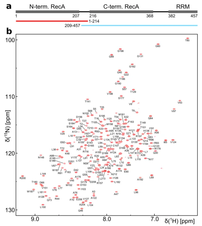
Fig. 1
DbpA domain organization and backbone resonance assign- ment of the N-terminal construct of DbpA. a The positions of the RecA and RRM domains and their domain boundaries are shown.
The N- (red) and C-terminal (blue) constructs that were used in the divide and conquer assignment approach are indicated below. b
1
H
15N-TROSY-HSQC spectrum of the
15N,
13C labeled N-terminal
construct (residues 1–214) recorded at 800 MHz. Assigned backbone
amide signals are labeled. The side chain amide signal of Trp 187 is
indicated by an asterisk
biogenesis (Sharpe Elles et al. 2009) and possesses a C-ter- minal RNA recognition motif in addition to the canonical RecA domains (Hardin et al. 2010). The RRM specifically binds to hairpin 92 of the 23S rRNA and thereby recruits DbpA to the nascent ribosome (Tsu et al. 2001). Hairpin 92 is part of the highly conserved peptidyl transferase center which remains unstructured until the final stages of 50S ribosomal subunit maturation (Nikolay et al. 2018). The hel- icase activity of DbpA and YxiN is strongly activated upon binding of hairpin 92 to the RRM (Diges and Uhlenbeck 2001; Samatanga et al. 2017), but the molecular mechanism behind this allosteric activation process is not clear.
We report here the ILMVA methyl group assignment of full length DbpA. These assignments will serve as the basis for further studies on the mechanism behind the allosteric activation of DbpA and on the RNA unwinding mechanism.
In this regard, it is noteworthy that neither the isolated C-ter- minal RecA domain nor the isolated RRM could be obtained in soluble form after expression in E. coli. A direct interac- tion between the RRM and the C-terminal RecA domain therefore seems likely and might be the basis for the allos- teric activation of the helicase activity upon binding of hair- pin 92 to the RRM.
Methods and experiments Construct design
The gene coding for full length DbpA and two constructs comprising the N-terminal RecA domain (residues 1–214) or the C-terminal RecA domain plus the RRM (residues 209–457) were PCR amplified from genomic E. coli DNA (strain BL21(DE3)) and cloned into a modified pET vector with a TEV-cleavable, N-terminal hexahistidine tag. Point mutations were introduced using the QuickChange approach and verified by sequencing.
Sample preparation
For protein expression the plasmids were transformed into E. coli BL21(DE3) cells. Cells were grown in M9 medium (H
2O based for protonated samples or D
2O based for deuter- ated samples) containing 0.5 g/l
15NH
4Cl and 2 g/l glucose (
1H
13C-labeled for protonated samples,
2H
13C-labeled for deuterated, uniformly
13C-labeled samples or
2H-labeled for deuterated samples with selective methyl group labelling).
In order to adapt the cells to D
2O-M9 medium the cells from an initial preculture in LB medium were transferred to 10%
of the final volume of D
2O-M9 medium to an OD
600of 0.15 and grown over night at 37 °C. This culture was used to inoculate the remaining D
2O-M9 medium. Cells were grown to an OD
600of 0.7–0.9 at 37 °C, then IPTG was added to
a concentration of 1 mM and proteins were expressed at 25 °C over night. Protonation of the methyl groups of Ile (Cδ methyl only), Val and Leu (ILV) in a
2H,
15N,
13C labeled background was achieved by addition of 60 mg/l 2-Keto- butyric acid-(
13C
4,3,3-d2) and 100 mg/l 2-Keto-3-methyl- butyric acid-(
13C
5, 3-d) to the medium. For
1H,
13C labeling of the methyl groups of Ile (Cδ methyl only), Val, Leu, Met and Ala (ILMVA) in a
2H,
15N labeled background 60 mg/l 2-Ketobutyric acid-(4-
13C,3,3-d2), 100 mg/l 2-Keto-3-me- thyl-butyric acid-(dimethyl-
13C
2, 3-d), 100 mg/l L-methio- nine-(methyl-
13C) and 100 mg/l L-alanine-(methyl-
13C, 2-d) were added to the medium. All precursors were added 1 h prior to induction except for alanine, which was added 20 min before induction (Kerfah et al. 2015; Schütz and Sprangers 2020). Isotopically labeled precursors and amino acids were obtained from Cambridge Isotope Laboratories (Ile and Leu/Val precursors) or Sigma-Aldrich (L-methio- nine and L-alanine).
After expression cells were harvested by centrifugation, resuspended in buffer A (400 mM NaCl, 50 mM sodium phosphate, pH 7.4, 10 mM imidazole) supplemented with 0.1% (v/v) triton x-100 and 1 mg/ml lysozyme and lysed by sonication. Cell debris was removed by centrifugation and the supernatant was loaded onto a gravity flow Ni–NTA colum equilibrated in buffer A. The column was washed with wash buffer (1 M NaCl, 25 mM sodium phosphate, pH 7.4) to remove nucleic acids bound to DbpA and with buffer A supplemented with 20 mM imidazole. DbpA was eluted with elution buffer (150 mM NaCl, 25 mM sodium phos- phate, pH 7.4, 300 mM imidazole). The hexahistidine tag was removed by TEV cleavage during dialysis over night at 4 °C against dialysis buffer (150 mM NaCl, 25 mM sodium phosphate, pH 7.4, 1 mM DTT). DbpA was loaded onto a NiNTA column equilibrated in dialysis buffer. The flow through was collected, mixed with ½ volume of 60% glyc- erol (v/v) and loaded onto a 5 ml HiTrap HP heparin column.
DbpA was eluted using 10–50% gradient over 50 ml (buffer A: 25 mM HEPES, pH 7.3, 20% (v/v) glycerol, buffer B:
as buffer A + 1 M NaCl). The heparin column was omitted for the N-terminal RecA domain as it lacks the positively charged RRM that is essential for binding to the heparin column. As the final purification step DbpA was subjected to size exclusion chromatography (SEC) using a Superdex 75 16/600 column (SEC buffer: 125 mM NaCl, 25 mM HEPES, pH 7.3, 1 mM DTT). NMR samples were prepared in SEC buffer supplemented with 5% (v/v) D
2O.
NMR experiments
The sample for the assignment of the N-terminal RecA
domain (residues 1–214) was
15N,
13C labeled at a con-
centration of 400 μM. Higher concentrations lead to a
strong decrease in signal intensity in the heteronuclear 3D
spectra. The assignment of the N-terminal RecA domain was based on the following spectra:
1H
15N-HSQC*,
1H
13C- HSQC, 3D-HNCA, 3D-HN(CA)CO*, 3D-CBCA(CO)NH, 3D-HNCO*, 3D-HNCACB*, 3D-HBHA(CBCACO)NH, 3D-(H)CCH-TOCSY, 3D-H(C)CH-TOCSY, 3D-(H)CCH- COSY and 3D-H(C)CH-COSY (Sattler et al. 1999) (aster- isks indicate TROSY based spectra (Salzmann et al. 1998)).
The assignments from the
15N,
13C labeled sample were extended and transferred to the deuterated, ILMVA labeled sample based on a 3D-CCH-NOESY spectrum (SOFAST- HMQC-based, (Rossi et al. 2016)) and a model of the struc- ture of the N-terminal RecA domain. The model was gener- ated using the SWISS-MODEL server (Waterhouse et al.
2018) and the structure of the DEAD-box helicase VASA as template (pdb identifier 2DB3, (Sengoku et al. 2006)).
The sample for the assignment of the C-terminal construct (residues 209–457) was protonated at the methyl groups of Ile (Cδ methyl only), Val and Leu in a
2H,
15N,
13C labeled background at a concentration of 740 μM. To increase the solubility and long term stability of the sample the NaCl concentration was increased to 250 mM and 25 mM Arg/
Glu was added to the sample buffer. The assignment of the backbone resonances and ILV methyl groups of the C-ter- minal construct was based on the following spectra:
1H
15N- HSQC*,
1H
13C-HMQC, 3D-HNCACB*, 3D-HN(CO) CACB*, 3D-HN(CA)CO*, 3D-HNCO*, 3D-(H)CC(CO) NH*, 3D-H(CCCO)NH*, 3D-HCH- and 3D-CCH-NOESY (SOFAST-HMQC-based, (Rossi et al. 2016)). The assign- ment was expanded to the Met and Ala methyl groups using a ILMVA
1H
13C-methyl group labeled sample in a
2H,
15N labeled background based on 3D-HCH-, 3D-CCH- and 3D-CNH-NOESY spectra in combination with models of the C-terminal RecA domain and the RRM. The models were generated based on the structures of the DbpA homolog YxiN from Bacillus subtilis (pdb identifiers 2HJV (Caru- thers et al. 2006) and 3MOJ (Hardin et al. 2010) for the RecA and RRM domains, respectively).
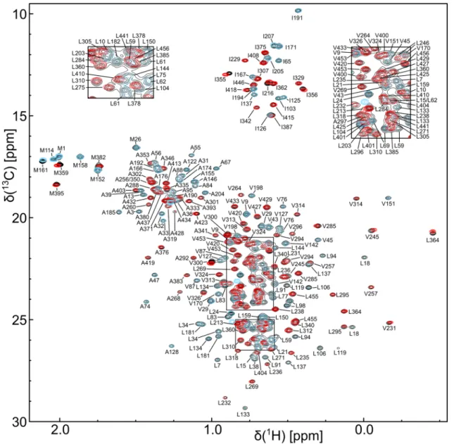
The methyl group assignments of the two constructs could then be transferred to full length DbpA (residues 1–457) as the
1H
13C-HMQC spectra of the individual domains overlay very well with the spectrum of the full length protein. Two methionine methyl group signals (M1 and M114) could not be assigned based on the recorded spectra. An M114E point mutation was thus introduced into full length DbpA. This allowed for the assignment of the M114 methyl group by comparison with the spectrum of wild type DbpA and also led to the assignment of the last unassigned methyl group signal as M1.
All spectra were recorded at 298 K on 600 and 800 MHz Bruker Neo Avance NMR spectrometers equipped with nitrogen (600 MHz) or helium cooled (800 MHz)
cryoprobes. Spectra were processed with Topspin 4.0.2 and analyzed using CARA (Keller 2004).
Assignments and data deposition
As the RecA domains of DEAD-box helicases have been shown to tumble independently in the apo state (Linder and Jankowsky 2011) we used a divide and conquer approach (Sprangers and Kay 2007) for the assignment (Fig. 1a). Two constructs of DbpA comprising the N-ter- minal RecA domain (residues 1–214) or the C-terminal RecA domain plus the RRM (residues 209–457) could be expressed and purified with high yield and showed well dispersed
1H
15N TROSY-HSQC spectra (Figs. 1b, 2). In addition the
1H
13C-HMQC spectra of the two constructs overlap very well the spectrum of full length DbpA (resi- dues 1–457) (Fig. 3). This indicates that the RecA domains of the two constructs indeed do not interact with each other and allowed us to transfer the methyl groups assign- ments to full length DbpA.
For a
15N,
13C labeled sample of the N-terminal RecA domain 88% of the H
N, N, Cα, Cβ and C’ backbone reso- nances could be assigned (Fig. 1b). These assignments were extended to the side chain methyl groups using 3D-TOCSY and 3D-COSY experiments. Despite the rela- tively small size of the construct (22 kDa) HCC(CO)NH experiments yielded very low signal intensities. This is most likely the result of transient, nonspecific intermo- lecular interactions as increasing protein concentrations also lead to decreasing signal intensities in heteronuclear 3D spectra. The methyl group assignments obtained for the
15N,
13C labeled sample were subsequently transferred to the perdeuterated, ILMVA labeled sample (Fig. 3) and extended to the Met methyl groups based on a 3D-CCH- NOESY experiment. In total 97% (134 out of 138) of the ILMVA methyl groups of the N-terminal RecA domain could be assigned.
For the assignment of the C-terminal construct (resi- dues 209–457) a
2H,
15N,
13C labeled sample (protonated at the ILV methyl groups) was used that allowed for the assignment of 93% of the H
N, N, Cα, Cβ and C’ back- bone resonances (Fig. 2). Based on these assignments the majority of the ILV methyl groups were assigned using HCC(CO)NH type spectra. These assignments were then used as starting points for the assignment of the remain- ing ILMVA methyl groups (Fig. 3) based on 3D-NOESY spectra (HCH-, CCH- and CNH-type) and a homology model of the RecA and RRM domains.
As the sum of the methyl group spectra of the two
assigned constructs is virtually identical to the spectrum
of full length DbpA (Fig. 3) the methyl group assignments could directly be transferred to the full length protein. In total 98% (260 out of 266) of the ILMVA methyl groups of DbpA were assigned. The assignments have been
deposited at the Biological Magnetic Resonance Bank under the accession numbers 50355 (N-terminal RecA domain), 50356 (C-terminal RecA domain plus RRM) and 50357 (full length DbpA).
Fig. 2
Backbone resonance assignment of the C-terminal con- struct of DbpA (residues 209–457) that comprises the C-terminal RecA domain and the RRM.
1H
15N-TROSY-HSQC spectrum of a
2
H,
15N,
13C labeled sample recorded at 800 MHz. Assigned backbone
amide signals are labeled. Assigned Trp side chain amide signals
are indicated by asterisks. The inset on the top left shows the boxed
region in the center of the spectrum
Fig. 3
ILMVA methyl group assignments of DbpA. An overlay of the
1
H
13C-HMQC spectra of full length DbpA (black), the N-terminal construct (residues 1–214, blue) and the C-terminal construct (resi- dues 209–257, red) is shown. The sum of the spectra of the isolated parts yields the spectrum of the full length protein. All spectra were
recorded at 800 MHz on perdeuterated samples that are
1H,
13C-
labeleled at the ILMVA methyl groups. Assigned methyl group sig-
nals are labeled. The insets on the top show close-ups of the two
boxed regions in the center of the spectrum
Acknowledgements
I am grateful to Remco Sprangers for access to lab space, spectrometer time and helpful discussions. I would like to thank Johanna Stöfl for great support in the wet-lab and Julia Haschlar and Katarzyna-Anna Glowacz for cloning, expression and purification of the M114E mutant. I would also like to thank Jan Overbeck for critical reading of the manuscript.
Funding
Open Access funding enabled and organized by Projekt DEAL. This work was funded by the Deutsche Forschungsgemein- schaft through grant number WU 988/1-1.
Open Access
This article is licensed under a Creative Commons Attri- bution 4.0 International License, which permits use, sharing, adapta- tion, distribution and reproduction in any medium or format, as long as you give appropriate credit to the original author(s) and the source, provide a link to the Creative Commons licence, and indicate if changes were made. The images or other third party material in this article are included in the article’s Creative Commons licence, unless indicated otherwise in a credit line to the material. If material is not included in the article’s Creative Commons licence and your intended use is not permitted by statutory regulation or exceeds the permitted use, you will need to obtain permission directly from the copyright holder. To view a copy of this licence, visit http://creat iveco mmons .org/licen ses/by/4.0/.
References
Aregger R, Klostermeier D (2009) The DEAD box helicase YxiN maintains a closed conformation during ATP hydrolysis. Bio- chemistry 48:10679–10681. https ://doi.org/10.1021/bi901 278p Caruthers JM, Johnson ER, McKay DB (2000) Crystal structure of
yeast initiation factor 4A, a DEAD-box RNA helicase. Proc Natl Acad Sci USA 97:13080–13085. https ://doi.org/10.1073/
pnas.97.24.13080
Caruthers JM, Hu Y, McKay DB (2006) Structure of the second domain of the Bacillus subtilis DEAD-box RNA helicase YxiN.
Acta Crystallogr Sect F Struct Biol Cryst Commun 62:1191–
1195. https ://doi.org/10.1107/S1744 30910 60446 42
Cheng Z, Coller J, Parker R, Song H (2005) Crystal structure and func- tional analysis of DEAD-box protein Dhh1p. RNA 11:1258–1270.
https ://doi.org/10.1261/rna.29209 05
Diges CM, Uhlenbeck OC (2001) Escherichia coli DbpA is an RNA helicase that requires hairpin 92 of 23S rRNA. EMBO J 20:5503–
5512. https ://doi.org/10.1093/emboj /20.19.5503
Fairman-Williams ME, Guenther U-P, Jankowsky E (2010) SF1 and SF2 helicases: family matters. Curr Opin Struct Biol 20:313–324.
https ://doi.org/10.1016/j.sbi.2010.03.011
Hardin JW, Hu YX, McKay DB (2010) Structure of the RNA binding domain of a DEAD-box helicase bound to its ribosomal RNA target reveals a novel mode of recognition by an RNA recogni- tion motif. J Mol Biol 402:412–427. https ://doi.org/10.1016/j.
jmb.2010.07.040
Henn A, Cao W, Hackney DD, De La Cruz EM (2008) The ATPase cycle mechanism of the DEAD-box rRNA helicase, DbpA. J Mol Biol 377:193–205. https ://doi.org/10.1016/j.
jmb.2007.12.046
Henn A, Cao W, Licciardello N, Heitkamp SE, Hackney DD, De La Cruz EM (2010) Pathway of ATP utilization and duplex rRNA unwinding by the DEAD-box helicase, DbpA. Proc Natl Acad Sci USA 107:4046–4050. https ://doi.org/10.1073/pnas.09130 81107 Keller R (2004) The computer aided resonance assignment tutorial.
Cantina Verl, Goldau
Kerfah R, Plevin MJ, Sounier R, Gans P, Boisbouvier J (2015) Methyl-specific isotopic labeling: a molecular tool box for solution NMR studies of large proteins. Curr Opin Struct Biol 32:113–122. https ://doi.org/10.1016/j.sbi.2015.03.009 Linder P, Jankowsky E (2011) From unwinding to clamping - the
DEAD box RNA helicase family. Nat Rev Mol Cell Biol 12:505–
516. https ://doi.org/10.1038/nrm31 54
Nikolay R, Hilal T, Qin B, Mielke T, Bürger J, Loerke J, Textoris-Taube K, Nierhaus KH, Spahn CMT (2018) Structural visualization of the formation and activation of the 50S ribosomal subunit dur- ing in vitro reconstitution. Mol Cell 70:881-893.e3. https ://doi.
org/10.1016/j.molce l.2018.05.003
Polach KJ, Uhlenbeck OC (2002) Cooperative binding of ATP and RNA substrates to the DEAD/H protein DbpA. Biochemistry 41:3693–3702. https ://doi.org/10.1021/bi012 062n
Putnam AA, Jankowsky E (2013) DEAD-box helicases as integrators of RNA, nucleotide and protein binding. Biochim Biophys Acta 1829:884–893. https ://doi.org/10.1016/j.bbagr m.2013.02.002 Rossi P, Xia Y, Khanra N, Veglia G, Kalodimos CG (2016) 15N and
13C- SOFAST-HMQC editing enhances 3D-NOESY sensitivity in highly deuterated, selectively [1H,13C]-labeled proteins. J Biomol NMR 66:259–271. https ://doi.org/10.1007/s1085 8-016-0074-5 Salzmann M, Pervushin K, Wider G, Senn H, Wüthrich K (1998)
TROSY in triple-resonance experiments: new perspectives for sequential NMR assignment of large proteins. Proc Natl Acad Sci USA 95:13585–13590. https ://doi.org/10.1073/pnas.95.23.13585 Samatanga B, Andreou AZ, Klostermeier D (2017) Allosteric regula- tion of helicase core activities of the DEAD-box helicase YxiN by RNA binding to its RNA recognition motif. Nucleic Acids Res 45:1994–2006. https ://doi.org/10.1093/nar/gkx01 4
Sattler M, Schleucher J, Griesinger C (1999) Heteronuclear multidi- mensional NMR experiments for the structure determination of proteins in solution employing pulsed field gradients. Prog Nucl Magn Reson Spectrosc 34:93–158. https ://doi.org/10.1016/S0079 -6565(98)00025 -9
Schütz S, Sprangers R (2020) Methyl TROSY spectroscopy: a versa- tile NMR approach to study challenging biological systems. Prog Nucl Magn Reson Spectrosc 116:56–84. https ://doi.org/10.1016/j.
pnmrs .2019.09.004
Sengoku T, Nureki O, Nakamura A, Kobayashi S, Yokoyama S (2006) Structural basis for RNA unwinding by the DEAD-box protein Drosophila Vasa. Cell 125:287–300. https ://doi.org/10.1016/j.
cell.2006.01.054
Sharma D, Jankowsky E (2014) The Ded1/DDX3 subfamily of DEAD- box RNA helicases. Crit Rev Biochem Mol Biol 49:343–360.
https ://doi.org/10.3109/10409 238.2014.93133 9
Sharpe Elles LM, Sykes MT, Williamson JR, Uhlenbeck OC (2009) A dominant negative mutant of the E. coli RNA helicase DbpA blocks assembly of the 50S ribosomal subunit. Nucleic Acids Res 37:6503–6514. https ://doi.org/10.1093/nar/gkp71 1
Sprangers R, Kay LE (2007) Quantitative dynamics and binding studies of the 20S proteasome by NMR. Nature 445:618–622. https ://doi.
org/10.1038/natur e0551 2
Steimer L, Klostermeier D (2012) RNA helicases in infection and dis- ease. RNA Biol 9:751–771. https ://doi.org/10.4161/rna.20090 Story RM, Li H, Abelson JN (2001) Crystal structure of a DEAD box
protein from the hyperthermophile Methanococcus jannaschii.
Proc Natl Acad Sci USA 98:1465–1470. https ://doi.org/10.1073/
pnas.98.4.1465
Theissen B, Karow AR, Köhler J, Gubaev A, Klostermeier D (2008)
Cooperative binding of ATP and RNA induces a closed confor-
mation in a DEAD box RNA helicase. Proc Natl Acad Sci USA
105:548–553. https ://doi.org/10.1073/pnas.07054 88105
Tsu CA, Kossen K, Uhlenbeck OC (2001) The Escherichia coli DEAD protein DbpA recognizes a small RNA hairpin in 23S rRNA. RNA 7:702–709. https ://doi.org/10.1017/s1355 83820 10101 35 Waterhouse A, Bertoni M, Bienert S, Studer G, Tauriello G, Gum-
ienny R, Heer FT, de Beer TAP, Rempfer C, Bordoli L, Lepore R, Schwede T (2018) SWISS-MODEL: homology modelling of
protein structures and complexes. Nucleic Acids Res 46:W296–
W303. https ://doi.org/10.1093/nar/gky42 7
Publisher’s Note