Regulation of human Argonaute proteins and its implications in disease
D I S S E R T A T I O N
zur Erlangung des Doktorgrades der Naturwissenschaften (Dr. rer. nat.) der Fakultät für Biologie und Vorklinische Medizin
der Universität Regensburg
vorgelegt von Daniela Zeitler
aus Regensburg
im Jahr 2019
Das Promotionsgesuch wurde eingereicht am: 11.01.2019 Die Arbeit wurde angeleitet von Prof. Dr. Gunter Meister
Daniela Zeitler
Meiner Familie
Not everything that counts can be counted, and not everything that can be counted counts.
Albert Einstein
Contents
Abstract
Zusammenfassung
Publications and Presentations
1 Introduction ... 1
1.1 Small RNA classes and their biogenesis ... 1
1.2 Argonaute proteins ... 3
1.2.1 Functional domains of Ago proteins ... 4
1.2.2 Binding of guide and target RNA... 5
1.3 GW182 proteins and their interaction with Ago proteins ... 6
1.3.1 Molecular basis of Ago-GW182 binding ... 6
1.4 Mechanism of GW182-mediated repression ... 7
1.5 Post-translational modifications of Ago proteins ... 8
1.5.1 Phosphorylation of Ago proteins ... 11
1.5.2 The role of Ago2 in cancer and neurological disease ... 12
1.6 Aims of the thesis ... 13
2 Results ... 14
2.1 Phosphorylation of endogenous Ago proteins... 14
2.1.1 Identification of endogenous phosphorylations in Ago proteins ... 14
2.1.2 Functional analysis of the 555:61 and 824:34 cluster phosphorylation of Ago2 17 2.1.3 Hyper-phosphorylation of the 824:34 cluster does not affect miRNA binding and localization ... 22
2.1.4 Phosphorylation at the 824:34 cluster is important for mRNA turnover ... 24
2.1.5 Cluster phosphorylation is essential for ALG-1 function in vivo ... 29
2.1.6 Modifying enzymes ... 31
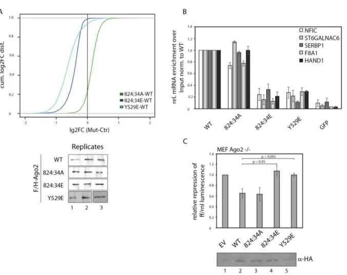
2.2 Ago2 mutations result in human neurodevelopmental disorders ... 35
2.2.1 Ago2 mutations and neurodevelopmental disorders ... 35
2.2.2 Functional analysis of the disease related mutations in Ago2 ... 36
2.2.3 Mutations affect mRNA binding ... 40
3 Discussion ... 43
3.1 Endogenous phosphorylation sites of Ago proteins ... 43
3.1.1 Conservation and positioning of phosphorylation in Ago protein ... 43
3.1.2 Functional analysis of Ago2 cluster phosphorylations ... 46
3.1.3 Cluster phosphorylation is essential for ALG-1 function in-vivo ... 48
3.1.4 Modifying enzymes acting on the Ago2 824:34 cluster ... 48
3.1.5 Phosphorylation of Ago proteins – outlook ... 51
3.2 Ago2 mutations and the relation to neurodevelopmental disease ... 53
3.2.3 Summary and outlook ... 57
4 Material and Methods ... 58
4.1 Consumables and chemicals ... 58
4.2 Buffers and solutions ... 58
4.3 Bacterial strain and cell lines ... 60
4.4 Vectors, constructs and oligonucleotides ... 60
4.4.1 Vectors ... 60
4.4.2 Constructs ... 61
4.4.3 Oligonucleotides ... 62
4.5 Antibodies ... 64
4.6 Technical Equipment ... 64
4.7 Producing DNA constructs ... 65
4.7.1 Polymerase chain reaction (PCR) ... 65
4.7.2 Transformation of Escherichia coli (E. coli) with plasmids ... 66
4.7.3 Preparation of plasmid DNA from E. coli ... 66
4.8 Protein-based methods ... 67
4.8.1 Cell culture... 67
4.8.2 Lysate preparation from harvested cells ... 68
4.8.3 Immunoprecipitation (IP) ... 68
4.8.4 SDS-PAGE, Western Blot and Coomassie staining ... 69
4.8.5 Mass Spectrometry ... 69
4.9 RNA-based methods ... 72
4.9.1 RNA isolation ... 72
4.9.2 RNA separation by Urea polyacrylamide gels and Northern Blotting ... 72
4.9.3 RISC cleavage assay ... 74
4.9.4 Autoradiographic sequencing gels ... 76
4.10 Tethering Assays ... 76
4.11 Dual Luciferase Assay ... 77
4.12 Immunofluorescence ... 77
4.13 GeneChip microarray assay ... 77
4.14 Bioinformatic analysis ... 78
4.14.1 Microarray ... 78
4.14.2 Sequence alignments... 79
Appendix ... 80
Mass spectrometry data – phosphorylation sites ... 80
List of Abbreviations ... 84
List of Figures ... 89
List of Tables ... 90 Bibliography ... 91
Abstract
Argonaute (Ago) proteins are key components of small RNA-guided gene silencing pathways. They interact with small non-coding RNAs, which guide them to specific RNA targets and are therefore referred as guide RNA. Some Ago proteins cleave RNA targets, which are perfect complementary to the bound guide RNA. In case of partial complementarity between guide and target RNA, Ago proteins recruit additional factors to mediate translational repression and mRNA degradation.
With such an important role in small RNA-guided gene silencing, it is necessary to understand the regulation of Ago proteins. Post-translational modifications, in particular phosphorylations, are an essential tool for protein regulation. Therefore, phosphorylations of Ago proteins from different species were analyzed using mass spectrometry. Here, multiple endogenous and highly conserved phosphorylation sites were identified. Moreover, five of them were organized in a phosphorylation cluster. This phosphorylation cluster was analyzed further regarding functionality and modifying enzymes. It turned out that this phosphorylation cluster is required for efficient target release and it is essential for early development C. elegans. By analyzing mass spectrometry data, one phosphatase was identified to be involved in the de-phosphorylation of this phosphorylation cluster.
Mis-regulated Ago expression levels or mutations of Ago proteins are known to result in various diseases or cancer. A collaboration partner found different heterozygous Ago2 mutations in patients showing the same neurological phenotype. In the second part of this thesis, all these detected mutations were characterized and checked regarding similarities.
Here, a common mis-regulation in target release was revealed and sheds light on the unexpected finding that heterozygous Ago2 mutations lead to neurodevelopmental disease.
Zusammenfassung
Argonaut (Ago)-Proteine zählen zu den Schlüsselkomponenten der kleinen RNS-gesteuerten Genstilllegung. Diese Proteine interagieren mit kleinen-nicht kodierenden RNS, die sie zu spezifischen Ziel-RNS leiten und deshalb als Führungs-RNS bezeichnet werden. Einige Ago Proteine sind in der Lage Ziel-RNAs zu spalten, die zu den Führungs-RNS perfekt komplementär sind. Im Falle einer nur teilweisen Komplementarität zwischen Führungs- RNS und Ziel-RNS, rekrutieren Ago Proteine zusätzliche Faktoren, um die Translationsrepression und den mRNS-Abbau zu vermitteln.
Mit dieser wichtigen Rolle bei der Genstilllegung mittels kleinen RNAs ist es notwendig, die Regulation von Ago Proteinen zu verstehen. Posttranslationale Modifikationen, insbesondere Phosphorylierungen, sind ein wesentliches Instrument für die Proteinregulierung. Daher wurden Phosphorylierungen von Ago Proteinen aus verschiedenen Spezies unter Verwendung von Massenspektrometrie analysiert. Hier konnten mehrere endogene und hochkonservierte Phosphorylierungsstellen identifiziert werden. Darüber hinaus waren fünf von diesen Phosphorylierungen in Phosphorylierungsclustern organisiert. Dieses wurde weiter auf Funktionalität und modifizierende Enzyme hin untersucht. Es stellte sich heraus, dass dieses Phosphorylierungscluster für eine effiziente Freisetzung der Ziel-RNS erforderlich ist und dass dieses auch für die frühe Entwicklung in C. elegans unerlässlich ist. Bei der Analyse der Daten generiert durch Massenspektrometrie wurde eine Phosphatase identifiziert, die an der Dephosphorylierung von diesem Phosphorylierungscluster beteiligt ist.
Es ist bekannt, dass falsch regulierte Expressionsniveaus von Ago Proteinen oder deren Mutationen zu verschiedenen Erkrankungen oder Krebs führen können. Ein Kooperationspartner fand bei Patienten, die einen ähnlichen neurologischen Phänotypen aufweisen, verschiedene heterozygote Mutationen in Ago2. Im zweiten Teil dieser Arbeit wurden all diese nachgewiesenen Mutationen charakterisiert und auf Ähnlichkeiten überprüft. Hier konnte eine Fehlregulierung bei der Freigabe der Ziel-RNS aufgedeckt werden und Licht auf die unerwartete Feststellung werfen, dass heterozygote Ago2 Mutationen zu einer neurologischen Erkrankung führen.
Publications and Presentations
Parts of this thesis were published in the following article:
Huberdeau M. Q.*, Zeitler D. M.*, Hauptmann J., Bruckmann A., Fressigne L., Danner J., Piquet S., Strieder N., Engelmann J. C., Jannot G., Deutzmann R., Simard M. J., Meister G.
Phosphorylation of Argonaute proteins affects mRNA binding and is essential for microRNA- guided gene silencing in vivo. EMBO J. 36, 2088–2106 (2017) *These authors contributed equally
Lessel D, Zeitler D. M., Kazantsev A., Ignatova Z., Meister G., Kreienkamp H. J. Heterozygous mutations in Ago2 result in neurodevelopmental disorders. Manuscript in preparation.
Additionally, I contributed to the following article:
Gust A., Jakob L., Zeitler D. M., Bruckmann A., Kramm K., Willkomm S., Tinnefeld P., Meister G., Grohmann D. Site-Specific Labelling of Native Mammalian Proteins for Single-Molecule FRET Measurements. Chembiochem.;19(8):780-783 (2018)
Parts of this thesis have been presented at international conferences:
Danner J., Hauptmann J., Zeitler D. M., Bruckmann A., Kremmer E., Deutzmann R., Meister G.
Exploiting peptide and antibody purification strategies to analyze post-translational modifications of endogenous Argonaute and TNRC6 proteins. Poster presentation, 11th Microsymposium on Small RNA Biology, 2016, Vienna, Austria.
Zeitler D. M., Huberdeau M. Q., Hauptmann J., Bruckmann A., Fressigne L., Danner J., Piquet S., Strieder N., Engelmann J. C., Jannot G., Deutzmann R., Simard M. J., Meister G. Hyper- Phosphorylation of Argonaute Proteins Affects mRNA Binding and is Essential for microRNA-guided Gene Silencing in vivo. Oral presentation, 2nd International Symposium on Frontiers in Molecular Science Non-Coding RNAs and Epigenetics in Cancer, 2017, Basel, Switzerland.
Zeitler D. M., Huberdeau M. Q., Hauptmann J., Bruckmann A., Fressigne L., Danner J., Piquet S., Strieder N., Engelmann J. C., Jannot G., Deutzmann R., Simard M. J., Meister G.
Phosphorylation of Argonaute Proteins Affects mRNA Binding and is Essential for microRNA-guided Gene Silencing. Poster presentation, SFB960-Symposium, The Biology of RNA-Protein Complexes, 2017, Regensburg, Germany.
Introduction
1
1 Introduction
In 1998, Andrew Fire and Craig Mello discovered RNAi in C. elegans and 8 years later they received the Nobel Price for their groundbreaking discovery1. It was shown that the injection of double stranded (ds) RNA molecules can lead to silencing of complementary genes much better than single stranded (ss) RNA molecules1. Since then, key insights of the role of small, non-coding RNAs in different cellular processes have been unraveled.
1.1 Small RNA classes and their biogenesis
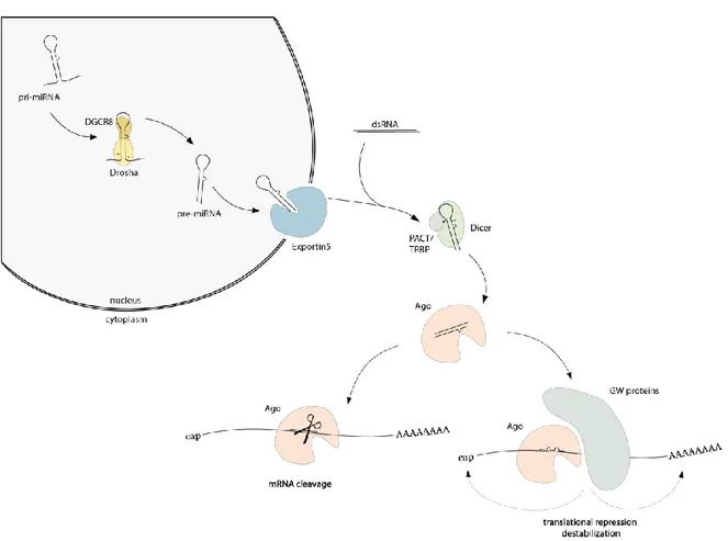
Small, non-coding RNAs that play roles in regulation of gene expression can be divided in three main classes based on the structure of the precursors, biogenesis, appearance, and mode of action. The classes are named microRNAs (miRNAs), small interfering RNAs (siRNAs) and the PIWI-interacting RNAs (piRNAs). They have their length of 20-35 nucleotides (nt) in common and that they are all loaded into a member of the Argonaute protein family. Loaded in Argonaute proteins (Ago), small RNAs guide the repression of gene expression in various organisms2–4. miRNAs are mainly transcribed by polymerase II or III and form a stem-loop structure, called primary-miRNA (pri-miRNA)5–7. The primary transcript is processed in the nucleus by the RNase III enzyme Drosha and cofactor DiGeorge syndrome critical region gene 8 (DGCR8)8–11. The stem-loop structure is recognized by DGCR8 and Drosha crops the pri- miRNA to generate precursor-miRNA (pre-miRNA)8–16. After the first processing step, pre- miRNAs are exported by Exportin5 from the nucleus into the cytoplasm17–20. In the cytoplasm, a second processing step occurs mediated by the RNase III enzyme Dicer and its cofactors PACT (protein activator of the interferon-induced protein kinase) and TRBP (TAR RNA binding protein)21–28. PACT and TRBP recognize dsRNA and promote the substrate interaction29–33. The loop of the pre-miRNA is cut off and a small RNA duplex with a length of about 22 nt with 5’ phosphates and 2 nt 3’ overhangs is generated9,21,22,34–38. One strand (guide strand) is bound by Ago (see chapter 1.2.1). The other strand of this intermediate miRNA structure, often referred to as miRNA*, is removed from Ago and degraded in most cases2,39–
42 (Figure 1). Dicer, TRBP and Ago form a stable complex, which is referred to as loading complex of the RNA-induced silencing complex (RISC loading complex, RLC). This complex allows pre-miRNA processing and Ago-loading21,23–25.
siRNAs are independent of Drosha cleavage and are processed by the RNase III enzyme Dicer from long, dsRNAs to about 21 nt long RNA duplexes. Their sources can be exogenous (exo- siRNAs) or endogenous (endo-siRNAs). Exo-siRNAs can be generated by the cleavage of viral RNAs. Endo-siRNAs derive for example from transposable elements, natural antisense transcripts or hairpin RNAs43–46. The duplexes are composed of a guide strand, which is perfectly complementary to its target (antisense strand), and the complementary passenger strand (sense strand)3. Like miRNAs, siRNA duplexes are separated and the guide is incorporated into the RISC (Figure 1). This strand guides the complex to perfect complementary targets, which are cleaved by sequence-specifically RISC. The passenger strand is unstable and degraded after unwinding2–4. In contrast to the biogenesis of miRNAs or siRNAs, piRNAs originate from ssRNA precursors47. They are transcribed from large piRNA clusters, which also represent a pool of transposon sequences4,47. piRNAs are associated with a member of the PIWI clade of the Argonaute protein family and are exclusively expressed in the germline where they silence transposons48,49.
Figure 1 miRNA/siRNA biogenesis pathway. miRNAs are transcribed by RNA polymerase II or III to form stem- loop structures, called pri-miRNA. In the nucleus, a first processing step to pre-miRNA takes place, which is performed by Drosha and its cofactor DGCR8 (cleavage sites are marked by arrowheads). After export into the cytoplasm by Exportin5, the precursor is processed a second time. Dicer and its cofactor TRBP cut off the loop (cleavage sites are marked by arrowheads). siRNAs derived from exogenous or endogenous sources are also
Introduction
3
processed by Dicer. One strand of the duplex is then incorporated into Ago proteins. In case of perfect complementarity, Ago2 in humans can cleave the mRNA. In case of partial complementarity between guide RNA and target RNA, GW182 proteins are recruited. These proteins serve as binding platforms for other proteins or complexes which lead to translational repression and destabilization8,9,50–52.
1.2 Argonaute proteins
Argonaute proteins play an essential role in small RNA-guided gene silencing. They are specialists in binding small RNAs, which guide them to the targets. Argonaute proteins are named after the characteristic phenotype of an Ago-knockout in Arabidopsis thaliana that looks like the octopus Argonauta argo53. Argonaute proteins can be found in various organism but differ in their number. For example, there are twenty-six Ago proteins present in Caenorhabditis elegans (C. elegans), ten in Arabidopsis thaliana, eight in mammalia, five in Drosophila melanogaster and one in Schizosaccharomyces pombe2. The Argonaute protein family can be classified in three clades: The Ago proteins, referred after Ago1 of Arabidopsis thaliana, the PIWI proteins, named after PIWI of Drosophila melanogaster, and the worm- specific WAGO proteins54–56. Four of the eight mammalian Ago proteins are part of the PIWI subfamily, called HIWI1-3 and HILI, the remaining four Ago proteins belong to the Ago clade, and are termed Ago1-44. PIWI proteins are mainly expressed in the germline and interact with piRNAs to protect the genome from mobile genetic elements via transcriptional silencing processes47,57,58. The members of the Ago protein clade are ubiquitously expressed and key players of the small RNA-guided gene silencing pathway. Some of them are able to silence targets or to mediate target repression by endonucleolytic cleavage (see chapter 1.2.1).
Another way to achieve silencing is the recruitment of other factors that destabilize the target RNA59,60 (see chapter 1.4). In addition, Argonaute proteins are present in archaea and bacteria. Bacterial and archaeal Ago proteins show a domain organization, which similar to the eukaryotic Ago proteins61–64. Of note, all characterized Ago proteins in prokaryotes target DNA, indicating that they are involved in DNA silencing61–63,65–68. Some studies hypothesized that prokaryotic Ago proteins are involved in protection against invasive genetic elements63,67,69.
1.2.1 Functional domains of Ago proteins
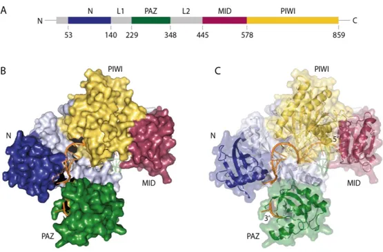
Ago proteins are two-lobe proteins, which comprise four different domains: the (N)-terminal domain, the Piwi-Argonaute-Zwille (PAZ) domain, the middle (MID) domain and the P- element-induced wimpy testes (PIWI) domain (Figure 2). Two linkers (L1 and L2) link the N- terminal with the PAZ and the MID with the PIWI domain. The 3’ nucleotide of the small RNA is bound by the PAZ domain, its 5’ end anchors in the MID and PIWI domain68,70–72.
Figure 2 Crystal structure of human Ago2. (A) Domain organization of human Ago2. Color code: N domain – blue, PAZ – dark green, MID – magenta, PIWI – yellow, linker domains - grey (B)/(C) Crystal structure of human Ago2 with co-crystalized miRNA (orange) and parts of mRNA (light green). The domains are indicated like (A).
The 5’ end of the miRNA anchors in the MID domain, its 3’ end is bound to the PAZ domain. The structure is based on the model 4W5O73.
Ago proteins associate with one strand of the miRNA duplex. In consequence, there must be a mechanism to unwind the duplex and to decide, which strand is bound by Ago. It is suggested that the N-terminal domain functions as the initiator for duplex unwinding of the miRNA during RISC assembly74. But the detailed mechanism of duplex-unwinding is not completely clear. The stability of the miRNA ends is one important determinant for strand selection, which is called ‘asymmetry rule’. The strand with the less thermodynamically stable 5’-terminus is preferentially loaded into Ago 75,76.
Introduction
5 The PIWI domain contains an RNase-H like fold that catalyzes cleavage of complementary RNA targets endonucleolytically, a process often referred to as ‘slicing’56. This slicing ability is based on a catalytic center, which contains a tetrade of Asp-Glu-Asp-His70. Only human Ago2 is able to cleave targets in case of fully complementarity between guide and target RNA77,78. In addition to its slicing function, the PIWI domain contains two binding pockets, which allow for binding to a member of the glycine-tryptophan protein of 182 kDa (GW182) protein family50,71.
1.2.2 Binding of guide and target RNA
Small RNAs are bound by Ago proteins via their phosphate-sugar backbone. This is the basis for guide target interaction, because the bases of the guide RNA protrude into the binding channel for target RNA68,71. Experiments of the DNA-binding Ago protein of Thermus thermophilus showed that base pairing of the guide strand at positions 4-16 is necessary for DNA cleavage. Only mismatches between position 2-8 reduced cleavage activity and mismatches at the cleavage site abolished slicing function because the catalytic center is not reached79.
The position 2-8 is referred to as seed sequence and its complementarity is the main criterion for target-site prediction in the miRNA pathway72,80–83. Overall, miRNA target sites are located in the 3’ UTR of mRNAs83. Structural investigations revealed that the strongest canonical target sites with seed complementarity contain an adenine opposite the first nucleotide of the guide. This adenine is recognized by a specific binding pocket within Ago proteins73,81,84. Target recognition occurs in two steps: first, the seed nucleotides 2-6 are pre-organized by MID and PIWI domains, resulting in a stacked and helical conformation with exposed nucleotides 2-471. This enables a fast and primary binding but it only leads to stable target binding, when the sites are complementary to the full miRNA seed85. With no full complementary seed, Ago either laterally diffuses along the mRNA to scan for potential targets or dissociate completely85. Crosslinking of Ago and its targets revealed also non- canonical targets, which do not have a full seed match86–89. Additionally to seed interaction, the guide’s 3’ end is also able to contribute to target recognition. Here, in particular the nucleotides 13-16 are involved81,90. Recent publications have shown that also the 3’ half of the guide can direct the miRNA family members with the same seed to different target sites in C.
elegans91 and mouse brain86.
1.3 GW182 proteins and their interaction with Ago proteins
In mammals, only a minor part of miRNA-guided gene silencing is based on endonucleolytic cleavage of target RNAs. The main part depends on Ago-miRNA binding to 3’ UTR and gene silencing through translational repression and mRNA decay of partially complementary target RNAs92,93. To fulfill these duties, Ago proteins recruit GW182 proteins24,94–96. GW182 proteins are large and unstructured proteins, which in humans are referred to as trinucleotide repeat-containing protein 6 A, B, C (TNRC6A-C). They serve as binding platforms for other effector complexes that are required for repression24,97 (see chapter 1.4, Figure 3).
Of note, GW182 proteins can repress a reporter gene without Ago, when artificially tethered to a target by recruiting effector complexes, which are responsible for deadenylation and decapping of the target RNA, indicating that this step is independent of Ago98. It is assumed that translational repression and/or storage is performed in structured protein networks of various size called processing bodies (p-bodies)94,99–101.
1.3.1 Molecular basis of Ago-GW182 binding
The three human GW182 paralogs are structurally similar. They consist of two main regions, an unstructured N-terminal Ago binging domain and the C-terminal silencing domain. The silencing domain mediates the recruitment of mRNA deadenylation machinery like CCR4- NOT or PAN2-PAN3102–104 (see chapter 1.4). As their name suggests, GW182 proteins contain many glycine-tryptophan (GW)-repeats, distributed over the whole protein50,92,102,105. Deadenylase complexes CCR4-NOT and PAN2-PAN3 are bound by tryptophans of GW182 proteins103,104,106. In addition, Ago proteins interact also with tryptophans of GW182 proteins.
Several binding sites for Ago could be identified in TNRC6, but only two Ws in the region comprising residues 599-683 in TNRC6B might be important for binding50,107. In contrast, a recent study reported three Ago proteins that are interacting with TNRC6A at the same time at three different motifs with high affinity108. Notably, all four human Ago proteins can be bound in the same fashion107,109–112. It is suggested that the Ws need 10 specific amino acids, which serve as spacers between the two Ws, for efficient Ago binding50,107.
Introduction
7
1.4 Mechanism of GW182-mediated repression
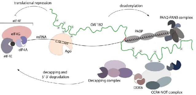
GW182 proteins interact with polyadenylate-binding protein (PABP) and thereby recruit the poly(A)-nuclease deadenylation complex subunit 2 (PAN2)-PAN3 and the carbon catabolite repressor protein 4 (CCR4)-NOT complexes (Figure 3)98,102–104,113. These recruitments catalyze deadenylation of the mRNA. Deadenylation is followed by decapping by mRNA- decapping enzyme subunit 1 (DCP1)-DCP2 complex113. The loss of the cap enables rapid degradation by the 5’-3’ exoribonuclease 1 (XRN1)114. In addition, the recruitment of the CCR4-NOT complex also leads to translational repression. The CCR4-NOT complex recruits probable ATP-dependent RNA helicase (DDX6), which inhibits translation (Figure 3)92,115. In addition, translational repression occurs through inhibition of translation initiation. The eukaryotic initiation factor 4A (eIF4A) is an RNA helicase that is needed for unwinding of secondary structures within the 5’ UTR of mRNAs116. This process is necessary for the ribosomal scanning, wherein the small ribosomal subunit attaches to the 5’-end of the target RNA and then searches for the start codon116,117. There are discrepancies about the mechanisms of the interference with eIF4AI/II but the consensus suggest that miRNA indicate the dissociation of these factors from the mRNA72,92,118–120. This might lead to blocking of the ribosomal scanning process and the assembly to eIF4F complex, which is composed of the eIF4A (eIF4AI/II), the eukaryotic initiation factor 4G (eIF4G) and the eukaryotic initiation factor 4E (eIF4E)72,92,118–120. In Drosophila, translational repression was also reported to be independent of GW182 proteins118. This underlines that the detailed molecular mechanisms of miRNA-guided gene silencing are still not fully understood.
Figure 3 Schematic model of miRNA-mediated gene silencing in animals. Ago proteins are guided by miRNA to their target mRNAs and GW182 proteins are recruited, which bind via two tryptophans to the binding pockets of Ago. GW182 proteins interact with PABP, the cytoplasmic deadenylase complexes PAN2-PAN3 and CCR4-NOT.
These complexes promote deadenylation of the mRNA. After deadenylation, decapping and degradation occurs.
Additionally, translation is repressed. Therefore, miRNAs block translational initiation by interfering with the activity of eukaryotic initiation factor 4F (eIF4F) complex. The cap structure is shown as a black circle. PABP- interacting motif is depicted in red. (modified from 92)
1.5 Post-translational modifications of Ago proteins
It is known that Ago proteins are post-translationally modified at multiple sites and in many ways, e.g. by ubiquitination, hydroxylation, SUMOylation, PARylation and phosphorylation.
Ubiquitinations of Ago proteins leads to destabilization by the ubiquitin-proteasome pathway and were detected in mouse embryonic stem cells and also in Drosophila Ago1121,122. One reported E3 ubiquitin ligase interacting with Ago is mLin41 (Trim71) and is involved in the regulation of Ago2 turnover122. Empty Drosophila Ago1 is selectively ubiquitinated by the RING-type E3 ubiquitin ligase Iruka (Iru) to trigger its degradation and to ensure correct Ago function121. The reduction of the abundance of Drosophila Ago1 and mouse Ago2 is regulated by the ubiquitin-proteasome system123. Additionally, Ago1 and Ago2 are reported to be downregulated during T cell activation by ubiquitination and proteasomal degradation124. In addition, SUMOylations are known to regulate Ago proteins. Here, a member of the SUMO (small ubiquitin-like modifier) family is attached to lysines of specific proteins125. SUMOylation at position L402 of Ago2, has been reported by two labs but with contrary effects: on the one hand it is suggested that SUMOylation leads to destabilization of Ago2126, on the other hand it might be required for full Ago2 siRNA-mediated activity127(Figure 4). In
Introduction
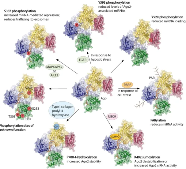
9 contrast, an increase of Ago2 stability in mouse and human cells has been observed with a hydroxylation at proline (P) 700128. This hydroxyl group is attached to Ago2 by the type I collagen prolyl-4 hydroxylase128. Both, mutation of P700 to A and depletion of the hydroxylase result in destabilization of Ago2, indicating a link between hydroxylation and protein stability128. Because the P700 position is located close to a W-binding pocket in Ago2 that ensures an interaction with TNRC6 proteins71,108, it remains speculative if the hydroxylation at this position affect miRISC assembly72 (Figure 4). In human cells, all Ago proteins undergo poly (ADP-ribosylation) (PARylation)129. Poly(ADP-ribose) (PAR) is an important regulatory molecule in the nucleus and regulates transcription, chromosome structure and DNA damage repair. This macromolecule is also reported to be added to Asp (D), Glu (E) and Lys (K) residues of Ago proteins. PARylation is involved in stress regulation, especially in the formation of cytoplasmic stress granules and therefore regulates mRNA translation and stability 129,130. It has been published that PARylation of Ago proteins reduced translational repression and slicer activity, which might come from an impeded target accessibility129. In addition, downregulated RNAi activity was monitored shortly after viral infection via PARylation of the RISC131 (Figure 4).
Figure 4 Post-translational modifications of Ago2. Phosphorylation of Y529, which is located near the 5’
binding pocket of the miRNA in the MID domain, prevent the miRNA loading132. Phosphorylation of Y393 is located in the linker 2 region and reduces the levels of Ago2 associated miRNAs133,134. The neighboring phosphorylation of S387was published to be mediated by MAPKAPK2135 or AKT3136 in vitro. This phosphorylation is responsible for increased miRNA activity by stimulating the assembly of RISC. In addition, it leads to the translocation of Ago2 to multivesicular endosomes and secretion of exosomes137. Some other phosphorylations (S253, T303, T307 and S798) were identified but the function of these have not been assigned yet132. Additionally, modifications like hydroxylation at P700 were measured. This hydroxylation increases the Ago2 stability128. SUMOylation at K402 was described either to destabilize Ago2126 or to be necessary for siRNA activity127. Also poly (ADP-ribosylation) (PARylation) was found, which blocks miRNA activity by decreasing target accessibility129,131. EGFR: epidermal growth factor receptor; OH: hydroxyl group; P: phosphate; PAR: poly (ADP-ribose); PARP: poly (ADP-ribose) polymerase; SUMO: small ubiquitin-like modifier; UBC9: ubiquitin carrier protein 9. Modified from 72.
Introduction
11 1.5.1 Phosphorylation of Ago proteins
Phosphorylations are probably among the best-investigated post-translational modifications.
This might be due to its important role in various signaling pathways. Several phosphorylated sites were also found in Ago proteins.
Some studies detected a phosphorylation at S387, which is located in the L2 region of Ago2132,135,136 (Figure 4). In vitro studies discovered MAP kinase-activated protein kinase 2 (MAPKAPK2) in the p38 MAPK pathway-mediated stress response135 and the proto-oncogene RACγ serine/threonine-protein kinase (AKT3)136 to phosphorylate S387 in Ago2. Different results of localization within the cell are reported with the mutation of S387 into A in Ago2, which resembles a phospho-lacking mutant, indicating a not fully clear function of the phosphorylation in localization135,136,138. Phosphorylation at S387 is required for the interaction between Ago and LIM domain-containing protein 1. This protein bridges the Ago- TNRC6 interaction139. S387A mutation and the resulting blocking of phosphorylation also minimize trafficking of Ago2-miRNA complexes to multivesicular endosomes and reduce the secretion of exosomes137. Taken together, the data indicate that phosphorylation supports miRNA function by stimulating the miRISC assembly72.
A further well characterized phosphorylation is located at position tyrosine (Y) 393 in the L2 region and neighboring to S387132–134(Figure 4). Hypoxic stress stimulates the phosphorylation at this residue and is modulated by epidermal growth factor receptor (EGFR)133. Another study revealed that overexpressed RAS reversibly blocks protein–
tyrosine phosphatase 1B. This inhibition leads to a hyper-phosphorylation of Ago2 at position Y393134. Both groups describe a reduced interaction between Ago and Dicer with a phosphorylation at Y393. In addition, phosphorylation at this position prevents miRNA loading of Ago2, impairs the regulation of p21 mRNA and induces senescence133,134. In the same line, phosphorylation of Ago2 at Y529 inhibits miRNA loading (Figure 4). This specific residue binds the 5’ phosphate and the first nucleotide of guide RNA71,140,141. Phosphorylation at Y529 prevents miRNA binding and is known to associate with reduced miRNA binding during macrophage activation142. Further phosphorylation sites were detected in the PAZ domain (S253, T303, T307) and in the PIWI domain (S798) but their functions still remain unknown (Figure 4)132.
1.5.2 The role of Ago2 in cancer and neurological disease
It is known that miRNA regulation is critical in tumorgenesis. As the main interactor of miRNAs, an overexpression of Ago proteins were detected in several carcinomas, like colon cancer143, head and neck squamous cell cancer144, and glioma145. In these studies, a relation between Ago2 overexpression and tumor growth and survival of cancer patients could be revealed145,146. They also discovered that Ago2 was overexpressed in hepatocellular carcinoma tissues and cells and that the overexpression enables oncogenic miRNAs, e.g. miR- 21, to repress targets146,147. In contrast, the regulatory capacity of tumor suppressive miRNAs, like let-7, was unaffected146,147. In addition, a regulatory feedback loop was identified, in which miR-99a can inhibit Ago2 reciprocally. This miRNA was found to be remarkably decreased in hepatocellular carcinoma tissues146. Moreover, a decreased expression level of Ago2 protein but not mRNA was found in melanoma148,149. Additionally, reduced miRNA and siRNA functionality and phenotypic effects, like higher migration potential of melanoma cells were observed148. Interestingly, overexpression of Ago2 protein could block cell and tumor growth148,149. Due to different miRNA expression pattern in tissues and association of Ago2 with various processes in cancer, the expression level of Ago2 is variable in different types of cancer150,151. According to The Cancer Genome Atlas (TCGA, www.cancergenome.nih.gov), various mutations of Ago2 were identified in cancers, e.g. the mutation S831P was found in melanoma.
Despite cancer, a recent study uncovered an important role of Ago2 in neurological disease152. Pircs et al.152 published recently that in Huntington’s disease Ago2 accumulates as a result of impaired autophagy in neurons, which express aggregating mutant Huntingtin. The Ago2 accumulation leads to a global alteration of miRNA activity152. Taken together, these observations of Ago2 levels and mutations in cancer and neurological disease indicate that the correct Ago2 expression level in different tissues and cells but also intact Ago2 proteins are essential in maintaining appropriate miRNA activity and to ensure health.
Introduction
13
1.6 Aims of the thesis
Ago proteins are central components of the small RNA-guided gene silencing pathway. To fulfill their functions, Ago proteins interact with small non coding RNAs, which guide them to specific target RNAs. On the one hand, some Ago proteins are endonucleolytically active and are therefore able to cleave their targets. On the other hand, Ago proteins recruit additional proteins and complexes to mediate translational repression and mRNA decay.
Since Ago proteins play such a pivotal role in gene silencing, it is important to understand the regulation of these proteins by post-translational modifications. It’s already known that Ago proteins are targets for post-translational modifications. The best characterized modification on Ago proteins is phosphorylation. However, most phosphorylation sites were measured from overexpressed Ago proteins. The first focus of this thesis is the mass spectrometric identification of endogenous and conserved phosphorylation sites. Additionally, these phosphorylation sites will be characterized regarding their functionality and modifying enzymes will be identified.
Mis-regulation in Ago expression levels or Ago mutations result in diseases, like Huntington’s disease or several cancers. A genome screening analysis of various patients showing the same neurological phenotype revealed different, heterozygous point mutations or deletions of Ago2. The second focus of this thesis is the functional characterization of these mutations.
2 Results
2.1 Phosphorylation of endogenous Ago proteins
As a key player of the gene silencing machinery, Ago proteins underlie extensive regulation.
Many post-translational modifications of Ago proteins are known (see chapter 1.5), like ubiquitination, hydroxylation and phosphorylation. Here, a systematic and unbiased screen using different purification methods of endogenous Ago proteins are described, followed by the identification of phosphorylation sites using mass spectrometry.
2.1.1 Identification of endogenous phosphorylations in Ago proteins
Since overexpression of tagged proteins can lead to unspecific phosphorylation events, different purification strategies to purify endogenous Ago proteins from human cell lysates were established. On the one hand, a purification with specific monoclonal antibodies against Ago2 or a co-immunoprecipitation (co-IP) with antibodies against TNRC6 proteins was used.
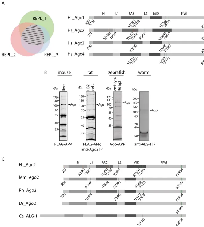
On the other hand, a purification strategy, termed Ago-APP (affinity purification by peptide) was chosen to precipitate all Ago proteins107,154. Shortly, for the immunoprecipitations, a specific monoclonal antibody against either Ago2 or TNRC6 proteins present in the cell lysate were coupled to protein-G sepharose beads. After washing, lysates, which contain phosphatase inhibitors, were added to the beads, followed by Ago protein elution and analysis using mass spectrometry. The second purification method, the Ago-APP, was performed with a short FLAG-GST-TNRC6B-peptide (T6B_599-683)107, which was coupled to anti-FLAG M2 affinity agarose gel. The lysates were incubated on the affinity gel, washed, eluted with Lämmli-sample buffer and separated on a SDS-PAGE. Ago bands were excised and the samples were prepared for mass spectrometric analysis. The measurements were performed at least in triplicates. The overlapping potential phosphorylation sites are highlighted in Figure 5 A. The left part of this figure shows a Venn diagram with three biological replicates.
The striped area represents the overlap of the phosphorylation sites of Ago in all three replicates. In the right part of this figure, the human Ago proteins are depicted schematically with their domain structure. Within this figure, the green bars represent the phosphorylation sites, which were measured in at least three biological replicates. The orange bars depict the phosphorylation sites measured in two biological replicates. Several phosphorylation sites
Results
15 are highly conserved between the four human Ago proteins. At the N-terminus, there is one serine (S) as well as one tyrosin (Y), which were phosphorylated on the same peptide. The phosphorylation of a Y, in Ago2 the Y322, which is located in the PAZ domain seems to be phosphorylated in all four human Ago proteins.
Figure 5 Identification and conservation of potential phosphorylation sites in Ago proteins. (A) Mass spectrometric measurements of human Ago proteins. Left: Venn diagram shows three replicates with an overlapping part, which is marked by stripes. Right: Schematic depiction of all four human Ago proteins and their domains. Green bars represent phosphorylation sites, which were measured at least in three biological replicates.
The orange bars represent phosphorylation sites, which were measured in two biological replicates. Those
phosphorylation sites were detected via mass spectrometric analysis after an anti-TNRC6-IP or Ago-APP. (B) Endogenous Ago proteins were purified from human, mouse, rat, zebrafish and C. elegans with the indicated method. Here, SDS gels are shown which were stained with Coomassie. (C) Schematic representation of the Ago proteins and their domain structure of various species. The phosphorylation sites are shown as green bars (measured in at least three biological replicates) and orange (measured in two biological replicates). Hs: Homo sapiens, Mm: Mus musculus, Rn: Rattus norvegicus, Dr: Danio rerio, Ce: Ceanorhabditis elegans. The sample preparation of mouse, zebrafish and C. elegans were performed by Dr. Judith Hauptmann.
Prominent phosphorylations, in which single amino acids were detected to be phosphorylated, is found between the residues 530 and 556. Lastly, a cluster phosphorylation at the very C-terminus with five potential residues from 824-834 could be identified up to four times phosphorylated at the same time and within the same peptide. These residues are very well conserved in all four human Ago proteins and appears in a phosphorylation cluster.
Only Ago4 showed less phosphorylation sites but this might be due to low abundance of Ago4.
The well-characterized phosphorylation site on S387 could only be detected in human Ago2 in our measurements135,136,139.
To see if those phosphorylation sites are also conserved between different species, material from mouse, rat, zebrafish and C. elegans was analyzed as described above (Figure 5 B). All phosphorylation events found on human Ago proteins were also found phosphorylated in these species except for Y322 and S136, which appeared to be human specific (Figure 5 C).
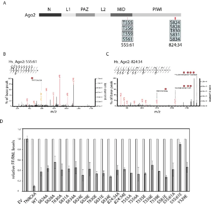
Because of the high conservation, two multiple phosphorylations were analyzed further. First, the peptide around 555-561 (555:61) in human Ago2, which contains four potential residues but was only found to be single phosphorylated in each individual measurement but at different residues. These amino acids are located in the MID domain in close proximity to the binding pocket of the target mRNA that is opposite to the first nucleotide of the miRNA73,108. The second phosphorylation cluster was the 824-834 cluster (824:34). This cluster is located on an exposed and flexible loop, which is not resolved in the known human Ago2 crystal structure71.
Results
17 2.1.2 Functional analysis of the 555:61 and 824:34 cluster phosphorylation of Ago2
The 555:61 cluster contains four closely located potential phosphorylation sites, but only single phosphorylation events were measured on individual peptides (indicated by the individual boxes, Figure 6 A). In the phosphorylation cluster at residues 824:34, hyper- phosphorylation of single peptides could be detected by mass spectrometry (indicated by one box, Figure 6 A). These phosphorylation events of single or multiple phosphorylated amino acids were readily detected in the spectra of mass spectrometry analysis (Figure 6 B and C).
To learn more about those phosphorylated clusters and their potential functions, the phosphorylated residues were mutated into glutamate (E), a phospho-mimicking mutant, and into alanine (A), a non-phosphorylatable mutant, mimicking the unphosphorylated state.
In a first experimental characterization, the constructs were fused to a Lambda N (λN)- peptide and a HA-tag to physically tether them to a luciferase reporter mRNA containing the boxB RNA segment allowing for high affinity binding to λN 155,156 (Figure 6 D). Here, single as well as multiple mutants were analyzed. In this analysis, the single mutants behaved like wild- type (WT) Ago2. Moreover, the cluster mutations which all have potential phosphorylated residues of 824:34 mutated into either E or A, did not show an effect in this tethering assay (Figure 6 D). This approach demonstrates that mutations in the cluster region did not abolish silencing activity and recruitment of downstream factors when artificially tethered to a reporter mRNA target using the λN/boxB system.
Only the phospho-mimicking mutant, in which all residues of the 555:61 cluster were mutated to E, showed a strongly impaired silencing activity (Figure 6 D). This leads to the assumption that downstream factors of the gene silencing machinery cannot be recruited when artificially tethered to a reporter mRNA target using the λN/boxB system.
Figure 6 Phosphorylation cluster and their spectra. Schematic representation of human Ago2 and its domain structure. The blue individual boxes represent single phosphorylations on the residues 555-561. One blue box with the residues 824-834 indicates multiple phosphorylations on this peptide. (B) Representative CID fragment spectra of the mono phosphorylated at T556 of human Ago2 peptide (TpTPQTLSNLCLK). This spectrum originates from a LC-UHR-QTOF run. Monoisotopic mass: 1397.65 Da, Mascot ion score: 45, expectation value: 3.2e-005, Δ mass: 0.01 Da. (C) Spectra of a four times phosphorylated peptide of human Ago2 (YHLVDKEHDpSAEGpSHTpSGQpSNGR). The phosphorylated sites are S824, S828, S831 and S834 according to Mascot. Monoisotopic mass: 2829.98 Da, Mascot ion score: 31, expectation value: 0.0085, Δ mass: 0.0017 Da. (D) Tethering assay with HA-tagged Ago2 wt and mutants. Renilla luciferase (RNL) activity was measured in extracts of HeLa cells. Cells were co-transfected with Ago2 constructs, either with or without λN-peptide, RNL-reporter with 5boxB aptamers, and Firefly (FF) luciferase. TNRC6A served as positive control. The expression levels of RNL was normalized to the FF signals (n ≥ 3, SEM).
In a second approach, TNRC6 binding of Ago2 mutants was analyzed. TNRC6 proteins are the main interactors of Ago proteins in context of translational repression and mRNA decay.
Therefore, FLAG/HA-tagged constructs immunopurified via anti-FLAG-IP. The beads with the coupled proteins were split for either RNA isolations, followed by Northern Blotting, or eluted
Results
19 for Western Blot analysis. Co-precipitated TNRC6 proteins were detected by a specific anti- TNRC6 antibody. The TNRC6 binding of the 555:61A mutant is undistinguishable from WT (Figure 7 A, lane 1-2). In contrast, the phospho-mimicking mutant showed an impaired interaction with TNRC6 (Figure 7 A, lane 3). The reduced TNRC6 binding results in impaired silencing activity, as additional factors like deadenylation and decapping complexes will not be recruited. Consistently, the reduced silencing activity could be seen in Figure 6 D.
In a next assay, binding of miRNA was tested. Ago proteins need miRNAs to guide them to specific targets, which are then repressed. After the anti-FLAG-IP, which is mentioned above, Northern Blotting was performed. A probe against the endogenous miR-19b was used to detect the bound miRNA (Figure 7 A). A signal for the bound miR-19b could detected in Ago2 WT and 555:61A mutant (Figure 7 A, lane 1-2), whereas in the 555:61E mutant, only weak signal of the bound miR-19b is visible (Figure 7 A, lane 3). This is consistent with the reduced binding of TNRC6 proteins. With a reduction in miRNA binding, 555:61E mutant could not be guided to the target and TNRC6 proteins could not be recruited. In conclusion, the silencing function of the phosphorylated 555:61 Ago2 protein is inhibited.
To get a complete picture of the silencing activity of the 555:61 phosphorylation mutants, target RNA interaction was tested using qRT-PCR. After an anti-FLAG-IP of the Ago2 proteins, RNA was isolated and cDNA generated. The direct binding of the distinct mRNA of High Mobility group protein HMGI-C (HMGA2), which is a target of let-7, was checked. 555:61A mutant binds this mRNA like Ago2 WT (Figure 7 B). In contrast, 555:61E mutant had a very low affinity to the target, which is correspond with the impaired miRNA binding (Figure 7 B).
Y529E mutant, which does not bind miRNAs132, and GFP served as negative controls and showed no mRNA interaction (Figure 7 B). A decreased miRNA binding of the hyper- phosphorylated 555:61 Ago2 protein results in a low mRNA affinity.
After checking for miRNA binding, these phosphorylation mutants were additionally tested for cleavage activity. After immunoprecipitation of the Ago2 proteins, they were incubated with a radiolabeled substrate RNA. In this in vitro cleavage assay, the endogenous miR-19b binds the perfect complementary substrate and cleaves it at a distinct position (indicated by the black bar in Figure 7 C). A signal for the cleavage product was detectable in the lane of 555:61A mutant and is undistinguishable from WT (Figure 7 C, lane 1-2), contrary to 555:61E mutant, which showed only a very weak cleavage product (Figure 7 C, lane 3). The result of impaired cleavage activity of the 555:61E mutant was also shown by Dr. Judith Hauptmann154. Consistently with the assays described above, phosphorylation at positions 555:61 results in
an impaired miRNA binding and can therefore interact with mRNA or even cleave perfect complementary targets only in a reduced manner.
Finally, the localization of Ago2 555:61A/E mutants was examined within the cell using immunofluorescence. P-bodies are sites in the cell where RNA metabolism takes place157 and it is known that Ago proteins localize to p-bodies as well24,94. With the anti-HA antibody, Ago2 proteins were detected in green under confocal microscope. An antibody against U6 snRNA- associated Sm-like protein LSm4 (LSm4) detects the endogenous in red (Figure 7 D). LSm4 is a part of the cellular RNA decay machinery and served as a p-body marker. DAPI was used for nuclei staining (blue). While 555:61 un-phosphorylatable mutant showed a co-localization with LSm4 in p-bodies (Figure 7 D, upper panel), 555:61E mutant was not detectable in these structural elements (Figure 7 D, lower panel). In conclusion, the Ago2 555:61 phospho- mimicking mutant is not located in p-bodies, indicating that hyper-phosphorylation at positions 555:61 affect localization within the cell and may also reduce silencing activity. This finding is consistent with the reduced binding of TNRC6 proteins (Figure 7 A), miRNAs (Figure 7 A), mRNAs (Figure 7 B) and the hindered silencing activity (Figure 6 D).
However, in all these approaches the 555:61 non-phosphorylatable mutant behaves like WT (Figure 7). Since only mono-phosphorylated peptides were measured in this peptide and there were no phenotypes with single mutants in the tethering assay (Figure 6 D), this cluster was not characterized further, because effects of multiple phosphorylations could be artificial.
Results
21
Figure 7 Functional analysis of the 555:61 phosphorylation cluster. (A) Immunofluorescence of HeLa cells transfected with F/H-tagged Ago2 555:61 phosphorylation mutants. Ago2 constructs were detected via anti-HA antibody (green), the endogenous LSm4 is stained in red and serves as p-body marker. The nuclei are stained via DAPI (blue). (B) HEK 293T cells were transfected with F/H-tagged Ago2 constructs, immunopurified with anti- FLAG antibodies, and separated on an SDS-PAGE or urea-gel and analyzed by either Western Blotting or Northern Blotting. Co-precipitated TNRC6 proteins were detected by pan-TNRC6-antnibody, clone 7A9. Ago2 proteins were detected by anti-HA antibody. For detection of the bound miRNAs, radioactively labeled probes against the endogenous miR-19b was used. (C) Cleavage activity of the 555:61 cluster mutants. F/H-Ago2 WT and mutants (GFP as negative control) were transfected in HEK 293T cells, immunopurified via anti-FLAG IP, incubated with a radioactively labeled substrate, which is perfect complementary to the endogenous miR-19b, followed by RNA isolation and separation on an 8 % sequencing gel. The black bar indicates the binding site of miR-19b. RNase T1 digest is labeled with ‘T1’. (D) mRNA binding of Ago2 mutants. HeLa cells were transfected with F/H-Ago2 WT or 555:61 A/E mutant. Ago2 Y529E or GFP were transfected as negative controls. After anti-FLAG-IP, RNA isolation and cDNA synthesis, the enrichment of HMGA2 was measured by qRT-PCR over input and normalized to WT.
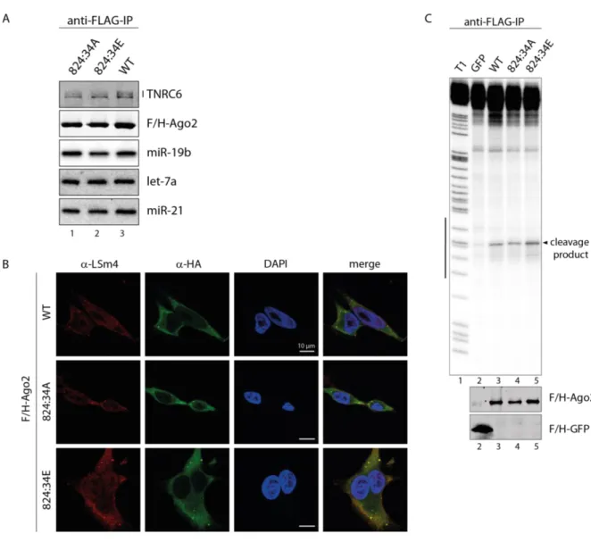
2.1.3 Hyper-phosphorylation of the 824:34 cluster does not affect miRNA binding and localization
Since there was no effect of the A or E mutant of 824:34 cluster in silencing activity, downstream steps were analyzed next. In a first approach, the miRNA and TNRC6 binding was checked. Therefore, the mutants were overexpressed in HEK 293T cells, immunoprecipitated by anti-FLAG antibodies from lysates, and either RNA isolated for Northern Blot analysis or interacting proteins eluted for Western Blot analysis. Both mutants, the phospho-mimicking and the phospho-lacking mutant, are able to bind TNRC6 proteins like WT suggesting that recruiting of additional factors for translational repression and mRNA degradation are not affected by phosphorylations at these positions (Figure 8 A, lane 2 and 3). These mutants can also interact with miRNAs, like miR-19b, miR-21 and let-7a, as efficient as WT indicating that phosphorylations at 824:34 is not important for small RNA binding and also demonstrate that the mutants are not globally unfolded (Figure 8 A, lane 2 and 3).
Next, the cellular localization of the 824:34A and E mutant was checked. Here, the Ago2 proteins were stained with an antibody against its HA-tag in green, LSm4 functions as p-body marker are shown in red and DAPI was used for nuclei staining in blue. Both mutants co- localize with LSm4 in p-bodies and are indistinguishable from WT (Figure 8 B). This result indicates that hyper-phosphorylation at the 824:34 cluster does not affect localization.
Results
23
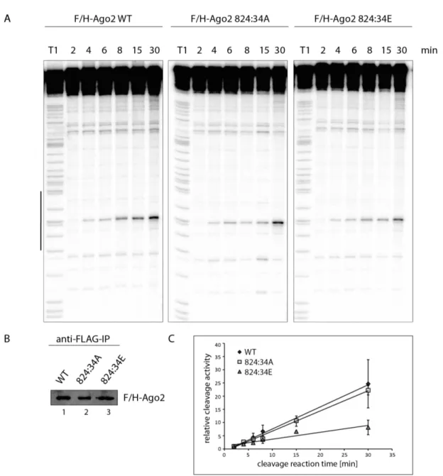
Figure 8 Functional analysis of the 824:34 phosphorylation cluster. (A) Immunofluorescence of HeLa cells transfected with F/H-tagged Ago2 WT and 824:34 phosphorylation mutants as described in Figure 7 A. (B) HEK 293T cells were transfected with F/H-tagged Ago2 constructs, immunopurified with anti-FLAG antibodies, and separated on a SDS-PAGE or urea-gel and analyzed by either Western Blot or Northern Blot. Co-precipitated TNRC6 proteins were detected by pan-TNRC6-antnibody, clone 7A9, and Ago2 proteins were detected by anti-HA antibody. For detection of the bound miRNAs, radioactively labeled probes against the endogenous miR-19b, miR- 21 and let-7a were used. (C) Cleavage activity of the 824:34 cluster mutants as described in Figure 7 C. Western Blot was performed for control.