Gametogenesis-related small RNAs and Argonaute Proteins
in Arabidopsis thaliana
DISSERTATION ZUR ERLANGUNG DES DOKTORGRADES DER NATURWISSENSCHAFTEN (DR. RER. NAT.)
DER FAKULTÄT FÜR BIOLOGIE UND VORKLINISCHE MEDIZIN DER UNIVERSITÄT REGENSBURG
vorgelegt von Marc Urban
aus Hannover
im Februar 2016
Das Promotionsgesuch wurde eingereicht am: 12.02.2016
Die Arbeit wurde angeleitet von: PD Dr. S. Sprunck
Unterschrift:
I
TABLE OF CONTENTS
1 SUMMARY ... 1
2 ZUSAMMENFASSUNG ... 4
3 INTRODUCTION ... 7
3.1 Gametogenesis in Arabidopsis thaliana ... 7
3.2 Argonaute proteins ... 10
3.2.1 Structure of Argonaute proteins ... 11
3.2.2 Subcellular localization ... 12
3.2.3 Functions of Argonaute proteins ... 13
3.2.4 The microRNA pathway ... 14
3.2.5 The siRNA pathway ... 15
3.2.6 Argonaute proteins of Arabidopsis thaliana ... 15
3.2.7 RNA-directed DNA-methylation and transposon silencing in plants ... 19
3.2.8 Epigenetic reprogramming in plant sporo- and gametogenesis ... 22
3.2.9 The role of AGO proteins in plant reproduction ... 24
3.3 Aims of this work ... 27
4 MATERIAL & METHODS ... 30
4.1 Plant materials and growth conditions... 30
4.2 Callus generation ... 30
4.3 Bacterial work ... 31
4.3.1 Generation and transformation of chemically competent Escherichia coli cells... 32
4.3.2 Generation and transformation of chemically competent Agrobacterium tumefaciens cells ... 32
4.4 DNA molecular biology work ... 33
4.4.1 Isolation of total DNA from leaves ... 33
4.4.2 Isolation of high quality total DNA from plant material ... 33
4.4.3 Polymerase chain reaction (PCR) ... 34
4.4.4 Gel extraction of DNA fragments ... 35
4.4.5 Isolation of plasmid DNA from E.coli ... 36
4.4.6 Isolation of plasmid DNA from A.tumefaciens ... 36
4.4.7 Analytical and preparative restriction digest ... 36
4.4.8 DNA Ligation ... 36
4.4.9 Gateway®-based cloning ... 37
4.4.10 Cloning of PCR-fragments for the analysis of AGO9 transcript variants ... 37
4.4.11 Cloning of AGO9 minigene constructs ... 37
4.4.12 Sequencing ... 38
4.5 RNA work ... 39
4.5.1 Isolation of total RNA ... 39
4.5.2 mRNA isolation and cDNA synthesis ... 39
4.5.3 Northern Blot hybridization ... 39
II
4.5.4 Quantitative real-time PCR Assay ... 41
4.5.5 Library Construction and Illumina Next-Generation-Sequencing ... 42
4.5.6 mRNA library preparation... 42
4.5.7 Small RNA library preparation ... 42
4.6 Bioinformatic analysis of RNA sequencing data ... 43
4.6.1 Small RNA library analysis ... 43
4.6.2 Prediction of small RNA targets ... 43
4.7 Protein Methods ... 44
4.7.1 Total protein extracts from Arabidopsis thaliana ... 44
4.7.2 Bradford protein assay ... 44
4.7.3 SDS-PAGE ... 44
4.7.4 Western Blot ... 45
4.7.5 Peptide competition assay ... 45
4.8 Cell biological work ... 46
4.8.1 Transient transformation of Tobacco BY2 cells by particle bombardment ... 46
4.8.2 Whole mount-immunolocalization ... 47
4.9 Microscopy ... 47
4.9.1 Transmitted light and fluorescence microscopy ... 47
4.9.2 Confocal microscopy ... 48
5 RESULTS ... 49
5.1 Generation of an RKD2-induced cell line with egg cell-like identity ... 49
5.2 Analysis of Argonaute expression patterns ... 52
5.2.1 Differential expression of Argonaute genes in different plant tissues ... 52
5.2.2 Differential expression of AGO proteins in different plant tissues ... 54
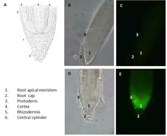
5.3 Analysis of AGO8 and AGO9 promoter activities ... 55
5.4 Expression and subcellular localization of AGO9 proteins ... 59
5.4.1 Subcellular localization after transient expression ... 60
5.4.2 Subcellular localization in stably transformed plants ... 61
5.4.3 Expression of AGO8 transcript variants under control of their native promoter ... 62
5.4.4 Expression of GFP-AGO9 under the control of the AGO9 Promoter ... 63
5.5 Ectopic overexpression of GFP-AGO9 in the egg cell ... 66
5.5.1 Localization of AGO8p:GFP-AGO9 ... 67
5.5.2 Localization of EC1.1p:GFP-AGO9 ... 68
5.6 Whole mount-immunolocalization of AGO9 ... 72
5.6.1 Control experiments ... 72
5.6.2 Immunolocalization of AGO9 ... 75
5.7 Transcript variants of AGO8 and AGO9 ... 78
5.7.1 Expression pattern of AGO8 transcript variants ... 78
5.7.2 AGO8 transcript variants in RKD2-induced callus ... 80
5.7.3 Expression pattern of AGO9 transcript variants ... 81
III
5.7.4 Systematic approach to identify other AGO9 transcript variants ... 83
5.7.5 AGO9 minigene approach ... 86
5.8 Deep sequencing of mRNA and small RNAs in RKD2-induced cells with egg cell-like fate ... 89
5.8.1 Transcriptome profiling of RKD2-induced callus with egg cell-like fate ... 89
5.8.2 RNA quality control and library generation ... 89
5.8.3 Data validation by qRT PCR ... 91
5.8.4 Data validation by analysis of the Köszegi dataset ... 92
5.8.5 Differentially expressed mRNAs in the RKD2-induced cell lines, compared with the CIM callus 94 5.8.6 Small RNA pathway components ... 97
5.8.7 Pol IV and Pol V components ... 99
5.8.8 Auxin and Cytokinin-related genes ... 100
5.8.9 Small RNA profiling of RKD2-induced cells with egg cell-like fate ... 105
5.8.10 Small RNA length distribution and annotation ... 105
5.8.11 Analysis of 5′ terminal nucleotide frequency ... 106
5.8.12 Identification of known miRNAs and analysis of differential expression ... 107
5.8.13 Identification of miRNA targets ... 110
5.8.14 Novel miRNAs and their targets ... 114
5.8.15 Validation of miRNA expression by Northern Blot analysis ... 117
5.8.16 Transposable elements in RKD2-induced callus and CIM callus ... 118
6 DISCUSSION ... 122
6.1 The role of AGO8 and AGO9 in female gametogenesis ... 123
6.2 AGO9 protein expression and subcellular localization in ovules before and after fertilization ... 124
6.3 A cell line with egg cell-like expression profile as a tool to identify new small RNA pathway components in the female germline ... 125
6.4 Small RNA profiling reveals differential expression of known and novel miRNAs ... 130
6.5 Regulation of egg cell-expressed genes via miRNAs ... 131
6.6 Transposon silencing and epigenetic modifications in the egg cell-like callus ... 133
6.7 Outlook ... 133
7 PUBLICATIONS ... 135
8 BIBLIOGRAPHY ... 136
9 APPENDIX ... 147
9.1 Antibodies ... 147
9.2 Oligonucleotides ... 147
9.3 Plasmids ... 150
9.3.1 Summary of used vectors ... 150
9.3.2 Plasmid sequences ... 152
IV
9.4 RNA sequencing results ... 155 9.4.1 mRNA sequencing data ... 155 9.4.2 Small RNA sequencing data... 157
1
1 SUMMARY
The life cycle of flowering plants alternates between a diploid multicellular sporophyte generation and a highly reduced haploid gametophyte generation located within the reproductive tissues of the sporophyte. Emerging evidence indicates that the transition from sporophyte to a gametophytic life phase and the acquisition of reproductive fate is marked by extensive epigenetic reprogramming employing distinct members of the ARGONAUTE (AGO) family of proteins with roles in RNA-directed DNA methylation (RdDM) and heterochromatin formation. Nevertheless, almost nothing is known about RNA silencing machinery components and small noncoding RNAs associated with epigenetic reprogramming and the acquisition of gametic cell fate.
The aim of this work was the detailed investigation of two RdDM-associated Argonaute genes (AGO8 and AGO9) regarding their expression pattern and function in the female reproductive lineage of Arabidopsis thaliana. Furthermore, a specialized Arabidopsis cell line with an egg cell-like transcriptome was generated to enable the identification of small RNAs and RNA silencing machinery components potentially associated with female reproductive fate.
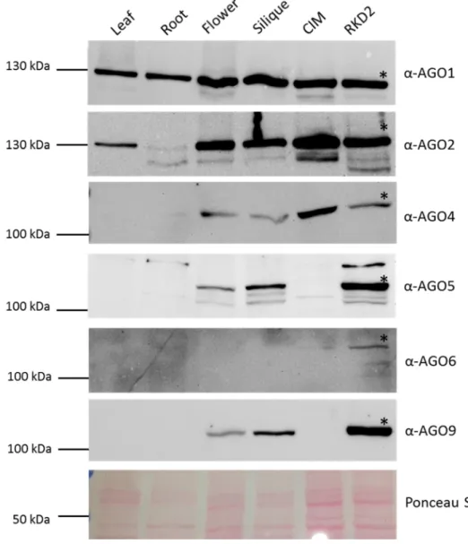
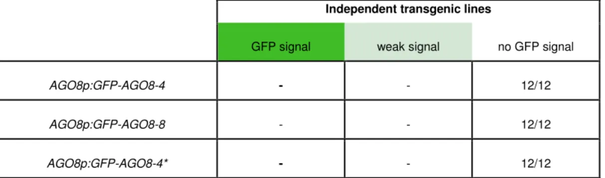
RT-PCR based expression studies were performed for all ten Arabidopsis AGO genes, confirming that AGO5, AGO8 and AGO9 are predominantly expressed in floral tissues containing reproductive lineages, while other AGO genes are more or less ubiquitously expressed. Protein abundance was investigated by Western Blots using peptide antibodies available for six AGOs andp09 revealed that AGO5 and AGO9 are strongly represented in reproductive organs and in the egg cell-like callus. To investigate AGO8 and AGO9 promoter activities and the presence of AGO8 and AGO9 proteins in more detail during female gametophyte development, transgenic Arabidopsis reporterlines were generated and whole- mount immunolocalization experiments were performed. These studies showed that the AGO8 promoter is only active in the egg cell. Nevertheless, AGO8 expression only yielded aberrant spliced mRNA and a GFP-AGO8 fusion protein was not detectable in egg cells expressing AGO8p:GFP-AGO8, suggesting that AGO8 represents a pseudogene.
AGO9 promoter-reporter lines showed activity in the nucellus cells, the megaspore
mother cell (MMC) and the functional megaspore (FM) of young ovules. In the mature female
gametophyte, AGO9 promoter activity was detectable in the egg cell, the central cell and the
chalazal region of the ovule. This is in remarkable contrast to results obtained by AGO9
2
whole-mount immunolocalization and expression of the GFP-AGO9 fusion protein: AGO9 was neither detectable in the MMC and FM nor in the egg cell nor central cell. The occurrence of aberrant splicing by intron retention in AGO9 mRNA, emerging during ovules maturation, and the results obtained from ectopic expression of correctly spliced AGO9 cDNA in the egg cell suggests that both, developmentally regulated differential splicing and decreased AGO9 protein stability is responsible for the lack of AGO9 protein in the egg cell.
A few plants were obtained expressing GFP-AGO9 in the egg cell, which did, however, not correlate with developmental defects in the female gametophyte or in developing seeds. The impact of ectopic GFP-AGO9 on DNA methylation in the egg cell remains to be investigated.
To identify protein coding genes and small noncoding RNAs possibly involved in the acquisition of female reproductive fate or in egg cell specification a specialized transgenic Arabidopsis cell line with egg cell-like expression profile was established by ectopically expressing the transcription factor RKD2. Compared to the control cell line 5,511 genes were classified as differentially expressed by RNA-seq. Quantitative real-time PCR with eighteen selected genes confirmed mRNA-Seq expression data. A global look at genes involved in small RNA pathways revealed several differentially expressed genes in RKD2-induced cell line, including upregulated genes encoding the DNA methyltransferase DOMAINS REARRANGED METHLYTRANSFERASE 1 (DRM1), the DOUBLE-STRANDED RNA BINDING PROTEIN 3 (DRB3), the HISTONE DEACETLYLASE 18 (HDA18), the putative chromatin remodeling protein CHROMATIN REMODELING 34 (CHR34) and a so far undescribed DNA-directed RNA polymerase V subunit 5A-like gene.
Small RNA profiling in the RKD2-induced cell line revealed noticeable differences
compared to the control callus. The most abundant group of small RNAs in the control callus
are miRNAs, whereas the biogenesis of siRNAs from transposable element (TE) transcripts is
dominant in the egg cell-like callus. TE-derived siRNAs of the Ty3/Gypsy superfamily of
long terminal repeat retrotransposons, containing members of the ATHILA, ATLANTYS, and
ATGP family, form the largest fraction of siRNAs, supporting the idea that the plant
reproductive lineage is protected by siRNAs against the activity of TEs. This is in line with
the observation that aligned reads to respective TE transcripts are not present in the mRNA-
Seq data of the egg cell-like cell line. Differential expression analysis of miRNAs revealed 96
miRNAs being at least up- or down regulated with a log2 fold change of 2, including only 9
miRNAs upregulated in the RKD2-induced callus. Furthermore, 32 novel miRNAs were
discovered, with 20 miRNAs being derived from known miRNA precursors while 12
3
miRNAs are completely novel. Target gene predictions of differential expressed miRNAs revealed several inverse correlations between miRNA and mRNA target expression levels in both the egg cell-like callus and the control callus.
The discovery of so far undescribed transcripts in the egg cell-like cell line encoding
small RNA pathway components involved in chromatin modification, together with the
identification of differentially expressed known and novel small noncoding RNAs open up
interesting possibilities for future investigations regarding their contribution to the acquisition
of reproductive fate and egg cell specification.
4
2 ZUSAMMENFASSUNG
Der Lebenszyklus der Blütenpflanzen ist durch einen Generationswechsel geprägt. Der diploide und mehrzellige Sporophyt wechselt sich mit dem stark reduzierten, haploiden Gametophyten ab, welcher sich in den reproduktiven Geweben des Sporophyten befindet.
Neuere Erkenntnisse weisen darauf hin, dass der Übergang vom Sporophyten zur gametophytischen Lebensphase und der Erwerb der Fähigkeit zur Reproduktion durch eine weitgehende epigenetische Neuprogrammierung des Genoms gekennzeichnet sind. Dabei sind bestimmte Mitglieder der ARGONAUTE (AGO) Proteinfamilie beteiligt. Diese spielen eine Rolle beim Prozess der RNA gesteuerten DNA-Methylierung (RdDM, RNA-directed DNA methylation). Trotz dieser Erkenntnisse ist wenig darüber bekannt, welche Komponenten der RNA- silencing Maschinerie und kleinen nicht-kodierenden RNAs an der epigenetischen Reprogrammierung in reproduktiven Geweben und am Erwerb eines gametischen Zellschicksals beteiligt sind.
Ziel dieser Arbeit war die detaillierte Untersuchung zweier RdDM-assoziierter AGO Proteine (AGO8 und AGO9) bezüglich ihres Expressionsmusters und ihrer Funktion in der weiblichen Keimbahn von Arabidopsis thaliana. Darüber hinaus wurde eine spezialisierte Arabidopsis Zelllinie mit einem eizellähnlichen Transkriptom erzeugt, um die Identifizierung von kleinen RNAs und RNA-silencing Komponenten bei der weiblichen Gametenbildung zu ermöglichen.
Für alle zehn Arabidopsis AGO Gene wurden RT-PCR basierte Expressionsstudien
durchgeführt, diese bestätigten dass AGO5, AGO8 und AGO9 überwiegend in
Blütengeweben, welche reproduktive Zellen enthalten, exprimiert sind. Im Gegensatz dazu
kommen alle anderen AGOs mehr oder weniger ubiquitär vor. Die Präsenz von AGO-
Proteinen wurde mittels Western Blot untersucht, wozu verfügbare Peptidantikörper gegen
sechs AGO Proteine verwendet wurden. Diese Untersuchung ergab, dass AGO5 und AGO9 in
reproduktiven Organen und in der eizellähnliche Zelllinie verstärkt vertreten sind. Um die
Promoteraktivitäten und die Proteinpräsenz von AGO8 und AGO9 während der Entwicklung
des weiblichen Gametophyten eingehender zu untersuchen, wurden transgene Arabidopsis
Reporterlinien generiert und eine immunhistologische Untersuchung (whole mount-
immunolocalization) durchgeführt. Diese Studien ergaben, dass der AGO8 Promoter
ausschließlich in der Eizelle aktiv ist. Nichtsdestotrotz ergab die Expression von AGO8
lediglich aberrant gespleißte mRNA. Auch in Eizellen die AGO8p:GFP-AGO8 exprimierten,
5
konnte kein GFP-AGO8 Fusionsprotein nachgewiesen werden, was darauf hinweist, dass es sich bei AGO8 um ein Pseudogen handelt.
AGO9 Promoter-Reporter-Linien zeigten Aktivität in den Nucelluszellen, in der Megasporenmutterzelle (megaspore mother cell, MMC) und der funktionellen Megaspore (FM) von jungen Samenanlagen. Im reifen weiblichen Gametophyten konnte AGO9 Promoteraktivität in der Eizelle, der Zentralzelle und der chalazalen Region der Samenanlagen nachgewiesen werden. Dies ist ein bemerkenswerter Unterschied zu den Ergebnissen der Immunolokalization und der Expression des GFP-AGO9 Fusionsproteins.
AGO9 war weder in der MMC oder FM noch in der Eizelle oder Zentralzelle nachweisbar.
Das Auftreten aberranten Spleißens mit Intron Retention in der AGO9 mRNA während der Reifung der Samenanlagen und die Ergebnisse der ektopischen Expression der korrekt gespleißten AGO9 mRNA in der Eizelle weisen darauf hin, dass sowohl entwicklungsspezifisch reguliertes alternatives Spleißen als auch verminderte Proteinstabilität für das Fehlen des AGO9-Proteins in der Eizelle verantwortlich sind. Nur wenige Pflanzen mit GFP-AGO9 Expression in der Eizelle konnten erzeugt werden, diese Expression korrelierte aber nicht mit Entwicklungsdefekten im weiblichen Gametophyten oder im sich entwickelnden Samen. Der Einfluss von ektopisch exprimiertem GFP-AGO9 auf die DNA- Methylierung in der Eizelle bleibt noch zu untersuchen.
Um protein-kodierende Gene und kleine nicht-kodierende RNAs mit möglicher Beteiligung an Prozessen der Zellspezifizierung im weiblichen Gametophyten identifizieren zu können, wurde eine spezialisierte transgene Arabidopsiszellline mit eizellähnlichem Expressionsprofil erzeugt, indem der Transkriptionsfaktor RKD2 ektopisch exprimiert wurde.
Im Vergleich zur Kontrollzelllinie wurden 5.511 Gene als differentiell exprimiert identifiziert.
Quantitative real-time PCR Analysen mit achtzehnzehn ausgewählten Genen bestätigte die
mRNAseq-Expressionsdaten. Ein allgemeiner Blick auf Gene der RNA-silencing Maschinerie
enthüllte einige differentiell exprimierte Gene in der RKD2-induzierten Zelllinie, so zum
Beispiel hochregulierte Gene die für Proteine kodieren wie die DNA-methyltransferase
DOMAINS REARRANGED METHLYTRANSFERASE 1 (DRM1), das Doppelstrang-RNA-
Bindeprotein the DOUBLE-STRANDED RNA BINDING PROTEIN 3 (DRB3), die HISTONE
DEACETLYLASE 18 (HDA18), das mögliche Chromatinremodellierungsprotein
CHROMATIN REMODELING 34 (CHR34) und ein bisher nicht beschriebenes Gen welches
der Untereinheit 5A der RNA-Polymerase V ähnelt.
6
Die Untersuchung der kleinen RNAs in der RKD2-induzierten Zelllinie zeigt eindeutige Unterschiede im Vergleich zum Kontrollkallus. Die häufigste Gruppe kleiner RNAs im Kontrollkallus sind miRNAs, wohingegen im eizellähnlichen Kallus die Biogenese von siRNAs aus mRNAs transposabler Elemente (TEs) vorherrscht. TE-abgeleitete siRNAs der TY3/Gypsy Überfamilie (TEs mit long terminal repeats), welche Mitglieder der ATHILA, ATLANTYS, und ATGP Familie beinhalten, stellen dabei den größten Teil dar, was die Vorstellung unterstützt, das pflanzliche Keimzellen durch siRNAs gegen Transposonaktivität geschützt sind. Die Beobachtung, dass in den mRNA Sequenzdaten keine reads der entsprechenden TEs zu finden sind unterstützt ebenfalls diese Idee. Analyse der differentiellen Expression von miRNAs ergab 96 miRNAs die mindestens um einen log2 Faktor von zwei hoch- oder runterreguliert sind, wobei lediglich neun miRNAs im RKD2- induzierten Kallus hochreguliert sind. Weiterhin wurden 32 neuartige miRNAs entdeckt, wovon 20 aus bereits bekannten miRNA-Vorläufern entstehen, während 12 komplett neuartig sind. Die Voraussage möglicher Zielgene differentiell exprimierter miRNAs ergab einige inverse Korrelationen zwischen miRNA und mRNA Expressionslevels im eizellähnlichen und auch im Kontrollkallus.
Die Entdeckung von bisher nicht beschriebenen Transkripten in der eizellähnlichen
Zelllinie, die für sRNA Maschinerie-Komponenten codieren und die an
Chromatinmodifikationen beteiligt sind, zusammen mit der Identifizierung differentiell
exprimierter bekannter und neuartiger kleiner nicht-kodierender RNAs eröffnet neue,
interessante Möglichkeiten für zukünftige Untersuchungen. Diese könnten neue Erkenntnisse
darüber hervorbringen, wie Zellen ein reproduktives Zellschicksal annehmen und wie die
Spezifizierung der Eizelle vonstattengeht.
7
3 INTRODUCTION
3.1 Gametogenesis in Arabidopsis thaliana
In the life cycle of a plant, two generations are involved: the diploid sporophyte and the haploid gametophyte. In flowering plants, the sporophyte is the dominant generation and the gametophyte is reduced to a few cells: the three-celled male gametophyte (pollen), and 8 nucleated female gametophyte (embryo sac), which are characteristic of most flowering plants. In general, gametogenesis starts with the specification of a spore mother cell, which then meiotically divides into four spores.
Figure 1: Sexual reproduction in Arabidopsis thaliana.
In the flower of A.thaliana, male and female reproductive tissues produce pollen mother cells and megaspore mother cells. They are generated in a position-dependent manner from somatic tissues.
During the process of sporogenesis, meiosis takes place, microspores are generated from pollen mother cells and megaspores from megaspore mother cells. In male gametogenesis, the mature pollen is formed. Female gametogenesis results in the formation of the mature female gametophyte, which consists of the egg cell, two synergids, three antipodal cells and a diploid central cell. The egg cell and the central cell are each fertilized by one sperm cell to produce the zygote and the endosperm, respectively. The zygote the elongation of the zygote precedes the first cell division, which initiates a proembryo that develops into the globular-shaped embryo and then the heart-shaped embryo to become the mature seed. Image taken from Kawashima and Berger, 2014.
8
In male gametogenesis this process is also called microsporogenesis, because the spore mother cell divides into four haploid microspores which first form a tetrad. After separation of the microspores each microspore develops into one pollen grain. The nucleus of a microspore undergoes nuclear migration and asymmetric division to generate the bicellular pollen grain (Berger and Twell 2011), which is composed of a generative and a vegetative cell. In Arabidopsis the generative cell divides once more before anthesis and the mature tri-cellular pollen is formed (Figure 1).
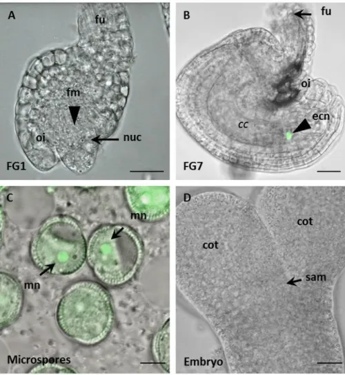
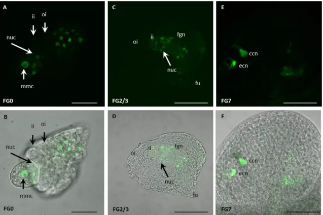
The generation of female gametes occurs within the female gametophyte (or embryosac) during ovule development. First, an archaesporial cell is specified from a cell from of the sporophytic nucellus tissue, which is called the megaspore mother cell (MMC).
The MMC is localized directly under the nucellar epidermis and is relativly large compared to the other cells. It then undergoes meiosis to generate four haploid cells, from which three undergo programmed cell death. The remaining cell is called the functional megaspore (FM) and is the progenitor of the whole female gametophyte.
Figure 2: Schematic and microscopic view of a mature Arabidopsis ovule.
(A) Cartoon of an ovule, showing the position of the female gametophyte (FG) within the embedding tissues of the ovule. Within the curved morphology of the female gametophyte the egg cell nucleus (ecn) is always oriented towards the chalazal pole (chz), close to the nucleus of the much larger central cell (ccn). The micropylar region (mp) is the entry site of the pollen tube. The vacuole of the egg cell is oriented towards the micropylar pole. The large central cell vacuole (cv) occupies the chalazal part of the female gametophyte. Next to the egg cell are two synergid cells (in orange), which undergo cell death upon fertilization. The synergid nuclei (sn) are oriented towards the micropylar pole of the FG. On the chalazal pole lie the three antipodal cells (ap), which are a result of the mitotic divisions starting from the functional megaspore. (B) Differential interference contrast (DIC) microscopy image of an ovule showing the same cells as in (A). Picture taken from Sprunck et al. 2011.
9
After three rounds of nuclear divisions the female gametophyte has eight nuclei within a syncytium. The nuclei have to be repositioned and after cell specification and cellularization the female gametophyte consists of seven cells: three antipodal cells at the chalazal end, the homo-diploid central cell (resulting from fusion of two polar nuclei), and one egg cell and two synergid cells at the micropylar end (Figure 2). Recent studies showed that the antipodal cells persists through fertilization (Song et al. 2014).
During this whole process of gametogenesis, cell specification events are necessary, but little is known about the genes and molecular mechanisms involved. For example, the mechanisms of how somatic cells aquire a reproductive cell fate are not fully understood (reviewed in Yang et al. 2010). However, a few genes that influence cell fate during gametogenesis have been identified. The SPOROCYTELESS (SPL) gene of Arabidopsis has been reported to play a role in megasporogenesis, as spl mutants are able to initiate formation of MMCs but the meiosis of the MMCs does not occur, resulting in a complete lack of a germline. Later during the process of cellularization and cell specification, the nuclei within the embryo sac need positional information to aquire their cell fate. This positional information can be provided by polar expression of proteins. DEMETER (DME), a DNA glycosylase, which can also remove 5-methylcytosine, is localized in the micropylar region of the embryo sac before cellularization but restricted to the central cell after cellularization (Choi et al. 2002). Furthermore, an auxin gradient was suggested to provide positional information during female gametophyte development (Pagnussat et al. 2009), but a recent study provided evidence that only a shallow auxin gradient can be maintained in the female gametophyte (Lituiev et al. 2013). Mutagenesis of an egg cell-specific marker line identified three mutants which show misregulation of the egg cell reporter gene expression: lachesis (lis), clotho (clo) and atropos (ato). Loss of lachesis function results in expression of the egg cell marker in the synergid and the central cell (Groß-Hardt et al. 2007). Notably, lachesis, clotho and atropos are all genes encoding components of the splicing machinery. This is a hint that RNA splicing may play a role in cell specification during gametogenesis.
A transcriptome analysis of isolated egg cells revealed the enrichment of certain
functional groups of proteins in the egg cell (Wuest et al. 2010). Especially genes involved in
RNA interference are highly abundant in the egg cell, including genes encoding factors
involved in biogenesis of microRNAs like DICER-LIKE 1 (DCL1) and HYPONASTIC
LEAVES (HYL1), but also ARGONAUTE 1, a protein involved in gene silencing mediated by
small RNAs (Wuest et al. 2010).
10
3.2 Argonaute proteins
A forward genetic screen for genes involved in developmental processes generated a mutant with extraordinary appearance (Böhmert el al. 1998). Seedlings had pointy, unexpanded cotyledons and a dark green hypocotyl. This phenotype reminded the researchers of a tiny squid and so this mutation was called Argonaute. Many homologues of Argonaute proteins were found in other species and it became clear that AGOs form a conserved family of proteins, which can be found in Archaea, Bacteria and almost all eukaryotes.
Figure 3: The three clades of the Argonautes.
The Ago-like clade, shown in black, is based on similarity to AtAGO1. The Piwi-like clade, shown in green, is based on similarity to Drosophila melanogaster Piwi and the Wago’s, which is a worm-specific clade, is shown in red. Argonaute proteins that contain a complete catalytic motif are underlined. Image taken from Joshua-Tor and Hannon 2010.
AGOs can be grouped into three big subfamilies; the AGO group which is close to the
Arabidopsis AGO1, the PIWI group containing the P-element Induced Wimpy testis protein
from Drosophila melanogaster, and a third group which can be only found in Caenorhabditis
11
elegans, the WAGO or Worm AGO group (Hutvagner and Simard 2008, Joshua-Tor and Hannon 2010). Although almost every organism has at least one Argonaute protein but the total number of proteins varies strongly between species (Figure 3): the human genome contains eight Argonaute proteins, four from every subgroup. In Drosophila, five Argonaute proteins can be found (Höck and Meister, 2008), three AGOs and two PIWI-like proteins.
Saccharomyces cerevisiae, a widely used model organism has no Argonaute protein, while its close relative Schizosaccharomyces pombe possesses one. The nematode Caenorhabditis elegans has 5 AGO, 4 PIWI and 18 WAGO proteins. The model plant Arabidopsis thaliana possesses ten different AGO proteins, but no members of PIWI-clade. Plant Argonautes will be discussed in detail in Chapter 3.2.6.
3.2.1 Structure of Argonaute proteins
The domain structure within the Argonaute protein family is highly conserved. In general, four domains characterize those proteins, the N-terminal, the PAZ domain (named after the PIWI, Argonaute and Zwille proteins containing it), the MID domain and the PIWI (P-element Induced Wimpy testis) domain (Figure 4A). Crystallization studies with prokaryotic AGOs provided insight into the overall protein structure (Tolia and Joshua-Tor 2007, Yuan et al. 2005). The tertiary structure of AGOs allows them to bind small RNAs and DNAs in a bilobal structure which is formed by the N-terminal and PAZ-domain on one side and the MID and PIWI domain on the other side. Figure 4 shows the domains and the tertiary structure of human AGO2 (Schürmann et al. 2013).
Each of the domains has its own specific function: the N-terminal domain takes not only part in forming the bilobal structure, but it also contains regulatory motifs, which can influence the activity of catalytic domains (Schürmann et al. 2013). The PAZ domain has been proven to bind small RNAs via the 3´ end, which was first shown for the Drosophila AGO2 protein (Lingel et al. 2003, Lingel et al. 2004). The MID domain is located between the PAZ and the PIWI domain. This domain binds small RNAs via the 5´ phosphate (Ma et al.
2005). Additionally, a highly conserved (m7G) cap-binding motif can be found in the MID domain, which is generally found in eukaryotic translational factor 4E (Kiriakidou et al.
2007). The PIWI domain has a strong similarity to RNAse H proteins, which are
endonucleases. Some AGOs have an endonucleolytic activity and can cleave RNA targets that
show perfect complementarity to the AGO bound small RNA. This activity is also referred to
12
as slicer activity. Three catalytic residues were found to be necessary for this endonucleolytic activity, the DDX motif consists of two aspartic acid residues (DD) and one histidine or another aspartic acid residue (X) (Liu et al. 2004, Rivas et al. 2005). Lately, it has been revealed that a fourth residue is important for slicer activity: crystallization studies of Kluyveromyces polysporus Argonaute together with its bound RNA showed that a conserved a glutamate residue, which protrudes into the catalytic pocket, is needed to stabilize the active catalytic center of the PIWI domain (Nakanishi et al. 2012b). Although the PIWI domain and its catalytic residues are needed for the slicing activity of Argonaute proteins, they are not the only requirement: also the N-terminal domain contributes to the slicing activity. Studies with chimeric human AGOs revealed the importance of two short sequence elements within the N- terminal domain (Hauptmann et al. 2013).
3.2.2 Subcellular localization
Argonaute proteins localize to cytoplasm as well as to the nucleus. In the cytoplasm they are often concentrated in so-called processing bodies (P-bodies), in those defined regions they colocalize with proteins involved in mRNA decay, silencing and translational repression, like for example XRN1 (5'-3' exoribonuclease 1), an exonuclease, decapping enzymes and
Figure 4: Structure of human AGO2.
(A) Domain structure of human AGO2 showing the N (purple), PIWI, Argonaute and Zwille =PAZ (navy), MID (green), P-element Induced Wimpy testis=PIWI (grey) domains and linkers L1 (teal) and L2 (B) Front and top views of AGO2 A generic guide RNA (red) can be traced for nucleotides 1-8 and 21. Tryptophan molecules (orange) bind to tandem hydrophobic pockets in the PIWI domain. Image taken from Schirle and McRae 2012.
13
deadenylases (Eulalio et al. 2007a). PIWI-clade AGOs are also present in a diffuse manner in the cytoplasm (Eulalio et al. 2007b). Mammalian AGO2 was reported to be concentrated in another cytoplasmic structure called stress-granule (Leung et al. 2006).
The presence of AGOs in the nucleus and their function in this cellular compartment has remained unclear for a long time and especially the presence of AGOs in the human nucleus has been a great matter of debate. A recent study revealed that human AGO2 colocalizes in the nucleus with other components of the RNAi machinery like for example DICER and GW182 (Gagnon et al. 2014). In plants, the Arabidopsis AGO4 is localized in the nucleus in specialized structures within the nucleolus; the so-called Cajal bodies (Li et al.
2006). Arabidopsis AGO1 has been reported to be at least partially associated the endoplasmic reticulum (Li et al. 2013).
3.2.3 Functions of Argonaute proteins
Argonaute proteins play a central role in regulatory pathways guided by small RNAs (sRNAs). Based on the sequence of the sRNA, the AGO/sRNA particle can bind to its complementary target and then regulate genes in different ways. AGOs can degrade mRNA via their slicer activity, but are also often incorporated into protein complexes, which are called RNA-induced silencing complexes or RISC (Hammond et al. 2001). Within those RISC complexes AGOs interact with other effectors of RNA silencing, for example poly(A)- binding proteins, decapping enzymes, deadenylases and 5´ to 3´ exonucleases (Braun et al.
2012). The interaction of AGOs with those effector protein within RISC complexes is mediated by the class of TNRC6 proteins (also known as GW proteins or GW182 family proteins), which interact with AGOs via conserved WG/GW motifs (Eulalio et al. 2008).
Different types of RISC complexes can be distinguished considering the type of small
RNAs they bind and the protein components they consist of. For example the miRISC
contains micro RNAs (miRNAs) and the siRISC incorporates small interfering RNAs
(siRNAs). The role of miRISCs and siRISC is explained in the following sections.
14 3.2.4 The microRNA pathway
The biogenesis of microRNAs begins with a primary transcript produced by RNA- polymerase II. Those transcripts exhibit a characteristic structure, which contains one or more hairpins. The mature miRNA sequence is located within this hairpin. The primary transcript is processed by the microprocessor complex, which separates the hairpin from the transcript.
Figure 5: Biogenesis of miRNAs and siRNAs.
miRNAs and siRNA are generated by different mechanisms. Biogenesis of microRNAs (miRNAs) begins with a hairpin structure which processed in the nucleus by the microprocessor complex. The pre-mRNA is exported into the cytoplasm, processed by DICER and after degradation of the passenger strand the miRNA is incorporated into the RNA-induced silencing complex (RISC, or miRISC for miRNA-containing RISC or miRNP for microribonucleoprotein), Small interfering RNAs (siRNAs) are processed in the cytoplasm from double-stranded RNAs with the help of DICER. The guide strand is incorporated into RISC (or siRISC for siRNA-containing RISC). DGCR8, DiGeorge syndrome critical region gene 8. Image taken from Meister 2013.
Within the microprocessor complex the RNAse III like protein DROSHA is the
catalytic component and it requires the cofactor DiGeorge syndrome critical region gene
(DGCR8). After being transported into cytoplasm via Exportin 5, the pre-miRNA undergoes
15
further processing by Dicer, another RNAse III like protein Dicer removes the loop of the hairpin and leaves a double-stranded RNA of 21-22 nucleotides (nt) length. One strand of this dsRNA (miRNA*) is then degraded and the remaining strand (the mature miRNA) is incorporated into a miRNA-induced silencing complex (miRISC) and bound by an Argonaute. The miRISC can then be active as an RNP and degrade mRNAs, which have to be perfectly complementary to the bound miRNA. miRISCs can also mediate translational repression by preventing interaction of initiation factors with mRNAs (Humphreys et al.
2005) and cause deadenylation and subsequent decapping and degradation of the target mRNA (Fabian and Sonenberg 2012).
3.2.5 The siRNA pathway
Primary transcripts of small interfering RNAs (siRNAs) are generated from intergenic repetitive elements, pseudogenes, or endogenous siRNA clusters. They form long dsRNAs, which are transported into the cytoplasm and processed by DICER to generate a short double- stranded RNA of 20 to 30 nucleotides length (Kim et al. 2009). Only one strand of this dsDNA (the guide strand) is incorporated into an Argonaute within a siRISC effector complex (Figure 5). This siRISC targets and cleaves mRNAs with perfectly complementarity to the bound siRNA (Meister and Tuschl 2004). siRNAs from endogenous origins are also called endo-siRNAs. Exogenous siRNAs (exo-siRNAs) can be derived from viruses or transgenes (Meister 2013). Drosophila melanogaster, for example, uses a siRNA mechanism to defend itself against double stranded RNA viruses (Kingsolver et al. 2013). Small interfering RNAs take part in post-transcriptional and transcriptional gene silencing. In Schizosaccharomcyes pombe the only AGO protein forms a silencing complex at centromere regions, leading to histone methylation (Volpe et al. 2002).
3.2.6 Argonaute proteins of Arabidopsis thaliana
The model plant Arabidopsis thaliana possesses ten different AGO proteins. In the
context of eukaryotic Argonautes, they all belong to the AGO subfamily. Phylogenetic
analyzes group the Arabidopsis AGOs into three protein clades (Vaucheret 2008), the AGO1,
AGO5, and AGO10 clade; the AGO2, AGO3, and AGO7 clade; and the AGO4, AGO6,
16
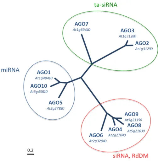
AGO8, and AGO9 clade (Mallory and Vaucheret, 2013) A phylogenetic tree comprising all Arabidopsis Argonautes is shown in Figure 6.
Figure 6: Phylogenetic tree illustrating the three clades of Arabidopsis Argonautes.
AGO1 and AGO10 act in the classic canonical miRNA pathway and the close relative AGO5 is involved in a novel miRNA pathway in the plant germline (Chapter 3.2.9). AGO7 binds miR390 is involved in the generation of trans-acting small interfering RNAs (ta-siRNAs), AGO2 is involved in antiviral and antibacterial defense and binds miRNAs beginning with an adenosine. AGO3 is part of a tandem repeat with AGO2 and it function is unknown. AGO4, AGO6 and AGO9 predominantly bind 24 nt siRNAs with 5’ adenosine and are involved in the mechanism of RNA-directed DNA-methylation (RdDM). AGO8 is generally regarded as a pseudogene. Image by Stefanie Sprunck.
AGO1 is the main effector in the classic canonical miRNA pathway in plants, which
follows similar principles as described above for animals. One difference of the miRNA
pathway in plants is the fact that the microprocessor complex of Drosophila and mammals is
not part of it (Denli et al. 2004). Instead, the Pol II transcribed miRNA hairpin is directly
processed by a protein complex containing DCL1 (Figure 7A), which is one of four
Arabidopsis homologues of the human DICER protein, methylated at the 2´O by HEN1, and
17
transported out of the nucleus with the assistance of HASTY, a homolog of Exportin 5 (Xie 2015). Finally, this results in 21-22 nucleotides long miRNAs which are bound by AGO1. In plants, microRNAs bound by AGO1 are characterized by a 5´ uracil (U) nucleotide and indeed, the identity of this nucleotide is important for sorting different small RNAs into their respective AGOs (Mi et al. 2008, Havecker et al. 2010). Like in other eukaryotes, AGO1 can, as part of a RISC complex, cleave mRNA targets (Baumberger and Baulcombe 2005) or mediate translational repression (Li et al. 2013). Beside these canonical pathways, AGO1 can also trigger the biogenesis of secondary siRNAs (Creasy et al. 2014). MiR/AGO1 complexes are involved in an initial cutting step which generates a template for an RNA-dependent RNA polymerase (RDR6), the resulting dsRNA is then processed into secondary siRNAs. This pathway is important for the defense against epigenetically reactivated transposons (Creasey et al. 2014).
AGO10 has been proposed to compete with AGO1 for miRNAs and thus keep up the expression of the HD-ZIP-III transcriptions factors, which is important for the maintenance of cell identity in the region of the shoot apical meristem (Zhang and Zhang 2012).
AGO5 shows a specific expression pattern in reproductive tissues of Arabidopsis (Mallory and Vaucheret 2010). The role of AGOs in plant reproduction will be discussed in detail in Chapter 3.2.9. Besides that, a recent work from Brosseau and Moffett (2015) showed that AGO5 expression is induced by infection with potexvirus and is needed, together with AGO2, to restrict the viral infection.
AGO7 is the prominent member of the second clade, the so-called ta-siRNA-clade, which stands for trans-acting small interfering RNA. AGO7 is involved in the specialized ta- siRNA silencing pathway, in which the respective AGOs interact with their target via miRNA/mRNA interaction and exert an initial cleavage step on the transcript (Figure 7B).
This processed transcript serves then as a template for an RNA-dependent RNA polymerase
(RDR6), the resulting double-stranded RNA is cut by DCL4 resulting into a number of small
dsRNAs of 21 to 22 nucleotides length. In the end, those RNAs can be incorporated into
AGO1-RISC again to attack mRNA targets. In case of AGO7 only one miRNA is bound,
which is miR390. The AGO7/miR390 complex then cleaves the mRNA of the TAS3 gene, the
resulting ta-siRNAs act via the miRNAs pathway to inhibit the auxin response factors ARF2,
ARF3 and ARF4, which are involved in regulation of lateral root growth (Marin et al. 2010).
18
Figure 7: Trans-Silencing: the miRNA and ta-siRNA pathways.
(A) Micro RNA genes are transcribed by RNA polymerase II (PolII) and form a hairpin structure which is directly processed by a complex containing DCL1 and DRB1. The miRNA is then incorporated into an AGO1 or AGO10 containing RNA-induced silencing complex (RISC), which mediates translational repression or mRNA cleavage. (B) TAS genes are transcribed from PolII to form single-stranded RNAs, which are targeted by microRNAs, cleaved by AGO1 or AGO7 and copied into dsRNA by RDR6, SGS3, and SDE5, The dsRNA is processed into 21 to 22 nucleotide ta-siRNAs by the DRB4/DCL4 complex. The ta-siRNAs direct cleavage of complementary mRNAs. PolII, RNA-polymerase II; CBP20/80; cap binding protein20/80; SE, Serrate; DDL, DAWDLE; DCL1, dicer-like 1; DRB1; dsRNA-binding protein1; SDN, small RNA degrading nuclease; HEN1, HUA enhance 1; AGO1/10, Argonaute 1/10; SQN, squint; FWB2, F-BOX WITH WD-40 2; VCS, varicose, KTN1, katanin 1; THO/TREX, THO/Transcription export, SGS3, suppressor of gene silencing 3; SDE5, silencing defective 5; RDR6, RNA-dependent RNA polymerase 6; DCL4, dicer-like 4; DRB4, dsRNA-binding protein1. Image taken from Mallory and Vaucheret 2010.
Considering the fact that those small RNAs can trans-act on their target molecules and share the biogenesis of siRNAs, they are called ta-siRNAs.
AGO2 is reported to play a role in antiviral (Harvey et al. 2011) and antibacterial
defense (Zhang et al. 2011). It predominantly binds miRNAs with a bias for a 5’ adenosine
(A) (Takeda et al. 2008, Mi et al. 2008). Additionally, it associates with virus-derived siRNAs
(Harvey et al. 2011) and is involved in repair of DNA double strand breaks (Wei et al. 2012).
19
AGO3 is located closely to AGO2 on chromosome 2 and those two proteins are very similar to each other, which is a hint towards recent gene duplication (Vaucheret 2008).
The AGO4 clade AGOs are functionally associated with RNA-directed DNA methylation (RdDM), an epigenetic process in which 21-24 nucleotide siRNAs guide methylation of homologous DNA loci. For AGO4, AGO6 and AGO9 the predominant binding of siRNAs has been shown (Havecker et al. 2010), while AGO8 is regarded as a pseudogene (Takeda et al. 2008). The AGO4 clade AGOs will be discussed in detail in the next Chapter.
3.2.7 RNA-directed DNA-methylation and transposon silencing in plants
The AGO4 clade of Arabidopsis comprises AGO4, AGO6, AGO8, and AGO9. AGO4, AGO6 and AGO9 are characterized by the fact that they predominantly bind siRNAs of 24 nucleotides length (Mi et al. 2008, Borges and Martienssen 2015). Those siRNAs represent a central component of a plant specific siRNA pathway, which creates epigenetic modifications through siRNA guidance. This was first discovered in Arabidopsis plants which were infected with RNA viroids and it could be shown that viral RNA could direct DNA methylation towards specific genomics loci (Wassenegger 1994). This phenomenon was called RNA- directed DNA methylation (RdDM). Later it was discovered that components of the RNA silencing machinery are required for this mechanism (Chan 2004). A study by Zilbermann et al. (2003) identified AGO4 as a central component of this pathway. RdDM requires the plant specific RNA polymerases Pol IV and Pol V. Pol V generates the precursor transcripts, which are then converted into a double-stranded RNA (dsRNA) by RDR2 and processed by DCL3 (Haag and Pikaard 2011). The resulting small dsRNA is bound by an AGO4-clade Argonaute and the passenger strand is degraded (Ye et al. 2012). For AGO4 is has been shown that been shown that a nuclear localization sequence in the N-terminus of the protein is then accessible for nuclear import factors and AGO4 is imported back into the nucleus (Ye et al. 2012). The AGO4–RNP then interacts with Pol V transcripts and forms a complex with several RdDM–
factors, like for example the largest Pol V subunit, the DNA and RNA-binding protein KOW
DOMAIN-CONTAINING TRANSCRIPTION FACTOR 1 ( KTF1), which is also known as
SUPPRESSOR OF TY INSERTION 5-LIKE (SPT5L), and the DOMAINS REARRANGED
METHYLTRANSFERASE2 (DRM2) which catalyzes the de novo methylation of DNA
(Figure 8).
20
Figure 8: The RdDM pathway in Arabidopsis thaliana.RNA-directed DNA methylation is a complex pathway of transcriptional silencing, preferentially directed towards transposons and other repetitive elements. The canonical RdDM pathway involves POL IV transcripts, which are converted into double-stranded RNA by the RNA-dependent RNA-polymerase RDR2. This dsRNA is processed into 24 nt long siRNA which cooperate with AGO protein and Pol V transcripts to direct DNA methylation. The Pol II/RDR6 pathway uses siRNAs generated by POLII, AGO1/7 and RDR6 to guide an AGO protein to the Pol V transcript. In both cases, DNA is methylated by DOMAINS REARRANGED METHYLTRANSFERASE 2. (DRM2). SHH, SAWADEE HOMEODOMAIN HOMOLOG; RDR2, RNA- DEPENDENT RNA-POLYMERASE 2; CLSY1, CLASSY1; DCL3, DICER-LIKE 3; AGO4, ARGONAUTE 4;
MORC, MICRORCHIDIA; SUVH, SU(VAR)3-9 HOMOLOG; DMS3, DEFECTIVE IN MERISTEM SILENCING 3; RDM1, RNA-DIRECTED DNA METHYLATION 1; DRD1, DEFECTIVE IN RNA- DIRECTED DNA METHYLATION 1; SPT5L, SUPPRESSOR OF TY INSERTION 5-LIKE; IDN2, INVOLVED IN DE NOVO 2; IDP, IDN2 PARALOG, SWI/SNF, SWITCH/SUCROSE NON- FERMENTABLE; HDA6, HISTONE DEACETYLASE 6, JMJ14, JUMONJI 14, UBP26, UBIQUITIN- SPECIFIC PROTEASE 14, DCL, dicer-like.Image taken from Matzke et al. 2015.
This mechanism especially silences genes where POL V is active, such as transposons
and other repetitive elements like telomeres (Verdel et al. 2004). The role of AGO6 is
partially redundant with that of AGO4, it is reported to take part in transcriptional gene in root
and shoot meristems (Havecker et al. 2010, Eun et al. 2011). A variant of this canonical
RdDM pathway relies on RNA polymerase II (PolII) transcripts, which are subjected to an
21
initial cleavage step by an AGO, followed by generation on a dsRNA by RDR6, like in the TASI pathway (Figure 8).
Each of the of the four DCLs can generate 21 to 22 nt long ta-siRNAs, which are bound by one of the RdDM AGOs in order to initiate de novo DNA methylation via DRM1.
In spite of this redundancy there seem to be differences in subnuclear localization and AGO6 seems to be even more connected with POL V than AGO4 (Figure 9). McCue et al. (2014) proposed that AGO6 plays a role in establishment of transposable element silencing. AGO9 is also a member of the AGO4 clade and binds 24 nt siRNAs, with a bias to a 5´ A.
Figure 9: Distinct roles of AGO4 and AGO6 Proteins
AGO4 and Pol V are involved in RNA-directed DNA methylation (RdDM) in perinucleolar foci. RdDM activities in the nucleoplasm are mediated by AGO6 and Pol V, and by AGO4 and Pol II. DRM2, DOMAINS REARRANGED METHYLTRANSFERASE 2; DDR, a protein complex formed by DEFECTIVE IN RNA- DIRECTED DNA METHYLATION 1 (DRD1), RNA-DIRECTED DNA METHYLATION 1 (RDM1) and DEFECTIVE IN MERISTEM SILENCING 3 (DMS3). KTF1, KOW DOMAIN-CONTAINING TRANSCRIPTION FACTOR 1. Image taken from Duan et al. 2014.
Although its role in RdDM has not been clarified, AGO9 has been shown to silence
repetitive elements (Olmedo-Monfil et al. 2010). Additionally, the enhanced sensitivity of the
ago9-1 mutant against reagents that cause DNA double strand breaks could indicate a role of
AGO9 in DNA repair (Oliver et al. 2014). AGO8 is found in close proximity to AGO9 on
chromosome 5 and is generally believed to be a pseudogene (Takeda et al. 2008). Silencing of
22
TEs is not only achieved by RdDM, which is a mechanism for de novo DNA-methylation occurring either after removal of DNA-methylation marks or after fresh insertion of TEs in the genome. Silencing of TEs is achieved by setting epigenetic marks on TE sequences and also other epigenetic mechanisms play a role in keeping TEs under control (see next Chapter).
3.2.8 Epigenetic reprogramming in plant sporo- and gametogenesis
The term “epigenetics” refers to heritable changes in gene expression, which are not caused by a change in DNA sequence. The biochemical modifications within an organism related to epigenetic changes are often called epigenetic marks. Silencing of transposons by modification of histones and DNA is only one part of the process of epigenetic silencing.
Those marks can consist of methylation of DNA and modification of histones and have an
influence on transcriptional activity and chromatin structure (Feng et al. 2010). DNA
methylation occurs at specific DNA sites: CG, CHG, and CHH (where H = C, T, or A). In
plants, DNA methylation in a GC-context is maintained by MET1 (DNA
METHYLTRANSFERASE 1), with the help of the cofactor VIM (VARIATION OF
METHYLATION). CHG-methylation is performed by CMT3 (CHROMOMETHYLASE 3),
which interacts with the histone methyltransferase KYP (KRYPTONITE), forming a positive
feedback loop. The maintenance of DNA methylation by CMT3 and MET1 can be
distinguished from the de novo mechanism of RdDM (Chapter 3.2.6), which is active in all
sequence contexts. In general, epigenetic marks have an important role in the formation of
male and female gametes in plants as well. For example, the trimethylation of histone 3 at
lysine 4 (H3K4) as an epigenetic mark is important for the maintenance of certain chromatin
structures and is needed for gametophyte development (Berr et al. 2010). Until now, the state
of epigenetic marks in plant meiocytes is unknown, but the increase of expression of
transposable elements in male meiocytes indicates a change in histone modification and non-
CG DNA-methylation, which normally keep TE expression under control (Kawashima and
Berger 2014). In the central cell of the mature female gametophyte, MET1 is repressed, while
DEMETER (which removes 5-methylcytosine) is active (Jullien et al. 2012). Thus, a general
state of DNA-hypomethylation in the central cell is likely. The consequence of this
hypomethylated state would be the transcriptional activation of transposable elements and
other elements, like imprinted genes. In the egg cell, the maintenance DNA
methyltransferases CMT3 and MET1 are barely detectable (Jullien et al. 2012). But on the
23
other hand, the de novo DNA methyltransferases DRM1 and DRM2 can be found in the egg cell, with DRM1 exclusively expressed in the egg cell (Jullien et al. 2012, Kawashima and Berger).
Figure 10: Transposon silencing in Arabidopsis male and female germline.
(A) In the Arabidopsis male gametophyte the chromatin remodeler DDM1 is repressed in the vegetative nucleus, allowing transposons to be expressed and mobile siRNAs to be generated. 24 nt siRNAs move to the sperm nuclei to trigger post-transcriptional gene silencing (B) In the female gametophyte, the central cell shows a general state of hypomethylation caused by repression of the DNA-methyltransferase MET1 and the demethylase DEMETER (DME1). This allows transposon activation and generation of mobile siRNAs. 21 nt siRNAs move to the egg cell nuclei to trigger post-transcriptional gene silencing. PTGS, post-transcriptional gene silencing; DDM1, DECREASED DNA METHYLATION 1; AGO, Argonaute; TGS, transcriptional gene silencing; 5meC, 5-methylcytosine, DME1, DEMETER; MET1, METHYTRANSFERASE1. Image taken from Castel and Martienssen 2012.
De novo DNA methylation via the RdDM pathway thus seems to play an important role in the egg cell. In the male gametophyte, de-repression of TEs in the vegetative nucleus of the pollen grain correlates with the repression of TEs in the sperm cell (Calarco et al.
2012). In plants, this involves movement of siRNAs from the vegetative nuclei to the sperm nuclei in the male gametophyte and movement of siRNAs from egg to central cell in the female gametophyte ( Figure 10).
A
B
24
3.2.9 The role of AGO proteins in plant reproduction
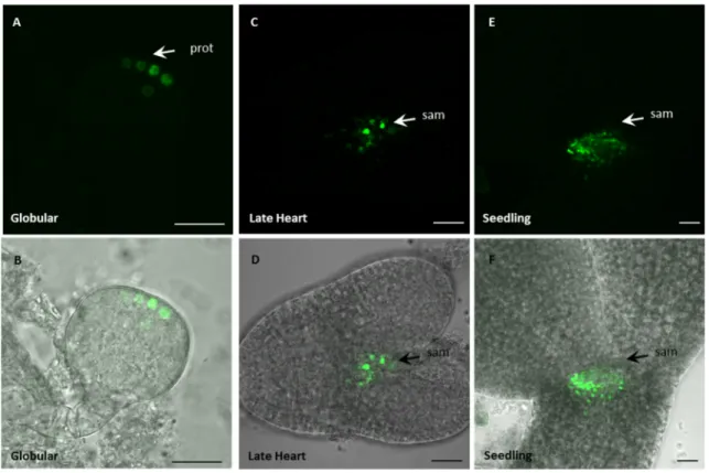
Transcriptomic analyzes revealed that some AGO genes show a highly specific expression pattern in different cell types and developmental stages of plant development (Schmid et al. 2005, Mallory and Vaucheret 2013). For example, AGO5 and AGO9 transcripts are strongly present in reproductive tissues. In case of AGO5, the mutant ago5-4 shows severe defects in the initiation of megasporogenesis (Tucker et al. 2012).
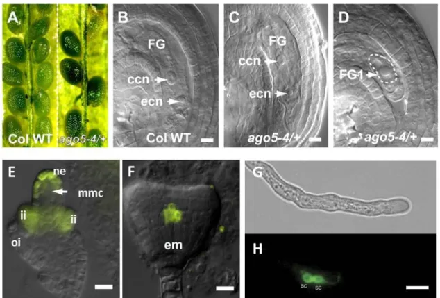
Figure 11: Role of AGO5 in ovule development and presence of AGO5 fusion proteins in developing ovules and in sperm cells.
(A) Comparison of seeds in mature WT (left) and ago5-4/+ (right) siliques. (B) WT anthesis ovule. (C) WT-like ago5-4/+ anthesis ovule. (D) Phenotypic ago5-4/+ anthesis ovule showing FG1 abortion (dashed line). (E) and (F) Expression of the YFPer reporter under control of the AGO5 promoter. In immature ovules the AGO5 promoter is active in the nucellar epidermis cells (ne) surrounding the functional megaspore (fm), and the developing inner integuments (ii). (G) and (H) Transgene expression of AGO5-eGFP in sperm cells (sc). GFP was C-terminally fused to the genomic AGO5 sequence, under the control of the AGO5 promoter. Images (A) to (F) taken from Tucker et al. 2012 and Borges et al. 2011 (G). Scale bars (A) to (F) are 10 µM. Scale bars (G) and (H) are 5 µM.
About 50 % of ago5-4/+ heterozygous plants show a developmental arrest at stage
FG1 (Figure 11D), meaning that the functional megaspore mother cell does not progress to
mitotic divisions. However, an YFP-AGO5 fusion protein expressed under control of the
AGO5 promoter is detected in the inner integuments and nucellus of the developing ovule but
25
neither in the functional megaspore nor in later stages of the developing female gametophyte (Tucker et al. 2012). In pollen, AGO5-GFP fusion protein is localized in the sperm cell cytoplasm (Figure 11G).
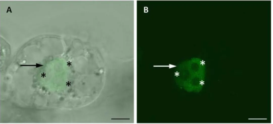
Arabidopsis ovules that carry a mutation in AGO9 show a higher number of additional cells with germline identity (Olmedo-Monfil et al. 2010, Figure 12A). In these mutants, the expression level of otherwise silenced TEs is elevated, suggesting that AGO9 is involved in the silencing of TEs in the female germline.
Figure 12: Function of AGO9 homologues in Arabidopsis and maize.
(A) In Arabidopsis ago9 mutants, a high percentage of young ovules with additional megaspore mother cells (MMC) was observed. L1, L1 layer of the nucellus tissue. In wildtype plants, 94.2 % of the ovule had one MMC whereas 5.8% showed additional MMCs (left two pictures). In the mutants lines ago9-2 and ago9-3 47.7% and 37.2% of ovules with additional MMCs were found, respectively (the two pictures on the right). Arrows indicate the position of additional MMCs. (B) In immunohistochemical staining experiments, AGO9 protein was detected in the nucellar tissue surrounding the MMC. Blue color indicates AGO9 expression. (C) Zea mays loss of function mutant ago104-5 and ago 104-6 show kernel abortion (left images). In ago104-5 and ago 104-6 mutants defects in meiosis II, visualized using DAPI staining, lead to the production of abnormal tetrads, including triads and microspores with multiple nuclei (fluorescence images on the right). Images (A) and (B) (modifed) taken from Olmedo-Monfil et al. (2010). Images (C) and (D) taken from Singh et al. 2011. Scale bars (A) are 10 µM.
Scale bars (B) are 50 µM.
Analysis of small RNAs bound to AGO9 showed a high number of sRNA derived
from TEs and repetitive elements such as centromeres and telomeres (Duran-Figueroa and
Vielle-Calzada, 2010). Recently, Rodríguez-Leal et al. (2015) linked AGO9 activity to
variations in cell differentiation in different Arabidopsis ecotypes. However, the maize
26
homologue of AGO9, which is called AGO104, seems to have an opposite function: egg cells
of maize ago104 mutant plants fail to undergo meiosis and generate diploid gametes (Singh et
al. 2011), leading to kernel abortion. Those mutants showed an altered expression profile in
ovules compared to the wild type, affecting genes as well as centromeric repeats and certain
types of transposable elements.
27
3.3 Aims of this work
Small RNA pathways have been implicated to play an important role in plant reproduction. Argonaute proteins, which are the main effectors of such pathways, show a highly specific expression pattern, and transcripts of certain AGOs are highly abundant in the cells of the female gametophyte. In a previous work using isolated egg cells, central cells and synergid cells, microarray-based expression studies were performed (Soljic 2012).
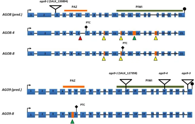
Figure 13: The AGO8 locus and cloned AGO8 transcript variant.
Cloning and sequencing of AGO8 cDNA revealed the presence of alternative 3´ splice sites in exon 10 and exon 12. Orange boxes and letters represent exons. Blue lines and letters represent introns. The arrow indicates the translation start site. Black dots stand for a stop codon. Black bars: PAZ and PIWI domain (chapter 3.2.1).
Triangles point at the position of amino acids needed for catalytic activity (DDH). Image taken from Takeda et al. 2008.
In these studies the expression of the putative AGO8 pseudogene was found to be
restricted to egg cells, while AGO5 and AGO9 are expressed in egg cells, central cells and
sperm cells. In this work the expression of AGO8 and AGO9 genes and proteins should be
investigated in more detail in transgenic plants, after generating suitable promoter-reporter
constructs and constructs in which fluorescent AGO fusion proteins were expressed under
control of their own promoters. Aspects such as expression pattern during female
gametophyte development and the subcellular localization of AGO fusion proteins and native
AGOs in the ovule and female gametophyte were one focus of this thesis. An approach of
ectopically expressing AGO8 and AGO9 was chosen to address questions of AGO8 and AGO9
28
function, since the generation of ago8/ago9 double mutants is not possible due to their close physical proximity on chromosome 5 (Wartlsteiner 2010).
Figure 14: Alignment of AGO8 and AGO9 genomic DNA sequence with the cloned AGO8 and AGO9 transcript variants.
Genomic DNA and annotated cDNA sequence from AGO8 and AGO9 were retrieved from The Arabidopsis Information Resource (TAIR) and compared to previously discovered transcript variants. Blue bars represent exons as annotated by TAIR. Triangles indicate alternative splicing events. Red triangles stand for alternative 5’splice, yellow triangles represent alternative 3’splice sites and green triangles indicate intron retention. Orange bars indicate intronic sequences contained in the transcript. Image modified from Wartlsteiner 2010.