Wallerian degeneration and Wallerian-related axon loss in disease
Inaugural-Dissertation zur
Erlangung des Doktorgrades
der mathematisch-naturwissenschaftlichen Fakultät der Universität zu Köln
vorgelegt von Weiqian Mi
aus Köln Köln 2003
Berichtserstatter: Prof. Dr. Sigrun Korsching Prof. Dr. Michael Coleman Tag der Mündlichen Prüfung: 07/11/2003
ERKLÄRUNG
Ich versichere, daß ich die von mir vorgelegte Dissertation selbständig angefertigt, die benutzten Quellen und Hilfsmittel vollständig angegeben und die Stellen der Arbeit - einschließlich Tabellen, Karten und Abbildungen -, die anderen Werke im Wortlaut oder dem Sinn nach entnommen sind, in jedem Einzelfall als Entlehnung kenntlich gemacht habe; daß diese Dissertation noch keiner anderen Fakultät oder Universität zur Prüfung vorgelegen hat; daß sie - abgesehen von unten angegebenen Teilpublikationen - noch nicht veröffentlicht worden ist sowie, daß ich eine solche Veröffentlichung vor Abschluß des Promotionsverfahrens nicht vornehmen werde.
Die Bestimmungen dieser Promotionsordnung sind mir bekannt. Die von mir vorgelegte Dissertation ist von Prof. Dr. S. I. Korsching betreut worden.
Köln, den 26. Juli, 2003 Weiqian Mi
Acknowlegment
ACKNOWLEGMENT
This study was performed in the laboratory of Prof. Dr. Michael Coleman and Prof. Dr.
Sigrun Korsching in the Institüt für Genetik, Universität zu Köln, Germany and was supported by Center for Molecular Medicine, University of Cologne (CMMC) and a Wellcome Trust Biomedical Collaboration Grant, I would like to thank you for the constant support and the intellectual enlightenment during my Ph.D. work.
I would like to thank Prof. Dr. Michael Coleman, Prof. Dr. Mats Paulsson, Prof.
Dr. Sigrun Korsching and Dr. Mathias Cramer for being in my thesis committee.
My special thanks go to my dear parents, for supporting me all way along and encouraging me going through any difficulties in life and study, especially my great thanks to my Mum, for those Chinese periodicals and food from home…
My thanks go to all my colleagues, who gave me all kind of helpful advices and technical supports, thus enabling me to complete my bench work with utmost ease, namely, Dr. Robert Adalbert, Dr. Laura Conforti, Dr. Livia Adalbert, Dr. Till G.A. Mack, Dr. Heike Laser, Dr. Arzu Celik, Diana Wagner, Daniela Grumme, Vu Hangoc, Bogdan Beirowski, and our collaborators in Edinburgh, Prof. Dr. Richard R. Ribchester, Dr. Tom H. Gillingwater and Derek Thomson.
In every work undertaken, there are many whose contributions are intangible but are no less significant. This thesis and the work reported therein is no exception, and my deepest heartfelt thanks go to everyone who in his or her own unique way has made its completion so successful and so much rewarding.
List of publications related to the Ph.D. thesis
Manuscripts in peer reviewed journals
I. Mi W., Beirowski B., Gillingwater T.H., Coleman M.P. et al. Wallerian degeneration shares a regulatory step with axonal spheroid pathology caused by defective ubiquitin metabolism. Submitted.
II. Mi W., Glass J.D., Coleman M.P. Stable inheritance of an 85-kb triplication in C57BL/WldS mice. Mutat. Res. May 15; 526(1-2): 33-7 (2003).
III. Samsam M., Mi W., Wessig C., Zielasek J., Toyka K.V., Coleman M.P. and Martini R. The WldS mutation delays robust axonal loss of motor and sensory axons in a genetic model for myelin-related axonopathy. J. Neurosci. Apr 1;
23(7): 2833-9 (2003).
IV. Mi W., Conforti L., Coleman M.P. A genotyping method to detect a unique neuroprotective factor for axon (WldS). J. Neurosci. Meth. Jan 30; 113(2):
215-8 (2002).
V. Mack T.G.A., Reiner M., Beirowski B., Mi W., Coleman M.P. et al.
Wallerian degeneration of injured axons and synapses is delayed by a Ube4b/Nmnat chimeric gene. Nat. Neurosci. Dec; 4(12): 1199-1206 (2001).
VI. Conforti L., Tarlton A., Mack T.G.A., Mi W., Buckmaster E.A., Wagner D., Perry V.H., and Coleman M.P. A chimeric protein and overexpression of Rbp7 in the slow wallerian degeneration (WldS) mouse. Proc. Natl. Acad. Sci.
Oct 10; 97 (21): 11377-11382 (2000).
Chapter Contents
1. Introduction………1
1.1 Wallerian degeneration and slow Wallerian degeneration mouse, C57BL/WldS……….2
1.2 Molecular genetics of WldS………4
1.3 Triplication and its genomic stability………5
1.4 The chimeric gene is the WldS gene………...5
1.5 Slow Wallerian degeneration mouse, C57BL/WldS and human disease……...7
1.6 The gad mouse (gracile axonal dystrophy)………9
1.7 Ubiquitin metabolism in neurodegeneration………10
1.8 XFP (especially YFP-H) mice in studying Wallerian degeneration…………12
2. Material and methods………..27
2.1 Molecular and cellular biology………28
2.1.1 Extraction of genomic DNA and preparation of DNA agarose plugs for PFG………..28
2.1.2 Genotyping method to track WldS triplication……….28
2.1.3 Genotyping of transgenic mice and gad mice………..30
2.1.4 Generation of transgenic mice……….30
2.1.5 Cloning and expression of recombinant protein………..33
2.1.6 Purification of recombinant protein (N70 and WldS)………..34
2.1.7 Generation of polyclonal antisera………35
2.1.8 Western blot……….35
2.1.9 Cell transfection and fixation………...36
2.2 Animal breeding protocols and behavioral analysis………37
2.2.1 Animal breeding protocols………...37
2.2.2 Rotarod analysis………...39
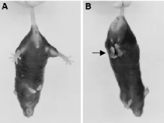
2.2.3 Clasping………...39
2.2.4 Foot splay test………..39
2.2.5 Peripheral nerve lesion (PNL)……….39
2.2.6 Processing YFP nerve………..40
2.3 Histological analysis………40
2.3.1 Paraffin embedding of brain/spinal cord………..40
2.3.2 Haematoxylin/eosin staining (H&E)………41
2.3.3 Luxol fast blue staining………41
2.3.4 Anti-GFAP immunostaining………42
2.3.5 Statistical analysis of histopathological results………43
2.3.6 Analysis of neuromuscular pathology……….43
Appendix……….48
1. Principle of Nucleon II kit………...48
2. Principle of plasmid Mini kit………...48
3. Principle of QIAquick extraction……….48
4. Principle of ProBondTM Resin column………48
5. Reagents for PCR……….48
6. Principle for Southern blot………...49
7. Pulse field gel electrophoresis……….49
8. DNA extraction using phenol/chloroform………...49
9. Ligation reaction………..50
10. PCR screening………..50
11. Optimization of conditions for the expression of N70 or full-length chimeric protein (WldS)……….50
12. Western blot……….51
13. Buffer for SDS-polyacrylamide gel electrophoresis………51
3. Genotyping method to track the WldS triplication………53
3.1 Introduction………..54
3.2 Result………...54
3.2.1 Genotyping methods for the WldS triplication……….54
3.2.2 The application of genotyping method to track the WldS triplication……….55
3.3 Discussion………59
4. The 85 kb triplication in C57BL/WldS is stably inherited………....74
4.1 Introduction………..75
4.2.1 Sources of WldS tissue………75
4.2.2 Stable inheritance of an 85 kb triplication in C57BL/WldS…………....76
4.3 Discussion………77
5. The WldS mutation reduced axonal spheroid pathology in gad mutation…..84
5.1 Introduction………..85
5.2 Result………...86
5.2.1 WldS can function to protect axons in gad mice……….…86
5.2.2 Genotyping of gad and WldS status………87
5.2.3 Axonal spheroid pathology is reduced by WldS………..…88
5.2.4 Secondary myelin loss and astrocyte activation are reduced by WldS………88
5.2.5 Behavioral changes due to WldS are complex……….…89
5.2.6 gad/WldS motor nerve terminals degenerate extensively by 15 weeks……….…90
5.3 Discussion………90
6. Study of the WldS mechanism………...117
6.1 Introduction………118
6.2 Result……….118
6.2.1 Identifying the WldS gene and its initial characterization…………..118
6.2.2 Transgenic N70-NLS or NES does not show WldS phenotype………..121
6.3 Discussion………..122
7. Summary……….138
8. Reference………141
List of Figures
Figure 1.1 A schematic diagram shows undergoing Wallerian degeneration…………..14
Figure 1.2 Location of exons within the 85-kb WldS triplication repeat unit…………...16
Figure 1.3a WldS express novel and wild type proteins……….18
Figure 1.3b Sequence of the chimeric cDNA and the predicted coding sequence………18
Figure 1.4 Scheme of the deleted region of Uchl1 in gad mice………..23
Figure 1.5 Four spectrally distinct XFPs serve as vital stains in transgenic mice……...25
Figure 2.1 Sequence of NLS, NES and Flag-tag……….44
Figure 2.2 pET-28 a-c (+) vectors map………45
Figure 2.3 Abnormal hind limbs clasping in mice………...47
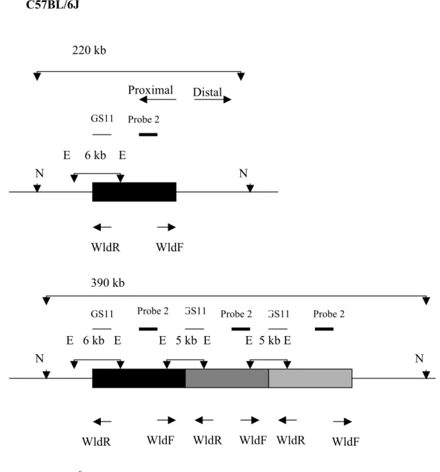
Figure 3.1 Tandem triplication schematic diagram model………..61
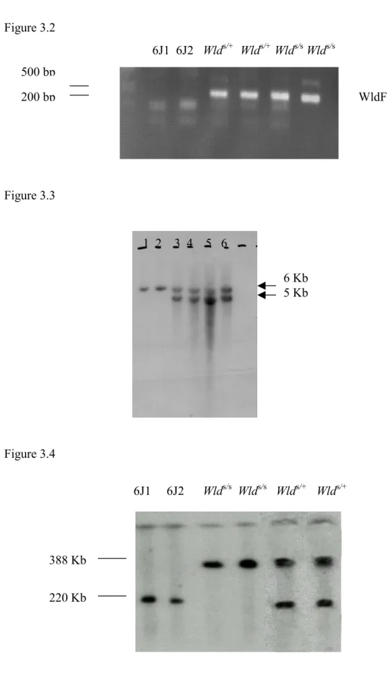
Figure 3.2 PCR for genotyping WldS………. .63
Figure 3.3 Southern blotting for genotyping WldS………...63
Figure 3.4 Pulsed-field gel analysis for genotyping WldS………...63
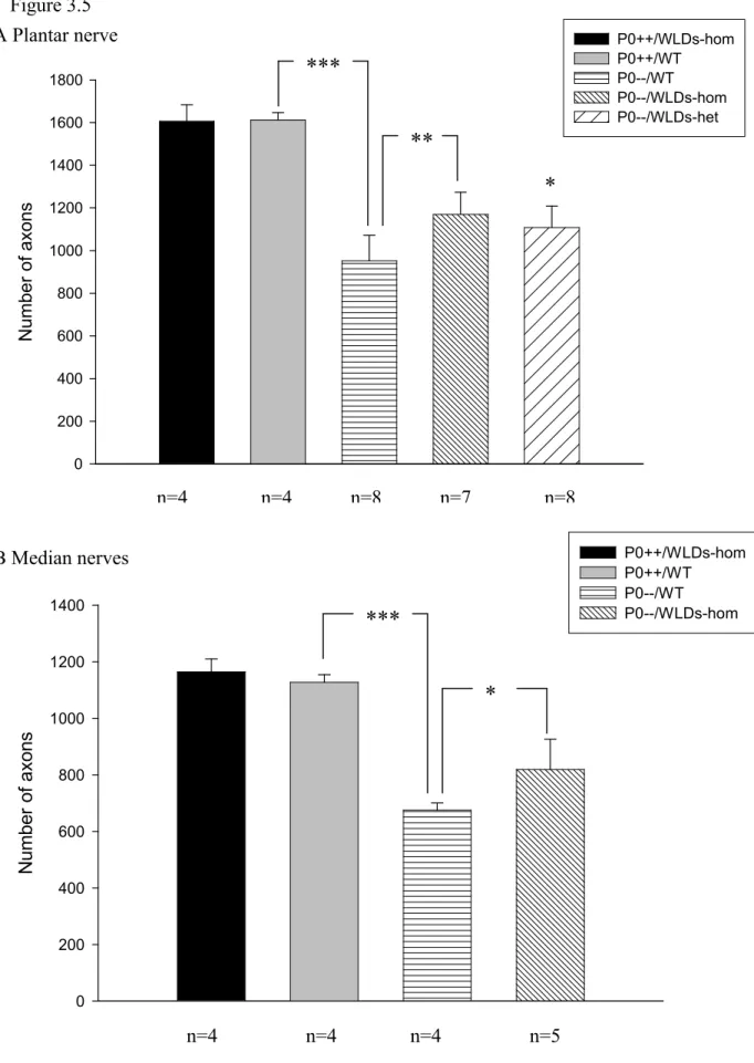
Figure 3.5 Schematic representation of numbers of axons in plantar (A) and median (B) nerves of 3-month-old P0+/+ and P0-/- mice with or without the WldS mutation……….65
Figure 3.6a Schematic representation of amplitudes of compound action potentials from small foot muscles of 3-month-old P0+/+ and P0-/- mice with and without the WldS mutation………..67
Figure 3.6b Schematic representation of retrogradely labeled spinal motoneurons of 3-month-old mice using Fluorogold………...67
Figure 3.7 Schematic representation of muscle strength of P0+/+ and P0-/- mice with and without the WldS mutation………..70
Figure 3.8 Western blot analysis of young versus old P0-/-/WldS double mutants……..72
Figure 4.1a Pulsed field gel analysis of C57BL/WldS chromosomes from 3 diverged breeding colonies………...80
Figure 4.1b Identification of 170- kb (triplication) and 85-kb (duplication) insertions in C57BL/Wlds………80 Figure 4.2 Unequal crossing-over between tandem head-to-tail
Figure 5.1 Southern blot genotyping of gad………..95
Figure 5.2 Slow Wallerian degeneration in WldS heterozygotes carrying homozygous gad alleles………...97
Figure 5.3 WldS reduces spheroid body numbers in the gracile tract of gad mice….…99 Figure 5.4 WldS reduces also other measures of gracile tract pathology………..101
Figure 5.5 gad mice perform worse on Rotarod when they carry a WldS allele……...103
Figure 5.6 There is no significant difference on footsplay test in gad mice with WldS allele or without WldS allele………...105
Figure 5.7 There is no significant difference on hind limbs clasping test between gad mice with WldS allele or without WldS………107
Figure 5.8 Denervation at the neuromuscular junction……….109
Figure 5.9 Axonal spheroids are present widely in degenerative pathology…………111
Figure 5.10 Evidences show that Wallerian degeneration and axonal spheroid are related………...113
Figure 5.11 WldS delays a central step of axonal pathology………...115
Figure 6.1a The transgenic construct of Ube4b/Nmnat………..124
Figure 6.1b The transgenic construct of N70 plus NLS……….124
Figure 6.1c The transgenic construct of N70 plus NES………..124
Figure 6.2 The intracellular location of the WldS protein……….126
Figure 6.3 Western blotting shows Ube4b, WldS and β-tubulin expression in WldS mice of different ages compared to a 6J mouse control…………128
Figure 6.4 Increased Nmnat activity and unaltered NAD+ content in WldS brain……130
Figure 6.5 Transfection of COS-7 cell with pHbApr-1-N70-NLS or pHbApr-1-N70-NES………...132
Figure 6.6 Transgenic N70-NLS and N70-NES do not show WldS phenotype………134
List of Tables Table 4.1 Triplication results of WldS chromosomes examined by PFGE……….79
Table 6.1 Results of preservation of axons in transgenic N70-NLS or N70-NES mice by western blot or visualizing YFP signal………..136
CHAPTER 1
INTRODUCTION
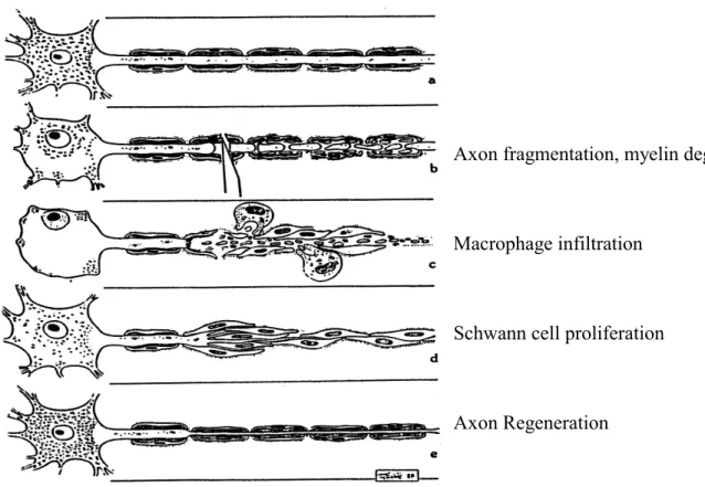
1.1 Wallerian degeneration and slow Wallerian degeneration Mouse, C57BL/WldS Wallerian degeneration is the degeneration of the distal stump of an injured axon (Waller, 1850). It normally occurs over a time course of around 24-48 hr of a lesion, such as mechanical (transection, crush, blunt trauma), chemical toxic (acrylamide), or metabolic (ischemia) injuries. It includes disintegration of the axonal cytoskeleton and fragmentation of the axon, followed by breakdown of myelin sheath, macrophage infiltration and Schwann cell proliferation (Figure 1.1). It is not known how Wallerian degeneration is initiated, but the mechanism clearly involves an active and regulated program of self-destruction and is distinct from neuronal cell body apoptotic degeneration: in WldS, neurites of sympathetic ganglia undergo slow Wallerian degeneration after deprivation of their physiological trophic factor (nerve growth factor), however the neuronal soma degenerates normally as wild type (Deckwerth & Johnson, 1994); overexpression of Bcl-2, a anti-apoptosis protein, prevents motoneuron cell body loss but not axonal degeneration in a mouse model of motor neuron disease (progressive motor neuronopathy, pmn/pmn)(Sagot et al, 1995); Wallerian degeneration and NGF- withdrawal-induced axonal degeneration do not activate classical caspase cascade happening in cell body’s apoptosis, and caspase inhibitors do not block axonal degeneration (Finn et al, 2000).
The slow Wallerian degeneration mouse, C57BL/Wlds, carries a dominant mutation that delays Wallerian degeneration in the distal stump of an injured axon. It is a spontaneous mutant that was revealed by experiments to investigate the role of macrophages in peripheral-nerve repair (Lunn et al, 1989). Transected axons from the PNS (such as sciatic nerve and tibial nerve) or CNS (such as optic nerve) of the Wlds retain the ability to transmit a compound action potential for up to 3 weeks (Lunn et al 1989; Ludwin and Bisby, 1992; Watson et al, 1993), and continue the anterograde and retrograde transport of proteins for a similar length of time (Smith & Bisby, 1993; Glass & Griffin, 1994). In fact, all PNS and CNS neuronal subtypes studied show delayed Wallerian degeneration, including motor and sensory neurons, sympathetic neurons, and retinal gangalion cells (Lunn et al, 1989; Perry et al, 1991; Deckwerth & Johnson, 1994). The neuroprotective phenotype is dominant and intrinsic to the axon (Perry et al, 1990; Glass et al, 1993;
Deckwerth & Johnson, 1994; Buckmaster et al, 1995). The existence of a putative
regulatory molecule in axons suggests that Wallerian degeneration is not a passive process, as previously thought, but an active one that removes damaged axons (Buckmaster et al, 1995). Surprisingly, such mutant is developmentally normal, with unaltered axon numbers or synapse elimination (Perry et al 1990; Parson et al, 1997).
Thus WldS offers an attractive route to therapy that may have few side effects.
Axonal degeneration, including Wallerian degeneration, is considered to occur through the activation of a local self-destruct program, which is distinct from apoptosis (programmed cell death) (Raff and Finn, 2002). So far, apoptosis is considered as responsible for neuronal cell death in the developing nervous system and probably in certain pathological states such as ischemia and neurodegenerative diseases. It relies on a cascade of enzymes. Most of these are caspases, enzymes that are expressed as inactive precursors (procaspases) and are activated by being cleaved at specific peptide bonds.
Likewise, activated caspases in turn activate procaspases at the next point in the enzyme chain by cleaving them at specific bonds. Caspases early in the cascade are activated after a death-inducing signal propagates to the mitochondria, which release several proteins including cytochrome c. Cytochrome c, the adaptor protein Apaf-1, the enzyme precursor procaspase-9 and dATP form the ‘apoptosome’ death complex. Procaspase-9 is cleaved to form the active enzyme, caspase-9, which activates caspases 3, 6 and 7 in the same way. These enzymes cleave several intracellular substrates, such as ICAD (resulting eventually in DNA fragmentation) and other cell-death regulators, ensuring the death of the cell. Several different proteins acting as ‘roadblocks’ prevent unwanted cell death by controlling the cytochrome c release or the ability of caspases to bind to linker molecules.
For example, Bcl-2 proteins regulate the release of proteins from mitochondria; inhibitor- of-apoptosis proteins (IAPs) bind procaspases (to prevent them being activated) and active caspases (to inhibit their activity).
Wallerian degeneration was, until recently, considered as a passive mechanism, with axons degenerating through loss of the inhibitory influence of neuronal trophic substance on the Schwann cell due to the nerve interruption, loss of protein synthesis in the cell body or activation of Ca2+-dependent protease (Lubinska, 1977; Schlaepfer and Hasler, 1979). The picture has changed dramatically with the discovery of the WldS mutant and
(Ube4b/Nmnat) within the region (Lunn et al, 1989; Coleman et al, 1998; Conforti et al, 2000; Mack et al, 2001). Although the molecular mechanism of Wallerian degeneration is still not clear, WldS offers an attractive route into this problem because identifying the gene should lead to regulators of Wallerian degenration. In addition to that, the Wlds mutant also gives a great potential to investigate its role in protecting or alleviating neurological diseases, such as myelin-related neuropathy, multiple sclerosis and amyotrophic lateral sclerosis (Coleman and Perry, 2002).
1.2 Molecular genetics of WldS
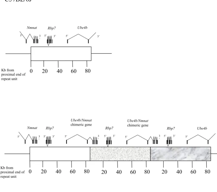
The WldS mutation has been mapped to distal mouse chromosome 4 (Lyon et al, 1993), and a tandem triplication of an 85-kb genomic region has been identified within a genetic candidate interval (Coleman et al, 1998). Exons of three genes were identified within the 85-kb tandem triplication unit (Conforti et al, 2000) (Figure 1.2). Ubiquitination factor E4B (Ube4b) and nicotinamide mononucleotide adenylytransferase (Nmnat), span the distal and proximal boundaries of the repeat unit, respectively. They have the same chromosomal orientation and form a chimeric gene when brought together at the boundaries between adjacent repeat units in WldS. The chimeric mRNA is abundantly expressed in the nervous system and consists of the N-terminal 210 coding nucleotides of Ube4b, the entire 855 coding nucleotides of Nmnat, and 54 nucleotides formed at the junction (Figure 1.2 and Figure 1.3b). The third gene altered by the triplication, Rbp7, is a novel member of the cellular retinoid-binding protein family. Although the function of Rbp7 is unknown, cellular retinoid-binding proteins, most of which are belonging to the fatty acid-binding protein/cellular retinol-binding protein family, have been well characterized in controlling the concentration of free retinoids and in directing protein- bound retinoids to key enzymes responsible for their metabolism. For example, the cellular retinol-binding protein, CRBP, has been implicated in retinol uptake, retinol esterification, mobilization of retinyl esters, and the initial oxidation of retinol to retinaldehyde. It is also conceivable that an alternation in retinoid metabolism could influence axonal survival given that retinoic acid can induce neuronal differentiation, increase nNOS (neuronal nitric oxide synthase) levels in vitro, and participate in the cholinergic potentiation of synaptic activity (Sidell et al, 1983; Ando et al, 1996;
Personett et al, 2000). However, while Rbp7 is highly expressed in white adipose tissue
and mammary gland, it is undetectable on northern blots of WldS brain. This suggests that it is unlikely to play a role in delaying Wallerian degeneration of axons, but does not rule it out.
1.3 Triplication and its genomic stability
Germ-line triplication is a rare event. However, Reddy and Logan (2000) found a triplication with an inverted middle repeat in 13q22q33 in a new born boy with multiple complications including meconium aspiration syndrome (Reddy and Logan, 2000) and also found triplications in 15q11q13 and 2q112q21. Thus they suggest that intrachromosomal triplication may be more prevalent than previously assumed and can sometimes be mistaken for duplications. Some rare triplications have also found within the α globin gene cluster (Villegas et al, 1995). An 85-kb tandem triplication has been identified in the WldS mouse (Coleman et al, 1998). There are very few reports of tandem triplications in a vertebrate. Perhaps the best-studied tandem triplication is that of the Drosophila double Bar mutant, which was shown to arise following a recombination event of the original Bar duplication, using polytene banding patterns long before the advent of molecular genetics (Sturtevant et al, 1925; Muller et al, 1936; Bridges et al, 1936). Such triplication is mediated by homologous, but unequal, crossing-over between tandem head-to-tail repeated DNA sequences (Jeffreys et al, 1988; Tartof, 1988).
Very little is known about genomic instability of triplications, although it is clear that duplications of similar repeat unit length can be unstable in mammals, for examples the 70-kb pink-eyed unstable (pun) duplication that frequently reverts to wild type (Gondo et al, 1993). Initial experiments in Wlds mice showed that some duplication alleles also existed in two out of seven mice analyzed (Coleman et al, 1998). So, like double Bar, the tandem triplication in the WldS mouse is very likely to have developed from duplication, which, like the mouse pink-eyed unstable (pun) mutation, was unstable. However, unlike pink-eyed, WldS contains no genes that are harmful at the increased dosage.
A study of the genomic instability of the triplication in WldS is important to understand the gene dosage that is present in WldS mice and to facilitate the study of the potential role of Wld in neuroprotection, such as crossing WldS with mouse model of neurodegenerative diseases.
During the course of the experiments described, it has been confirmed that the Ube4b/Nmnat chimeric gene confers the slow Wallerian degeneration phenotype using transgenic mice expressing the Ube4b/Nmnat cDNA from an b-actin promoter. The WldS phenotype was reproduced fully in one line (line 4836) and partially in three other lines (lines 4830, 4858 and 4839) with a phenotype strength determined by transgene expression level, thus proving that the chimeric gene is the Wld gene (Mack et al, 2001).
The protective gene thus confers a dose-dependent block of Wallerian degeneration.
Expression of Wld needs to reach a threshold level to exert a significant protective effect:
transected distal axons (69%-73%) survived for two weeks in transgenic line 4836 homozygotes which has the highest expression level, however the protection was considerably weaker with only 7% surviving in line 4836 hemizygotes. In addition, motor nerve conduction, synaptic transmission, vesicle recycling and motor nerve terminal morphology were also preserved in a dose-dependent manner.
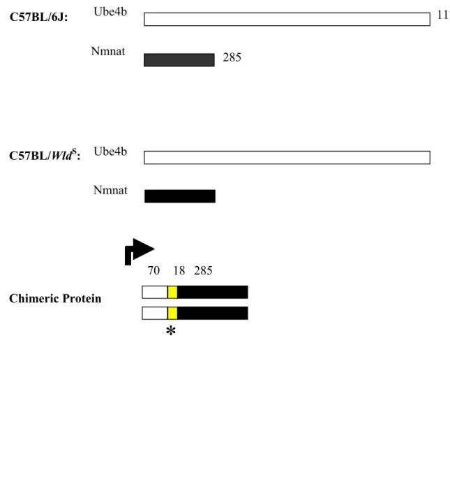
Ube4b/Nmnat chimeric gene encodes a 43-kDa in-frame fusion protein consisting of the N-terminal 70 amino acid of Ube4b, the C-terminal 285 amino acids of Nmnat, and 18 amino acids formed at the junction (Figure 1.3).
The protective mechanism is unknown. Using an antibody described in section 6.2.1, the WldS protein was detected predominantly in the nucleus of WldS and transgenic neurons, indicating an indirect protective mechanism. Although we could not rule out that a possible extremely low and undetectable level of WldS protein might exist in axons, it is unlikely that the predominantly nuclear localization of WldS does not play a role in protecting axons. The yeast homologue (Ufd2) of Ube4b is required to multi-ubiquitinate some substrate proteins (Koegl et al, 1999) and a direct link between ubiquitination and axon degeneration comes from the Uch-l1 mutation in gracile axonal dystrophy (Saigoh et al, 1999 and see section 1.6). However, the WldS protein contains only 70 of 1,173 amino acids from Ube4b, and these are absent from the yeast homologue Ufd2. So far ubiquitin-protein ligases (E3s) had been classified into several established families including HECT, RING-finger families and U-box proteins (Hershko and Ciechanover, 1998; Joazeiro and Weissman, 2000; Hatakeyama and Nakayama, 2003). HECT domain E3 family was first characterized by its representative member, the E6 associated protein (E6-AP). The U-box is a domain of approximately 70 amino acids that is present from
yeast to humans. The prototype U-box is present in the C-terminal region of yeast Ufd2, an ubiquitin chain assembly factor (E4), however absent in the Ube4b-derived N-terminal 70 amino acids of chimeric protein in WldS. They are therefore unlikely to confer multi- ubiquitination activity, but may have a related role, such as substrate binding or regulation of E3 activity. The protective mechanism may be linked to the nuclear location of the WldS protein. Possibilities include sequestering of ubiquitination factors by protein-protein interactions and ubiquitination within the nucleus altering transcription factor stability or RNA processing, leading to an axon effect mediated by unknown proteins. Nmnat is a nuclear protein and has two isoforms of mammalian enzymes catalyzing the reaction NMN + ATP ® NAD+ + Ppi (Magni et al, 1999; Raffaelli et al, 2002), a reaction generally assumed not to be at equilibrium because of the constitutive action of pyrophosphatases. We confirmed that WldS protein has Nmnat enzyme activity and found a fourfold increase of Nmnat activity in WldS brain homogenates compared to C57BL/6J, but the total content of NAD+ was not significantly altered (see section 6.2.1).
Thus, the increase in Nmnat activity in WldS should increase NAD+ synthesis, and the maintenance of normal steady-state levels suggests that the putative additional NAD+ is metabolized. The product of a compensatory reaction could itself be involved in axon protection. For example, poly-ADP ribosylation uses NAD+, influencing protein activity and cellular NAD+ and ATP content, especially in response to stress (Smith, 2001; Ha and Snyder, 1999). Mild activation of PARP-1 without NAD+ depletion can be neuroprotective (Nagayama et al, 2000). Another metabolite, NADH, is a coenzyme for nitric oxide synthase, an enzyme linked to axon damage, and synthesis of the signaling molecule cyclic ADP ribose from NAD+ regulates calcium release from intracellular stores, potentially influencing calcium activated proteases in Wallerian degeneration (Smith et al, 2001; Di Lisa & Ziegler, 2001).
1.5 Slow Wallerian degeneration Mouse, C57BL/WldS and human disease
The morphological features of Wallerian degeneration – the granular disintegration and beading of an axon distal to a site of injury – can be triggered by neurotoxins, and by defects in myelin, axonal transport or oxygen delivery (Waller, 1850; Cliffer et al, 1998;
Frei et al, 1999; Probst et al, 2000; Oosthuyse et al, 2001; Garbern et al, 2002).
diseases of the central and peripheral nervous system such as multiple sclerosis, amyotrophic lateral sclerosis and peripheral neuropathy (Waller, 1850; Dal & Gurney, 1995; Fujimura, 1991). In addition, other diseases, such as dying-back type neuropathies, are traditionally considered distinct from Wallerian degeneration, although there is no evidence that their mechanisms are unrelated (Saigoh et al, 1999; Frei et al, 1999;
Cifuentes-Diaz et al, 2002). Recent data shows that Wallerian degeneration and dying- back axon degeneration might share some mechanism in common (Wang et al, 2002);
and this could be extended to other neurodegenerative disorders, see section 3.2.2. Since axon loss is a major cause of symptoms, even in disorders where the primary defect lies elsewhere, identification of the WldS gene facilitates studies to determine whether it protects axons in clinically relevant situations. The WldS mutation is already known to protect neuronal processes on primary sensory neuronal culture in vitro from the toxic effects of vincristine, suggesting a possible role in combating neurodegeneration induced by chemotherapy (Wang et al, 2001). Studies in which I collaborated indicate that distal axon loss in myelin protein zero deficient mutants (P0) is partially rescued by WldS (Samsam et al, 2003 and see section 3.2.2), and that WldS protects axon loss in Pmn, a mouse model of motor neuron disease (Ferri et al, 2003 and see section 3.2.2). In addition to that, WldS should shed light on the roles of axon loss in multiple sclerosis (MS), amyotrophic lateral sclerosis (ALS), axonal transport defects (such as Charcot- Marie-Tooth disease type 2A, peripheral neuropathy) (Zhao et al, 2001; Cliffer et al, 1998), and acute disorders (such as brain trauma and stroke) (Sherriff et al, 1994;
Graham et al 2000; Stys, 1998). WldS-derived knowledge might even lead towards a therapy for axons in such clinical situations. Since axon injury has recently emerged as a substantial, and perhaps the key component of MS pathology, manipulating the survival of axons, such as WldS does, could lead more axons to survive a period of demyelination and then recover, thus arresting the decline from a relapsing-remitting disease to a progressive one (Narayanan et al, 1997; Sherriff et al, 1994). As in MS, there is considerable axonal loss in human ALS and in animal models of ALS and spinal muscular atrophy (Oosthuyse et al, 2001; Cifuentes-Diaz et al, 2002; Dal & Gurney, 1994). It has been reported, that prevention of axonal loss in pmn does improve the phenotype (Sagot et al, 2000; Ferri et al, 2003). Delaying Wallerian degeneration might
well preserve axons, although could not unblock axonal transport, while another treatment removes the blockage. In brain trauma, axon transection is not necessarily instantaneous upon injury and can occur hours later, giving a window of opportunity in which to save axons (Jafari et al, 1997). In stroke, there is large component of damage in white matter, thus minimizing the loss of the axons could offer new therapeutic opportunities (Dewar et al, 1999).
Those observations above have many important implications: 1) a pathway related to Wallerian degeneration occurs in disease-related processes. Thus, understanding the Wallerian degeneration mechanism is relevant to non-injury disorders such as dying-back neuropathies; 2) WldS is a valuable and unique experimental tool with which to probe the contribution of axonal degeneration to numerous neurodegenerative models; 3) such studies could lead to the development of novel therapeutic strategies.
1.6 The gad mouse (gracile axonal dystrophy)
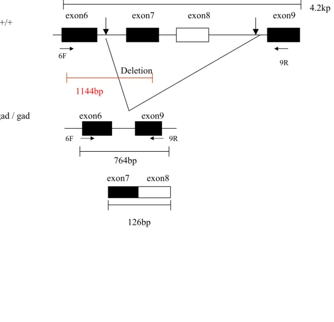
The gracile axonal dystrophy mouse is an autosomal recessive mutant that shows sensory ataxia at an early stage (first detectable at 30 days after birth), followed by motor ataxia (detectable about 60 days after birth) (Yamazaki et al, 1988). Pathologically, the mutant is characterized by ‘dying-back’ type axonal degeneration in both the central and peripheral distal ends of primary sensory neurons and the lower motor neurons and formation of spheroid body in the central distal ends of gracile tract (Mukoyama et al, 1989; Kikuchi et al, 1990; Oda et al, 1992; Miura et al, 1993). Recent pathological observations have associated neurodegenerative diseases with progressive accumulation of ubiquitinated protein conjugates (Arnold et al, 1998; Alves-Rodrigues et al, 1998). In gad mice, accumulation of amyloid b-protein and ubiquitin-positive deposits occur retrogradely along the sensory and motor nervous system (Ichihara et al, 1995; Wu et al, 1996). The gad mutation is caused by an in-frame deletion including exons 7 and 8 of Uchl1, encoding the ubiquitin carboxy-terminal hydrolase (UCH) isozyme (Uch-l1) selectively expressed in the nervous system and testis (Day & Thompson, 1987;
Wilkinson et al, 1989; Day et al, 1990; Kajimoto et al, 1992; Saigoh et al, 1999)(Figure 1.4). The gad allele encodes a truncated Uch-l1 lacking a segment of 42 amino acids containing a catalytic residue (Larsen et al, 1996). Uch-l1 is known to be involved in the
adducts and ubiquitin to generate free monomeric ubiquitin (Dang et al, 1998; Larsen et al, 1998). Recent data also suggests this neuronal-abundant protein (Uchl1) mediates the ubiquitin stability and function in neurons (Osaka et al, in press). Thus, UCHs are essential in continuing the function of the ubiquitin system, and the defect in Uch-l1 in gad mutation appears to affect protein turnover (Saigoh et al, 1999). The gad mouse is the first mammalian model of neurodegeneration with a defect in the ubiquitin system. In addition to that, dying-back degenerative process is thought to underlie several neurodegenerative diseases in human such as motor neuron disease and peripheral neuropathy (Schmalbruch et al, 1991, Samsam et al, 2003), and gad mutant is characterized by dying-back type degeneration in both sensory and motor nerve fibers, thus it could be also expected to provide important information on the pathogenesis of some neurodegenerative diseases.
1.7 Ubiquitin metabolism in neurodegeneration
The integrity of cellular processes depends upon the proper balance of different proteins.
In cells, there are two major destruction pathways to regulate such activities involving either a collection of proteolytic enzymes within the lysosome or the multicatalytic proteolytic core of the proteasome. The majority of proteins that are destined for proteasome degradation are marked by the covalent attachment of multiple ubiquitin molecules, which provide a recognition signal for the 26S proteasome. Such ubiquitin- proteasome pathway involves two discrete and successive steps: a specific recognition process, employing the ubiquitin conjugation cascade, and secondly, an indiscriminate destruction process, mediated by the proteolytic proteasome core. The ubiquitin conjugation cascade uses E1 (ubiquitin-activating enzyme), E2 (ubiquitin-conjugating enzyme), E3 (ubiquitin ligase) and E4 (ubiquitin elongation factor) to shuttle ubiquitin and eventually link ubiquitin to the protein substrates, which then targeted by proteasome for degradation (Myung et al, 2001; Koegl et al, 1999).
Such ubiquitin-proteasome pathway is considered to play a central role in the processing of damaged and toxic protein, and thus impairing such a pathway could lead to the abnormal accumulation and processing of mutant or damaged intra- and extracellular proteins and finally cause neuronal dysfunction and cell death. In a variety of neurodegenerative diseases, such ubiquitin-positive protein aggregates or inclusion
bodies could be found, for example in the prion protein (PrP) plaques in prion disease, amyloid plaques and neurofibrillary tangles in Alzheimer’s disease (AD), Lewy bodies in Parkinson’s disease (PD) and nuclear inclusion in the poly-glutamine repeat diseases such as Huntington’s disease (HD), spinocerebellar ataxias (SCA) (Hardy and Gwinn- Hardy, 1998; Alves-Rodringues et al, 1998; Kenward et al, 1994). The mutation of two genes (Uch-l1 and Parkin) within the ubiquitin-proteasome pathway in hereditary PD and recent proof of a mutation in Usp14 encoding a ubiquitin-specific protease in ataxia mice, clearly indicate that ubiquitin-proteasome pathway plays a crucial role in the pathogenesis of neurodegenerative diseases (Kitada et al, 1998; Leroy et al, 1998; Wilson et al, 2002). A missense mutation (Ile93Met) in ubiquitin carboxy-terminal hydrolase L1 (UCH-L1) gene was identified in a small German pedigree composed of two affected family members (Leroy et al, 1998). Since such mutation causes a partial loss of the catalytic activity of this protease, which involves in the release of ubiquitin from the degraded end products containing small molecules (amines and thiols) and has a potential role in mediating ubiquitin stability in axons (Osaka et al, in press), the mutation could impair the overall efficiency of the ubiquitin system leading to aggregation of proteins.
The deletion of exon7 and exon8 of Uchl1 was found to be the causative gene in the first mice model (gad) of neurodegeneration with defect in ubiquitin system as well, and such ablation of Uch-l1 expression causes the primary defect in the gracile tract of gad mutant, which will be discussed in detail in chapter 5. As described before, E3 ubiquitin-ligase is involved in recognizing the substrates and mediating the attachment of polyubiquitin chains to the substrates, thus it is an important factor in the regulation and selectivity of substrates targeted for degradation. Missense mutations in Parkin were identified in familial Parkinson disease and further parkin was found to be a ubquitin-protein ligase E3 (Kitada et al, 1998; Shimura et al, 2000). Familial-associated mutations in parkin cause impaired binding to E2 and defective E3 ligase activity, which might cause the autosomal recessive PD (Shimura et al, 2000; Imai et al, 2000; Zhang et al, 2000). Several potential substrates for parkin have recently been identified: CDCrel-1 (potential role in regulating synaptic vesicle release in nervous system) (Beites et al, 1999), α-synuclein interacting protein synphilin-1 (Chung et al, 2001), Pael-R (parkin-associated endothelin-receptor-
synuclein)(Shimura et al, 2001). Interestingly, ubiquitinated synphilin-1 is enriched in Lewy bodies, the hallmark of PD, and thus suggests that familial-associated mutations in parkin involve in the pathogenesis of PD. Recently, a mutation in Usp14, encoding an ubiquitin-specific protease was identified as the causative gene for ataxia mice (Wilson et al, 2002). The function of Usp14 might be associated with cleaving the mono-ubiquitin side chain from substrate, and the removal of such mono-ubiquitin would regulate processes such as protein localization and protein activity (Katzmann et al, 2001; Hicke, 2001; Shih et al, 2000). Although neither ubiquitin-positive protein aggregates nor neuronal cell loss is detectable in the central nervous system (CNS) of ataxia mice, there are defects in synaptic transmission in both the central and peripheral nervous systems, and thus suggest that ubiquitin proteases are important in regulating synaptic activity in mammals.
It is clear that the defects in the components of ubiquitin-proteasome pathway play a major role in neurodegenerative diseases, thus a full understanding of such intricate system could contribute to understand the pathogenesis of these disorders.
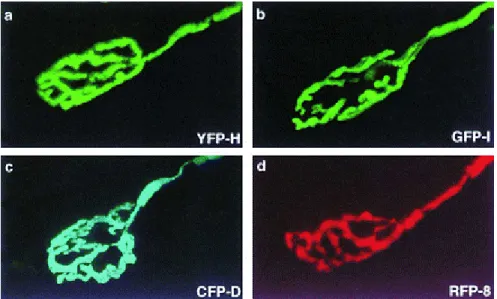
1.8 XFP (especially YFP-H) mice in studying Wallerian degeneration
A series of methods have been developed before to image individual neurons: from Golgi stain to electron microscopy to anterograde and retrograde labeling methods to immunohistochemical labeling protocols to confocal microscopy. The introduction of the jellyfish green fluorescent protein (GFP) as a vital stain has rendered several useful features: GFP is a protein, so the cells could be rendered fluorescent stably and heritably by introduction of a cDNA; the GFP chromophore is derived entirely from the polypeptide chain without a need of exogenous cofactors or substrates, thus it can be used to view living cells with minimal perturbation; GFP can be fused to other proteins without loss of fluorescence, thus it can be directed to specific subcellular compartments;
GFP can be mutated to generate variants (named as XFP) with altered spectral properties and improved translational efficiency, thermostability, and quantum yield. As a result of these favorable properties, GFP and its variants have been used to follow molecules and cells in a lot species (Chalfie et al, 1994; Chalfie and Kain, 1998; Tsien, 1998; Conn, 1999) and recently, transgenic mice in which red, green, yellow, or cyan fluorescent proteins (together termed XFPs, termed RFP, GFP, YFP and CFP respectively) were
selectively expressed in neurons were also generated to image individual neurons (Feng et al, 2000)(Figure 1.5). All four XFPs labeled neurons in their entirety, including axons, nerve terminals, dendrites, and dendritic spines (XFPs label axons over centimeter-long distances and dendrites over millimeter-long distances). Expression of XFP for up to 9 months has no discernible effect on synaptic structure and that multiple imaging of XFP- labeled neurons in vivo is not detectably toxic. Although there is a remarkable variability in patterns of XFP expression among mice generated from the same construct (under the neuron-specific thy1-derived regulatory element), expression is unique and heritable among offspring of each transgenic founder, indicating the difference due to integration site and/or copy number.
Since YFP-fluorescence is intense in both live and paraformaldehyde-fixed tissues, shows good quality of staining axons and nerve terminal and is commercial available, it was chosen as a useful tool to study Wallerian degeneration in following attempts: 1) to study how Wallerian degeneration initiates and progresses in the axotomised axons.
Although Wallerian degeneration was described as a process starting from proximal to distal at the transection site with fragmentation of axons, the evidence is indirect;
introducing YFP into WldS mice should give us a clear picture of it; 2) to study synaptic changes in WldS mice. Since XFP could label neurons in their entirety including synapses, CFP-expressing mice were crossed with WldS mice to study the undergoing mechanism of synapse withdrawal and degeneration (Gillingwater et al, 2002); 3) to study synaptic and axonal change in mice model of neurological disorders with WldS. Crossing YFP-H mice with gad/WldS mice could give us the information of the difference in the nerve terminal of gad with WldS or without WldS and thus give us a better picture to understand the potential rescue-function of WldS in neurodegenerative disorders (see 5.2.1 and 5.2.6);
4) to facilitate the mechanism study of Wallerian degneration. Crossing YFP with transgenic mice with the expression of N-terminal of 70 amino acid of the chimeric gene with NLS or NES could make it easy to visualize the preservation of the axon (see section 6.2.2 in detail).
Figure 1.1 A schematic diagram shows undergoing Wallerian degeneration:
disintegration of axonal cytoskeleton and fragmentation of the axon, followed by breakdown of the myelin sheath, macrophage infiltration and Schwann cell proliferation.
Figure 1.1 Schematic diagram of Wallerian degeneration.
Axon fragmentation, myelin degradation
Macrophage infiltration
Schwann cell proliferation
Axon Regeneration
Figure 1.2 Location of exons within the 85-kb WldS triplication repeat unit. In WldS, three adjacent repeat units are shown to indicate (i) how N-terminal of Ube4b and Nmnat are brought together to form a chimeric gene (ii) that normal copies of Ube4b and Nmnat exist still at either end of the repeat array (the complete genes are not shown), and (iii) how Rbp7 is present at three time the normal copy number.
Figure 1.2 Location of exons within the 85-kb WldS triplication repeat unit.
C57BL/6J
C57BL/WldS
Kb from proximal end of repeat unit
0 20 40 60 80
Rbp7
Nmnat Ube4b
5’ 3
’
5’ 3’ 5’ 3’
0 20 40 60 80
Rbp7
5’ 3
’
5’ 3’ 5’
20 40 60 80
Rbp7
3
’ 5’ 3’ 5’
20 40 60 80
Rbp7 Ube4b
3
’ 5’ 3’
5’ 3’
Ube4b/Nmnat
chimeric gene Ube4b/Nmnat
chimeric gene
Kb from proximal end of repeat unit
Nmnat
Figure 1.3a WldS express novel and wild type proteins: (i) a 43-kDa WldS-specific chimeric protein; (ii) Ube4b and Nmnat which express both in wild type C57BL/6J and C57BL/WldS.
Figure 1.3b Sequence of the chimeric cDNA and the predicted coding sequence. The vertical arrow indicates the junction between Ube4b and Nmnat (starting from 71aa and ending at 88aa), and the amino acid shown in bold are those used to make peptides for generation of polyclonal antisera 183 (Ile-325 to Lys-339) and 185 (Thr-64 to His-79) (see section 3.2.1 and section 6.2.1).
Figure 1.3a WldS express novel and wild type proteins.
C57BL/6J:
C57BL/WldS:
Ube4b Nmnat
Chimeric Protein
*
1174
285 70
Ube4b Nmnat
285
18
Figure 1.3b Sequence of the chimeric cDNA and the predicted coding sequence.
ATGGAGGAGCTGAGCGCTGACGAGATTCGACGGAGGCGCCTGGCACGACTTGCTGGTGGA MetGluGluLeuSerAlaAspGluIleArgArgArgArgLeuAlaArgLeuAlaGlyGly
CAGACCTCCCAGCCGACCACCCCGCTTACATCTCCCCAGAGGGAGAACCCTCCGGGACCT GlnThrSerGlnProThrThrProLeuThrSerProGlnArgGluAsnProProGlyPro
CCAATAGCTGCATCAGCCCCAGGCCCCTCCCAGAGTCTTGGTCTCAATGTCCACAACATG ProIleAlaAlaSerAlaProGlyProSerGlnSerLeuGlyLeuAsnValHisAsnMet
ACCCCAGCTACCTCCCCCATAGGTGCAGCAGACAACATCGCTGTCAGAGGGTTGCATGTA ThrProAlaThrSerProIleGlyAlaAlaAspAsnIleAlaValArgGlyLeuHisVal
GGTCAACACCACCAACTTCTCCCCATGGACTCATCCAAGAAGACAGAGGTGGTTCTCCTG GlyGlnHisHisGlnLeuLeuProMetAspSerSerLysLysThrGluValValLeuLeu
GCCTGTGGCTCTTTTAACCCCATCACCAACATGCACCTCAGGCTGTTCGAGCTGGCCAAG AlaCysGlySerPheAsnProIleThrAsnMetHisLeuArgLeuPheGluLeuAlaLys
GACTATATGCATGCTACAGGAAAATACAGTGTTATCAAAGGCATTATCTCACCGGTCGGT AspTyrMetHisAlaThrGlyLysTyrSerValIleLysGlyIleIleSerProValGly
GATGCGTACAAGAAGAAAGGGCTCATCCCAGCCCACCACCGAATCATCATGGCAGAACTT AspAlaTyrLysLysLysGlyLeuIleProAlaHisHisArgIleIleMetAlaGluLeu
21 1
41
101
121
141 61
81
GCCACCAAGAACTCACACTGGGTGGAAGTGGATACGTGGGAAAGTCTTCAGAAGGAGTGG AlaThrLysAsnSerHisTrpValGluValAspThrTrpGluSerLeuGlnLysGluTrp
GTGGAGACTGTGAAGGTGCTCAGATACCATCAGGAGAAGCTGGCAACTGGCAGCTGCAGT ValGluThrValLysValLeuArgTyrHisGlnGluLysLeuAlaThrGlySerCysSer TACCCACAAAGCTCACCTGCACTGGAAAAGCCTGGGCGGAAGAGGAAGTGGGCTGATCAA TyrProGlnSerSerProAlaLeuGluLysProGlyArgLysArgLysTrpAlaAspGln
AAGCAAGATTCTAGCCCACAGAAGCCCCAAGAGCCCAAACCAACAGGTGTGCCCAAGGTG LysGlnAspSerSerProGlnLysProGlnGluProLysProThrGlyValProLysVal
AAATTGCTGTGTGGGGCAGATTTACTGGAGTCCTTCAGCGTGCCCAACTTGTGGAAGATG LysLeuLeuCysGlyAlaAspLeuLeuGluSerPheSerValProAsnLeuTrpLysMet
GAGGACATCACGCAAATCGTGGCCAACTTTGGGCTCATCTGTATCACTCGGGCTGGCAGT GluAspIleThrGlnIleValAlaAsnPheGlyLeuIleCysIleThrArgAlaGlySer
GACGCTCAGAAATTCATCTACGAGTCCGATGTGCTGTGGAGACATCAGAGCAACATCCAC AspAlaGlnLysPheIleTyrGluSerAspValLeuTrpArgHisGlnSerAsnIleHis
CTGGTGAACGAGTGGATCACCAATGACATCTCGTCCACCAAGATCCGGAGGGCGCTCAGG LeuValAsnGluTrpIleThrAsnAspIleSerSerThrLysIleArgArgAlaLeuArg
161
181
201
221
241
261
281
301
AGGGGCCAGAGCATCCGCTACTTGGTACCGGACCTGGTCCAAGAGTACATTGAGAAGCAT ArgGlyGlnSerIleArgTyrLeuValProAspLeuValGlnGluTyrIleGluLysHis
GAGCTGTACAACACGGAGAGCGAAGGCAGGAATGCTGGGGTCACCCTGGCTCCTCTGCAG GluLeuTyrAsnThrGluSerGluGlyArgAsnAlaGlyValThrLeuAlaProLeuGln
AGGAACGCCGCAGAGGCCAAGCACAACCATTCCACTCTGTGA ArgAsnAlaAlaGluAlaLysHisAsnHisSerThrLeu*** 373
321
341
361
Figure 1.4 Scheme of the deleted region of Uch-l1 in gad mice. gad mutation is caused by an in-frame deletion including exon7 and exon 8 of Uchl1, encoding the ubiquitin carboxy- terminal hydrolase l1.
Figure 1.4 Scheme of the deleted region of Uchl1 in gad mice.
Deletion
exon6 exon7 exon8 exon9
exon6 exon9
exon7 exon8
126bp +/+
gad / gad
4.2kp
1144bp
764bp
6F 9R
6F 9R
Figure 1.5 Four spectrally distinct XFPs serve as vital stains in transgenic mice.
Neuromuscular junctions from thy1-YFP line H (a), thy1-GFP line H (b), thy1-CFP line D (c), and thy1-RFP line 8 (d) transgenic mice.
Figure 1.5 Four spectrally distinct XFPs serve as vital stains in transgenic mice.
CHAPTER 2
MATERIAL AND METHODS