cytoskeletal proteins horizontally transferred to a cyanobacterium
D I S S E RTAT I O N
zur Erlangung des akademischen Grades d o c t o r r e r u m n a t u r a l i u m
(Dr. rer. nat.)
im Fach Biologie eingereicht an der
Mathematisch-Naturwissenschaftlichen Fakultät I der Humboldt-Universität zu Berlin
von
Diplom-Biologe Arthur Guljamow
Präsident der Humboldt-Universität zu Berlin:
Prof. Dr. Jan-Hendrik Olbertz
Dekan der Mathematisch-Naturwissenschaftlichen Fakultät I:
Prof. Dr. Andreas Herrmann
Gutachter: 1. Prof. Elke Dittmann-Thünemann 2. Prof. Conrad W. Mullineaux 3. Prof. Harald Saumweber
Tag der mündlichen Prüfung: 21. Oktober 2011
Abstract
The cyanobacterium Microcystis aeruginosa PCC 7806 expresses two unique proteins that show a very high degree of sequence identity with key components of the eukaryotic cytoskeleton. One is actin itself, the building block of microfilaments, the other is profilin, an important actin binding protein. Both proteins are remnants of a rarely observed horizontal gene transfer from eukaryotes to bacteria and their functions are unknown.
By employing a wide range of in vivo and in vitro methods in complementary experimental approaches, both proteins were characterized in great detail during the course of this work. The purification of both proteins after heterologous expression in E.coli allowed for the determination of key biochemical and structural parameters and the comparison with the eukaryotic archetype. In contrast to bona fide actin, for instance, its cyanobacterial counterpart does not inhibit DNase I. It forms polymers that can be visualized with labeled phalloidin, resembling eukaryotic actin in that respect. However, confocal laser scanning microscopy reveals key differences between polymers of eukaryotic and cyanobacterial actin. Whereas the former appear as thin cylindrical filaments about 100 µm in length, the latter are shorter and wider arresting polymerization at 5-10 µm. A more refined structural elucidation was achieved by Small-angle X-ray scattering showing that polymers of cyanobacterial actin are more ribbon-shaped.
Furthermore, this work shows fundamental differences between cyanobacterial and eukaryotic profilin. Restricted to actin monomer binding in eukaryotes, a number of experiments described herein show that cyanobacterial profilin decorates actin filaments. Additionally, confocal microscopy and SAXS suggest that cyanobacterial profilin mediates the bundling of cyanobacterial actin polymers into extended heteropolymeric sheets.
These co-polymers may be the basis of the large hollow enclosures observed in E.coli cells co-expressing GFP-labeled cyanobacterial profilin and actin. These hollow structures resemble the shell-like distribution of actin in Microcystis aeruginosa PCC 7806 and in colonies sampled from its original habitat, as shown in this work.
The findings of this work show that as part of the proteome of a natural bacterial community, both cyanobacterial proteins have gained properties unknown from their eukaryotic ancestors. Furthermore, the results have led to the hypothesis that the adaptation to the confined space of a bacterial cell devoid of binding proteins usually regulating actin polymerization in eukaryotes has driven the co-evolution of cyanobacterial actin and profilin, giving rise to an intracellular entity of potential structural relevance.
Deutsche Zusammenfassung
Das Cyanobakterium Microcystis aeruginosa PCC 7806 kodiert zwei Proteine, welche einen ungewöhnlich hohen Grad an Sequenzverwandschaft mit essentiellen Komponenten des eukaryotischen Zytoskeletts aufweisen. Bei einem dieser Proteine handelt es sich um Aktin, den Grundbaustein der Mikrofilamente, das andere ist Profilin, ein wichtiges Aktinbindeprotein. Beide Proteine resultieren aus einem selten beobachteten horizontalen Gentransfer von Eukaryoten zu Bakterien.
Unter Anwendung einer Reihe von in vivo und in vitro Methoden in einander ergänzenden experimentellen Ansätzen wurden beide Proteine im Verlauf dieser Arbeit detailliert charakterisiert. Die heterologe Expression in E.coli und die anschließende Aufreinigung beider Proteine erlaubten die Bestimmung charakteristischer biochemischer und struktureller Parameter sowie deren Vergleich mit bekannten eukaryotischen Vertretern. So inhibiert das cyanobakterielle Aktin im Gegensatz zu seinem eukaryotischen Verwandten zum Beispiel nicht das Enzym DNase I. Hingegen ähnelt es
„echtem“ Aktin in der Hinsicht, dass es Polymere bildet, welche mit farbmarkiertem Phalloidin visualisiert werden können. Konfokale Laserscanning Mikroskopie offenbart jedoch grundlegende Unterschiede in den Polymerstrukturen. Filamente von eukaryotischem Aktin sind zylindrisch und erreichen typische Längen von 100 µm, wohingegen Polymere cyanobakteriellen Aktins eine Länge von 5-10 µm nicht überschreiten und zudem breiter sind. Detailliertere Strukturaufklärungen mittels Röntgen- Kleinwinkelstreuung zeigen, dass cyanobakterielle Aktinpolymere in ihrer Form am ehesten einem Band mit annähernd viereckigem Querschnitt ähneln.
Desweiteren förderte diese Arbeit grundlegende Unterschiede zwischen eukaryotischem und cyanobakteriellem Profilin zu Tage. Während Profilin in Eukaryoten ausschließlich monomerisches Aktin binden kann, bestätigen verschiedene hier durchgeführte Experimente, dass sich cyanobakterielles Profilin an Aktinfilamente anlagert. Zudem deuten Erkenntnisse aus konfokaler Mikroskopie und Kleinwinkelstreuung darauf hin, dass
cyanobakterielles Profilin die Bündelung von cyanobakteriellem Aktin zu ausgedehnten Heteropolymeren vermittelt.
Diese Co-Polymere bilden möglicherweise die Grundlage für die verhältnismäßig großen, hohlen Einschlüsse, welche in genetisch modifizierten Stämmen von E.coli beobachtet werden konnten. Diese Zellen exprimierten sowohl ein Fusionsprodukt aus cyanobakteriellem Profilin und dem grün fluoreszierenden Protein GFP, als auch cyanobakterielles Aktin. Die dort beobachteten hohlen Strukturen erinnern an die hüllenartige Lokalisation von Aktin, welche sowohl für den Microcystis aeruginosa Stamm PCC 7806, als auch für Microcystis Kolonien aus dessen ursprünglichen Habitat in dieser Arbeit gezeigt wurden.
Die Erkenntnisse dieser Arbeit verdeutlichen, dass beide cyanobakteriellen Proteine als Teil der Proteinausstattung einer natürlich vorkommenden Bakterienpopulation Merkmale erworben haben, die ihre eukaryotischen Vorläufer nicht zeigen. Darüber hinaus konnte anhand der hier gewonnenen Ergebnisse folgende Hypothese aufgestellt werden: Die Anpassung an die begrenzten räumlichen Bedingungen einer Bakterienzelle, welche außerdem keine der für die Regulierung der Polymerisation notwendigen Aktinbindeproteine enthält, war offenbar die Triebkraft für eine Co-Evolution von cyanobakteriellem Aktin und Profilin. Dieser Prozess gipfelte möglicherweise in der Entstehung eines neuartigen intrazellulären Gebildes von potentiell struktureller Bedeutung.
Abbreviations
aa amino acid
ABP actin binding protein
ADF actin depolymerizing factor Amp ampicillin
APS ammonium persulfate
Arp actin related protein
bp base pair(s)
BSA bovine serum albumin
CIAP calf intestine alkaline phosphatase Cm chloramphenicol
CTAB cetyl-trimethyl-ammonium-bromide DTT 1,4-dithiothreitole
DMF dimethylformamide
DNA deoxyribonucleic acid
dNTP any triphosphates of the naturally occurring coding deoxy- nucleosids
E. coli Escherichia coli
EDTA ethylene diamine tetra-acetic acid
EM electron microscopy
F-actin filamentous actin FITC fluorescein isothiocyanate
Fts filamentous temperature sensitive protein G-actin globular, monomeric actin
HEPES [4(2-hydroxyethyl)-1-piperazino]-ethanesulfonic acid HGT horizontal gene transfer
IFM immunofluorescence microscopy
hsp heat shock protein
IGS intergenic spacer
IPTG isopropyl β-D-1-thiogalactopyranoside
kb kilo base pairs
kDa kilo Dalton
Mbp mega base pairs
mre murein cluster e
MW molecular weight
NCBI The National Center for Biotechnology Information nt nucleotide
OD optical density
ORF open reading frame
PAA polyacrylamide
PAGE polyacrylamide gel electrophoresis PCC Pasteur Culture Collection PCR polymerase chain reaction PEG polyethylenglycol
pI isoelectric point
PIP2 phosphatidylinositol (4,5) bisphosphate PMSF phenyl-methyl-sulphonyl-fluoride PVP polyvinyl-pyrrolidon
RING-FISH recognition of individual genes – fluorescence in situ hybridization
RNA ribonucleic acid
rpm rounds per minute
RT room temperature
SAXS small-angle X-ray scattering SDS sodium dodecyl sulfate SSC salt-sodium-citrate
TAE tris-acetate-EDTA buffer
TEMED N’,N’,N’,N’-tetramethyl-ethylene-diamine
Tris-HCl tris-(hydroxymethyl)-aminomethane-hydrochloride
UTR untranslated region
UV ultraviolet light
WASp Wiskott-Aldrich-Syndrome protein
WT wild type
X-Gal 5-bromo-4-chloro-3-indolyl-β-D-galactopyranoside
Table of contents
Abstract ... II Deutsche Zusammenfassung ... IV Abbreviations ... VI Table of contents ... VIII
1 Introduction ... 1
1.1 Cytoplasmic actin in eukaryotes ... 1
1.1.1 General features ... 1
1.1.2 The functions of the eukaryotic actin cytoskeleton ... 6
1.1.2.1 The role of microfilaments in cell stabilization ... 6
1.1.2.2 Cell motility ... 8
1.1.3 Actin binding proteins ... 10
1.1.3.1 Actin monomer binding proteins ... 10
1.1.3.1.1 Inhibitors and activators of actin polymerization ... 10
1.1.3.1.2 Profilin ... 11
1.1.3.1.3 DNase I ... 14
1.1.3.2 Actin filament binding proteins ... 15
1.1.3.3 Concluding remarks on actin binding proteins ... 17
1.2 Prokaryotic actins ... 18
1.2.1 Bacterial cytokinesis: FtsA... 19
1.2.2 Plasmid segregation: ParM and AlfA ... 20
1.2.3 Wide-spread and multifunctional: MreB ... 22
1.2.3.1 General features of MreB ... 22
1.2.3.2 MreB and cell shape determination ... 23
1.2.3.3 The role of MreB in DNA replication and segregation ... 24
1.2.3.4 Additional functions of MreB and unresolved issues ... 25
1.2.4 More actins: MamK, BARP, Alps and Ta0583 ... 25
1.3 Concluding remarks on actins ... 26
1.4 Horizontal gene transfer ... 27
1.5 Cyanobacteria ... 29
1.5.1 General aspects ... 29
1.5.2 Cyanobacterial actins ... 31
1.5.3 Microcystis aeruginosa ... 31
1.6 Eukaryotic actin and profilin in Microcystis aeruginosa ... 32
1.7 Aims of this study ... 34
2 Materials and methods ... 35
2.1 Materials ... 35
2.1.1 Chemicals ... 35
2.1.2 Enzymes ... 37
2.1.3 PCR primers ... 37
2.1.4 Plasmid vectors ... 39
2.1.5 Antibodies ... 39
2.1.6 Kits ... 40
2.1.7 Membranes, papers, films and filters ... 40
2.1.8 Technical appliances ... 41
2.1.9 Miscellaneous materials ... 42
2.1.10 Biological material ... 43
2.2 Methods ... 43
2.2.1 Cultivation of bacteria ... 43
2.2.1.1 Cultivation of Microcystis aeruginosa ... 43
2.2.1.2 Cultivation of Escherichia coli ... 43
2.2.2 Collection of field samples ... 44
2.2.3 Molecular biological techniques ... 44
2.2.3.1 Preparation of genomic DNA from cyanobacteria ... 44
2.2.3.2 Preparation of metagenomic DNA from field samples ... 45
2.2.3.3 Preparation of plasmid DNA from Escherichia coli ... 46
2.2.3.4 Quantification of nucleic acids by spectro-photometry ... 46
2.2.3.5 Digestion of DNA with restriction endonucleases ... 46
2.2.3.6 Agarose gel electrophoresis of DNA ... 47
2.2.3.7 Elution of DNA fragments from agarose gels ... 47
2.2.3.8 Primer design and polymerase chain reaction ... 47
2.2.3.9 Inverse PCR ... 48
2.2.3.10 Biotin pull-down assay... 49
2.2.3.11 Quantitative PCR (qPCR) ... 50
2.2.3.12 Generation of labeled RNA-probes for RING-FISH ... 50
2.2.3.13 Purification of PCR fragments and other DNA ... 51
2.2.3.14 Ligation of linear DNA fragments into plasmid vectors ... 51
2.2.3.1 Transformation of Escherichia coli ... 52
2.2.3.2 Generation of GFP-fusion proteins ... 52
2.2.3.3 Construction of plasmid vectors for heterologous expression ... 53
2.2.3.4 Generating E.coli-strains for co-expression of proteins ... 54
2.2.3.5 Construction of genomic fosmid libraries ... 54
2.2.3.6 Sequencing of DNA fragments ... 54
2.2.4 Proteo-biochemical methods ... 55
2.2.4.1 Preparation of proteins from bacterial cells ... 55
2.2.4.2 Heterologous expression and purification of proteins ... 56
2.2.4.3 Purification and concentration of protein solutions ... 57
2.2.4.4 Quantification of protein extracts after BRADFORD ... 57
2.2.4.5 SDS Polyacrylamide gel electrophoresis of proteins ... 57
2.2.4.6 “Western blot” – transfer of proteins from PAA gels ... 58
2.2.4.7 Immunodetection on Western blot membranes ... 58
2.2.4.8 DNase I assay ... 59
2.2.4.9 Binding and co-elution assays ... 59
2.2.4.10 PfnM antibody generation ... 60
2.2.4.11 Preparation of polymerized actin ... 60
2.2.4.12 Phalloidin staining of actin ... 60
2.2.4.13 FITC-labeling of PfnM ... 61
2.2.4.14 Co-polymerization of ActM and PfnM ... 61
2.2.5 Fluorescence microscopy ... 62
2.2.5.1 Fixation and permeabilization of bacterial cells ... 62
2.2.5.2 Immunostaining of fixed cyanobacterial cells ... 63
2.2.5.3 RING-FISH of bacterial cells ... 63
2.2.5.4 Live-cell imaging ... 64
2.2.5.5 Image acquisition and processing ... 64
2.2.6 Small-Angle X-ray Scattering (SAXS) ... 65
2.2.6.1 SAXS data acquisition ... 65
2.2.6.2 Modeling analysis of SAXS data ... 65
2.2.6.3 Model-free analysis of SAXS data ... 67
2.2.7 In silico analyses ... 67
3 Results ... 69
3.1 Characterization of ActM and PfnM in vitro ... 69
3.1.1 Heterologous expression of ActM and PfnM ... 69
3.1.2 Characterization of PfnM ... 71
3.1.2.1 Antibody generation and Western immunoblots ... 71
3.1.3 Characterization of ActM ... 72
3.1.3.1 Quantitative Western blot analysis ... 72
3.1.3.2 DNase I assay ... 73
3.1.3.3 Polymerization and ultracentrifugation ... 75
3.1.3.4 Phalloidin staining of ActM and rabbit actin ... 76
3.1.3.5 SAXS analyses of rabbit F-actin and ActM polymers ... 77
3.1.4 Interaction of ActM and PfnM ... 79
3.1.4.1 Co-elution assay ... 79
3.1.4.2 ActM-PfnM Phalloidin assay ... 81
3.1.4.3 Polymerization and ultracentrifugation of ActM and PfnM ... 83
3.1.4.4 SAXS assay ... 84
3.2 Visualization of ActM and PfnM in vivo ... 87
3.2.1 Immunofluorescence microscopy ... 87
3.2.2 GFP fusions of ActM and PfnM ... 88
3.3 Presence of ActM and PfnM in field samples ... 95
3.3.1 Immunofluorescence microscopy with Braakman colonies ... 96
3.3.2 Metagenomic analyses ... 98
3.3.2.1 DNA extraction and PCR ... 98
3.3.2.2 Analyzing the regions flanking the genomic island ... 101
4 Discussion ... 103
4.1 ActM and PfnM have distinct and unique properties ... 104
4.2 Biological relevance of ActM and PfnM ... 109
4.3 Towards the function of ActM and PfnM ... 111
4.4 Concluding remarks and future outlook ... 116
5 Appendix ... 118
5.1 List of figures ... 118
5.2 List of tables ... 119
6 References ... 120
Publications, awards, conference contributions ... 137
Acknowledgement ... 138
Eigenständigkeitserklärung ... 139
1 Introduction
The actin family of proteins is an evolutionarily ancient group whose ancestral genes can be traced back to the origins of life on Earth (Erickson, 2007; Pollard & Cooper, 2009). The first actins have presumably arisen from a primal protein marked by a bi-lobed, flexible structure and the ability to catalyze nucleotide hydrolysis. These, indeed, are the characteristics contemporary actins share with their distant relatives, the sugar kinases and the hsp70 heat shock proteins widely distributed throughout the tree of life (Bork et al, 1992). However, actins diverged from this line rather early in evolution by developing a substantially new trait: the self-aggregation into polymeric filaments. Being one of their signature features, it forms the basis for the remarkable functional versatility and the resultant extensive prevalence of actins in the living world. The adjustability of actin filament assembly, stability and outward appearance has consequently led to actins being utilized by numerous evolutionarily remote organisms for a multitude of key physiological processes.
1.1 Cytoplasmic actin in eukaryotes
1.1.1 General features
Eukaryotic actin is an essential protein that, apart from its prominent role in muscle contraction, is the building block of what is known as the microfilament cytoskeleton. Microfilaments are found in every eukaryotic cell where actin frequently is the most abundant protein species, typically found in concentrations between 0.2 mM and 0.5 mM (8 µg/µl – 20 µg/µl), making up 1-5% of the total protein amount of non-muscle cells (Lodish et al, 2000;
Purich & Allison, 1999). Whereas unicellular eukaryotes usually encode and express only one actin gene, most vertebrates possess three tissue-specific isoforms, α-actin in muscle cells and the “non-muscle” β- and γ-actins, constituents of the cytoplasmic microfilaments (Bhattacharya & Weber, 1997;
Drouin et al, 1995). Multicellular organisms usually encode multiple isoform
variants, quite frequently amounting up to 10 functional actin genes (Chang et al, 1984; Engel et al, 1982; Engel et al, 1981; Ponte et al, 1983). Warm- blooded vertebrates, for instance, typically express four different α-actins from a single copy gene each and contain multiple copies (totaling four to six) of the β and γ isoforms. Actins are among the most conserved proteins in evolution; α-actins from human, mouse, rat, rabbit and chicken are identical, sharing a 90% sequence identity with fission yeast actin. Plant actins tend to show a higher degree of sequence diversion with many plants possessing up to 100 different actin isoforms. The basis for this exceptional situation is poorly understood (An et al, 1999; Diaz-Camino et al, 2005; Kandasamy et al, 1999;
McDowell et al, 1996).
The actin polypeptide is usually composed of about 375 amino acids and has a molecular weight of 43 kDa. After its discovery in skeletal muscle, a high- resolution structure of actin had long remained elusive, mainly because of difficulties obtaining crystals suitable for X-ray crystallography. Therefore, taking from low-resolution data, the monomeric, or “globular”, form is referred to as “G-actin”. However, as detailed atomic models have eventually become available, the organization of actin into one large and one small domain, forming a deeply divided, roughly “U-shaped” protein has become apparent (see Figure 1.1A) (Kabsch et al, 1990; Otterbein et al, 2001). Each of the two domains is further divided into two subdomains, with subdomain 1 and 2 forming the small domain and subdomains 3 and 4 representing the large domain (Kabsch et al, 1990). A nucleotide binding pocket and the binding site for a catalytic cation (Mg2+ in vivo) - both required for nucleotide hydrolysis - are situated at the bottom of the major cleft where the domains are connected by a flexible hinge region (Kabsch et al, 1990; Otterbein et al, 2001). Owing to this flexibility and in response to its association with its many binding partners, the actin molecule shows a great variability in surface structure and three-dimensional appearance, the most common of which being referred to as the “open” and the “closed” state (Dedova et al, 2002;
Fujii et al, 2010; Pollard, 1999; Schutt et al, 1993; Schutt et al, 1995).
Figure 1.1. Atomic models of monomeric and filamentous actin.
A: Crystal structure of uncomplexed actin from rabbit skeletal muscle in the “conventional view”. The four subdomains of actin are represented in different colors. The polypeptide chain termini are indicated.
Four Ca2+ ions bound to the actin monomer in the crystals are represented as red spheres.
Tetramethylrhodamine-5-maleimide (TMR) covalently attached to Cys374 binds in a hydrophobic pocket near the C-terminus. Adapted from Otterbein, 2001.
B: “Holmes” model of one F-actin protofilament. Subunit numbers and the axial repeat are indicated.
Adapted from van den Ent et al, 2001.
C: The original “Holmes” model of F-actin as published in 1990 (Holmes et al, 1990) can be interpreted as two protofilaments winding around each other in a two-start right handed helix. Actin monomers are shown in individual colors. Each amino acid is represented as a sphere of 27 nm radius, green spheres represent potential myosin binding regions.
It has been know since its early description that solutions of G-actin spontaneously adopt a gelatinous, highly viscous state under physiological ion concentrations (Straub, 1942). This process can be reversed in a low-salt environment. Closer investigations have shown that these changes in viscosity are brought about by a dramatic reorganization of the actin structure: the linear assembly into polymeric fibers with a diameter of 7-9 nm (Cowieson et al, 2008; dos Remedios et al, 2003). Although the fibrous nature of so called
“F-actin” has been established very early in the history of actin research, the determination of the exact physical dimensions and the orientation and conformation of the monomers in the filament pose a challenge to scientists even today. Structural data from low-resolution analyses such as small angle X-ray scattering (SAXS) of F-actin can be fit conveniently to a cylindrical model (Lepault et al, 1994; Norman et al, 2005). More refined low-resolution
models show that the cylindrical surface of the filament is periodically interspersed with structural entities protruding from the main longitudinal axis, confirming findings that were long known from electron microscopy data (Fujii et al, 2010; Hanson & Lowy, 1963; Moore et al, 1970). Although this information was taken as a clear indication for the helical nature of F-actin, definite experimental proof with atomic detail was long unavailable. The main obstacle in obtaining high-resolution structural data from crystallized filamentous material is the lack of precise contacts between filaments (dos Remedios et al, 2003; Hanson & Lowy, 1963). This inherent irregularity of oriented gels of F-actin does, on the one hand, produce characteristic fiber diffraction patterns which are, on the other hand inconclusive with regard to the atomic details of the structure. Therefore, the currently accepted atomic model of F-actin has originally been devised by combining the high-resolution structure of monomers and finding the best fit between its calculated fiber diffraction data and experimental findings (Holmes et al, 1990; Lorenz et al, 1993). According to this model, the actin filament consists of two protofilaments which align in parallel in the same orientation to wind together in a right handed helix forming a polar filament (see Figure 1.1B and C).
Although this “Holmes model” has gained wide acceptance, a competing model based on crystallization data of G-actin and the actin binding protein profilin has been vibrantly discussed for a number of years (Schutt et al, 1993;
Schutt et al, 1995). The observed co-crystals revealed a ribbon-shaped filamentous structure with actin-actin interactions that were proposed to be a representation of those found in F-actin. The “ribbon model” has mostly been regarded as biologically irrelevant and inferior to the helix model, however, with the lack of high-resolution structural data it could not be easily dismissed (Egelman, 1994; Orlova et al, 1994). Thus, it was only with the recent availability of high-resolution structures of F-actin that the helix model was ultimately confirmed (Fujii et al, 2010; Oda et al, 2009). Refinement of the fiber diffraction approach has yielded the first high-resolution model demonstrating that the actin monomers are in a flat and closed conformation in the filament as opposed to being twisted as in the G-actin crystals (Oda et
al, 2009). Another model obtained through cryoEM experiments is very similar and provides additional invaluable insight into the monomer-monomer contact sites showing, for instance, that the axial interactions are stronger than inter-strand bonds (Fujii et al, 2010).
The aggregation of G-actin en route to the filament always occurs in a specific head-to-tail fashion that seals the bound nucleotide inside the actin molecule. The formation of dimers and trimers is a comparably slow and rare process, however, if the assembly reaction is nucleated by pre-existing polymer ends, polymerization is rapid and consumes most of the free G-actin (Pollard & Cooper, 2009). Another important feature of the aggregation process is that the addition of subunits preferentially occurs at the end of the growing polymer that does not expose its bound nucleotide to the medium.
This has two critical implications. First, actin filaments are polar structures and second, they have considerable differences in terminal elongation rates.
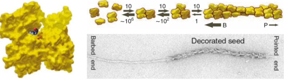
This can be illustrated with the S1 globular head domain of myosin that binds to F-actin in a specific, tilted manner, yielding a characteristic arrowhead-like appearance with a “barbed” and a “pointed” end, as observed via electron microscopy (Woodrum et al, 1975). By conducting such
“decoration” experiments with growing actin filaments it was shown that the assembly of monomers at the barbed end occurs up to 10 times faster than it does at the pointed end, where monomers dissociate from the polymer. This leaves the impression of a filament of roughly constant length moving in one direction (see Figure 1.2) (Pollard & Borisy, 2003; Pollard & Cooper, 2009).
Moreover, as ATP-G-actin has a higher affinity for elongating filament ends than ADP-G-actin and the ATPase activity is activated upon polymerization, ADP-bound subunits are predominately situated towards the pointed end.
The structural changes in ADP-actin monomers entail a lower affinity for the filament and they subsequently dissociate from its pointed end.
Figure 1.2. Nucleation, polymerization and treadmilling of polar actin filaments.
Model of G-actin (left) in the “conventional view” with the “barbed end” of the molecule facing down.
Cartoon diagram of the spontaneous nucleation and polymerization dynamics of actin (top right). Dimers and trimers are very unstable, numbers and widths of arrows indicate relative equilibrium constants of respective reactions. Monomers are added much faster to the barbed end (B) than to the pointed end (P). Electron micrograph of F-actin decorated with myosin heads and elongated with ATP-G-actin (bottom right). Adapted from Pollard & Cooper, 2009 and Pollard & Borisy, 2003.
Free ADP-G-actin is then available for nucleotide exchange and another round of polymerization (Pantaloni et al, 2001; Pollard et al, 2000). This feature of actin filaments is known as “treadmilling” and it provides a means for the cell to generate directed force and do mechanical work (Theriot &
Mitchison, 1991; Wang, 1985). The structural flexibility of actin filaments and the dynamic nature of the polymerization process provide an excellent foundation to regulate the overall appearance of the microfilament network.
Consequently, actin filaments adopt a variety of higher-ordered structures and form an extended intracellular network essential for many fundamental cellular processes revolving around the two main tasks of maintaining a rigid scaffold and ensuring cell motility (Pollard & Cooper, 2009).
1.1.2 The functions of the eukaryotic actin cytoskeleton 1.1.2.1 The role of microfilaments in cell stabilization
Although the dense system of cellular actin filaments appears to be randomly distributed in the cytosol, close examination reveals a principal organization.
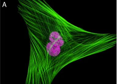
In situ fluorescent labeling of F-actin shows bundled fibers aligned in a parallel fashion, spanning the cell’s periphery where they play a structural role in maintaining cell shape (see Figure 1.3A). Special actin bundles, termed “stress fibers,” provide mechanical support in cells that are in direct
contact with a solid substratum (Walcott & Sun, 2010). Such cells are attached to the external surface by special cortical regions called “focal adhesions” which are connected to the actin cytoskeleton through direct contact with the end of stress fibers. From the bundles at the cell’s periphery, the network of microfilaments fans out into the cytosol to form a less dense three-dimensional network that gives the cytoplasm its gel-like properties.
Figure 1.3. Visualizations of the microfilament network.
A: Fibroblast labeled with probes for actin and the nucleus. The actin cytoskeleton was visualized using phalloidin conjugated with green-fluorescent Alexa Fluor 488. The nucleus (purple) was stained with the TO-PRO-3 reagent. Adapted from www.invitrogen.com/site/us/en/home/References/Molecular-Probes- The-Handbook/.html
B: Transmission electron micrograph of the cortical region of a motile keratocyte. Regions with different filament organizations are indicated, branched filaments face the cell membrane. Scale bar: 500 nm.
Adapted from Pollard & Borisy, 2003 and Svitkina & Borisy, 1999.
A much higher concentration of actin polymers is found in close vicinity to the inner face of the cell membrane (see Figure 1.3B). In this narrow cortex of the cell, microfilaments form a complex three-dimensional network which excludes most cytosolic organelles and is closely connected to the cell membrane via membrane-microfilament binding proteins (Svitkina & Borisy,
1999). These integral membrane proteins serve as attachment sites for the membrane to the cytoskeletal framework, thereby determining the distinctive shape of each cell (Kabsch & Vandekerckhove, 1992; Lodish et al, 2000).
Notwithstanding its essential structural role in determining and maintaining cell shape, the most intriguing property of the actin cytoskeleton is undoubtedly its ability to confer what has become known as “cell motility”.
1.1.2.2 Cell motility
The term “cell motility” encompasses a variety of cellular processes involving the generation of mechanical force to move the whole cell or objects within. In eukaryotic cells, movement is generated in two principle ways: either by harnessing the force generated through the regulated assembly and disassembly of cytoskeletal elements or by employing motor proteins which move along filaments, carrying various cellular components along these tracks.
The only known actin motor proteins belong to the large myosin family (Hodge & Cope, 2000). These multi-chain proteins share a specific overall structure, organizing the myosin complex into three characteristic functional units. The actin binding head domain harbors an ATPase activity, the adjacent neck domain plays a regulatory role for the head domain’s function and the tail domain mediates myosin binding to various cellular structures.
Myosins can “walk” along actin filaments in discrete steps harnessing the energy of ATP hydrolysis by the head domain. The most prominent example of force generation by actin and myosin is the process of muscle contraction in muscle tissue achieved by making long polar filaments of actin and myosin slide past each other in opposite directions. The same principle is applied in myosin-containing contractile stress fibers of non-muscle cells. During cytokinesis, actin and myosin accumulate at the cell’s equator to form a contractile ring which forms the cleavage furrow and whose ingression into the cell’s interior eventually leads to the separation of the two daughter cells (Scholey et al, 2003). While long distance intracellular transport of large cargo, such as organelles, is achieved by the microtubule system, short distance transport of smaller load such as membrane coated vesicles, proteins
and even nucleic acids within the peripheral regions of the cell is carried out by myosins moving along actin filament tracks (Boldogh & Pon, 2006;
Estrada et al, 2003; Pollard & Cooper, 2009; Takizawa & Vale, 2000).
A remarkable property of many eukaryotic cells is the ability to move across a surface in an “amoeboid crawling” motion. Essentially, this is a three-step process of controlled rapid change of cell morphology: first, the membrane extends to form protrusions known as “pseudopodia”. In the second phase, the pseudopodium attaches to the surface via focal adhesions and the protruding compartment rapidly fills with cytosol. Finally, the rear of the cell is detached from the surface and retracts in the direction of the cell’s movement. The key event in cell locomotion is the directed forward movement of a defined patch of the membrane. This is achieved by a rapid elongation of the actin filaments of the cortical network of the respective region. Although the cortex filaments form a complex, cross-linked three-dimensional network, each fiber is generally oriented in the same way, with the barbed end facing the membrane. Upon stimulation, the cell initiates rapid addition of new subunits at these ends, generating enough mechanical force to push forward large portions of the membrane. To allow the interior of the pseudopodium to be filled with cytosol shortly after its attachment to the substratum, the underlying actin network quickly decondenses from the pointed ends. The same principle mechanism is employed by eukaryotic cells to engulf and absorb extracellular objects during endocytosis (Pollard & Borisy, 2003;
Pollard & Cooper, 2009; Welch & Mullins, 2002).
The actin network’s contrasting tasks of conferring both stability and motility are reconciled by the multitude of mechanisms eukaryotic cells have developed for precisely controlling the rapid rearrangement of well-defined portions of the network in space and time. It is apparent that these rearrangements cannot be provided by the chance fluctuations of the dynamic filaments alone.
Instead, a concerted effort and interplay of many factors of control and regulation is required. That, essentially, is the task of the many actin binding proteins.
1.1.3 Actin binding proteins
For the control of the actin network architecture, eukaryotes employ more than 100 actin binding proteins (ABPs) (dos Remedios et al, 2003; Winder &
Ayscough, 2005). These proteins are involved in virtually every step of the assembly process leading from single actin monomers to the specific shape of the filamentous network required to carry out its multiple functions.
Generally, ABPs can be classified according to their capacity to either bind to actin monomers or filaments, although some ABPs are able to bind both (Winder & Ayscough, 2005).
1.1.3.1 Actin monomer binding proteins
1.1.3.1.1 Inhibitors and activators of actin polymerization
Under physiological conditions, free G-actin quickly polymerizes into F-actin (Pollard & Borisy, 2003). However, filaments near the leading edge of protruding membrane regions were found to elongate up to 250 times faster than observed at typical steady-state conditions while G-actin concentrations generally remain constant (Pollard & Borisy, 2003). Thus, cells must maintain a pool of “silent” actin monomers that can be rapidly recruited to elongate filaments if circumstances necessitate such. The protein thymosin β4 is believed to be the principal actin-monomer-sequestering protein, binding as much as 70% of all cellular monomeric actin, inhibiting nucleotide exchange and blocking polymerization (Hertzog et al, 2004; Paavilainen et al, 2004).
Acting in a way similar to thymosin β4, the protein twinfilin suppresses nucleotide exchange and the inherent tendency of G-actin to polymerize (Palmgren et al, 2002).
To circumvent the thermodynamically unfavorable nucleation of polymerization by the formation of actin trimers, the cell utilizes nucleation factors such as Arp2/3 and formin. The Arp2/3 complex contains two actin related proteins that are believed to have evolved from an ancestral actin (Machesky et al, 1994; Mullins et al, 1996; Mullins et al, 1997; Schroer et al, 1994). Binding one actin monomer, the Arps form a stable trimeric nucleus
and a free barbed end primed for polymerization. The Arp2/3 complex also binds laterally to F-actin and is the main factor responsible for the generation of characteristical 70° branched filaments (Mullins et al, 1998). The nucleation activity of the Arp2/3 complex needs to be activated by members of the WASp family of proteins (Wiskott-Aldrich-Syndrome protein) which also bind G-actin and stimulate polymerization (Miki et al, 1998; Paavilainen et al, 2004; Pollard et al, 2000). Because of their multiple interactions with numerous proteins, WASp proteins are major intermediaries between the signaling pathways triggered by internal and external stimuli and the microfilament network (Paavilainen et al, 2004).
While the Arp2/3 complex mainly produces branched F-actin networks, formin is responsible for the formation of straight, unbranched actin filaments (Evangelista et al, 2003; Pollard & Cooper, 2009; Yang et al, 2007). Like most ABPs, formin contains multiple sites of interaction for many proteins. Binding two actin monomers, it nucleates filament formation and elongation. After that, it remains bound to the barbed end, prevents its capping and recruits further actin monomers (Xu et al, 2004).
1.1.3.1.2 Profilin
Another key G-actin-binding protein is profilin. Like thymosin β4 and twinfilin, this ubiquitously expressed protein specifically binds G-actin in a strict 1:1 molar ratio (Yarmola et al, 2008). However, profilin differs in one crucial aspect as it is a nucleotide exchange factor, facilitating the replacement of G-actin-bound ADP with ATP (Goldschmidt-Clermont et al, 1991; Witke, 2004). Since ATP-G-actin has a higher affinity for the elongating ends of filaments, the nucleotide exchange activity of profilin further promotes barbed end growth of F-actin (Vinson et al, 1998). Profilin very effectively recruits G-actin from the silent thymosin β4 pool and keeps it in a polymerization-prone state (Pantaloni & Carlier, 1993; Pollard & Borisy, 2003; Yarmola & Bubb, 2009). In fact, its name derives from its tendency to keep actin pro-filamentous (Carlsson et al, 1977). In the absence of free filament barbed ends, profilin merely sequesters G-actin and inhibits
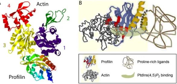
spontaneous polymerization (Pantaloni & Carlier, 1993). The crystal structure of the 12-16 kDa profilin has been solved both in the uncomplexed and in the actin:profilin heterodimeric state (Chik et al, 1996; Eads et al, 1998; Schutt et al, 1993; Vinson et al, 1993). The residues involved in actin binding are well established, consistent with its high affinity for G-actin, profilin was found to make extensive contact with a hydrophobic cleft situated opposite the nucleotide binding site at the barbed end of the actin molecule (see Figure 1.4A) (Chik et al, 1996; Schutt et al, 1993). Profilin also interacts with the membrane-anchored phospholipid phosphatidylinositol (4,5) bisphosphate (PIP2) and with poly-L-proline sequences found on many proteins (see Figure 1.4B) (Paavilainen et al, 2004; Pollard & Quirk, 1994;
Schmidt & Hall, 1998; Witke, 2004). PIP2-bound profilin is unable to bind G- actin, thus facilitating the release of actin monomers to the cortical regions which usually display high polymerization activity.
Figure 1.4. The crystal structure of profilin and its binding sites.
A: Crystal structure of a β-actin:profilin complex. Actin is shown in the “conventional view”, numbers and colors indicate the subdomains. Modified from Chik et al, 1996, PDB code 1HLU.
B: Molecular model of the major binding sites on the profilin molecule. The binding sites for actin (red) and poly-L-proline (yellow) are distinct, whereas the phosphatidylinositol (4,5)-bisphosphate-binding area occupies a larger part of the surface of the molecule (light green). Adapted from Witke, 2004.
Phosphorylation of PIP2 is regulated by a kinase/phosphatase system which is controlled by various signaling cascades responding to a broad range of stimuli. However, profilin also inhibits hydrolysis of PIP2 by phospholipase C,
thereby providing a possible mechanism for feedback auto-regulation and further signal transduction to other target systems (Machesky et al, 1990). To accelerate F-actin polymerization in the cortical region, G-actin-profilin complexes are directed to the plasma membrane by high-affinity binding to a large number of membrane-associated signaling proteins through their poly-L- proline regions (Mahoney et al, 1997; Perelroizen et al, 1994; Witke, 2004).
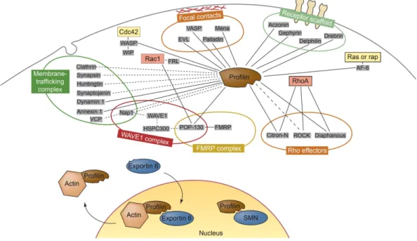
This is possible because the actin-profilin binding is not influenced by the interaction with poly-L-proline (Paavilainen et al, 2004; Witke, 2004). To date, more than 50 profilin ligands are known and this figure is steadily increasing. Thus, in addition to its crucial binding to G-actin, profilin is also one of the principle signal integrators and mediators at the interface of the cellular signaling system and the actin cytoskeleton (see Figure 1.5).
Figure 1.5. Network of molecular interactions of profilin.
Proteins that are known to interact with profilin are grouped according to their cellular location or the complexes in which they are found. Several links exist to small GTPases such as Rac1, RhoA, cdc42, Ras and Rap that are part of pathways that signal to the actin cytoskeleton. Direct interactions between profilin and the ligands are indicated by unbroken lines, whereas potentially direct interactions are indicated by broken lines. Abbreviations: AF-6, All-1 fusion partner from chromosome 6; EVL, Ena VASP like; FMRP, fragile X mental retardation protein; FRL, formin-related gene in leukocytes; HSP, heat-shock protein; Mena, mouse homolog of Drosophila enabled; POP, partner of profilin; SMN, survival of motor neuron; VCP, valosine-containing protein; WIP, WASP-interacting protein. Adapted from Witke, 2004.
1.1.3.1.3 DNase I
The enzyme deoxyribonuclease I (DNase I) is a secretory protein highly conserved in vertebrates. Its most prominent function is the endonucelolytic cleavage of DNA. However, the extracellular distribution throughout many tissues suggests additional functions (Lazarides & Lindberg, 1974; Peitsch et al, 1993; Yonezawa et al, 1990). Most intriguingly, DNase I specifically binds G-actin with a very high affinity (dos Remedios et al, 2003). The crystal structure of the actin:DNase I complex has been solved at high resolution (see Figure 1.6) (Kabsch et al, 1990). DNase I tightly binds to the eponymous loop on subdomain 2 at the pointed end of actin, thereby inhibiting the nuclease activity of DNase I (Hitchcock, 1980; Lazarides & Lindberg, 1974).
Figure 1.6. The crystal structure of actin in complex with DNase I.
The actin molecule (grey) is shown in the conventional view, the DNase I binding loop and subunit numbers are indicated. DNase I (red) binds to subdomain 2 at the pointed end of actin. Modified from Kabsch et al,1990.
This interaction has made DNase I an important analytical tool, as measuring the inhibition of DNase I allows the determination of the ratio of G-actin to F-actin with high precision (Blikstad et al, 1978; Malicka-Blaszkiewicz, 1986).
Fluorescently labeled DNase I is frequently used to visualize G-actin in live cells (Cramer et al, 2002). Remarkably, the physiological relevance of the actin:DNase I interaction is still largely unknown. As the major fraction of
DNase I is found in extracellular space where actin is scarce, it has been discussed that actin plays a role in protecting the cell from uncontrolled DNA degradation by intracellular DNase I prior to its secretion (dos Remedios et al, 2003).
1.1.3.2 Actin filament binding proteins
Once a specific manifestation of the microfilament cytoskeleton has been established, the cell must be able to control its stabilization or its quick remodeling, depending on the given situation. To stabilize filaments of constant lengths, proteins such as CP (capping protein) or tropomodulin are employed (Winder & Ayscough, 2005). While the former caps barbed ends, the latter is a potent pointed-end-capper. Moreover, capped filaments are frequently stabilized by the highly conserved tropomyosins which bind laterally over an extended range of the whole filament and protect it from the influence of depolymerizing, severing and branching factors (Blanchoin et al, 2001; Maciver et al, 2000).
Stable single filaments can be shaped into a dense network both by Arp2/3 mediated branching and by proteins such as filamin, spectrin and transgelin.
These cross-linking factors create a very dense, intricate meshwork of actin filaments (Winder & Ayscough, 2005). The parallel alignment and lateral stabilization of filaments into bundles and cables is brought about by bundling proteins. While α-actinin and fascin are responsible for the formation of loose filamentous bundles, other proteins, such as plastin and fimbrin produce bundles with higher stiffness and rigidity (Bartles, 2000;
Claessens et al, 2006; Winder & Ayscough, 2005).
F-actin interacting proteins also play an important role in the connection and anchoring of filaments to the other major cytoskeletal elements and the cytoplasmic membrane. Spectrin and plectin, for instance, link F-actin to intermediate filaments and microtubules. Anchoring to lipid membranes can occur by the binding to integral membrane proteins, mediated e.g. by dystrophin, vinculin and talin (Le Rumeur et al, 2010; Xu et al, 1998).
Alternatively, some proteins (e.g. annexins) can connect actin filaments
directly to phospholipids of the membrane (Hayes et al, 2004).
The importance of the rapid initiation of polymerization and the stabilization of existing filaments is equaled by the regulated disassembly of distinct portions of the actin network. To this end, cells usually follow a three-way approach of removing the tropomodulin cap, accelerating depolymerization at the pointed end and severing filaments. The major proteins facilitating the depolymerization of F-actin belong to the highly conserved ADF (actin depolymerizing factor)/cofilin (cosediments with filamentous actin) family of proteins that bind to F-actin and induce the dissociation of ADP-bound monomers from the pointed end (Lappalainen et al, 1998; McGough et al, 1997). While ADF/cofilins also exhibit a weak filament-severing activity, the main protein employed to break actin filaments in two is gelsolin. This highly flexible multi-domain protein is a very potent regulator of filament assembly and disassembly (Sun et al, 1999). After severing a filament, gelsolin caps the newly generated barbed end and prevents its elongation. Additionally, this activity of gelsolin is regulated through the interaction with various intracellular factors (Chou et al, 2002; Hartwig et al, 1995).
Many F-actin binding proteins do not directly influence the structure of the filamentous network, they rather interact with signaling pathways and mediate reconstruction by the interaction with other shape-modulating ABPs.
VASP (vasodilator stimulated phosphoprotein), for instance, is often found in cellular regions with a high actin turnover rate. It interferes with capping proteins thus ensuring the availability of free barbed ends for continuous polymerization (Bear et al, 2002). Through the interaction with profilin, VASP recruits polymerization-competent ATP-G-actin (Reinhard et al, 1995).
Another important signal intergrator is WIP (WASp interacting protein). It was discovered via its regulation of the WASp mediated control of filament nucleation, effectively blocking actin polymerization (Martinez-Quiles et al, 2001; Vetterkind et al, 2002). This activity can be reversed by the interaction with cortactin (Kinley et al, 2003). Covering F-actin along its entire length, cortactin connects many actin related signaling pathways to the filament network through its multi-domain modular organization (Pant et al, 2006).
Recently, cortactin has been found to modulate the appearance of actin filaments on its own by initiating the formation of flat sheets of F-actin (Cowieson et al, 2008).
1.1.3.3 Concluding remarks on actin binding proteins
A recurring theme of ABPs is their modular polypeptide structure and their ability to interact not only with actin but also with a number of other cellular factors creating a complex system of functional interdependence, cross-talk and feedback regulation. The most potent proteins responsible for dramatic changes of the network architecture are particularly well-embedded in signaling and regulatory networks. Profilin, the Arp2/3-WASp system and formin as the main factors promoting actin polymerization are tightly regulated, multi-modular proteins. The same holds true for the potent F-actin disassemblers, ADF/cofilin and gelsolin and also for the large class of myosin motor proteins. The involvement of actin in a multitude of protein-protein interactions exerts significant evolutionary constraint on its structure and sequence. Indeed, the observation that no other known protein participates in more protein-protein interactions than actin (Dominguez, 2004) and the fact that actin is among the most conserved proteins in evolution are intimately related (Erickson, 2007). This notion has even been further substantiated in the last decade by the discovery and characterization of prokaryotic actins.
1.2 Prokaryotic actins
Long believed to be restricted to eukaryotes, prokaryotic actin homologs have been discovered by the identification of conserved amino-acid residues derived from structure-based alignments of core motifs comprising what is now called the “actin fold” (Bork et al, 1992). This ground-breaking study was initiated bearing in mind the observation that the similarity actin shares with sugar kinases and hsp70 chaperones is immediately recognizable at the structural level while being virtually undetectable in amino acid sequence comparisons.
Therefore, a database search based on three-dimensional structural alignments was performed (Bork et al, 1992). Prokaryotic proteins identified to be structurally related to actin were FtsA, MreB and ParM. The first two were then known to be involved in the regulation of cytokinesis and in cell shape determination of rod-shaped cells, respectively. ParM had been found necessary for the faithful replication and segregation of the E. coli plasmid R1. Early investigations into the newly discovered actin-like proteins, primarily, the elucidation of high-resolution crystal structures, revealed that all three proteins are true actin homologs (see Figure 1.7). Despite only showing a degree of sequence identity of about 14% with each other and eukaryotic actin, each of the three bacterial actin candidate proteins adopts the typical bi-lobed actin fold with 4 clearly distinguishable subunits. Thus, their homology with actin was widely accepted. This finding soon triggered the search for the roles these newly found actins played in their host organisms.
Figure 1.7. Crystal structures of actin, MreB, ParM and FtsA.
Structures are shown in the conventional view, corresponding subdomains are in the same color, subdomain classifications are given. Data are from Otterbein et al, 2001 (actin); van den Ent et al, 2001 (MreB); van den Ent et al, 2002 (ParM) and van den Ent & Löwe, 2000 (FtsA).
1.2.1 Bacterial cytokinesis: FtsA
Although FtsA shows the typical bi-lobed actin structure, subdomain 2 (here termed 1C) is misplaced when compared to the actin archetype, apparently having “swung out” towards the barbed face of the protein (van den Ent &
Löwe, 2000). In many bacteria, FtsA is involved in positioning the Z-ring during cytokinesis. The main component of the Z-ring is FtsZ, a bacterial homolog of the eukaryotic microtubule-forming protein tubulin (see Figure 1.8A) (Ben-Yehuda & Losick, 2002; Erickson, 1998; van den Ent et al, 2001a).
FtsZ is targeted to the future site of cell constriction through an elaborate cascade of many protein-protein interactions. Being part of that cascade, FtsA bridges FtsZ to the cytoplasmic membrane and possibly to other components of the cytokinesis machinery (see Figure 1.8D) (Pichoff &
Lutkenhaus, 2005; van den Ent & Löwe, 2000; Yan et al, 2000). Despite clear indications that FtsA monomers interact with each other, the question whether polymeric filaments are formed is still unresolved. There have been reports of corkscrew-like aggregations formed in vitro, however, their physiological relevance remains speculative and no FtsA polymers have been confirmed in vivo (see Figure 1.8B and C) (Lara et al, 2005; van den Ent &
Löwe, 2000).
Figure 1.8. FtsA and the cell division protein FtsZ.
A: GFP-tagged Z-rings at the division site in Bacillus subtilis. The membrane is stained red, scale bar: 1 µm. Adapted from Ben-Yehuda & Losick, 2002.
B and C: Electron micrographs of FtsA polymers formed in vitro. B shows an overview, scale bar: 100 nm. C shows filaments in detail, scale bar: 20 nm. Adapted from Lara et al, 2005.
D: Model for the role FtsA plays in the assembly of the Z-ring. FtsZ polymers are linked to the membrane through FtsA. Elongation of membrane-linked polymers is subject to an elaborate control system. Growth is restricted to the division plane and to a direction perpendicular to the long cell axis.
Adapted from Pichoff & Lutkenhaus, 2005.
1.2.2 Plasmid segregation: ParM and AlfA
By the time of its identification as a putative bacterial actin, ParM was known to be essential for the faithful segregation after replication of the E.coli plasmid R1. The crystal structure of ParM and its ability to polymerize into double-stranded helical filaments have demonstrated some similarities with eukaryotic actin (van den Ent et al, 2002). In analogy to actin, ATP-ParM shows a higher tendency to polymerize than ADP-ParM. Also, ATPase activity is induced by filament formation and ADP-ParM rapidly dissociates from filament ends (Garner et al, 2004). Further analyses have shown, however, that the evolutionary distance between actin and ParM is reflected in a number of fundamental differences. The spontaneous nucleation of ParM filaments, for instance, occurs rather rapidly and is believed to constantly take place in vivo. Moreover, ParM filaments have, in contrast to eukaryotic actin, a left-handed helical twist, they grow bidirectionally at equal rates and show a length adaptation to the bacterial cell, terminating elongation at about 1.5 µm in vitro (Garner et al, 2004; Garner et al, 2007; Orlova et al, 2007). Three plasmid loci are required for the partitioning after replication:
parM (the “partition motor”), parR and parC.
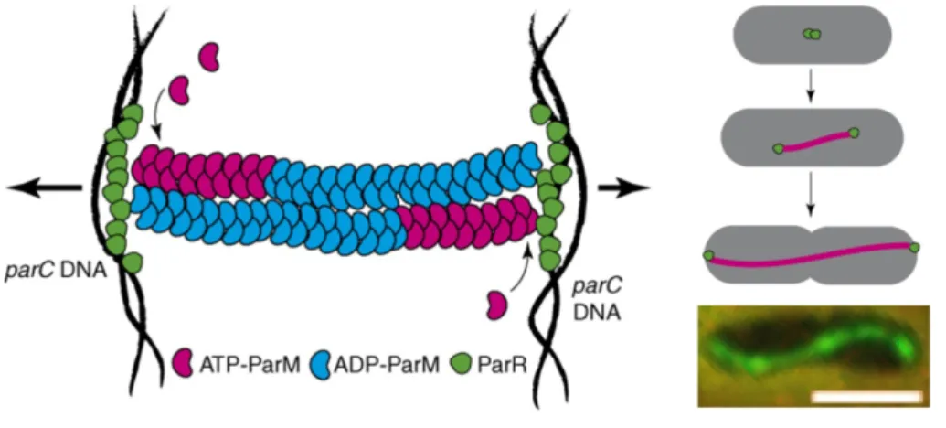
Figure 1.9. ParM filament formation and plasmid DNA segregation.
ParM forms a filament bundle in-between duplicated plasmids that elongates as plasmids segregate towards the poles. Filaments are bound through ParR (green) to a specific site on the plasmid, parC. Insertion of ATP-ParM (red) at the growing tip of filaments is proposed to exert force on the plasmids, pushing the copies apart (arrows). Owing to its hydrolysis activity ADP-ParM (blue) accumulates in the filament. The right hand side shows an in vivo model (top) and an anti-ParM immunofluorescence image of an E.coli cell (bottom, scale bar: 2 µm). Modified from Dye & Shapiro, 2007 (schematic models) and Møller-Jensen et al, 2002 (micrograph).
The ParR protein binds to ParM filament ends thus terminating depolymerization while actively adding new ParM monomers, displaying a
“formin-like” activity (see 1.1.3.1.1 Inhibitors and activators of actin polymerization, p10) (Garner et al, 2004; Garner et al, 2007; Moller-Jensen et al, 2002). Additionally, ParR binds to the centromer-like locus parC forming a simple analog to the eukaryotic mitotic spindle (see Figure 1.9) (Dye &
Shapiro, 2007; Møller-Jensen & Gerdes, 2004; Møller-Jensen et al, 2002).
The recently discovered AlfA protein from Bacillus subtilis has some properties strongly resembling ParM and apparently segregates plasmids in a very similar way. However, AlfA filaments show considerable structural differences compared to ParM (see Figure 1.10) (Becker et al, 2006; Polka et al, 2009; Popp et al, 2010a). Recently, the plasmid encoded loci alfB and alfC were discussed to perform functions similar to ParR and parC, respectively (Popp et al, 2010b).
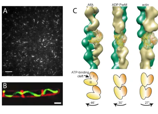
Figure 1.10. Filaments formed by AlfA.
A: TIRF micrograph of Cy3-labeled AlfA showing stable filamentous bundles. Scale bar: 10 µm.
B: GFP-AlfA filaments (green) formed in Bacillus subtilis cells. Membranes are stained with FM 4-64 (red). Scale bar: 1 µm.
C: Architecture of filaments of AlfA (left), ADP-ParM (middle) and actin (right). AlfA and ParM are left-handed, actin is right-handed. A single filament subunit is transparent, with the crystal structure of ParM fitted into the model. Cartoons (bottom) indicate the orientations of subunits in the filaments; the magnitude and direction of the rotation between subunits moving up each strand are indicated by labeled arrows. A and C modified from Polka et al, 2009, B adapted from Becker et al, 2006.
1.2.3 Wide-spread and multifunctional: MreB
MreB was originally characterized as part of the mre (murein cluster e) locus involved in cell shape regulation in E.coli (Doi et al, 1988). Although it had been identified as a potential actin homolog, research interest had long been hesitant owing to MreB’s low sequence similarity to actin. A dramatic turnaround was brought about by the discovery that MreB forms filaments in a nucleotide dependent manner (Jones et al, 2001; van den Ent et al, 2001b).
Many laboratories have since worked at uncovering the functions and molecular mechanisms connected with MreB. A thorough understanding of MreB similar in extent to the comparably simple ParM system is impeded by the fact that MreB is the most prevalent prokaryotic actin involved in a broad range of sometimes species-specific cellular processes (Shaevitz & Gitai, 2010).
1.2.3.1 General features of MreB
An mreB gene is found in the vast majority of non-spherical bacteria, with some species, mainly gram-positives, encoding multiple homologs (Cabeen &
Jacobs-Wagner, 2010; Daniel & Errington, 2003). While some rod-shaped bacteria (e.g. Rhizobiae, Mycobacteria, Mycoplasmas) lack MreB, it is found in some spherical members of the Cyanobacteria and Planctomycetes (Shaevitz & Gitai, 2010). The considerable sequence variation of MreBs commands caution in generalizing its function. Rather, the view has emerged that some very basic properties of MreB are applied by different bacteria to specific cellular processes. The unifying feature, of course, is the ability to form filamentous polymers. MreB protofilaments seem to assemble in a straight fashion rather than exhibiting the typical actin twist and have the tendency to spontaneously align into bundles and ribbons with mixed polarities (Shaevitz & Gitai, 2010; van den Ent et al, 2001b). Depending on the polymerization conditions, MreB can also form ring-like assemblies and sheets of diagonally interwoven filaments of about 1-5 µm (see Figure 1.11A) (Esue et al, 2005; Popp et al, 2010c). Finally, a general feature of MreB essential for its physiological function is its high turnover rate with the tendency to treadmill (Carballido-Lopez & Errington, 2003a; Defeu Soufo &
Graumann, 2004; Defeu Soufo & Graumann, 2006; Jones et al, 2001; Kim et al, 2006; Srinivasan et al, 2007).
1.2.3.2 MreB and cell shape determination
Mutational studies have shown that an MreB knock-out is either lethal or severely impairs cell viability (Cabeen & Jacobs-Wagner, 2010; Daniel &
Errington, 2003). At any rate, mreB mutants usually display aberrant cell shapes, such as the formation of spherical cells in rod-shaped bacteria (Graumann, 2004; Hu et al, 2007). Many insights into the role of MreB cell shape determination stem from Bacillus subtilis where two additional proteins with homology to MreB are found: Mbl and MreBH. All three assemble in vivo into dynamic helical “cables” that are, much as the filaments formed by actin, being constantly remodeled by treadmilling, moving with a velocity of about 0.1 µm per second just beneath the cellular surface (see Figure 1.11B and C) (Amos et al, 2004; Carballido-Lopez & Errington, 2003a; Defeu Soufo
& Graumann, 2005; Graumann, 2004; Jones et al, 2001). The helical superstructures formed by the individual MreB homologs in Bacillus subtilis show variations in length, pitch and cellular localization suggesting that they control different aspects of growth and cell shape (Daniel & Errington, 2003;
Defeu Soufo & Graumann, 2004; Graumann, 2004; Jones et al, 2001).
Figure 1.11. MreB filaments in vivo and in vitro.
A: Electron micrographs of MreB filaments formed in vitro. Single protofilaments assemble into pairs but do not twist around each other (bottom), the inset is an enlarged filtered image, the arrows show surface borders of individual filaments. Protofilaments can form flat sheets (top left) or ring-like structures (top right). Scale bars: 100 nm; adapted from Amos et al, 2004.
B: Dynamic GFP-Mbl helices in Bacillus subtilis. Scale bar: 4 µm; adapted from Carballido-Lopez &
Errington, 2003a.
C: Schematic representation of dynamic MreB helices in rod-shaped bacteria. Adapted from Graumann, 2004.
MreB and Mbl are thought to exert their influence on cell shape mainly through the regulation of the synthesis of new cell wall material whose insertion into a growing cylindrical peptidoglycan layer also follows comparable helical patterns (Carballido-Lopez & Errington, 2003b; Daniel &
Errington, 2003; Figge et al, 2004; Kruse et al, 2005; Scheffers et al, 2004; van den Ent et al, 2010). Similar observations were made with MreB in E.coli and Caulobacter crescentus suggesting a general role in cell shape determination (Kim et al, 2006; Takacs et al, 2010; Uehara & Park, 2008; Varma et al, 2007a; Varma & Young, 2009). It has been shown recently that in addition to its influence on cell wall synthesis the inherent stability of the helical MreB bundles mechanically contribute to the bending stiffness of E.coli cells (Wang et al, 2010).
1.2.3.3 The role of MreB in DNA replication and segregation
Knock-out strains have shown that MreB is required for proper segregation of chromosomes after replication (Gitai et al, 2004; Kruse et al, 2003; Soufo &
Graumann, 2003). However, this matter remains controversial as its exact function and the underlying mechanisms are still unclear. Since they bind to chromosomal origins of replication, MreB filaments have been proposed to actively push chromosomes apart in a ParM-like fashion (Gerdes et al, 2004;
Gitai et al, 2005; Graumann, 2004). Alternatively, MreB filaments may recruit the segregation machinery to the vicinity of the chromosome (Defeu Soufo & Graumann, 2005; Kruse et al, 2006). An example of this may be the suggested motor-like DNA segregating function of RNA polymerase that interacts with and is localized by MreB (Kruse et al, 2006). It is difficult, however, to directly attribute segregation defects in MreB-deficient cells to a primary role MreB plays in chromosome partitioning, as it is also involved in DNA replication (Defeu Soufo & Graumann, 2005; Munoz-Espin et al, 2009;
Shebelut et al, 2009). Additionally, MreB regulates DNA decatenation through the interaction with topoisomerase IV (Madabhushi & Marians, 2009).