Roth: Automated amino acid analysis with sensitive fluorescence detection 361 J. Clin. Chem. Clin. Biochem:
Vol. 14,1976, pp. 361-364
Automated Amino Acid Analysis with Sensitive Fluorescence Detection
By M RothLaboratoire central, Höpital cantonal, 1211 Geneva 4 (Switzerland) (Received November 3, 1975/April 5, 1976)
Summary:The fluorigenic ö-plithalaldehyde-mercaptoethanol reagent gives good reproducibility with a very stable baseline when applied to the automated analysis of amino acids at the rianomole level. Determination of even smaller quantities is possible; basic amino acids are then preferably eluted separately at constant pH (for example pH 6.0); this eliminates the baseline irregularities that occur with single column systems at high sensitivity settings.
The reagent gives excellent results in the assay of small quantities of biological fluids such as blood plasma.
Automatische Aminosäuren-Analyse mit empfindlichem Fluoreszenznachweis
Zusammenfassung: Das Fluoreszenzreagenz o-Phthalaldehyd-Mercaptoethanol liefert gut reproduzierbare Resultate und eine sehr stabile Basislinie bei der automatischen Aminosäurenanalyse im Nanomolbereich. Auch noch kleinere Mengen lassen sich bestimmen; basische Aminosäuren werden dann am besten bei konstantem pH-Wert (z. B. 6,0) eluiert; dadurch werden Unregelmäßigkeiten in der Basislinie, die im Einsäulenverfahren bei höchster Empfmdlich- keitseinstellung auftreten, vermieden. Das Reagenz eignet sich vorzüglich zur Analyse kleinster Mengen biologischer Flüssigkeiten wie Blutplasma.
Introduction
New fluorigenic reactions developed in recent years open interesting perspectives in the automatic analysis of amino acids. Some of them are much more sensitive than the colorimetric riinhydrin reaction; this is especially useful in new microanalytical systems operating with small particles of ion exchanger and microbore columns and in cases where only small quantities of sample are avail- able. Among the different reagents that have been reported to give fluorescent products with amino acids (1—8), the one consisting of a mixture of o-phthalalde- hyde and 2-mercaptoethanol in alkaline buffer is parti- cularly suitable for automatic amino acid analysis, because it combines the advantages of good solubility in water, fast rate of reaction at ordinary temperature and high sensitivity (5--7); The present paper presents a procedure using this reagent.
Materials and Methods Instruments
Amino acid analyses were performed with a Liquimat automatic analyzer (Labotron, 8191 Getting, W. Germany) equipped with two pumps and a 345 X 4.5 mm column filled with a Durrüm DC-6 strongly acidic ion exchanger. Average pumping rates were
19 mi/h for buffers and 23 ml/h for the phthalaldehyde reagent.
The detector was an Aminco fluoro-colorimeter equipped with a 70 quartz flow-cell (0.2 cm inside diameter) and a General Electric F4J4/BL source lamp. The liquid was made to flow up- wards through the cell. The primary and secondary filters were Corning 7-60 and 3-73, respectively. A W + W linear recorder (W+ W Electronics, Basle) was used with sensitivity set between 2.5 and 20 mV. In a few experiments, a 3380 A Hewlett-Packard calculating integrator was also connected to the fluorimeter.
Elution buffers
The first and second buffer (pH 3.07 and 4.25, respectively) contained 19.4 g trisodium citrate dihydrate, l g phenol, 200 mg Brij-35, 900 ml water, HC1 to adjust the desired pH, and water to make 1 litre. The third buffer contained 14.7 g trisodium citrate dihydrate, 37.4 g NaCl, l g phenol, 200 mg Brij-35, 900 ml water, HC1 to bring the pH to 6.0, and double distilled water to make 1 litre.
o-phthalaldehyde reagent
The concentration of mercaptoethanol was higher than in the previously reported reagent (6), since this has been shown by Benson I Hare (7) to improve the sensitivity for certain com- pounds. In addition to borate, disodium phosphate and sodium Kdroxide were incorporated in the solution to increase its
l. After disso- ution, 250
Na^HPO* and NaOH to give pH 10.0), 25
alid water to m^e 500 ml were added. This reagent may be kept S Äee daTat room temperature. For the separate analysis
J. Clin. Chem. Clin. Biochem. / Vol. 14,1976 / No. 7
362 Roth: Automated amino acid analysis with sensitive fluorescence detection
Tab. 1. Elution and regeneration programs for amino acid analysis
Solution pumped Temper- Program Program
ature 1 2 pH 3.07 citrate buffer
pH 3.07 citrate buffer pH 3.07 citrate buffer pH4.25 citrate buffer pH 6.00 citrate buffer pH 6.00 citrate buffer 0.2 mol/lNaOH pH 3.07 citrate buffer
25°C 43°C58°C 58°C 43°C58°C 58°C 25°C
8 min 20 min 40min 44min -- 24min 32min
8 min 20min 40 min 44 min 40 min 68 min 24 min 32min of basic amino acids, the 25 ml of 1 mol/1 NaOH are omitted and 500 mg Brij-35 are incorporated in the reagent.
Reference mixture
A Beckman solution containing 2.5 μπιοΐ/πύ each of the following substances was used: Asp, Thr, Ser, Glu, Gly, Ala, V2Cys, Val, Met, He, Leu, Tyr, Phe, Lys, His, NH4+ and Arg.
It was diluted before injection with an appropriate volume of citrate buffer, pH 2.2.
Procedure
A single elution system for all amino acids has proved satis- factory for amounts > 1 nanomole. It includes a temperature decrease step in order to remove an ammonia plateau (due to traces of ammonia in the first and second buffers) from the lysine and histidine peaks. The program is shown in table 1.
To ensure a fast and reproducible temperature decrease, the water bath of the instrument incorporates a coil cooled by tap water. When quantities less than one nanomole are analyzed with this program, satisfactory results are still obtained for acidic and neutral amino acids, but the higher sensitivity setting causes troublesome fluctuations of the baseline during elution of basic amino acids with the third buffer, and the evaluation of these peaks becomes less precise. Separate elution of basic amino acids at constant pH (5.8 or 6.0), on the other hand, pro- vides a stable baseline from which the emergence of extremely small peaks is easily detected.
The flow diagram corresponds to that reported previously (6).
Injections were made with the Labotron loop system, with volumes of 27 or 57 μ\.
Results
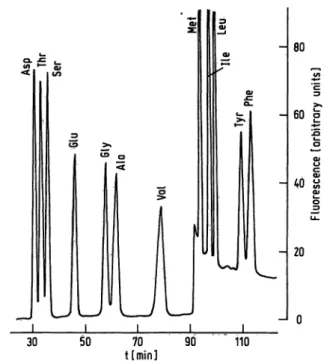
Figure 1 shows the Chromatographie separation of 0.5 nanomole each of the acidic and neutral amino acids from the reference mixture. Proline and cysteine, also present in the sample, are not detected. Excellent base- line stability is observed during elution with the first buffer. A sharp baseline rise appearing at 91 min is due to material released from the column by the second buffer. Its excitation and fluorescence characteristics are identical with those given by amino acids or primary amines. This jump is therefore not due to ammonia, which would yield a different fluorescence (9).
Figure 2 shows the record after injection of 0.5 nmol each of the same amino acids into a constant elution buffer stream of pH 6.0. After 18 min, when the major portion of the acidic and neutral amino acids have been eluted, tyrosine, phenylalanine, lysine, histidine, tryptophan and arginine appear as well separated peaks emerging from a stable baseline. The recovery of tryptophan is
80
60 σ
to y, I
20
-J 0
30 50 70
timin] 90 110
Fig. 1. Analysis of 0.5 nmol each of acidic and neutral amino acids. Program No. 1. Sample: reference mixture.
-180
60
e
S
«•2
20
0L
Fig. 2.
20 40 60
t [min] 100 120
Analysis of 0.5 nnioi each of aromatic and basic amino acids. Single elution buffer of pH 6.O. Constant temper- ature of 59°C. Sample: reference mixture.
much better than in a three-buffer system, because its destruction by the more acidic buffers is avoided.
Ammonia appears after 77 min as a small peak. The response is low because the filters do not correspond to the optimal excitation and fluorescence wavelengths shown by ammonia, which are 410 and 470 nm, respec- tively (9). If one is specifically interested in ammonia, appropriate filters may be used.
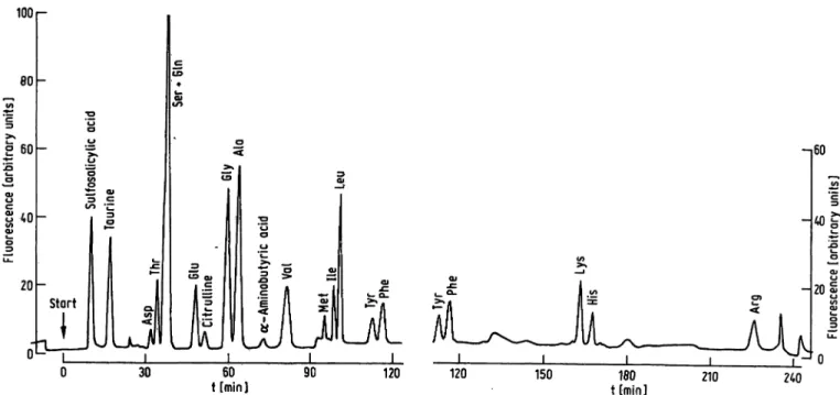
The results obtained with a sample of human blood plasma are shown in Figure 3 a and 3b. The first peak appearing in the chromatogram is due to sulfosalicylic acid, which was used as the deproteinizing agent.
Although its excitation and fluorescence spectra are quite different from those obtained with amino acids, sufficient interfering light passed the filters to produce this peak. The figure demonstrates the good resolution
J. Clin. Chem. Clin. Biochem. / Vol. 14,1976 / No. 7
Roth: Automated amino acid analysis with sensitive fluorescence detection 363
1001-
-,60
30 t [min] 1ΘΟ
t [min]
Fig. 3 a and 3b. Analysis of amino acids in normal human blood plasma. To 100 μΐ heparinized blood were added 3 mg of s u If ο- salicylic acid. After mixing and centiifuging, an aliquot of the supernatant was mixed with 4 volumes of citrate buffer, pH 2.2, and 57 μΐ of the resulting solution were injected into the Chromatograph. Single-column procedure with program No. 2.
and sensitivity achieved in the analysis of small quantities of biological samples.
Substances other than α-amino acids, e.g. taurine, phosphoserine and phosphoethanolamine, react fairly well with the 0-phthalaldehyde-mercaptoethanol reagent.
This is also the case with -2-thienylalanine and a-amino-
£-guariidinopropionic acid which can be conveniently used as internal standards in the procedure. On the other hand, urea does not interfere.
For quantitative analysis, one can use the same principles as with systems using ninhydrin, with the advantage, how- ever, that the response of the detector does not need to be linearized, since, within defined limits, fluorescence is proportional to concentration.
A Hewlett-Packard electronic integrator, which could be made available for a short period, was used in some ex- periments. It was not possible to perform a systematic study of the reproducibility, but the data obtained pro- vide nevertheless an idea of the precision attainable.
Thus, in two consecutive runs with 1.4 nanomole of each aiiiino acid, differences in relative peak areas (in- ternal standard: glutamic acid) were: < 2% for Thr, Ala;
2-3% for Ser, Leu, Tyr; 3-4% for Asp, Val, He, Phe;
and 4—5% for Gly and Met. Seven chromatograms carried out on a shorter column (25 cm) with 1.4 nanpmole of each amino acid gave a less satisfactory resolution, but provided relative standard deviations of the same order:
al nme 1.5%; serine 2.1 %; valine 3.2%; aspartic acid 3.3% and threonine 3.6%. The methionine peak must be sufficiently distinct from the preceding buffer peak, otherwise the instrument will integrate both areas to- gether.
The fluorescence yield particular to each amino acid depends on the composition of the phthalaldehyde
Tab. 2. Relative fluorescence from amino acids as eluted with program 2. The values are the relative integrated peak areas with glutamic acid taken as 100. Sample: refer- ence mixture with cysteic acid and 0-2-thienylalanine added; 2 nmol of each compound.
Substance Cysteic acid Aspartic acid Threonine Serine Glutamic acid Glycine Alanine Valine Methionine Isoleucine Leucine
j3-2-thienylalanine Tyrosine
Phenylalanine Lysine Histidine Arginine
Relative fluorescence 9781
11394 101100 10185 11285 103111 10880 105109 89
reagent and the pH of reaction. It is established by calibration of the system with a reference mixture of amino acids. Since the final pH is determined by the proportions of effluent and reagent, it is essential that the flow rates be the same both for calibration and analysis of unknowns. Table 2 gives the relative fluorescence intensities provided by such a calibration run.
Discussion
The o-phthalaldehyde-mercaptoethanol reagent offers several advantages over ninhydrin for the automatic analysis of amino acids. Its high sensitivity permits J. Clin. Gherh. Clin. Bipchem. / Vol. 14,1976 / No. 7
364 Roth: Automated amino acid analysis with sensitive fluorescence detection
precise determinations to be made at the nanomole level, and quantities of the order of 50 picomoles to be detected. Since the reaction is fast and does not require heating, the time taken by an amino acid to travel from the column to the detector is short. This prevents enlargement of the peaks by diffusion. The solubility of o-phthalaldehyde in water is better than that of ninhydrin, and we never experienced clogging of the tubes or the pump by the reagent.
The precision of quantitative analysis depends greatly on the stability of the baseline. Difficulties may occur with buffer peaks if they appear simultaneously with a compound to be determined. For the quantitative determination of lysine and histidine at the level of 500 pmol or less, elution at constant pH therefore proves to be more convenient than stepwise pH gradient elution.
Since fluorescence intensity depends on the intensity of the excitation source, the stability of the lamp is essen- tial for reproducible results. We did not observe marked changes in source intensity during single Chromato- graphie runs. A small drift, however, is always possible.
Better reproducibility may be expected from the use of a ratio fluorimeter. An internal standard should be included in every chromatogram, since the values provided by the fluorimeter are not absolute.
When applied to blood plasma, the procedure detects the same compounds as ninhydrin, except proline, hydroxy- proline and cystine. The advantages are the small amount of sample required and the ease with which compounds present in only minute amounts are detected.
The good precision obtained with the 0-phthalaldehyde reagent at the nanomole level will presumably prove very
useful for the analysis of protein and peptide hydrolyzates, for their valuable assistance.
especially when only a few micrograms of polypeptide are available for analysis. A comparison of the o-phthalalde- hyde reagent with fluorescamine has been made by Benson
&Hare (7). They found phthalaldehyde to be more con- venient because of its solubility and stability in water.
The present disadvantage of the reagent over ninhydrin is that it does not detect proline and hydroxyproline, and that the reaction with cysteine gives only poor fluor- escence. Lysine yields a good fluorescence if a surfactant like Brij is added to the buffer or the reagent (7,10). The failure of cysteine to provide sufficient fluorescence may be overcome by oxidation to cysteic acid, which yields a highly fluorescent peak early in the chromatogram.
Proline and hydroxyproline, if subjected to oxidation by active chlorine or Pb02, are transformed into com- pounds giving the fluorescence reaction. There is a problem in the control of the extent of oxidation, which should be made reproducible. Another approach which we are presently investigating uses the reagent 7-chloro- 4-nitro-benzofurazan first described by Ghosh & White- house (1). This gives a fairly good fluorescence with proline and hydroxyproline and can be added to the effluent subsequent to the reaction with 0-phthalalde- hyde. The sensitivity achieved is superior to that with ninhydrin, although not so high as that yielded by the Orphthalaldehyde reagent with -amino acids.
One can, thus, hope that a sensitive automatic fluori- metric analysis of all protein amino acids will be pos- sible in the near future.
Acknowledgement
I thank Miss Sylviane Jaccoud and Mrs. Mary-Claude Verlyck
References
1. Ghosh, P. E. & Whitehouse, M. W. (1968), Biochem. J. 108, 155-156.
2. Samejima, K., Dairman, W., Stone, J. & Udenfriend, S.
(1971), Anal. Biochem. 42, 237-247.
3. Stein, S., Bohlen, P., Stone, J., Dairman, W. & Udenfriend, S. (1973), Arch. Biochem. Biophys. 755, 203-212.
4. Lange, H.-W., Lustenberger, N. & Hempel, K. (1972), Z. Anal. Chem. 261, 337-342.
5. Roth, M. (1971), Anal. Chem. 43, 880-882.
6. Roth, M. & Hampai, A. (1973), J.' Chromatogr. 83, 353 -356.
7. Benson, J. R. & Hare, P. E. (1975), Proc. Nat. Acad. Sei.
USA 72, 619-.622.
8. Maeda, M. Tsuji, A., Ganno, S. & Onishi, Y. (1973), J.
Chiomatogr. 77, 434-438.
9. Taylor, S., Ninjoor, V., Dowd, D. M. & Tappel, A. L. (1974), Anal. Biochem. 60,153-162.
10. Schwabe, C. & Catlin, J. C. (1974), Anal. Biochem. 61, 302-304.
PD Dr. M. Roth Laboratoire central Höpital cantonal CH-1211 Geneve 4
J. Glin. Chem. Clin. Biochem. / Vol. 14,1976 / No. 7