Isolated flexor pollicis longus nerve fascicle lesion – a rare differential diagnosis of thumb flexion deficiency
Isolierte Nervenläsion des Flexor pollicis longus-Faszikels – eine seltene Differentialdiagnose bei Ausfall der Daumenbeugung
Abstract
A rare differential diagnosis of thumb flexion deficiency is an isolated flexor pollicis longus (FPL) nerve fascicle lesion. We present a 42-year-
Eva Glauser
1Andreas Gohritz
1old otherwise healthy female patient who developed a weak thumb-to-
Jan Fridén
2index pinch and deficient right thumb flexion following the removal of
Dirk J. Schaefer
1osteosynthesis plates after a forearm fracture. Clinically,the flexor pol- licis longus function was absent, yet index flexion and sensibility were unimpaired. Tendon rupture was excluded using a tenodesis test and
1 Klinik für Plastische, Rekonstruktive, Ästhetische the electro-physiological result of isolated interosseus nerve fascicle
lesion was confirmed intraoperatively by inspection and electrostimula-
und Handchirurgie, tion. Tendon transfer using the extensor carpi radialis longus reconstruct Universitätssspital, Basel,
Switzerland strong thumb flexion during pinch. In summary, due to its specific loca-
tion and anatomy, the FPL branch is more prone to isolated neuropathy,
2 Handchirurgie, Schweizer Paraplegiker-Zentrum, Nottwil, Switzerland e.g. by injections or operations, than to other fascicles of the anterior
interosseus nerve. When confronted with sudden and isolated thumb flexion deficiency, specialists should be aware of this rare phenomenon.
Keywords:anterior interosseous nerve, thumb flexion, flexor pollicis longus, isolated
Zusammenfassung
Bei Verlust der Daumenbeugung ist die isolierte Läsion des Nervenastes zum Flexor pollicis longus (FPL) eine seltene Differentialdiagnose. Wir stellen eine 42-jährige, sonst gesunde Patientin vor, die nach Entfernung von Osteosynthese-Platten infolge einer komplizierten Unterarm-Fraktur eine Greifschwäche zwischen Daumen und Zeigefinger mit komplettem Ausfall der Daumenendgelenksbeugung entwickelte. Klinisch lag ein Ausfall der FPL-Funktion vor, Zeigefingerbeugung und Sensibilität waren unbeeinträchtigt. Eine Sehnenruptur konnte per Tenodese-Test ausge- schlossen werden, das elektophysiologische Untersuchungsergebnis einer isolierten FPL-Faszikelläsion wurde durch intraoperative Inspektion und Elektrostimulation bestätigt. Eine motorische Ersatzoperation mittels Extensor carpi radialis longus-Transposition stellte einen kräftigen Kneifgriff wieder her. Zusammenfassend ist der FPL-Faszikel aufgrund seiner exponierten Lage und spezifischen intraneuralen Anatomie an- fälliger für isolierte Schädigung, z. B. durch Injektionen oder Operationen, als andere Anteile des N. interosseus anterior. Im Falle eines plötzlichen Verlust der Daumenbeugung sollte dieses seltene Phänomen jedem Spezialisten bewusst sein.
Schlüsselwörter:Nervus interosseus anterior,
Daumenendgelenksbeugung, Flexor pollicis longus, isoliert
1/4 GMS German Plastic, Reconstructive and Aesthetic Surgery 2016, Vol. 6, ISSN 2193-7052
Case Report
OPEN ACCESS
Introduction
Loss of pinch power and thumb flexion can be caused by many traumatic or atraumatic origins, most frequently by carpal tunnel syndrome, flexor tendon injuries or anterior interosseous nerve (AIN) compression. A rare, but relevant differential diagnosis is an isolated flexor pollicis longus (FPL) nerve fascicle lesion. The objective of this paper is to illustrate this phenomenon with a clinical case and provide a discussion of its anatomical, clinical background and differential diagnosis of incomplete AIN syndrome in the literature.
Clinical case
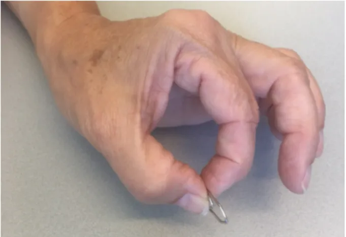
A 42-year-old otherwise healthy secretary was referred to our hand surgery department with the diagnosis of carpal tunnel syndrome for operative carpal tunnel re- lease of the non-dominant left hand. She presented with weakend pinch and impaired control of the right dominant thumb due to deficient interphalangeal flexion. Symptoms had started after the removal of osteosynthesis plates about a year after a complicated ulna and radius fracture in 2008. Clinically, FPL muscle function was entirely lost while index finger flexion was intact (incomplete O-sign) (Figure 1) and sensibility was clinically unimpaired with a two-point discrimination of 5–6 mm.
Figure 1: Incomplete O (or circle) sign due to paralysis of the FPL, yet preserved distal interphalangeal flexion of the index
finger
The FPL tendon was found intact on ultrasound but an atrophy of the muscle belly was noted. A tenodesis test furtherly excluded a rupture of the FPL tendon. Neuro- electrophysiology revealed preserved interosseus nerve function, yet complete paralysis of FPL muscle which could be confirmed during intraoperative inspection and electro-stimulation of the muscle.
Due to the atrophy of the FPL several years after dener- vation, tendon transfer was performed to restore FPL function. The extensor carpi radialis longus (ECRL) was chosen due to its proximity, favourable myo-architectural properties and synergistic action.
At follow-up after 15 months postoperatively the patient demonstrated interphalangeal flexion with fine motor skills and and pinch of 7 kg restored (Figure 2).
Figure 2: Restored thumb interphalangeal flexion (complete circle sign) and strong pinch after ECRL-pro-FPL tendon transfer
Discussion
This case report illustrates an exceptional, but anatomic- ally interesting and clinically relevant cause of sudden loss of thumb interphalangeal flexion. Thumb to index pinch may become deficient due to a great variety of causes, most obviously by tendon ruptures due to acute trauma, inflammation, e.g. rheumatoid arthritis, degener- ation caused by exostosis with pseudoarthritis of the scaphoid bone, necrosis of the lunate bone, stenosing tendovaginitis or trigger finger and tenodesis sequelae [1]. Lesions of the anterior interosseus nerve (AIN) can result in various clinical manifestations, depending on location and degree of axonal damage. In 1918, Tinel [2]
first described paralysis of the AIN and in 1957, Turner and Parsonage [3] mentioned six cases of the syndrome in a review of 136 patients with neuralgic amyotrophy. In 1952, Kiloh and Nevin [4] published two cases of the syndrome as isolated neuritis which until today bears their names (Kiloh-Nevin syndrome). The main clinical sign is an awkward pinch grip and inability to form a circle (or O), by flexing the interphalangeal joints of the thumb and the distal interphalangeal joint of the index finger.
Especially in incomplete AIN paralysis where this pathognomonic sign may be partly absent, it has been described as difficult to distinguish between flexor tendon ruptures interosseus anterior neuropathy [5]. Misinter- pretation of the clinical picture, especially in post-traumat- ic situations, may even lead to unnescessary operations [6], as illustrated by two cases presented by Joist et al.
[7]: while in the first case of suspected interosseous nerve compression, a spontaneous rupture of flexor pollicis longus was detected intraoperatively, in the second pa- tient, surgical exploration of flexor pollicis longus tendon and profundus tendon to index finger was performed due to suspected rupture, but revealed intact tendons and only a second operation with neurolysis of the interosseus anterior nerve achieved full recovery of thumb-index
2/4 GMS German Plastic, Reconstructive and Aesthetic Surgery 2016, Vol. 6, ISSN 2193-7052
Glauser et al.: Isolated flexor pollicis longus nerve fascicle lesion ...
pinch. Notably, simple clinical tests regarding the ten- odesis effect at the interphalangeal joint were described by Mody [8] and Melton et al. [9] and can rule out tendon discontinuity, as we could in our patient.
Diagnosis and decision-making may still be challenging, above all in incomplete anterior interosseous nerve syn- drome. Seror [10] reported on 17 cases of abnormal pinch due to anterior interosseous nerve (AIN) palsy. The AIN lesion was complete with FPL and flexor digitorum profundus of the index (FDP 2) in nine, while isolated le- sion of either the FPL or FDP 2 was observed in four cases each. Three of those eight cases were initially considered to be tendon ruptures. Electro-diagnosis assessed AIN lesion and respect of the main median trunk in all cases.
Pronator quadratus examination provided the diagnosis in 14 of 17 cases and FPL or FPD 2 testing revealed the diagnosis in the remaining three cases. Additional nerve lesions were documented in four cases. Pinch grip spon- taneously recovered in nine of ten cases. The AIN lesion was due to compression in three cases and to mononeuritis such as Parsonage Turner neuralgic amyotrophy in 14 cases. As two out of three cases of compression resolved spontaneously, the author discour- aged surgical exploration before 12 to 16 months when the lesion is not clearly traumatic or with evidence of compression. This conservative approach is in line with other studies with recovery rates of 70 percent without operations [11]. Seki et al. [12] studied 21 patients aged between 17 and 65 (mean, 39) years with spontaneous onset of AIN palsy. Pain around the elbow or another re- gion (forearm, shoulder, upper arm, systemic arthralgia) was documented in 17 patients, typically lasting for two to three and always ending within six weeks. In ten cases the palsy began after the pain went away. Complete palsy occured in 13 cases, isolated palsy of FPL in five. All pa- tients were treated non-operatively using antiinflammatory agents and physiotherapy. The mean time to initial muscle recovery was nine months in FDP 2 palsy and ten months for FPL palsy. Grade 4 muscle strength or better was seen in all 15 patients with a FDP 2 palsy and in 16 of 18 with palsy of the FPL. Patient age was strongly correlated with recovery, as recovery occurred within 12 months in all patients under 40 years who achieved a final British Medical Research Council grade of 4 or better. Con- sequently, surgical decompression seemed unnescessary in young individuals with anterior interosseous syndrome.
In our patient with onset of isolated FPL palsy immediately postoperatively, we assumed a traumatic origin. Dolderer et al. [13] have investigated the anatomical reason for isolated neuropathy of the FPL branch. As they could demonstrate by dissection and immune-histochemical staining, the FPL fascicle runs within the anterior inter- osseous nerve in a common epineurium, but is located on the outer aspect without interneural cross-links from the main trunk of the median nerve. This explains why it is more vulnerable to injury and solitary paralysis, even with minor trauma, e. g. vein punctions or endoscopic elbow procedures, than other muscles with unremarkable motor activity (flexor digitorum profundus and pronator
quadratus) that are also innervated by the AIN. If neurot- mesis seems unlikely, they also favour an initial conser- vative approach. In our case, as more than two years had elapsed since the beginning of FPL denervation, we dir- ectly chose a tendon transfer to restore strong thumb flexion function as fast as possible.
Conclusions
AIN syndrome is uncommon and incomplete manifest- ations with isolated palsy of the flexor FPL, FDP 2 or pronator teres are even more seldom. Tendon ruptures have to be excluded by clinical tests preoperatively.
Conservative approaches may lead to functional recovery in 70 percent if there is no clear evidence of compression or injury beyond neuropraxia. Solitary lesion of the FPL motor branch is a rare, but clinically important differential diagnosis in case of sudden thumb flexion loss. Due to its specific topographical location on the outer aspect over a long distance without interneural crosslinks from the main trunk of the median nerve, the FPL nerve fas- cicle is more prone to isolated traumatic lesion and palsy and clinicians should be aware of this pathology when facing with unclear paralysis of FPL function.
Notes
Competing interests
The authors declare that they have no competing in- terests.
References
1. Grünert J, Beutel F. Das N. interosseus anterior-Syndrom [Anterior interosseous nerve syndrome]. Unfallchirurg. 1999
May;102(5):384-90. DOI: 10.1007/s001130050422 2. Tinel J. Nerve Wounds: Symptomatology of Peripheral Nerve
Lesions Caused By War Wounds. New York: W. Wood; 1918. p.
183-5.
3. Turner JW, Parsonage MJ. Neuralgic amyotrophy (paralytic brachial neuritis); with special reference to prognosis. Lancet.
1957 Aug 3;273(6988):209-12. DOI: 10.1016/S0140- 6736(57)91595-7
4. Kiloh LG, Nevin S. Isolated neuritis of the anterior interosseous nerve. Br Med J. 1952 Apr 19;1(4763):850-1. DOI:
10.1136/bmj.1.4763.850
5. Walliser M, Beer S, Mark G. Das Nervus interosseus anterior Syndrom als Differentialdiagnose der geschlossenen isolierten Beugesehnenläsion [Anterior interosseous nerve syndrome as differential diagnosis of closed isolated flexor tendon lesion].
Swiss Surg. 2001;7(5):218-21.
6. Duteille F, Amara B, Dautel G, Merle M. Atteinte isolée du long fléchisseur du pouce dans le syndrome du Nerf Interosseux Antérieur [Isolated palsy of the flexor pollicis longus in anterior interosseous nerve syndrome]. Rev Chir Orthop Reparatrice Appar Mot. 2000 May;86(3):306-9.
3/4 GMS German Plastic, Reconstructive and Aesthetic Surgery 2016, Vol. 6, ISSN 2193-7052
Glauser et al.: Isolated flexor pollicis longus nerve fascicle lesion ...
7. Joist A, Probst A, Böhm A, Sprakel B, Joosten U. Zur Differentialdiagnose des N.-interosseus-anterior-Syndroms [Differential anterior interosseous nerve syndrome diagnosis].
Nervenarzt. 1998 Apr;69(4):335-7.
8. Mody BS. A simple clinical test to differentiate rupture of flexor pollicis longus and incomplete anterior interosseous paralysis.
J Hand Surg Br. 1992 Oct;17(5):513-4. DOI: 10.1016/S0266- 7681(05)80233-5
9. Melton JT, Murray JR, Lowdon IM. A simple clinical test of flexor pollicis longus rupture. J Hand Surg Br. 2005 Dec;30(6):624-5.
DOI: 10.1016/j.jhsb.2005.06.005
10. Seror P. Un déficit de la pince pouce index par lésion du nerf interosseux antérieur. A propos de 17 cas [Pinch deficit of the thumb-index finger due to a lesion of the anterior interosseous nerve. Apropos of 17 cases]. Ann Chir Main Memb Super.
1997;16(2):118-22. DOI: 10.1016/S0753-9053(97)80029-4 11. Dawson DM, Hallett M, Millender LH. Entrapment Neuropathies.
2nd ed. Boston: Little, Brown; 1990. p.121-8.
12. Seki M, Nakamura H, Kono H. Neurolysis is not required for young patients with a spontaneous palsy of the anterior interosseous nerve: retrospective analysis of cases managed non-operatively.
J Bone Joint Surg Br. 2006 Dec;88(12):1606-9. DOI:
10.1302/0301-620X.88B12.17700
13. Dolderer JH, Prandl EC, Kehrer A, Beham A, Schaller HE, Briggs C, Kelly JL. Solitary paralysis of the flexor pollicis longus muscle after minimally invasive elbow procedures: anatomical and clinical study of the anterior interosseous nerve. Plast Reconstr Surg. 2011 Mar;127(3):1229-36. DOI:
10.1097/PRS.0b013e3182043ac0
14. Haflah NH, Rashid AH, Sapuan J. Partial anterior interosseous nerve palsy: isolated neuropraxia of the branch to flexor pollicis longus. Hand Surg. 2010;15(3):221-3. DOI:
10.1142/S0218810410004928
Corresponding author:
Dr. med. Eva Glauser
Klinik für Plastische, Rekonstruktive, Ästhetische und Handchirurgie, Universitätssspital, Spitalstr. 21, CH-4031 Basel, Switzerland
eva.glauser@usb.ch
Please cite as
Glauser E, Gohritz A, Fridén J, Schaefer DJ. Isolated flexor pollicis longus nerve fascicle lesion – a rare differential diagnosis of thumb flexion deficiency. GMS Ger Plast Reconstr Aesthet Surg. 2016;6:Doc09.
DOI: 10.3205/gpras000044, URN: urn:nbn:de:0183-gpras0000442
This article is freely available from
http://www.egms.de/en/journals/gpras/2016-6/gpras000044.shtml Published:2016-12-16
Copyright
©2016 Glauser et al. This is an Open Access article distributed under the terms of the Creative Commons Attribution 4.0 License. See license information at http://creativecommons.org/licenses/by/4.0/.
4/4 GMS German Plastic, Reconstructive and Aesthetic Surgery 2016, Vol. 6, ISSN 2193-7052
Glauser et al.: Isolated flexor pollicis longus nerve fascicle lesion ...