Structural and functional analysis of MscS-like channels from Nanoarchaeum equitans
DISSERTATION
ZUR ERLANGUNG DES
DOKTORGRADES DER NATURWISSENSCHAFTEN (DR. RER. NAT.) DER FAKULTÄT FÜR BIOLOGIE UND VORKLINISCHE MEDIZIN
DER UNIVERSITÄT REGENSBURG
vorgelegt von
PIA BERLIK
(geb. Wiegmann)aus Lübbecke
Das Promotionsgesuch wurde eingereicht am:
09.05.2018
Die Arbeit wurde angeleitet von:
Prof. Dr. Christine Ziegler
Unterschrift:
“Be (like) water, my friend!”
Bruce Lee
Für M.
Table of contents
Table of contents
List of abbreviations ... VI List of figures ... X List of tables ... XII ABSTRACT ... XIV
I INTRODUCTION ... 1
1 Mechanosensitive channels ... 1
1.1 Mechanosensitive channels of large conductance (MscL) ... 3
1.2 Mechanosensitive channels of small conductance (MscS) ... 5
1.2.1 MscS superfamily ... 5
1.2.2 Structure and function of MscS ... 7
1.2.3 Protein-lipid interactions ... 11
1.2.4 Physiological functions of MscS-like channels ... 13
1.3 Mechanosensitive channels in archaea ... 15
2 Nanoarchaeum equitans and Ignicoccus hospitalis: the unusual intimate association of two archaea ... 16
2.1 The “intimate association” ... 16
2.2 I. hospitalis: current knowledge ... 17
2.3 N. equitans: a putative ectoparasite? ... 18
3 Aim of this work ... 21
II MATERIALS AND METHODS ... 22
Table of contents
II
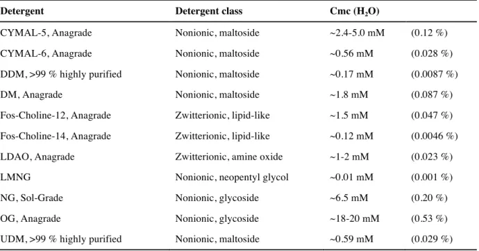
1.3 Detergents ... 23
1.4 Enzymes and inhibitors ... 24
1.5 Molecular weight markers ... 24
1.6 Reagent kits ... 24
1.7 Column materials ... 25
1.8 Antibodies ... 25
1.9 Media and antibiotics ... 26
1.9.1 LB media ... 26
1.9.2 2YT media ... 26
1.9.3 TB media ... 26
1.9.4 SOC media ... 27
1.9.5 CP minimal media ... 27
1.9.6 Antibiotics ... 28
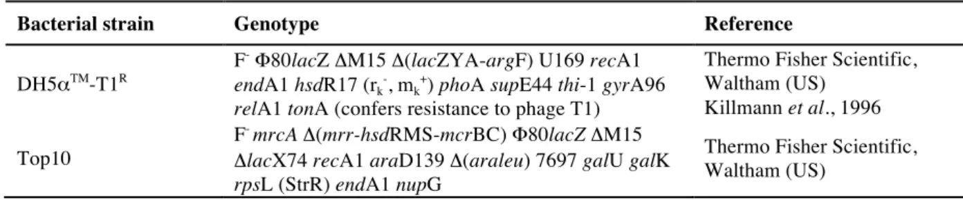
1.10 Bacterial E. coli strains ... 28
1.11 Plasmids and oligonucleotide primers ... 29
1.12 Archaeal strains ... 30
2 Molecular methods ... 31
2.1 Polymerase chain reaction (PCR) ... 31
2.2 Fusion PCR ... 32
2.3 Agarose gel electrophoresis ... 32
2.4 Digestion with restriction enzymes ... 33
2.5 Ligation ... 33
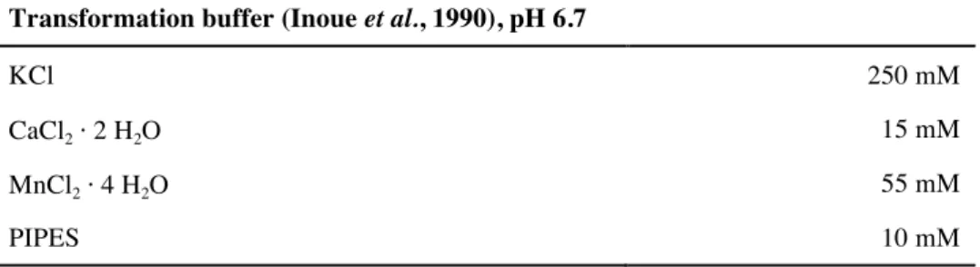
2.6 Preparation of chemical competent E. coli cells ... 34
2.7 Transformation of E. coli ... 35
2.8 Colony PCR ... 35
2.9 Isolation of plasmid DNA ... 36
2.10 Determination of DNA concentration ... 36
3 Biochemical methods ... 36
3.1 Determination of protein concentration ... 36
3.1.1 BCA assay ... 36
3.1.2 Bradford assay ... 36
Table of contents
3.1.3 Photometrical determination ... 37
3.2 SDS-Polyacrylamide gel electrophoresis (SDS-Page) ... 37
3.3 Blue Native Electrophoresis (BNE) ... 38
3.4 Western blot ... 39
3.5 Heterologous protein expression in E. coli ... 40
3.5.1 Expression ... 40
3.5.2 Cell disruption and membrane preparation ... 41
3.6 Protein purification ... 41
3.6.1 Solubilization ... 41
3.6.1.1 Detergent screening ... 41
3.6.1.2 Solubilization for purification ... 42
3.6.2 Immobilized metal affinity chromatography (IMAC) ... 42
3.6.2.1 Flow through-based purification ... 42
3.6.2.2 Gravity flow based method in batch ... 43
3.6.2.3 Proteolytic cleavage of histidine tag ... 43
3.6.3 Desalting and concentrating of protein samples ... 43
3.6.4 Size exclusion chromatography (SEC) ... 44
3.7 Reconstitution into membrane-mimicking environments ... 44
3.7.1 Reconstitution into nanodiscs ... 44
3.7.1.1 Expression and purification of the membrane scaffold protein MSP1E3D1 ... 44
3.7.1.2 Reconstitution procedure ... 46
3.7.2 Reconstitution into amphipol A8-35 ... 47
3.8 Lipid analysis by thin layer chromatography (TLC) ... 47
4 Electrophysiological analyses ... 48
4.1 Preparation of liposomes ... 48
4.2 Patch clamp recordings of proteoliposomes ... 49
4.3 Preparation of E. coli spheroplasts ... 49
4.4 Patch clamp recordings of E. coli spheroplasts ... 51
4.5 Hypoosmotic down-shock assay ... 51
Table of contents
IV
5.1.1 Preparation of carbon coated copper grids ... 52
5.1.2 Negative staining and electron microscopy ... 52
5.2 Cryo electron microscopy (cryo-EM) ... 53
5.3 Image-processing and single particle analysis ... 53
5.4 Map visualization and analysis ... 53
6 Bioinformatical analyses ... 54
6.1 Sequence alignments ... 54
6.2 Topology prediction ... 54
6.3 Homology modeling ... 54
III RESULTS ... 56
1 Biochemical analysis of Neq198 and Neq531 ... 56
1.1 Heterologous expression and purification ... 56
1.2 Reconstitution into membrane-mimicking environments ... 62
1.3 Lipid analysis by thin layer chromatography ... 68
2 Structural analysis of the MscS-like channels from N. equitans ... 70
2.1 Sequence analysis ... 70
2.2 Single particle analysis ... 73
2.3 Negative stain single particle analysis ... 73
2.3.1 Neq198 reconstituted into amphipol A8-35 ... 73
2.3.2 Neq531 reconstituted into amphipol A8-35 ... 77
2.3.3 Neq198 reconstituted into nanodiscs ... 80
2.4 Single particle analysis of a cryo-EM data set ... 84
2.5 Neq198: Fitting of atomic coordinates into cryo-EM density ... 92
3 Functional analysis of Neq198 and Neq531 ... 96
3.1 Hypoosmotic down-shock assay ... 96
3.2 Patch clamp analysis ... 97
IV DISCUSSION ... 102
1 Heterologous expression and purification of MscS-like channels from N. equitans ... 102
Table of contents
2 The role of lipids in mechanosensation ... 105
2.1 Bacteria and archaea: membranes and lipids ... 105
2.2 Towards a functional understanding of Neq198 and Neq531 ... 108
3 Single particle analysis using RELION ... 111
3.1 Protein quality and sample preparation challenges ... 111
3.2 Nanodiscs versus amphipol reconstitution ... 113
3.3 Preferred orientation and neighbouring particles ... 117
3.4 Heterogeneity within a data set ... 118
4 Structure of the first archaeal MscS-like channel ... 119
5 Conclusions and future directions ... 122
V REFERENCES ... 125
VI APPENDIX ... 148
1 Protein characteristics ... 148
1.1 General characteristics of Neq198 and Neq531 ... 148
1.2 Protein sequences of relevant MS channels ... 149
1.3 Accession numbers of relevant MS channels ... 151
1.4 Sequence alignments ... 151
2 Single particle data sets ... 155
2.1 Negative stain data sets ... 155
2.2 Cryo-EM data sets ... 155
3 Cryo-EM data sets: experimental set-up ... 156
3.1 Experimental set-up for “Feb2016” data set ... 156
3.2 Experimental set-up for “Jun2016” data set ... 157
ACKNOWLEDGMENTS ... 158
List of abbreviations
VI
List of abbreviations
2D two-dimensional
3D three-dimensional
Å Angström (s)
Aa amino acids
Amp ampicillin
APS ammonium persulfate
AU arbitrary unit (absorption unit)
BCA bicinchoninic acid
BCIP 5-bromo-4-chloro-3-indolyl-phosphate
BSA bovine serum albumin
CD cytoplasmic domain
Cfu colony forming units
Cm chloramphenicol
Cmc critical micelle concentration CIP alkaline phosphatase, calf intestinal
CTF contrast transfer function
CV column volume
CYMAL-5 5-Cyclohexyl-1-Pentyl-β-D-Maltoside CYMAL-6 6-Cyclohexyl-1-Hexyl-β-D-Maltoside
DDM n-dodecyl-β-D-maltoside
DM n-decyl-β-D-maltoside
DNA deoxyribonucleic acid
e.g. exempli gratia, for example
EM electron microscope/ microscopy
EMDB electron microscopy data bank
EPL E. coli polar lipids
et al. et alia, et alii
forw forward
List of abbreviations
Fos14 Fos-Choline-14 (n-Tetradecylphosphocholine) Fos12 Fos-Choline-12 (n-Dodecylphosphocholine)
xg acceleration of gravity
GOF gain-of-function
His6 hexa-histidine tag
HMW high molecular weight
i.e. id est, that is
IPTG isopropyl-β-D-thiogalactopyranosid IMAC immobilized metal affinity chromatography
Km kanamycin
LMNG Lauryl Maltose Neopentyl Glycol
LOF loss-of-function
MS mechanosensitive
MSP membrane scaffold protein
MWCO molecular weight cut-off
NBT nitro blue tetrazolium
NG n-Nonyl-β-D-Glucopyranoside
OD optical density
OG n-Octyl-β-D-Glucopyranoside
o/N overnight
PC phosphatidylcholine
PD periplasmic domain
PDB protein data base
PE Phosphatidylethanolamine
PELDOR Pulsed electron-electron double resonance
PG phosphatidylglycerol
pI isoelectric point
POPC 1-palmitoyl-1,2-oleoyl-sn-glycero-3-phosphocholine
POPG 1-palmitoyl-1,2-oleoyl-sn-glycero-3-phospho-(1’-rac-glycerol)
List of abbreviations
VIII
RT room temperature
SDS sodiumdodecylsulfate
SEC size exclusion chromatography
SEM scanning electron microscopy
SNR signal-to-noise ratio
TEM transmission electron microscope/ microscopy
TM transmembrane
TMD transmembrane domain
Tris 2-amino-2-hydroxymethyl-propane-1,3-diol
UAc uranyl acetate
UDM n-Undecyl-β-D-Maltoside
v/v volume per volume
w/ with
w/o without
w/v weight per volume
List of abbreviations
Amino acid One-letter code Three-letter code
alanine A Ala
cysteine C Cys
aspartic acid D Asp
glutamic acid E Glu
phenylalanine F Phe
glycine G Gly
histidine H His
isoleucine I Ile
lysine K Lys
leucine L Leu
methionine M Met
asparagine N Asn
proline P Pro
glutamine Q Gln
arginine R Arg
serine S Ser
threonine T Thr
valin V Val
trypthophan W Trp
tyrosin Y Tyr
variable X -
List of figures
X
List of figures
Figure 1: Physiological function of mechanosensitive channels in bacteria. ... 2
Figure 2: 3D crystal structure of MscL from M. tuberculosis. ... 4
Figure 3: Comparison of MscS homologues in E. coli. ... 6
Figure 4: 3D crystal structure of MscS from E. coli. ... 7
Figure 5: Proposed selectivity mechanism in MscS-like channels. ... 10
Figure 6: Lipid effect on membrane curvature. ... 11
Figure 7: Lipid exchange between the pockets and the lipid bilayer in MscS. ... 13
Figure 8: “Intimate association” of I. hospitalis and N. equitans. ... 16
Figure 9: Transmission electron micrographs of N. equitans. ... 19
Figure 10: Structure of amphipol A8-35. ... 47
Figure 11: Formation of E. coli spheroplasts. ... 50
Figure 12: Negative stain transmission electron micrograph of Neq531. ... 57
Figure 13: Western blot analysis of homogenized membranes. ... 58
Figure 14: Solubilization test of Neq198. ... 58
Figure 15: Buffer conditions for IMAC purification. ... 59
Figure 16: Immobilized metal affinity chromatography (IMAC) of Neq198. ... 60
Figure 17: Immobilized metal affinity chromatography (IMAC) of Neq531. ... 60
Figure 18: Homogeneity and oligomeric state of Neq198 in Fos14. ... 61
Figure 19: Membrane-mimicking sytems for membrane protein stabilization. ... 62
Figure 20: Determination of the optimal MP:APol ratio for Neq198. ... 63
Figure 21: Reconstitution of Neq198 into amphipol A8-35. ... 64
Figure 22: Reconstitution of Neq531 into amphipol A8-35. ... 65
Figure 23: Reconstitution of Neq198 into nanodiscs. ... 66
Figure 24: Quality control of Neq198 reconstituted into nanodiscs. ... 66
Figure 25: 1D thin layer chromatograms. ... 69
Figure 26: 2D thin layer chromatograms. ... 69
Figure 27: Prediction of TM helices of Neq198. ... 71
Figure 28: Prediction of TM helices of Neq531. ... 71
List of figures
Figure 29: Sequence alignment of EcMscS, Neq198 and Neq531. ... 72
Figure 30: Protein surface of EcMscS in different orientations. ... 73
Figure 31: Representative 2D class-averages in negative stain of Neq198 reconstituted into APol. .... 74
Figure 32: Putative domain organization of Neq198. ... 75
Figure 33: RELION-2.1 workflow overview of Neq198 reconstituted into APol in negative stain. .... 76
Figure 34: Representative 2D class-averages of Neq531 reconstituted into APol. ... 78
Figure 35: RELION-2.1 workflow overview of Neq531 reconstituted into APol in negative stain. .... 79
Figure 36: Surface representation of Neq531 reconstituted into APol. ... 80
Figure 38: RELION-2.1 workflow overview of Neq198 reconstituted into nanodiscs in negative stain. ... 82
Figure 39: Surface representation of Neq198 reconstituted into nanodiscs. ... 83
Figure 40: Cryo-EM micrographs of Neq198 reconstituted into nanodiscs. ... 85
Figure 41: Comparison of different data sets during 2D classification in RELION-2.1. ... 86
Figure 42: Representative 2D class-averages showing “defective” particles. ... 87
Figure 43: Representative 2D class-averages. ... 88
Figure 44: Representative final 3D volumes. ... 91
Figure 45: Surface representation of cryo-EM density with fitted atomic model of Neq198. ... 94
Figure 46: Profile of ion permeation pathway. ... 95
Figure 47: Hypoosmotic down-shock assay of Neq198 in MJF465 E. coli cells. ... 96
Figure 48: Patch clamp currents of Neq198 reconstituted into azolectin liposomes. ... 100
Figure 49: Patch clamp currents of Neq531 reconstituted into azolectin liposomes. ... 100
Figure 50: Comparison of archaeal and bacterial lipids and membranes. ... 105
Figure 51: Effect of internal pressure on the lipid membrane. ... 106
Figure 52: Structural comparison of Neq198 and EcMscS. ... 121
Figure 53: Electrostatic potential surface of Neq198 and EcMscS. ... 122
List of tables
XII
List of tables
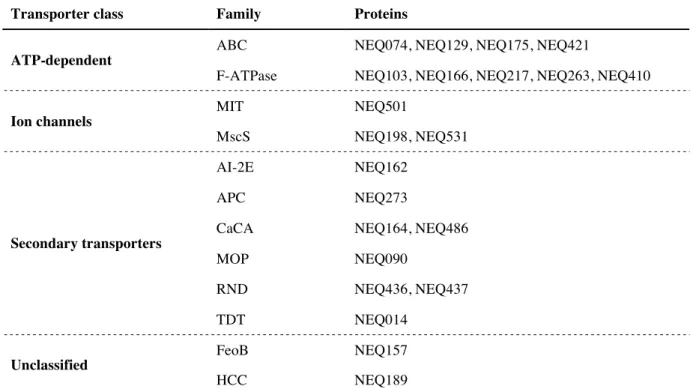
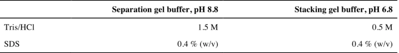
Table I.1: Solved structures for mechanosensitive channels of large conductance ... 4 Table I.2: Solved structures of mechanosensitive channels of small conductance ... 9 Table I.3: Annotated transporter classes and families in the genome of N. equitans. ... 20 Table II.1: Detergents and their characteristics ... 23 Table II.2: Enzymes ... 24 Table II.3: Protease inhibitors ... 24 Table II.4: Molecular weight markers ... 24 Table II.5: Reagent kits ... 25 Table II.6: Column materials ... 25 Table II.7: Primary and secondary antibodies ... 25 Table II.8: Antibiotics and their working concentrations ... 28 Table II.9: Bacterial strains for general cloning/subcloning and their characteristics ... 28 Table II.10: Bacterial strains for expression and their characteristics ... 29 Table II.11: General plasmids and their properties ... 29 Table II.12: Plasmid constructs for protein expression ... 30 Table II.13: Oligonucleotides ... 30 Table II.14: Archaeal strains ... 30 Table II.15: PCR mixture ... 31 Table II.16: Thermocycler program for PCR ... 31 Table II.17: Thermocycler program for Fusion PCR ... 32 Table II.18: Setup for digestion with restriction enzymes ... 33 Table II.19: Mixture for ligation of digested plasmids and DNA inserts ... 34 Table II.20: Transformation buffer for preparation of chemical competent E. coli cells ... 34 Table II.21: Colony PCR mixture ... 35 Table II.22: Thermocycler program for colony PCR ... 35 Table II.23: Separation and stacking gel buffer for SDS-Page ... 37 Table II.24: Laemmli running buffer and 6x loading buffer ... 37 Table II.25: SDS-Page gel composition ... 38
List of tables
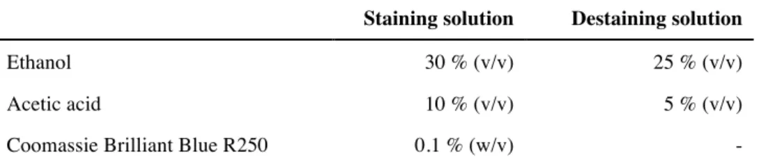
Table II.26: Composition of staining and destaining solution for SDS-Page ... 38 Table II.27: Blue Native gel buffer ... 39 Table II.28: Blue Native gel composition ... 39 Table II.29: Composition of Blue Native cathode and anode buffer ... 39 Table II.30: IMAC purification buffer ... 42 Table II.31: Buffers and columns used for size exclusion chromatography ... 44 Table III.1: Liposome preparations ... 98 Table III.2: Reconstitution of Neq198 into E. coli spheroplast ... 101 Table IV.1: Representative membrane proteins that have been reconsitituted into nanodiscs for
structure determination in cryo-EM ... 114 Table IV.2: Commonly used membrane scaffold protein constructs (Ritchie et al., 2009) ... 115
Abstract
XIV
ABSTRACT
Mechanosensitive (MS) channels are found throughout all domains of life since maintaining the intracellular homeostasis is crucial for all living cells. Bacterial MS channels represent the best- characterized force-sensing system. They mainly act as “emergency relief valves” by directly sensing elevated membrane tension as a consequence of osmotic down-shock. Upon opening, the channels release osmotically active solutes and ions from the cell and therefore prevent cell lysis.
In this thesis, two putative archaeal MscS-like channels from N. equitans - Neq198 and Neq531 - were investigated with respect to their structure and function. Major bottlenecks for the analysis of membrane proteins are the expression, solubilization, and purification that should yield sufficient amount of biologically active protein. Both channels were heterologously expressed in E. coli and successfully purified. Here, the N-terminal localization of the tag for purification turned out to be crucial. For Neq198 an optimized purification protocol was established. However, Neq531 is challenging to purify and the purification protocol still requires further optimization.
Membrane-mimicking systems such as lipid nanodiscs and amphipols are highly favored for the analysis of membrane proteins since they provide more native-like environments compared to detergents. Both MscS-like channels were reconstituted into amphipols or lipid nanodiscs for electron microscopy single particle analysis. Single particle analysis of negative-stained channels already gave a rough idea about the channel organization. The resulting 3D volume can be compared to EcMscS, whose structure was solved in 2002 by X-ray crystallography. Here, lipid nanodiscs were highly favored over amphipol for the structural analysis of Neq198. In amphipol reconstituted channels provided less detail than in lipid nanodisc reconstituted channels.
Abstract
Neq198 reconstituted into lipid nanodiscs was further analyzed in cryo-EM single particle that led to the first putative structure of an archaeal mechanosensitive channel at intermediate resolution so far.
The heptameric channel organization was confirmed. While the cytoplasmic domain of the channel is hardly changed compared to EcMscS, the membrane domain exhibits major differences. Two additional helices are located on top of the membrane domain building up a cap-like structure of unknown function. Whether this structural feature takes actively part in tension sensing and e.g.
provides interaction with the S-layer of N. equitans is open for speculation. The electrostatic potential surfaces indicate that the channel from N. equitans exhibits higher anion selectivity than its homologue in E. coli (EcMscS).
The functional analyses by patch clamping highlighted that these channels are highly dependent on their native environment. A proper characterization with respect to conductance, activation threshold and ion selectivity failed in liposomes and E. coli spheroplasts. So far, channels reconstituted into liposomes exhibited spontaneous and constitutive activity, although no pressure was applied. These difficulties were mainly contributed to the lipid environment, which is clearly different in N. equitans.
To characterize the channels, native lipids from N. equitans are required for future reconstitution and patch clamp analysis. However, hypoosmotic down-shock assays indicate that the channels are purified in active form and do not lose their functionality upon purification.
This thesis provides first structural insights into a MscS-like channel from N. equitans representing the first structure of an archaeal mechanosensitive channel from the MscS family. To increase the resolution of the membrane domain and cap-like structure and to refine the proposed model, more single particle analysis cryo-EM data is required.
I Introduction
I INTRODUCTION
1 Mechanosensitive channels
Mechanosensation as a physiological process describes the ability to convert a mechanical stimulus into electrical or biochemical signals and is believed to be one of the most ancient signal transduction mechanisms (Kloda and Martinac, 2001b). Mechanosensory transduction is involved in a wide range of physiological processes, such as touch, pain, hearing, proprioception, blood pressure control in animals, turgor control and gravitaxis in plants as well as regulation of cell shape and cell volume in bacteria (Hamill and Martinac, 2001; Martinac, 2004). As force-transducing molecules the so-called mechanosensitive (MS) channels have been identified. They can be found throughout all domains of life: archaea, bacteria (Pivetti et al., 2003; Kung, 2005), and eukarya (Árnadóttir and Chalfie, 2010), thus suggesting their early appearance during evolution on earth. This is further supported by the assumption that all aspects of cellular dynamics as growth, cell division, and differentiation involve changes in cell volume and shape (Kung et al., 1990; Martinac and Kloda, 2003).
Bacterial MS channels represent one of the best-characterized force-sensing systems and serve as model systems for mechanosensation (Kung, 2005). MS channels represent a structurally diverse group of proteins that have been classified according to their function rather than their sequence similarity or topology. In prokaryotes two families of MS channels have been identified. Those are classified according to their conductance: the mechanosensitive channels of large conductance (MscL)
I Introduction
2
(mechanosensitive channels of mini conductance) (Berrier et al., 1996). However, channels that exhibit MscM activity are structurally classified as members of the MscS family. Prokaryotic MS channels were first discovered 1987 in giant E. coli spheroplasts using patch clamp technique (Hamill et al., 1981; Martinac et al., 1987). Maintaining the intracellular homeostasis is imperative for all living cells so that the major function of MS channels can be assigned to act as an “emergency relief valve” upon hypoosmotic down-shock. The immediate release of solutes enables growth and survival even at changing external osmolarities (Figure 1) (Booth and Blount, 2012).
Figure 1: Physiological function of mechanosensitive channels in bacteria.
(a) At low osmolarity conditions, cells build up an outwardly directed turgor pressure, which is balanced by the cell wall and outer membrane; (b) upon hyperosmotic shock, cells shrink due to rapid water loss (c) but recover to full size by the accumulation of compatible solutes in the cytoplasm; MS channels remain closed; (d) in response to a reduction in external osmolarity (hypoosmotic shock), water floods into bacterial cells, resulting in swelling of the cell with a corresponding rise of cellular turgor and membrane tension. The rapid water entry is accompanied by the immediate activation of MS channels, allowing the efflux of intracellular osmolytes and relieving the membrane tension; (e) subsequently allowing normal growth; (f) in absence or fail in function of MS channels, the rise of membrane tension results in cell lysis as soon as the pressure exceeds the mechanical strength of the cell wall (Booth et al., 2007).
MS channels directly respond to changes in membrane tension. Osmotic down shift results in water influx into the cell causing the cell to swell. By the interaction with the surrounding lipid bilayer MS channels sense the increased membrane tension and temporarily open large pores in the membrane, by which compatible solutes and ions are released. By this, the pressure is relieved and cell integrity is preserved. Booth and co-workers showed that ΔmscL and ΔmscS double mutants were not able to maintain their cell integrity resulting in cell lysis upon down-shock (Levina et al., 1999). MscL and MscS have complementary function. They respond in a coordinated way to osmotic stress indicating a relationship between their activation threshold and their conductance (Berrier et al., 1989; Levina et
I Introduction
al., 1999; Perozo and Rees, 2003). MS channels exhibit a step-wise response to osmotic stress, where MscS open first and MscL remains closed as backup, opening just before the lytic limit of the cell. By this, as much metabolites as possible are retained within the cell sustaining their membrane gradients (Haswell et al., 2011). MS channels differ significantly with respect to their conductance ranging from 10-3,500 pS (Martinac et al., 1987; Sukharev et al., 1993) and their selectivity ranging from non- selective to potassium- and cation- or anion-selective channels (Sukharev et al., 1993; Li et al., 2002;
Zhang et al., 2012).
1.1 Mechanosensitive channels of large conductance (MscL)
Since its first discovery in E. coli in 1993 by Kung’s laboratory (Sukharev et al., 1993; Sukharev et al., 1994), the mechanosensitive channel of large conductance, MscL, was extensively studied and characterized. Compared to MscS, the MscL family is highly conserved with respect to structure and function. In general, the MscL family consists of non-selective ion channels, that exhibit an elevated conductance of ~3 nS. Members of the MscL family are pressure-activated and open at high membrane tension close to lytic limit of the bilayer (Berrier et al., 1989; Sukharev et al., 1993).
Interestingly, the TbMscL – a MscL homologue from Mycobacterium tuberculosis – exhibits an opening threshold that is significantly larger than that of EcMscL and even exceeds the membrane breaking point (Moe et al., 2000). This might be a result of the different lipid environments and underscores the importance of lipid-protein interactions in MS channel function (Perozo and Rees, 2003). However, in E. coli the mscL gene is not essential for growth and survival under hypoosmotic down-shock conditions. The MscL activity can be compensated by other mechanosensitive channels (Sukharev et al., 1994). MscL is commonly absent from marine organisms (Booth et al., 2015) and cannot be found in plants (Haswell, 2007).
To date, three MscL structures, two from bacterial and only one from an archaeal organism, have been solved by X-ray crystallography (Table I.1). As the first structure, the MscL from M. tuberculosis was solved in a closed conformation at a resolution of 3.5 Å (Chang et al., 1998). This 136-amino-acid protein exhibits a transmembrane (TM) domain and a cytoplasmic domain assembled to a homo- pentameric complex (Figure 2). Each subunit consists of two membrane-spanning α-helices (TM1 and TM2) connected by a periplasmic loop resulting in the location of N- and C-terminus in the cytoplasm.
I Introduction
4
Table I.1: Solved structures for mechanosensitive channels of large conductance
Organism Characteristics Resolution (Å) Method Reference/ PDB M. acetivorans/
M. jannashii (MaMscL-MjRS)
chimera, pentameric
open/closed 3.5/4.1 X-ray Li et al., 2015
4y7k, 4y7j S. aureus
(SaMscL)
tetrameric,
expanded intermediate state
3.82 X-ray Liu et al., 2009 3hzq
M. tuberculosis (TbMscL)
pentameric,
closed 3.5 X-ray
Steinbacher et al., 2007 Chang et al., 1998 2oar
The TM1 helix of each subunit lines the pore and forms a tightly packed bundle with its adjacent TM1 helices, whereas the TM2 helices interact with the surrounding lipid bilayer (Chang et al., 1998). The homo-pentameric complex forms a pore with a diameter of 30 Å, which can vary between 2 and 30 Å during gating (Perozo et al., 2002).
Figure 2: 3D crystal structure of MscL from M. tuberculosis.
Crystal structure of the pentameric TbMscL in closed conformation at 3.5 Å resolution (Chang et al., 1998), showing the overall structure (left) with one subunit highlighted in blue, a monomer (middle), and the top view from the periplasmic side of the membrane with one subunit highlighted in blue (right); N- and C-terminus are located in the cytoplasm; TM1 and TM2 helices are connected by a periplasmic loop. Structure was visualized in Chimera 1.12 (Pettersen et al., 2004), 2oar.pdb.
Deletion mutants lacking the cytoplasmic α-helical bundle were used for structure determination of the first archaeal MS channel, which also adopts a pentameric fold. The archaeal channel protein comprised only limited hydrophilic regions; therefore riboflavin synthetase from Methanocaldococcus jannashii (MjRS) was fused to the C-terminus of the MscL channel protein from Methanosarcina acetivorans (MaMscL) to increase potential crystal contacts (Table I.1) (Li et al., 2015). Surprisingly, the structure of the MscL homologue from Staphylococcus aureus (SaMscL) suggested a tetrameric
I Introduction
stoichiometry (Liu et al., 2009). Further experiments demonstrated that the detergents used in purification and crystallization caused the variable stoichiometry of SaMscL in vitro. In vivo studies suggested a pentameric oligomeric state (Dorwart et al., 2010).
1.2 Mechanosensitive channels of small conductance (MscS)
1.2.1 MscS superfamily
In contrast to the MscL family, the MscS family is relatively diverse with respect to structure and function. Members of the MscS superfamily show great diversity in sequence, size, and topology.
They are classified into 15 subfamilies according to their unique functional domains. Those can be further divided into larger subfamilies (~50 to 645 members each) like MscK, DUF3772-MscS, bCNG, BON-MscS, EF-MscS and smaller subfamilies (one to only few members) like MscCG, extended C-terminus MscS, MscS-DEP, YjeP, Glucose Transporter MscS, PBD-1-MscS, Cu-Heme- MscS, and concatenated MscS (Malcolm and Maurer, 2012). For the overall and most-common architecture of MscS applies the following: a large water-filled cytoplasmic domain that opens to the cytoplasm follows the transmembrane domain. Several members exhibit additional N- and/or C- terminal extensions/domains, which are assumed to affect the tension sensing mechanism or create additional regulatory sites. MscS subtypes exhibit varying numbers of transmembrane helices ranging from at least three (e.g. EcMscS) to 11 (e.g. MscK) (Booth et al., 2011; Malcolm and Maurer, 2012).
The pore lining helix TM3a is highly conserved within the MscS superfamily (Pivetti et al., 2003;
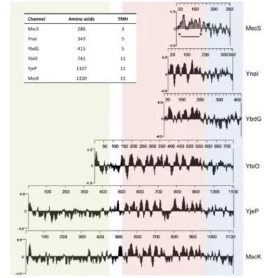
Balleza and Gómez-Lagunas, 2009). The ion selectivity that distinguishes different family members is mediated by the cytoplasmic domain (Gamini et al., 2011; Cox et al., 2014). Single organisms might encode multiple MscS members in their genome. In E. coli for example, six MscS-like channels of variable size are present in total: MscS (286 aa, EcMscS), MscK (1120 aa), YbdG (415 aa), YnaI (343 aa), YbiO (741 aa), and YjeP (1107 aa) (Figure 3) (Levina et al., 1999; Schumann et al., 2010;
Edwards et al., 2012). The EcMscS represents the MscS archetype (cf. I1.2.2). YbdG has a larger cytoplasmic domain than the other homologues due to an insertion of ~50 amino acids between the middle β and the αβ domains (cf. Figure 4). Moreover, YbG exhibits five putative TM helices. The expression level of YbdG in the membrane of E. coli is not sufficient to protect against severe hypoosmotic shock. Interestingly, overexpression of YbdG provides complete protection. YbdG is
I Introduction
6
class of channels, while YbiO, YjeP, and MscK comprising 11 putative TM helices and extensive periplasmic domains form the larger topological class (Pivetti et al., 2003). The potassium-dependent MscK represents the largest MscS-like channel in E. coli and has a conductance of ~0.875 nS (Li et al., 2002). YbiO, YnaI, and YjeP are only present in low levels in the membrane and therefore do not provide protection against hypoosmotic shock as well. NaCl especially induces YbiO, which exhibits a conductance of ~1000 pS. YnaI and YjeP have MscM activity with a conductance of ~100 pS and
~300 pS, respectively (Edwards et al., 2012).
Figure 3: Comparison of MscS homologues in E. coli.
Hydrophobicity blots (Kyte-Doolittle, w = 9) of the six MscS homologues in E. coli, aligned by the last three predicted transmembrane (TM) helices for each homolog with the three TM helices of MscS (*---*). Highlighted sequences:
periplasmic region – green, membrane region – red; cytoplasmic domain – blue (adapted from Schumann et al., 2010 and Naismith and Booth, 2012); inset: overview of amino acid length and predicted TM helices of each homolog.
Besides the six homologues in E. coli, various other members have been characterized electrophysiological, e.g. MscMJ and MscMJLR from M. jannashii (Kloda and Martinac, 2001d), MscSP from Silicibacter pomeroyi (Petrov et al., 2013), MSL10 from Arabidopsis thaliana (Maksaev and Haswell, 2013), and Ng-MscS from N. gonorrhoeae (Wang et al., 2018).
I Introduction
1.2.2 Structure and function of MscS
MscS fold as homo-heptamers with a sevenfold rotational axis perpendicular to the membrane normal through the center of the pore. The channel can be divided in a transmembrane domain and a large water-filled cytoplasmic domain (Figure 4) (Bass et al., 2002; Steinbacher et al., 2007).
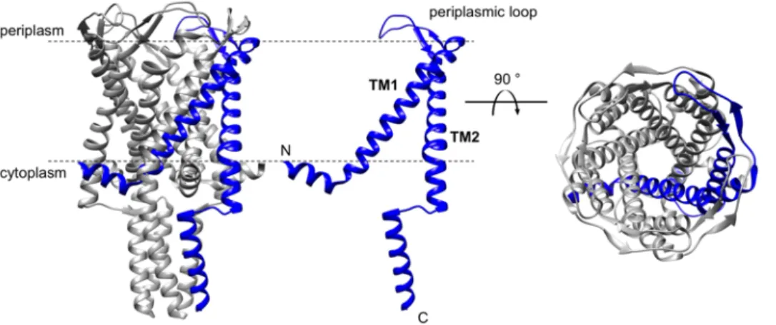
Figure 4: 3D crystal structure of MscS from E. coli.
Crystal structure of the homo-heptameric EcMscS in a closed conformation at a resolution of 3.7 Å (Bass et al., 2002;
Steinbacher et al., 2007), showing an overall structure (left) with one subunit highlighted in blue, a channel monomer (middle) and the top view of the channel (right) from the periplasmic side of the membrane with one subunit highlighted in blue; N-terminus is located in the periplasm, whereas the C-terminus is located in the cytoplasm. Structure was visualized in Chimera 1.12 (Pettersen et al., 2004), 2oau.pdb.
Each subunit of WT EcMscS consists of 286 amino acids and exhibits three transmembrane helices (TM1 29-57, TM2 68-91, TM3 96-127) with the N-terminus facing the periplasm (Bass et al., 2002).
The membrane-spanning helices TM1 and TM2 form a closely packed hairpin. They are tilted by 27°
to 35° with respect to the sevenfold axis, which results in the displacement of the helices TM1 and TM2 from the core of TM3 helices. A kink at Gly113 divides the TM3 helix into the pore-lining helix TM3a (residues 96-112) and the helix TM3b (residues 114-127) and marks the membrane boundary.
Seven TM3a helices are arranged in a helical barrel, which creates the central axis of the pore and thereby lines the route of ions through the membrane. Adjacent TM3a helices from neighboring subunits are tightly packed. A pattern of highly conserved alanine and glycine residues enables these
I Introduction
8
hydrophobic residues (Leu105 and Leu109) line the permeation pathway. In addition to the hydrophobic nature of the TM3a, these residues create a hydrophobic seal (Bass et al., 2002) that was also termed as “vapor lock” (Anishkin and Sukharev, 2004). The narrowest point is constricted to
~11 Å (Bass et al., 2002). TM3b connects the membrane-embedded channel part to the cytoplasmic domain and is orientated almost parallel to the membrane surface (Booth et al., 2011). The membrane channel opens to a large water-filled chamber with a diameter of 40 Å and is formed predominantly by β-sheets and αβ-sheet domains. Seven lateral portals with a diameter of ~14 Å between the interfaces of the single subunits connect the chamber to the cytoplasm. The last ~18 C-terminal amino acids of each subunit form a seven-stranded β-barrel as a distal pore with a diameter of ~8 Å (Bass et al., 2002). Deletion experiments have shown that a loss of the β-barrel does not influence channel assembly or gating mechanisms but severely impairs stability (Schumann et al., 2004). The overall structure of EcMscS is determined to dimensions of 120 Å in length parallel to the sevenfold axis and 80 Å in width parallel to the membrane (Bass et al., 2002).
To date, nine different structures of MscS were solved, among those six of EcMscS in distinct conformations and at different resolutions (Table I.2). The first crystal structure for the WT EcMscS was obtained at a resolution of 3.9 Å by Bass and co-workers in 2002 (Bass et al., 2002). They captured the protein in a non-conducting, closed conformation. For the subsequently solved structures, the cytoplasmic domain remained essentially unchanged whereas distinct conformations could be observed for the transmembrane region. The Ala106Val variant resulted in an open conformation at a resolution of 3.45 Å (Wang et al., 2008). Compared to the closed state, the helices TM1 and TM2 rotate clockwise as a rigid body by approximately 45° along the sevenfold axis. Moreover, the helices TM1 and TM2 increase their tilt by ~15° with respect to the sevenfold axis. The TM3a helices rotate clockwise around their axis by ~15° and move out from the central axis of the channel. As a result, the TM3a helices are parallel to each other, to the sevenfold axis, and to the membrane normal in the open structure. In the closed structure the TM3a helices arrange diagonal to the sevenfold axis, instead. It is proposed that the open-close-transition of the channel is enabled by an iris-like motion of the three TM helices (Wang et al., 2008). Intriguingly, the DDM solubilized WT EcMscS adopts an open conformation as observed for the Ala106Val EcMscS variant, which was solubilized in Fos14. This highlighted the general sensitivity of membrane proteins and their conformational states to variations in detergents, crystallization conditions, and point mutations (Lai et al., 2013). In 2015 the structure of another low-conductance MscS channel from E. coli, YnaI, was solved by cryo-electron microscopy to a resolution of 12.6 Å (Böttcher et al., 2015) - to date, the first and only structure of a
I Introduction
mechanosensitive channel of small conductance solved by cryo-EM. Furthermore, the structure of a MscS homologue from Helicobacter pylori (Lai et al., 2013), a gram-negative bacterium with potential pathogenicity, is currently available as well as a structure from Thermoanaerobacter tengcongensis, characterized as an anion-selective channel (Zhang et al., 2012) (Table I.2).
Table I.2: Solved structures of mechanosensitive channels of small conductance
Organism Characteristics Resolution (Å) Method Reference EMDB/PDB E. coli
(YnaI) DDM 12.6 cryo-EM Böttcher et al., 2015
3035 E. coli
(EcMscS)
D67R1
(R1 = MTSSL cysteine adduct), open
2.99 X-ray Pliotas et al., 2015 5aji
E. coli (EcMscS)
WT,
open, DDM 4.4
X-ray Lai et al., 2013 4hwa, 4hw9 H. pylori
(HpMscS)
WT,
closed, DDM 4.2
E. coli (EcMscS)
spin labeled D67C mutant,
open, DDM 4.84
X-ray Pliotas et al., 2012 4agf, 4age E. coli
(EcMscS)
spin labeled L124C mutant,
open, DDM 4.70
T. tengcongensis (TtMscS)
WT,
w/o ligand, w/ ligand LMT closed, DDM
3.36/3.46 X-ray Zhang et al., 2012 3udc, 3t9n E. coli
(EcMscS)
Ala106Val variant,
open, Fos14 3.45 X-ray Wang et al., 2008
2vv5 E. coli
(EcMscS)
WT,
closed, Fos14 desensitized/inactive
3.7/3.9 X-ray
Steinbacher et al., 2007 Bass et al., 2002 2oau
The helices TM1 and TM2 of EcMscS are thought to act as sensors for membrane tension by interacting with the surrounding lipid bilayer and therefore directly coupling changes in membrane tension to conformational rearrangements (Bass et al., 2002; Wang et al., 2008) (I1.2.3). PELDOR studies of EcMscS in proteoliposomes confirmed basic findings of the first crystal structure: the pronounced gap between the outer helices (TM1, TM2) and the pore lining helix TM3 is preserved in
I Introduction
10
In addition to its importance for stabilization and oligomerization (Schumann et al., 2004; Rasmussen et al., 2007), the cytoplasmic vestibule domain is involved in selectivity and gating. This domain is thought to act as a selectivity filter or sieve by controlling, which ions can enter through the portals and therefore exit the cells (Gamini et al., 2011). Interestingly, the filter function of the cytoplasmic domain stands in contrast to voltage-gated Cl-, Ca2+, K+ and Na+ channels, whose selectivity filters are located within the transmembrane region (Cox et al., 2014). For E. coli it is assumed that electronegative regions on the bottom of the cytoplasmic vestibule trap cations resulting in an environment that is more conducive to anion conduction (Figure 5) (Cox et al., 2014). In T. tengcongensis the lateral portals seem to be missing. Here, electronegative regions on the bottom of the cytoplasmic vestibule together with a residue outside the β-barrel result even in higher anion selectivity compared to E. coli (Zhang et al., 2012). The significant role of the β-barrel for anion selectivity in TtMscS was confirmed by Song et al., 2017.
Figure 5: Proposed selectivity mechanism in MscS-like channels.
Electronegative regions on the bottom of the cytoplasmic domains create an environment conducive to anion conduction. (A) Residues around E187 and E227 trap cations resulting in easier transit for anions; (B) in addition to the electronegative region on the bottom of the cytoplasmic domain, a residue on the outside of the β-barrel (E278) likely traps cations resulting in a higher anion selectivity in TtMscS compared to EcMscS (adapted from Cox et al., 2014).
Electrophysiological experiments characterize the EcMscS to exhibit a conductance of ~1 nS and a slight preference for anions over cations: (PCl/PK) of 1.2-3.0. The channel is activated by membrane tensions 1.4 times lower compared to MscL and shows additional voltage dependency (Martinac et al., 1987; Sukharev et al., 1993; Sukharev, 1997). Furthermore, EcMscS shows a complex inactivation/desensitization mechanism while sustaining membrane tension (Levina et al., 1999).
EcMscS reacts to sudden changes in membrane tension; however, if lateral pressure is applied slowly, the channel remains closed suggesting a relation between gating and pressure rate (Akitake et al., 2005). EcMscS measurements of gating transitions show that the channel opens within milliseconds to around two-thirds of its conductance and then opens slower to full conductance (Booth et al., 2007).
I Introduction
1.2.3 Protein-lipid interactions
Protein-lipid interactions have moved to the center of interest lately. Direct or indirect interactions with the surrounding membrane and thus conformational changes of the proteins have been investigated. Recently, a general model for tension sensing in MS channels was introduced (Pliotas and Naismith, 2017). Bacterial MS channels were first described to directly sense membrane tension in the lipid bilayer. This “bilayer model” or force-from-lipids (FFL) principle applies to both prokaryotic and eukaryotic ion channels (Hamill and Martinac, 2001; Perozo et al., 2002; Teng et al., 2015). An explanation towards the understanding of channel gating was established with the observation that an incorporation of conical lipids in the lipid bilayer results in the opening of MscS and MscL (Martinac et al., 1990; Perozo et al., 2002). The incorporation of conical lipids alters the curvature of the membrane and therefore changes intrinsic forces (Figure 6). Regardless of whether conical lipids are incorporated, or the internal pressure rises as a result of hypoosmotic shock, the membrane is dilated so that the membrane curvature decreases. Thereby the lateral pressure on the mechanosensitive channels is relieved resulting in channel opening.
Figure 6: Lipid effect on membrane curvature.
(A) Bilayer-forming phospholipids (shown in red), such as phosphatidylcholine (PC), can be approximated as rods. Micelle- forming lysophospholipids (blue), such as lysophosphatidylcholine (LPC) with only one fatty acid chain, can be regarded as cones. (B) The addition of cone-shaped lipids (or other amphipaths) into one of the leaflets of the bilayer can alter the shape/curvature, and therefore the intrinsic forces (modified from Kung, 2005).
These findings suggested that changes in the membrane tension alone or lateral pressure are sufficient for channel gating. Furthermore, prokaryotic MscS and MscL maintain their mechanosensitivity after purification and reconstitution into liposomes suggesting that the mechanical force is directly transmitted from the lipid bilayer to the protein (Sukharev et al., 1993; Sukharev, 2002; Nomura et al., 2006). MscS channels that have been studied in different lipid environments (giant E. coli
I Introduction
12
the hydrophobic core of the membrane (Perozo et al., 2002). Since changes of lipids determine MS channel activity, the interaction of MS channels with their surrounding lipid bilayer was identified as an important aspect of their function. Among membrane proteins, the MscS family offers probably the most dynamic set of interactions with the lipid bilayer (Booth et al., 2011).
EcMscS provides three principal lipid-protein interfaces. Originally, the TM1-TM2 helix pair was determined to sense membrane tension (Bass et al., 2002). These helices form a rigid sensor paddle that moves independently upon channel gating whereupon the tension is transmitted to the pore through the linker between TM2 and TM3a (Wang et al., 2008). Sukharev and co-workers showed that the inter-helical contact is inevitable for force transmission from the lipid-facing helices TM1/TM2 to the pore-lining helix TM3. The uncoupling of TM2 and TM3 appears to results in inactivation of the channels (Belyy et al., 2010). The extreme asymmetry with respect to their amino acid composition of TM1 and TM2 might additionally contribute to lipid tension sensing: whereas the residues in the periplasmic half of the helices are of low polarity, polar residues are observed in the cytoplasmic half. Thus, different interactions with lipid head groups on both sides of the membrane are induced (Booth et al., 2011). Scanning mutagenesis identified residues located at both ends of the helices TM1 and TM2 as essential for channel function (Nomura et al., 2006). In another study, lipid- facing hydrophobic residues, that are located within the TM1 and TM2 helices, were determined to be involved in tension sensing. Those residues interact with lipid tail groups in the closed state and are rotated inwards to form intra-protein hydrophobic interactions in the open state of the channel (Malcolm et al., 2011). Another lipid-protein interface is located within TM3b helix, which is proposed to lie along the inner membrane presenting a hydrophobic surface to the bilayer. Conserved basic residues (Arg128 and Arg131) at the C-terminal end of TM3b are proposed to build an anchor by the interaction with phospholipid head groups (Nomura et al., 2008; Booth et al., 2011). Finally, the N-terminal sequence (residues 1-26) seems to have a significant impact on gating characteristics.
Especially, the tryptophan residue (Trp16), which is located upstream of TM1 facing the periplasm, is proposed to have an anchoring function by the interaction with lipid head groups (Rasmussen et al., 2007).
The EcMscS crystal structure revealed that the helices TM1 and TM2 are displaced from the core of TM3 helices creating an apparent void in between (Bass et al., 2002; Pliotas et al., 2012; Ward et al., 2014). These voids have also been observed in other organisms (Zhang et al., 2012; Lai et al., 2013).
Intriguingly, alkyl chains inside this hydrophobic cleft have been identified in a new high-resolution structure (Pliotas et al., 2015). Moreover, it was shown that the void between TM3 and TM1/TM2 is
I Introduction
reversibly filled with lipids (lipid interdigitation) upon channel gating. The number of lipids decreases upon channel opening. Thus, the degree of lipid interdigitation determines open/closed conformations of MscS (Figure 7) (Pliotas et al., 2015). How lipids move in and out of a binding site and by this create sensitivity to membrane tension, was already shown for the human TRAAK K+ channel (Brohawn et al., 2014). Thus, reversible lipid interdigitation is proposed to represent an universal model for tension sensing by MS channels (Pliotas et al., 2015; Pliotas and Naismith, 2017).
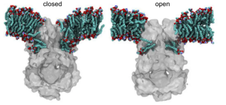
Figure 7: Lipid exchange between the pockets and the lipid bilayer in MscS.
Multiscale molecular dynamics (MD) simulations of the closed and open conformation of MscS in POPE/POPG (4:1) phospholipid bilayers. During simulations lipids migrated to fill the TM pockets and a strong local membrane curvature around MscS was observed (Pliotas et al., 2015).
The question of lipid-protein interaction for larger members of the MscS family is more complex.
Several homologues exhibit multiple additional N-terminal transmembrane helices, which result in isolating the aforementioned TM1-TM2 sensor domain from the surrounding lipid bilayer. Possibly, here the tension sensor is not constituted by the adjacent helices of the TM3a domain. Therefore, larger homologous channels require a more complex mechanism for tension sensing and transmission to the channel pore than it was described so far for MS channels exhibiting only three transmembrane helices (Booth et al., 2011). For a larger family member, YnaI from E. coli, lipid interdigitation into cavities and extended sensor paddles, i.e. four TM helices instead of two as in EcMscS, have already been discussed (Böttcher et al., 2015).
1.2.4 Physiological functions of MscS-like channels
Besides its well-known and characterized function as emergency relief valve upon hypoosmotic down- shock (Levina et al., 1999), there is emerging evidence that MS channels are not only limited to osmoregulation. Especially members of the MscS family play additional roles in bacterial and plant cell physiology. Those physiological functions range from osmoregulation to more specialized
I Introduction
14
MscK from E. coli represents an example for a highly specialized and tightly regulated channel, which is more sensitive to membrane tension than EcMscS but is also regulated by the ionic environment. Its activation requires external potassium ions and the channel is proposed to be essential for the survival at high potassium concentrations (Li et al., 2002).
In E. coli the interaction of FtsZ with the α/β domain of the cytoplasmic domain of MscS was identified as a possible non-channel function (Koprowski et al., 2015). FtsZ, a bacterial tubulin-like protein, is involved in the Z-ring formation that initiates cell division (Buddelmeijer and Beckwith, 2002; Adams and Errington, 2009). Overexpression of the soluble α/β domain as well as a deletion mutant missing the last 20 amino acids disabled cell division resulting in elongated (filamentous) cells.
Moreover, mutations in the α/β domain reduced its binding to FtsZ and its interaction was identified to play a crucial role in cell protection against antibiotic stress. Therefore, it was hypothesized that the α/β domain binds FtsZ and modulates FtsZ-dependent processes like cell wall synthesis and repair (Koprowski et al., 2015). This is further supported by the involvement of MSL2 and MSL3, two MscS-like channels from A. thaliana, in chloroplast division (Wilson et al., 2011; Wilson and Haswell, 2012).
With the cyanobacterial MscS homologue PamA, a protein was identified that is independent of osmotic shock but instead implicated in cellular signal transduction pathways. Here, the C-terminus of PamA interacts with the signaling protein PII, that coordinates several signal transduction pathways associated with carbon and nitrogen metabolism. Moreover, a deletion of pamA results in glucose sensitive mutants that show abnormal expression of genes involved in sugar and nitrogen signaling (Osanai et al., 2005).
Two MscS-like homologues from the fission yeast Schizosaccharomyces pombe are localized within the membrane of the endoplasmatic reticulum and have a putative EF-hand motif. They play a crucial role in regulation of cell volume and intracellular Ca2+ concentrations (Nakayama et al., 2012).
Furthermore, new findings suggest that EcMscS is not only involved in osmotic shock response but also in the regulation of intracellular Ca2+ concentrations (Cox et al., 2013).
In microbial biotechnology the MscS-like channel from Corynebacterium glutamicum (MscCG) is of high interest. This channel plays a crucial role in the industrial production of amino acids, e.g.
glutamate or lysine. Under production conditions like biotin limitation or the treatment with penicillin, MscCG mediates the passive efflux of negatively charged glutamate down its electrochemical gradient (Nakamura et al., 2007; Becker et al., 2013). Interestingly, MscCG was also reported to act under
I Introduction
hyperosmotic stress conditions by fine-tuning the steady state concentration of compatible solutes (betaine) in the cytoplasm of C. glutamicum (Börngen et al., 2010).
Two putative MS channels from Campylobacter jejuni, responsible for bacterial gastroenteritis in humans, provide first evidence for the role of MS channels in pathogenicity. Especially one channel turned out to be essential for the survival upon hypoosmotic stress that is experienced during environmental transmission (Kakuda et al., 2012). With Ng-MscS another MscS-like channel from the human-specific bacterial pathogen Neisseria gonorrhoeae was identified and characterized. Ng-MscS contributes to in vivo colonization of the mucosal epithelia of the urogenital tract and survival. Here, gain-of-function mutations in Ng-MscS inhibited bacterial growth. Moreover, the survival of Ng-mscS deletion mutants was significantly reduced in hypoosmotic shock assays (Wang et al., 2018).
Recently, MscS-like channels have been repeatedly discussed as potential targets for antibiotics (Booth and Blount, 2012). In this regard, it is particularly advantageous that MscS-like channels have no homologues in humans or animals, are more widely distributed than MscL and can be numerously found among pathogenic bacteria, for example like H. pylori, Vibrio cholera, Neisseria meningitides and Haemophilus influenza (Lai et al., 2013; Cox et al., 2015).
1.3 Mechanosensitive channels in archaea
Archaea are adapted to extreme habitats, where they are exposed to high temperatures, extreme salinity or low pH. Therefore, it is not surprising that mechanosensitive channels are found within the archaea, since maintaining the intracellular homeostasis is imperative for all living cells for their growth and survival.
The existence of MS channels in archaea was first documented using patch clamp technique with the identification of two types of MS channels located in the cell membrane of Haloferax volcanii (Le Dain et al., 1998). Since then other archaeal mechanosensitive channels have been characterized electrophysiological, e.g. MscTA from the cell wall-less Thermoplasma acidophilum (Kloda and Martinac, 2001a), and MscMJ/MscMJLR from M. jannashii (Kloda and Martinac, 2001d; Kloda and Martinac, 2001c). Archaeal MS channels share structural and functional homology with bacterial MS channels regarding ion selectivity, voltage dependence and mechanism of activation. They are expected to carry out similar cellular functions (Kloda and Martinac, 2001a). However, archaeal lipids
I Introduction
16
link or an evolutionary intermediate between bacterial and eukaryotic MS channels (Kloda and Martinac, 2001b; Kloda and Martinac, 2002; Martinac and Kloda, 2003; Balleza, 2011). So far, only one structure of the mechanosensitive channel chimera of large conductance (MscL) from M. acetivorans has been solved by X-ray crystallography (Li et al., 2015) (Table I.1).
2 Nanoarchaeum equitans and Ignicoccus hospitalis: the unusual intimate association of two archaea
2.1 The “intimate association”
Ignicoccus hospitalis (“the friendly fire sphere“) and Nanoarchaeum equitans (“the riding dwarf”) represent the first described (meanwhile two further biocoenoses are known) cultivable natural biocoenosis of two archaea that is also designated as “intimate association” (Huber et al., 2002; Jahn et al., 2008). The strictly anaerobic and hyperthermophilic organisms were isolated from hydrothermal vents at the Kolbeinsey Ridge at the northern coast of Iceland (Huber et al., 2000).
In contrast to its host, the symbiotic partner N. equitans can only be cultivated in co-culture, at least under laboratory conditions, depending vitally on a direct cell-cell contact with an actively growing I. hospitalis cell (Figure 8) (Huber et al., 2002). So far, the explicit nature and mechanism of this relationship remains still unclear, although no beneficial effects for I. hospitalis have been found yet.
Figure 8: “Intimate association” of I. hospitalis and N. equitans.
SEMs illustrating different stages of N. equitans colonization of I. hospitalis; depending on the growth stage of the co- culture a variable number of N. equitans cells is attached to the surface of I. hospitalis ranging from one (exponential growth) to many (stationary phase); the close-up shows the interspecies membrane contact (Giannone et al., 2015).
I Introduction
Currently, the genus Ignicoccus includes three species: I. hospitalis (Paper et al., 2007), I. islandicus, and I. pacificus (Huber et al., 2000). Moreover, another species was isolated: Ignicoccus sp. MEX13A (“I. morulus”, Lange, 2009). Whereas a highly unusual cell anatomy can be seen as a common structural feature within this genus, cross infection studies have demonstrated that only I. hospitalis can serve as a host for N. equitans (Jahn et al., 2008). Related Nanoarchaea are widespread in marine and terrestrial thermal environments around the world (Hohn et al., 2002; McCliment et al., 2006;
Casanueva et al., 2008; Clingenpeel et al., 2013; Munson-McGee et al., 2015; Wurch et al., 2016).
Recently, a new terrestrial representative of the Nanoarchaeota was isolated at the Yellowstone National Park, which is assumed to live in a similar association with an archaeon (Acidolobus sp.) (Podar et al., 2013).
2.2 I. hospitalis: current knowledge
The genus Ignicoccus belongs to the Desulfurococcaceae within the Crenarchaeota. These strictly anaerobic and obligate chemolithoautotrophs have an optimal growth temperature at 90 °C. As energy source, elemental sulfur is reduced using molecular hydrogen as electron donor (Paper et al., 2007).
CO2, the sole carbon source, is fixed by the dicarboxylate/4-hydroxybutyrate pathway (Jahn et al., 2007; Huber et al., 2008). Due to its unique relationship to N. equitans, I. hospitalis represents the best known and most studied species. Still, general characteristics are believed to be applicable to all members of the genus Ignicoccus (Huber et al., 2012).
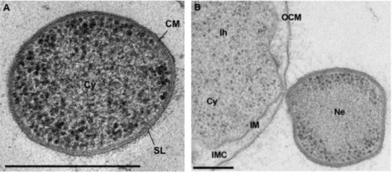
I. hospitalis forms cocci with a cell diameter of 1.5-5 µm and exhibit a new type of cell surface appendages. Those fibers (Ø 14 nm) are anchored by spherical structures located beneath the inner membrane in the cell and show features as in type IV pilus-like structures (Müller et al., 2009; Meyer et al., 2014). Intriguingly, I. hospitalis cells exhibit unusual cell architecture for a prokaryote. Unlike most other archaea, they lack an S-layer and are instead surrounded by two membranes: the inner and the outer cellular membrane (IM, OCM respectively) (Näther and Rachel, 2004; Huber et al., 2012).
Both membranes form a large inter-membrane compartment (IMC), which is completely separated from the cytoplasm (Figure 9B) (Rachel et al., 2002). The localization of different membrane proteins involved in energy conservation in the OCM leads to an energization of the outer cellular membrane, which is highly unusual. In contrast to other prokaryotes, the H2:sulfur oxidoreductase (primary proton
I Introduction
18
(Küper et al., 2010). This results in a structural as well as in a functional compartmentalization. With the Acetyl-CoA synthetase, which is associated with the OCM, a first ATP-consuming process in the IMC was identified (Mayer et al., 2012). Recently, another novel membrane-associated octa-heme cytochrome c was characterized, which is believed to be involved in detoxification or in respiratory energy conservation due to its nitrite reductase activity (Parey et al., 2016).
Both membranes differ in their lipid composition: whereas the OCM consists mainly of archaeol, the IM is made up of archaeol and caldarchaeol (Jahn et al., 2004). Moreover, a novel pore-forming complex Ihomp1 was identified in the OCM of I. hospitalis. It represents the most abundant protein in the OCM, for which no recognizable homologues have been found in other archaea (Burghardt et al., 2007; Burghardt et al., 2008). In earlier studies, numerous vesicles of different shape and size were observed within the IMC. Those vesicles were either budding from the IMC or undergoing fusion with the IMC or OCM (Rachel et al., 2002; Näther and Rachel, 2004). Recent studies for I. hospitalis identified a complex and highly dynamic endomembrane system, which consists of cytoplasmic protrusions and might have secretory function. Moreover, filamentous structures, similar to a cytoskeleton, were observed in the IMC (Heimerl et al., 2017).
The genome of I. hospitalis is the most minimal organized genome known to date that guarantees an independent way of living. With only a size of 1.3 Mbp it encodes for 1,444 genes, whereas less than 3 % of the proteome encodes for transport proteins (Podar et al., 2008). Interestingly, comparative analysis of the proteome of I. hospitalis in single and co-culture with N. equitans showed that the relative abundance of membrane proteins increases up to 50 % in co-culture. This up-regulation affected especially protein complexes involved in energy conservation as well as proteins involved in membrane stabilization and transporters (Giannone et al., 2015).
If the eukaryotic cell originated from an archaeal ancestor, as many believe, then an organism like I. hospitalis, with its large ATP-rich intermembrane compartment, is an ideal candidate for such an ancestor; providing easy ATP and other metabolites to an incorporated symbiont.
2.3 N. equitans: a putative ectoparasite?
N. equitans forms tiny cocci with cell diameters of only 0.35-0.5 µm and exhibits a typical archaeal cell architecture: cytoplasm, cytoplasmic membrane and S-Layer (Figure 9A) (Huber et al., 2002;
Huber et al., 2003). The cytoplasmic membrane consists of archaeol and caldarchaeol like the IM of I. hospitalis. The interaction between both organisms is mediated by filamentous structures or a direct cell-cell contact and is limited to a relatively small contact area of only 40-170 nm in diameter (Figure