Synthesis and biological evaluation of small molecule GLUT inhibitors that cause glucose starvation in cancer
Dissertation
For the achievement of the academic degree of the Doctor in Natural Sciences
(Dr. rer. nat.) Submitted to
Faculty of Chemistry and Chemical Biology TU Dortmund University
by
Elena Sabrina Reckzeh
from Hilden, Germany
Dortmund 2019
Die vorliegende Arbeit wurde zwischen März 2015 und Mai 2019 unter der Anleitung von Prof. Dr.
Dr. h.c. Herbert Waldmann und Dr. Slava Ziegler an der Fakultät für Chemie und Chemische Biologie der Technischen Universität Dortmund und an dem Max-Planck Institut für Molekulare Physiologie Dortmund angefertigt.
Dekan: Prof. Dr. Stefan M. Kast
1. Gutachter: Prof. Dr. Dr. h.c. Herbert Waldmann
2. Gutachter: Prof. Dr. Daniel Summerer
Results presented in this thesis contributed to the following publications:
Ceballos J, Karageorgis G, Schwalfenberg M, Reckzeh ES, Sievers S, Ostermann C, Pahl A, Ziegler S, Waldmann H, “Synthesis and Biological Evaluation of Indomorphan Pseudo Natural Products”, submitted.
Schwalfenberg M
#, Ceballos J
#, Reckzeh ES, Karageorgis G, Sellstedt M, Nowacki J, Carnero Corrales MA, Wilke J, Laraia L, Tschapalda K, Metz M, Sehr DA, Rahmann S, Winklhofer K, Janning P, Ziegler P, Waldmann H, “A Dual-Specific GLUT-1- and GLUT-3 Inhibitor Potently Suppresses Cancer Cell Growth”, submitted.
Reckzeh ES, Karageorgis G, Schwalfenberg M, Ceballos J, Nowacki J, Stroet MCM, Binici A, Knauer A, Brand S, Strohmann C, Ziegler S, Waldmann H. “Inhibition of Glucose Transporters and Glutaminase Synergistically Impairs Tumor Cell Growth.” Cell Chemical Biology, in press.
Reckzeh ES, Brockmeyer A, Metz M, Waldmann H, Janning P, “Target Engagement of Small Molecules: Thermal Profiling Approaches on Different Levels”, Methods in Molecular Biology, Systems Chemical Biology, Ed. Ziegler S, Ed. Waldmann H, New York: Humana Press, 2019, Vol.
1888, 73-98.
Karageorgis G, Reckzeh ES, Ceballos J, Schwalfenberg M, Sievers S, Ostermann C, Pahl A,
Ziegler S, Waldmann H. Chromopynones are “Pseudo Natural Product” Glucose Uptake Inhibitors
Targeting Glucose Transporters GLUT-1 and -3” Nature Chemistry, 2018, 10, 1103-11.
1 Table of content
1 TABLE OF CONTENT ... 1
2 ACKNOWLEDGEMENT ... 4
3 SUMMARY ... 6
4 ZUSAMMENFASSUNG ... 8
5 INTRODUCTION ... 10
5.1 CANCER ... 10
5.2 THE WARBURG EFFECT ... 11
5.3 GLUCOSE TRANSPORTERS ... 13
5.3.1 Glucose uptake inhibitors ... 17
5.4 METABOLIC REWIRING ... 21
5.4.1 KRas in metabolic rewiring ... 22
5.4.2 Metabolic flexibility and glutamine ... 24
5.5 FURTHER APPLICATIONS OF GLUCOSE UPTAKE INHIBITORS ... 25
6 AIM OF THE THESIS ... 26
7 EXPERIMENTAL PART ... 27
7.1 EXPERIMENTAL PART – CHEMISTRY ... 27
7.1.1 Materials ... 27
7.1.2 Instrumentation ... 27
7.1.3 Methods ... 28
7.1.3.1 Preparative HPLC-MS ... 28
7.2 EXPERIMENTAL PART – BIOLOGY ... 32
7.2.1 Material ... 32
7.2.2 Instruments and software ... 40
7.2.3 Cell biology methods ... 42
7.2.4 Biochemical methods ... 53
7.2.5 Methods to study target engagement of small molecules ... 57
7.2.6 Compound absorbance and fluorescence spectra ... 59
7.2.7 Data analysis ... 60
8 RESULTS ... 62
8.1 IDENTIFICATION OF SMALL-MOLECULE INHIBITORS OF GLUCOSE UPTAKE ... 62
8.2 CHEMICAL INVESTIGATION OF THE PIPERAZIN-2-ONE CLASS ... 64
8.2.1 Confirmation of the glucose uptake inhibitory activity of 1a ... 64
8.2.2 Synthesis of a piperazin-2-one-based compound library ... 66
8.2.3 Structure-activity relationship (SAR) studies of the piperazin-2-ones ... 69
8.3 BIOLOGICAL INVESTIGATION OF GLUTOR ... 77
8.3.1 Mode of action of Glutor ... 77
8.3.2 Drug-likeness of Glutor ... 79
8.3.3 Influence of Glutor on cancer cell growth ... 80
8.3.4 Glutor induces cell death in physiologically relevant spheroids ... 86
8.3.5 Metabolic profiles of different cell lines correlate with Glutor sensitivity ... 87
8.3.6 GLUT-1, GLUT-2 and GLUT-3 are targets of Glutor ... 89
8.3.7 Cancer cells upregulate GLUT-1 and GLUT-3 to escape glucose starvation ... 94
8.3.8 Nutrient dependencies of HCT116 cells ... 99
8.3.9 Influence of co-treatment on growth of HCT116 cells ... 110
8.4 BIOLOGICAL CHARACTERIZATION OF OTHER GLUT INHIBITORS ... 119
8.4.1 (+)-Glupin-1 ... 119
8.4.2 Chromopynones ... 129
8.4.3 BAY-876 ... 135
9 DISCUSSION ... 148
9.1 STRUCTURAL OPTIMIZATION OF GLUTOR ... 149
9.2 GLUT ISOFORM SELECTIVITY ... 152
9.2.1 Target identification, validation and off-targets ... 153
9.3 RESCUE EFFECT ... 154
9.3.1 Regulation of B2M ... 156
9.4 CANCER CELL GROWTH INHIBITION ... 157
9.5 RAS MUTATION AND GLUCOSE ADDICTION ... 160
9.6 METABOLIC FLEXIBILITY AND COMBINATORY TREATMENT APPROACH ... 162
9.6.1 Other combinatorial treatments ... 164
9.7 FURTHER INDICATIONS AND APPLICATIONS OF GLUCOSE UPTAKE INHIBITORS ... 167
10 OUTLOOK ... 170
11 ABBREVIATIONS ... 171
12 LITERATURE ... 175
13 APPENDIX ... 187
13.1 SUPPLEMENTARY FIGURES ... 187
13.2 SUPPLEMENTARY TABLES ... 194
13.3 COMPOUND CHARACTERIZATION ... 204
14 CURRICULUM VITAE ... 243
15 PUBLICATION LIST ... 246
2 Acknowledgement
Throughout all my studies I could not wait to dive deep into a project and start to take on responsibility, as well as to follow own ideas. The PhD phase was the most exciting and enriching time of my education and I truly enjoyed every day of it. I would like to thank all people that contributed to this important phase of my life.
First and foremost, I would like to thank Prof. Dr. Dr. h.c. Herbert Waldmann for the opportunity to obtain my PhD in his group. The versatile project, the independent way of working as well as the access to high-end equipment and the excellent academic environment contributed to my scientific maturation. I truly appreciate all the support that I received to grow on so many levels.
I am sincerely grateful to Dr. Slava Ziegler, my co-supervisor and group-leader, who was always helpful during professional and personal hurdles. Her valuable, constructive input contributed to the success of this project. Moreover, the personal feedback I received throughout these years clearly motivated me to always give my very best.
I would like to thank Prof. Dr. Daniel Summerer for taking over the function of the second examiner.
My personal thanks go to the students, Jessica Nowacki, Marc Stroet and Aylin Binici, that I supervised throughout these past four years during their Bachelor and Master thesis and who were always extremely dedicated to participating in this project with their scientific work and creative ideas. Moreover, I thank the talented trainees Laura Ney and Alexandra Brause for their assistance. I would also like appreciate my gratitude to Beate Schölermann for her support during the last months of my PhD.
I would like to thank Melanie Schwalfenberg, Dr. Javier de Ceballos and Dr. George Karageorgis
who worked together with me on this project. I would also like to thank Julian Wilke, Dr. Silke
Brand and Lena Knauer who contributed some of the experiments. Furthermore, I appreciated all
discussions with the people from the Lead Discovery Center about this project and sharing their knowledge and expertise.
I would like to particularly thank the mass spectrometry team of Dr. Petra Janning, Malte Metz, Jens Warmers and Andreas Brockmeyer for measuring samples, analyzing data and for their constructive feedback on experimental design and analysis. I am very thankful to the Compound Management and Screening Center, particularly Dr. Sonja Sievers, Claude Ostermann and Dr.
Axel Pahl for their support in all compound screenings, compound handling and computational analysis. I furthermore appreciate the continuous help in all administrative issues from Brigitte Rose.
I would like to express my gratitude for the financial, personal and scientific support that I received during my time in the International Max Planck Research School in Chemical and Molecular Biology. Here my special thanks go to Christa Hornemann and Dr. Lucia Sironi who were always highly encouraging and motivating and make the graduate school an outstanding place to grow.
The nice working atmosphere in the department would not have been possible without Elisabeth Hennes, Julian Wilke, Prof. Dr. Luca Laraia, Dr. Luis Bering, Beate Schölermann, Dr. Sumersing Patil, Dr. Lea Kremer, Prof. Dr. Shobhna Kapoor, Dr. Tim Förster, Michael Winzker, Andreas Christoforow, Dr. Hélène Adihou, Stefan Zimmermann, Jens Warmers, Annina Burhop, Tabea Schneidewind, Dr. Marjorie Rummelt, Melanie Schwalfenberg, Dr. Javier de Ceballos, Dr. Kirsten Tschapalda, Dr. Janine Schulte-Zweckel and Dr. Guillaume Garivet.
My deepest and heartfelt appreciation goes to my husband and best friend Michael Reckzeh for
all the support, understanding and encouragement as well as exciting journeys that will always
continue. All my achievements would not have been possible without my parents, Roland and
Barbara Heider, who supported me throughout my life in the best way imaginable. I also thank my
siblings, Janina and Damian Heider, as well as Markus Geimer for being my family.
3 Summary
Deregulation of cellular energy metabolism has been well established as a hallmark of cancer.
Increased aerobic glycolysis, termed the Warburg effect, is associated with an overexpression of different glucose transporter isoforms, especially GLUT-1 (ubiquitously expressed). To date, numerous GLUT inhibitors have been reported to reduce cancer growth, albeit improvement of selectivity and efficacy as well as unspecific toxicity of such inhibitors remain to be major challenges. In order to identify compounds that block glucose uptake of cancer cells, a cell-based screen that monitors 2-deoxy-D-glucose (2-DG) uptake by cancer cells was employed. This led to the discovery and further investigation of three distinct substance classes of which one was investigated in-depth within this dissertation. Chemical investigation of the main compound class, the piperazin-2-ones, involved detailed planning and execution of compound synthesis to improve potency and physicochemical properties and establish a conclusive structure-activity relationship (SAR). The thereby generated most potent, enantiomerically pure compound, termed Glutor, inhibited glucose uptake and reduced the glycolysis rate of cancer cells with low nanomolar potency. In a panel of 94 cell lines Glutor impeded the growth of 44 different cancer cell lines with an IC
50<100 nM, while non-malignant cells were unaffected. Interestingly, among the most sensitive cancer cells, mutations in KRas were enriched. Spheroids were employed to investigate the cellular potency in a physiologically more relevant cell model. Thereby, Glutor induced a strong increase in cytotoxic events after 24 hours of treatment, which even outreached the compound effect on monolayer-cultured cells.
Furthermore, among the class I GLUTs, a GLUT-1/-2/-3 isoform selectivity was determined for Glutor by employing overexpression experiments of the respective isoform, which lead to partial rescue of 2-DG uptake inhibition and verification by cellular thermal shift assays (CETSA®).
Subsequent studies revealed an upregulation of GLUT-1 and GLUT-3 expression levels under
glucose starvation conditions. This proved the necessity of a GLUT-1/-3 selectivity profile to
achieve a full inhibitory effect on glucose uptake. Numerous reports describe the dependence of in vitro cultured cancer cells on glutamine to maintain catabolism and viability. Therefore, the effect of varying extracellular glutamine concentrations on glucose dependence as well as Glutor treatment was investigated and revealed that physiological glutamine concentrations (0.5 mM) increased the potency of both compound treatment and glucose deprivation. In line with this, a strong synergistic effect in a combinatory treatment using the glutaminase inhibitor CB-839 was uncovered. These experiments prove the possibility of novel combinatory treatment approaches for anti-cancer therapy. In the scope of this dissertation, derivatives of the other two substance classes, (R,R)-Chromopynone-1/2 and (+)-Glupin-1, were investigated as well. Thereby, several findings made for Glutor could be determined as general for glucose uptake inhibitors.
The new small molecular GLUT inhibitors may ultimately pave the way for the investigation of glucose uptake inhibition as a new therapeutic anti-cancer strategy or in a combinatory treatment approach with other drugs.
Figure 1 Graphical abstract indicates the synergistic effect between Glutor and the glutaminase inhibitor CB- 839 as well as the observed rescue effect upon glucose starvation. GLS: glutaminase kidney isoform, TCA:
tricarboxylic acid cycle, GLUT: facilitative glucose transporter.
4 Zusammenfassung
Die Veränderung des zellulären Metabolismus ist ein Hauptmerkmal von Krebserkrankungen.
Diese schließt die aeroben Glykolyse ein, auch Warburg-Effekt genannt, die zur vermehrten Expression von Glukose-Transportern (GLUTs), insbesondere GLUT-1, führt und dadurch zur erhöhten Glukoseaufnahme in Krebszellen. Um die Glukosezufuhr von Krebszellen zu unterbinden, wurden bereits einige GLUT-Inhibitoren entwickelt, die jedoch mangelnde Selektivität, Wirksamkeit sowie unspezifischen Nebenwirkungen aufweisen. Um neue niedermolekulare Glukoseaufnahme-Inhibitoren zu identifizieren, wurde ein Zell-basiertes Testsystem eingesetzt, das die Aufnahme von 2-Desoxy-D-Glukose (2-DG) in Krebszellen nachweist. Aus einer Bibliothek von über 150.000 niedermolekularen Substanzen wurden drei Substanzklassen identifiziert, welche die 2-DG-Aufnahme im niedrigen mikromolaren Bereich inhibieren. Eine dieser Substanzklassen, die Piperazin-2-one, wurde im Rahmen dieser Arbeit näher untersucht. Mithilfe organisch synthetischer Methoden wurden weitere Piperazin-2-on- Derivate generiert, um die Wirksamkeit sowie physikochemische Eigenschaften verbessert. Durch eine umfassende Strukturwirkungsbeziehung konnte die aktivste Verbindung, das Enantiomer Glutor, identifiziert werden. Glutor hemmte die Glukoseaufnahme und reduzierte die Glykolyserate von Krebszellen im niedrigen nanomolaren Bereich. Zudem unterband Glutor das Wachstum von diversen Krebszelllinien mit einem IC
50<100 nM, wobei nicht entartete Zellen unbeeinflusst blieben. Auffällig war hierbei eine Anreicherung von KRas-Mutationen unter den sensitiven Zelllinien. In physiologisch relevanteren, dreidimensionalen Sphäroiden führte die Behandlung mit Glutor bereits nach 24 Stunden zu einem, im Vergleich zu einlagig kultvierten Zellen, stärkeren Anstieg der Zytotoxizität.
Die Überexpression von GLUT-1/-2 und -3 verringerte die Glutor-vermittelte Inhibition der 2-DG-
Aufnahme, was auf die Inhibition der Isoformen GLUT-1/-2/-3 hindeutet. Zudem erhöhte Glutor
die Schmelztemperatur von GLUT-3 in Zelllysaten, was die Bindung an GLUT-3 und somit GLUT-
3 als Zielprotein bestätigte. Unter Glukoseentzug konnte zudem ein Anstieg der GLUT-1- und GLUT-3-Expression verzeichnet werden. Dieser Befund verdeutlicht die Relevanz eines GLUT- 1/-3-Selektivitätsprofils, um eine vollständige Hemmung der Glukoseaufnahme von Krebszellen zu garantieren. In vitro kultivierte Zellen sind auf extern hinzugefügtes Glutamin angewiesen, und viele Krebszellen sind Glutaminabhängig. In Übereinstimmung damit wies Glutor eine erhöhte Wirksamkeit bei geringerer, physiologischer Glutaminkonzentration von 0,5 mM auf. Die kombinatorische Anwendung von Glutor und dem Glutaminase-Inhibitor CB-839 zeigte einen klaren synergistischen, inhibitorischen Effekt auf das Zellwachstum. Dies könnte einen wirkungsvollen, kombinatorischen Behandlungsansatz für die Krebsbekämpfung darstellen. Im Rahmen dieser Arbeit wurden auch Derivate der zwei weiteren identifizierten Substanzklassen, (R,R)-Chromopynone-1/-2 und (+)-Glupin-1, näher untersucht. Viele der Erkenntnisse, die durch die Untersuchungen mit Glutor gewonnenen wurden, konnten als allgemeingültig für Glukoseaufnahme-Inhibitoren bestätigt werden.
Die im Rahmen dieser Arbeit untersuchten niedermolekularen Inhibitoren haben das Potential,
den Grundstein für den therapeutischen Einsatz von Glukoseaufnahme-Inhibitoren in der
Krebsforschung allein oder in Kombination mit anderen Medikamenten zu legen.
5 Introduction
5.1 Cancer
Cancer is a collective term for a large group of diseases that can affect any tissue of the body. It is characterized by uncontrolled cell proliferation that arises from numerous genetic alterations and most often results in tumor formation. Cancer cells invade the surrounding healthy tissue and can migrate to distal sites through the blood stream to form new tumors (metastasis). These metastases are usually the cause of death. Cancer is the second leading cause of death worldwide, while 70% of the associated deaths occur in low- and middle-income countries because of limited access to diagnostic and treatment opportunities.
1,2Traditional cancer treatments include the operative removal of the tumor, radiation and
chemotherapy or a combination of these. Since all three procedures entail a large degree of side
effects, modern research focuses on two main areas: immunooncology and targeted cancer
therapy. The aim of immunooncology is to reactivate the patient’s immune system to antagonize
the cancer, whereas the latter focuses on the
(molecular) differences between normal and cancer
cells to selectively kill cancer. These differences have been grouped by Hannahan and Weinberg
in 2000 as the seven hallmarks that enable cells to evolve into a malignant state. These hallmarks
include resisting cell death, sustaining proliferative signaling, evading growth suppressors,
activating invasion and metastasis, enabling replicative immortality and inducing angiogenesis.
2Further studies have revealed a second layer of complexity which is combined in the second
generation of hallmarks published by Hannahan and Weinberg in 2011 (Figure 2). Hannahan and
Weinberg added two enabling characteristics (genome instability and tumor-promoting
inflammation) and two emerging characteristics (avoiding immune destruction and deregulating
cellular energetics).
3The adapted energy metabolism of cancer is one hallmark that is recently
gaining more attention for targeted cancer therapy and is the focus of this thesis.
Figure 2 The second generation hallmarks of cancer as postulated by Hannahan and Weinberg.3
5.2 The Warburg effect
Non-malignant, resting cells convert the highly abundant nutrient glucose during glycolysis to pyruvate. Pyruvate subsequently enters the tricarboxylic acid (TCA) cycle and mitochondrial oxidative phosphorylation to generate 36 mol adenosine triphosphate (ATP) per mole glucose that is consumed by the cell (Figure 3a). In the absence of oxygen, cells convert glucose into pyruvate, which is fermented into lactate, generating 2 mol ATP per mole glucose. In 1924, Otto Warburg was the first scientist to observe that cancer cells perform glycolysis regardless of oxygen availability (Figure 3b).
4Thus, this observation is also known as the Warburg effect or aerobic glycolysis.
At first glance, this adaptation is counterintuitive since cancer cells require energy for their rapid
proliferation and generate 9-fold less ATP during aerobic glycolysis (since mitochondria remain
functional and oxidative phosphorylation occurs at low levels aerobic glycolysis generates ~4 mol
ATP/mol glucose).
3,5However, the rate of glycolysis is 10-100 times higher than the rate of oxidative phosphorylation, which compensates for the low ATP yield per mole glucose.
6The faster kinetics of glycolysis provide an advantage, when cancer cells compete with surrounding stroma cells for glucose.
6Figure 3 Metabolism of differentiated tissue and tumor cells. Schematic representation of the metabolic phenotypes of differentiated tissue (a) and tumor cells (b). The contribution of the metabolic pathways to proliferation is depicted in c). PPP=pentose-phosphate pathway, TCA=tricarboxylic acid cycle, OXPHOS=oxidative phosphorylation, GLUT=facilitative glucose transporter, GLS=glutaminase kidney isoform.
Another hypothesis for this metabolic adaptation is that intermediates, which are generated during
glycolysis, serve as a starting material for e.g. nucleoside biosynthesis using the pentose-
phosphate pathway and are therefore essential for DNA replication and division.
6Also, the need
for reducing equivalents NADPH is much higher than ATP demand. NADPH is produced in high
amounts through the pentose phosphate pathway, which is a pathway branch of glycolysis (Figure
3b). Thus, until today the exact reason why cancer cells perform aerobic glycolysis remains
controversial.
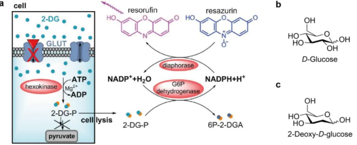
6To accelerate glucose uptake and thereby the rate of glycolysis, cancer cells overexpress the facilitative glucose transporters (GLUTs). High expression levels of the isoform GLUT-1 have been associated with poor patient prognosis and advanced tumor stages, which is why GLUT-1 was proposed as diagnostic marker.
7–9This phenomenon is now applied for tumor localization and diagnosis. Patients receive the glucose derivative 2-deoxy-2-(
18F)fluoro-D-glucose (FDG) intravenously, which is rapidly taken up by all tissues with high glucose consumption, such as tumors, within the human body.
10These can be detected with positron emission tomography (PET). 2-Deoxy-D-glucose (2-DG) itself reduces the glycolytic rate by means of hexokinase inhibition and is investigated in multiple clinical trials alone (trial I/II) and together with other anti- cancer treatment strategies, i.e. the chemotherapeutic agent docetaxel or radiotherapy.
11–13Naturally, targeting anaerobic glycolysis is a promising approach for cancer therapy. To date, no medication targeting metabolic differences in cancer cells has been introduced to the market, which underscores the high need for new therapeutic entities. In 2014, the Food and Drug Administration (FDA) approved the drug Enasidenib (AG-221), which inhibits a metabolic target.
This small molecule targets the cancer-associated mutated form of isocitrate dehydrogenase (IDH) 2 in order to reduce the production of the oncometabolite 2-hydroxyglutarate (2-HG) and does not target any metabolic adaptation of cancer directly.
145.3 Glucose transporters
Glucose is a hydrophilic molecule that cannot pass the plasma membrane of cells. Therefore,
glucose is transported via two main protein families, either via passive diffusion (facilitative glucose
transporters – GLUTs) or together with sodium by an electrochemical gradient (sodium-dependent
co-transporters - SGLTs). GLUTs are integral membrane proteins that passively transport glucose
according to the prevalent glucose gradient. There are 14 GLUT isoforms known so far that are
expressed in a tissue-specific manner and which are involved in central metabolic processes, e.g.
basal glucose uptake (GLUT-1). The isoforms are divided into three distinct protein classes I-III according to their amino acid similarity (Figure 4).
15Figure 4 GLUT isoforms of class I-III. a) CLUSTAL multiple alignment revealed amino acid sequence similarities between GLUT isoforms. Adapted from Scheepers et al.15 b) Function and main expression sites of all GLUTs.16,17
Class I GLUTs are the most studied isoforms and comprise five different proteins: GLUT-1, GLUT- 2, GLUT-3, GLUT-4 and GLUT-14, which is a duplicon of GLUT-3 and has not gained much attention. The function of most other GLUTs (class II and III) is still a matter of debate or unknown.
GLUT-1 is mainly expressed in erythrocytes and blood-tissue barriers to ensure glucose uptake
from the blood stream into organs. Furthermore, GLUT-1 is ubiquitously expressed and ensures
basal glucose supply of nearly all somatic cells. GLUT-2 is involved in glucose sensing in the
pancreas and GLUT-2-mediated glucose uptake is coupled to insulin secretion. Due to GLUT-2’s
low glucose affinity (Table 1), its major purpose is the postprandial glucose consumption.
18GLUT-
3 has the highest affinity for glucose and is expressed primarily in neurons. GLUT-4 is mainly
expressed in insulin-responsive tissues such as muscle or fat. Most expressed GLUT-4
transporters are stored in vesicles when insulin is absent. Upon insulin secretion GLUT-4
membrane translocation is induced.
Table 1 Overview of substrate specificity and affinity for the GLUT isoforms 1-4.18,19
GLUT isoform GLUT-1 GLUT-2 GLUT-3 GLUT-4
Characteristics High affinity
Low affinity High capacity
High velocity
High affinity
High capacity High affinity
K
mGlc 3-7 mM 17 mM 1.4 mM 6.6 mM
K
m2-DG 5 mM ~ 1.4 mM
K
mFru no ~ 76 mM yes
K
mGal ~ 17 mM ~ 92 mM ~ 8.5 mM
K
mMan yes ~ 125 mM yes
K
mGlcN 0.8 mM yes 3.9 mM
K
m3-OMe-Glc ~ 20 mM
DHA yes yes yes
Others GalN Maltose, Xyl
Highest substrate affinity is highlighted in grey. 2-DG=2-deoxy-D-glucose, Glc=glucose, Man=mannose, Mt=maltose, Xyl=xylose, DHA=dehydroascorbic acid, GlcN=glucosamine, Fru=fructose, GalN=galactosamine.
The structures of human GLUT-1
20(inward-open conformation) and GLUT-3
21(outward-open conformation) have been solved by means of X-ray crystallography. Other human GLUT isoform structures are based on computer homology models of the large-conductance mechanosensitive ion channel (MscL) from Mycobacterium tuberculosis, glycerol-3-phosphate transporter GlpT and GLUT-1-4 homologs of Escherichia coli.
22The sequences of GLUT-1-4 are highly conserved (Figure 4, Figure 5a) and the glucose binding site exhibits 100% sequence identifity among GLUT- 1-4 (Figure 5b). Different substrate affinities for glucose or susceptibility for other sugars (Table 1) must therefore originate from the surrounding amino acids. Variation between the amino acid sequences of these four isoforms occur within each of the 12 transmembrane helices.
Interestingly, GLUT-1 possesses unique sequence stretches in comparison to GLUT-2-4 at the
intracellular helix and within the extracellular loops (Figure 5a).
Figure 5 Crystal structure of GLUT-1 and similarity to GLUT-2-4. Structural similarity of GLUT-1-4 isoforms as illustrated on the inward-open crystal structure of GLUT-1 as overview (a) and within the glucose binding site (b). Amino acid alignment of GLUT-1-4 was reported by Zhao et al.23 PDB: 4PYP20.
The overexpression of all isoforms GLUT-1-4 has been described for cancers, mostly associated
with the tissue of their respective site of expression.
18The GLUT-1 isoform is upregulated in nearly
all malignant cells. The development of GLUT-1-selective inhibitors for targeted cancer therapy
was therefore a long standing goal that was only recently addressed by Siebeneicher et al..
24However, high expression levels of GLUT-3 were also determined in a wide range of cancer types,
among others endometrial and breast cancers, head and neck tumors, thyroid carcinomas, colon
and pancreatic, and non-small cell lung cancer.
25–30The required isoform selectivity profile of
GLUT inhibitors to potently target glucose uptake in cancer remains therefore uncertain. Multiple
selective and unselective inhibitors have been reported and will be discussed in the following
chapter.
5.3.1 Glucose uptake inhibitors
The basis of targeted cancer therapy is the discovery of molecules that perturb a biological process that is prevalent in cancer. Such molecules are identified in a process called chemical genetics. Compared to the traditional genetics approaches where certain genes are interrupted to elucidate their function via phenotype analysis, chemical genetics makes use of compounds that disrupt the same process. One disadvantage of the classical genomics approach is the poor transferability to human cells due to their complex genome and their slow reproduction rate compared to e.g. yeast and bacterial cells. Compounds that are utilized in chemical genetics can be natural products, peptides or small molecules. In order to identify compounds that selectively disrupt these biological processes, two main approaches are applied: forward chemical genetics and reverse chemical genetics (Figure 6).
31,32Figure 6 Overview of the classical genetics approach (top) and the chemical genetics approach (bottom). Both approaches can be performed in a forward or a reverse fashion. Figure adapted from Lehár et al.32
In a forward chemical genetics approach, the identification of small molecules that induce a certain
phenotype stands in focus (Figure 6, left to right). Usually phenotypic cell-based assays are
conducted in the presence of a large compound library to identify such hit compounds. Target
identification approaches are subsequently applied to determine the protein whose (altered)
function is responsible for phenotype induction. This way, novel interesting proteins and protein
functions can be elucidated. The reverse chemical genetics approach employs biochemical or cellular assays that investigate the manipulation of a specific gene or protein activity by means of small molecules. Subsequent studies deal with the investigation of the thereby caused phenotype (Figure 6, right to left).
Regardless of the approach, the discovery and further development of small molecules that can be used to perturb biological systems is of extreme importance not only to generate novel tool compounds, but also to pave the way for therapeutic applications. The year of 2018 broke all records for the Food and Drug Administration (FDA)-approved drugs: 59 new drugs (42 New Chemical Entities (NCE) and 17 biologics) were introduced to the drug market.
33Out of the 42 NCE, 47% are small molecules whereas 16% are natural products.
33This underscores the tremendous potential that lies in the discovery of novel small molecular tool compounds.
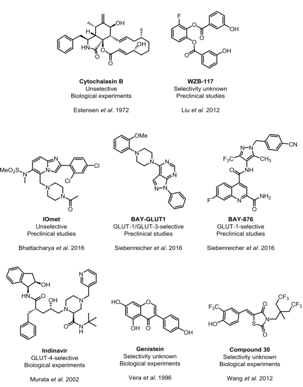
Glucose uptake inhibitors from diverse chemical classes have been investigated since the late 20
thcentury (Figure 7, Table 2). Cytochalasin B, a fungal metabolite that was originally described to inhibit cytokinesis, cell motility and disturbance of cell shape, was shown to inhibit glucose uptake by Estensen et al. in 1972 (Figure 7, Table 2).
34Since then, this compound was utilized as an unselective tool compound for glucose uptake inhibition in numerous studies.
35–37However, Cytochalasin B inhibits actin polymerization, which makes the compound unattractive for further clinical investigation due to the fundamental reliance of the human organism on this process.
38In the 21
stcentury, the systematic development of more specific glucose uptake inhibitors began.
Wang et al. reported a small molecule, compound 30, that inhibited glucose uptake and reduced
the viability of the prostate carcinoma cell line LNCaP with low micromolar IC
50value (Figure 7,
Table 2).
39Lui et al. discovered the tool compound WZB-117 that inhibits glucose uptake with
submicromolar potency (Figure 7, Table 2).
16Even though after 48 h of treatment the effect on
proliferation of the human lung cancer cell line A549 was rather weak (IC
50=10 µM), WZB-117
reduced the growth of A549 mouse xenograft models by 70% after daily intraperitoneal injection of WZB-117 at 10 mg/kg for 10 weeks.
16Figure 7 Selection of small molecule GLUT inhibitors. The potencies of all depicted compounds can be found in Table 2.
IOmet Pharma Ltd. filed a patent for GLUT inhibitors in 2014. One highly potent but unselective compound, herein referred to as IOmet, is illustrated in Figure 7 (Table 2).
40One described IOmet analog reduced lactate excretion and inhibited growth of A549 cells with nanomolar potency.
40In 2016, the Bayer corporation published two novel chemotypes, BAY-GLUT1 and BAY-876, that were discovered based on a high-throughput screening for inhibition of glycolysis-dependent ATP production (Figure 7, Table 2).
24,41BAY-876 was the first and until now the only GLUT-1-selective and highly potent (IC
50=2 nM for DLD-1 cells) glucose uptake inhibitor.
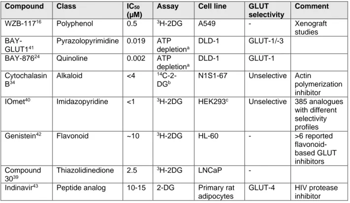
Table 2 Chemical class, potency and GLUT isoform selectivity of selected glucose uptake inhibitors (Figure 7).
Compound Class IC50
(µM)
Assay Cell line GLUT
selectivity
Comment
WZB-11716 Polyphenol 0.5 3H-2DG A549 - Xenograft
studies BAY-
GLUT141
Pyrazolopyrimidine 0.019 ATP depletiona
DLD-1 GLUT-1/-3 BAY-87624 Quinoline 0.002 ATP
depletiona
DLD-1 GLUT-1
Cytochalasin B34
Alkaloid <4 14C-2- DGb
N1S1-67 Unselective Actin
polymerization inhibitor IOmet40 Imidazopyridine <1 3H-2DG HEK293c Unselective 385 analogues
with different selectivity profiles Genistein42 Flavonoid ~10 3H-2DG HL-60 - >6 reported
flavonoid- based GLUT inhibitors Compound
3039
Thiazolidinedione 2.5 3H-2DG LNCaP - Indinavir43 Peptide analog 10-15 2-DG Primary rat
adipocytes
GLUT-4 HIV protease inhibitor
aATP depletion in the presence of Rotenone; bincorporation of 14C into lactate; cHEK293 cells overexpress human GLUT-1, GLUT-2, GLUT-3 or GLUT-4 transiently.
Over the past three decades, 15 flavonoids have been identified to inhibit glucose uptake. Among
these are Phloretin
44, Fasentin
35, Silybin
45, Apigenin
46and Genistein
42(Figure 7, Table 2).
47–49Genistein inhibited 2-DG uptake with micromolar potency. The HIV protease inhibitor Indinavir
was recently described as selective GLUT-4 inhibitor, which stopped 2-DG uptake with micromolar potency in primary rat adipocytes (Figure 7, Table 2).
43None of the above-mentioned GLUT inhibitors are currently investigated in clinical trials. Thus, there is still a high demand to develop novel GLUT inhibitors, which will pave the way for drug discovery.
5.4 Metabolic rewiring
Metabolic adaptations in cancer have been associated with the activation of proto-oncogenes such as the GTPase KRas (KRas), dysfunction of tumor suppressors like the cellular tumor antigen p53 (p53) and von Hippel-Lindau disease tumor suppressor (VHL), dysregulated expression of the Myc proto-oncogene protein (Myc) and stabilization of hypoxia-inducible factor 1-alpha (HIF-1- α).
50–52HIF-1-α regulates the transcription of different glycolytic enzymes, including GLUTs. Under normoxic conditions, HIF-1-α is hydroxylated via prolyl hydroxylases which increases the affinity to VHL, a E3 ubiquitin ligase, that induces the proteasomal degradation of HIF-1-α. Hypoxic conditions and mutations in VHL therefore stabilize HIF-1-α. Also, phosphatidylinositol-4,5- bisphosphate 3-kinase (PI3K) signaling that acts via RAC-alpha serine/threonine-protein kinase (AKT1) and serine/threonine-protein kinase mTOR (mTOR) activation as well as accumulation of reactive oxygen species (ROS) are associated with HIF-1-α stabilization.
53Inhibitors of glucose metabolism reduce growth of xenograft tumors that are derived from Myc- or
KRas-driven cancer cells.
54–56Therefore, targeting metabolism as an effector of signaling
pathways that control cell growth might be an effective strategy to address cancers that are driven
by these genetic alterations and cannot be targeted directly.
57Of note, several factors that are
involved in glucose addiction, such as Myc, also drive glutaminolysis. Thereby, cancer gains the
flexibility to switch between the two metabolic pathways, depending on the availability of glucose and glutamine.
5.4.1 KRas in metabolic rewiring
Mutated Ras is present in ca. 25% of all human cancers,
58with a strong accumulation in colorectal
cancers (CRC) (40% KRas mutated)
59, pancreatic ductal adenocarcinoma (PDAC) (>90% KRas
mutated)
60and non-small cell lung cancer (NSCLC).
5In general, the Ras isoform KRas is mutated
most frequently (85%), whereas GTPase NRas (NRas) (11%) and GTPase HRas (HRas) (4%)
are mutated less often.
61KRas, which is mostly associated with deregulation of cellular energetics,
but also HRas and NRas, are considered “undruggable”. This turns (K)Ras into the holy grail of
targeted cancer therapeutics. Associated metabolic transformations include enhanced nutrient
uptake, increased glycolysis (through expression of proteins involved in rate limiting steps of
glycolysis such as GLUT-1, hexokinase and lactate dehydrogenase)
60and upregulated
glutaminolysis (Figure 8). Furthermore, increased fatty acid and nucleotide synthesis, altered
expression of mitochondrial genes and reduced mitochondrial activity as well as increased
generation of ROS have been related to KRas mutation (Figure 8).
2,54,58,62,63Moreover, mutated
KRas funnels glucose intermediates into the hexosamine biosynthesis pathway (HBP) for O-
glycosylation of proteins and into the pentose phosphate pathway (PPP) to generate necessary
building blocks for nucleotide biosynthesis (Figure 8).
60However, the degree of metabolic rewiring
depends on the genetic background and on the tissue type.
64–66Kerr et al. reported that the level
of glucose addiction correlates with the copy number of KRas, specifically the heterozygous or
homozygous presence or a copy gain of mutated KRasG12D.
65Davidson et al. revealed that
NSCLC cells cultured in vitro exhibit high glutamine dependence, whereas the same NSCLC cell
line was highly glucose-addicted in a mouse xenograft model.
64The metabolic consequences in
cancers that carry NRas mutation remains unclear, even though about 20% of all melanomas are
affected and often display a highly glycolytic phenotype.
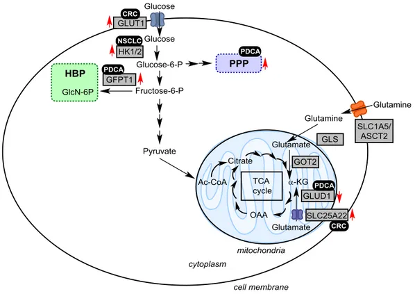
67,68Figure 8 Metabolic rewiring in KRas-driven cancers. Schematic representation of selected metabolic pathways.
Black box=cancer type, red arrows=change in expression level, grey boxes=enzymes; GLUT1: Glucose transporter 1, HK1/2: hexokinase 1/2, GFPT1: glucosamine-fructose-6-phosphate aminotransferase-1, GlcN: glucosamine, GLS:
glutaminase kidney isoform, GLUD1: glutamate dehydrogenase 1, GOT: glutamate–oxaloacetate transaminase, α-KG:
α-ketoglutarate, OAA: oxaloacetate, Ac-CoA: Acetyl-CoA, SLC: solute carrier, P: phosphate, BP: bisphosphate, ASCT2:
alanine, serine, cystein-preferring transporter 2, HBP: hexosamine biosynthesis pathway, PPP: pentose phosphate pathway, TCA: tricarboxylic acid, PDCA: pancreatic ductal cell carcinoma, CRC: colorectal cancer, NSCLC: non-small cell lung cancer. Adapted from Kawada et al.62
How mutated KRas coordinates the metabolic shift in cancer remains largely unknown.
62KRas is
a small GTPase that cycles between the active guanosine triphosphate (GTP)-bound and the
inactive guanosine diphosphate (GDP)-bound state. When activated, KRas mainly acts through
the RAF proto-oncogene serine/threonine-protein kinase (RAF) /mitogen-activated protein kinase
(MEK)/extracellular-signal-regulated kinase (ERK) signaling and the PI3K/AKT1/mTOR pathway,
which regulate distinct cellular processes such as proliferation, motility and survival.
69Independent
of oxygen availability, KRas can influence the expression level of HIF-1-α as well.
52,70,71Furthermore, Gaglio et al. reported a decoupling between glucose and glutamine metabolism in
KRas-mutated cancers.
72Conclusively, Ras-, especially KRas-mutated cancers, are largely
influenced in their metabolic phenotype, which drives nutrient, especially glucose addiction and makes them a highly attractive target for metabolic disruption via small molecules.
5.4.2 Metabolic flexibility and glutamine
It was long believed that cancer cells acquire an aerobic glycolytic metabolism and that they are disabled from performing oxidative phosphorylation. Recent evidence has emerged that some cancer cells possess a rather flexible metabolic phenotype and that the tumor itself is composed of cells with highly heterogeneous nutrient dependencies according to their perfusion.
73,74The most relevant nutrient besides glucose is the most abundant amino acid glutamine (Figure 3a).
75Glutamine fulfills a rather anabolic role in the biosynthesis of proteins, nucleotides, fatty acids and serves as a precursor to fuel the TCA cycle for energy production and for redox control.
Furthermore, glutamine is important for the uptake of essential amino acids.
76,77Under normal
conditions, glutamine is considered a non-essential amino acid since it can be generated in cellulo
from glucose.
78However, the necessity for biosynthetic building blocks during stress and
proliferation can increase the demand for glutamine, which outpaces glutamine availability, thus
rendering glutamine an essential amino acid.
79After cellular uptake via transporter proteins
(SLC1A5/ASCT2) (Figure 8), glutamine is converted to glutamate by glutaminase kidney isoform
(GLS), which fuels into the respective anabolic pathways (Figure 3a). Therefore, cancer
dependence on glutamine suggests the disruption of amidohydrolysis by glutaminase in order to
cut the glutamine supply. Indeed, a potent small molecule (CB-839) has been developed that
inhibits the cancer-specific glutaminase kidney isoform (GLS) in triple-negative breast cancer
(TNBC) cells (Figure 9).
80Also, cancer cell growth is reduced by CB-839 with low nanomolar
potency in the same cell lines.
80However, until now CB-839 has been investigated in multiple
clinical studies exclusively in co-administration with other cancer treatments, suggesting that
interrupting glutaminolysis alone is not sufficient to target this adaptive disease.
81Figure 9 Chemical structure of the inhibitor CB-839 against the glutaminase kidney isoform (GLS).
Targeting metabolic plasticity of cancer by disrupting glutamine and glucose supply simultaneously is therefore an attractive novel anti-cancer strategy. Low expression levels of GLUT-1 have already been associated with a sensitizing effect on lung cancer cells when glutamine is limited.
82Unspecific disruption of the glutamine supply via transaminase inhibition with aminooxyacetate (AOA) revealed a beneficial effect on growth inhibition of ovarian cancer when administered together with 2-DG.
83Therefore, a co-treatment approach with CB-839 and a GLUT inhibitor offers an attractive strategy to target metabolic flexibility in cancer.
5.5 Further applications of glucose uptake inhibitors
An adapted metabolism is found not only in cancer but also in other diseases that are associated
with stress or proliferation. Especially immunological diseases that involve the over-activation of
immune cells (e.g. T cells), infections or abnormal cell growth are associated with a strong glucose
dependence.
5This Warburg effect-like adaptation was observed for psoriasis (cytokine-driven,
inflammatory skin disease associated with hyperplasia),
84host cells infected with intracellular
bacteria such as Legionella pneumophila,
85fibrosis,
86graft-versus-host disease,
87colitis,
87systemic lupus erythomatheus,
88rhinoviral infections
89and persistence of the human
immunodeficiency virus (HIV).
90This broadens the potential applicability of glucose uptake
inhibitors for therapeutic indications.
6 Aim of the thesis
Curing cancer is a long-standing goal for which the development of novel therapeutics is necessary. Since cancer exhibits an altered metabolism and most often glucose addiction, glucose uptake inhibitors have the potential to be an effective cancer treatment.
An in-house medium throughput cancer cell-based screening was conducted in order to identify small molecules that inhibit glucose uptake. The piperazin-2-one hit substance class (Figure 10a,b) was selected according to its potency and structure. This small molecule class should be chemically and biologically investigated.
Figure 10 Chemical structure of the piperazin-2-one hit compound 1a (a) and general chemical structure of the piperazin-2-one substance class (b).
Chemical investigation should include the synthesis of a hit substance-based compound library.
Assessment of the biological activity of all derivatives should lead to subsequent analysis of the structure-activity relationship (SAR). The proposed protein target class and isoform selectivity within class I glucose transporters should be confirmed using well established, label-free methods.
Furthermore, the sensitivity of different cancer cell types towards a potent inhibitor as well as the
underlying reasons should be elucidated. Metabolic adaptations of cancer cell lines and potential
co-treatment opportunities should be explored.
7 Experimental part
7.1 Experimental part – chemistry
7.1.1 Materials
All chemicals were obtained from Acros Organics, Activate Scientific, Alfa Aesar or Sigma-Aldrich and used as provided, unless otherwise indicated. Dry solvents were stored over activated molecular sieves (MS 4 Å beads) and purchased from Sigma-Aldrich or Acros Organics.
7.1.2 Instrumentation
Proton and carbon nuclear magnetic resonance (
1H- and
13C-NMR) spectra were recorded on Varian Mercury 400 (400 MHz), Bruker Avance DRX 500 (500 MHz), INOVA 500 (500 MHz), Bruker AV600 (600 MHz) and Bruker AV700 (700 MHz) NMR spectrometer at ambient temperature. Proton chemical shifts are indicated as parts per million (ppm, δ-scale) and are referenced to residual protium in the NMR solvent (CHCl
3, δ7.26 ppm; CH
2Cl
2, δ5.30 ppm;
(CH
3)
2SO, δ2.50 ppm)
91. Data are represented as follows: chemical shift, multiplicity (s = singlet, bs = broad singlet, d = doublet, dd= doublet of doublets, ddd= doublet of doublet of doublets, dt = doublet of triplets, t = triplet, td = triplet of doublets, q = quartet, m = multiplet), coupling constant (J) in Hertz (Hz) and integration. Carbon chemical shifts are expressed in parts per million (ppm, δ-scale) and are referenced to the carbon resonances of the NMR solvent (CDCl
3, δ77.16 ppm;
CD
2Cl
2, δ53.84 ppm; (CH
3)
2SO, δ39.52 ppm). All NMR spectra were analyzed using MastReNova
Version 12.0.0. High resolution mass spectra (HR-MS) were recorded on a LTQ Orbitrap mass
spectrometer 5 coupled to an Acceka HPLC-System (HPLC column: Hypersyl GOLD, 0 m x 1 mm,
particle size 1.9 μm, ionization method: electron spray ionization). Systematic names for
molecules according to IUPAC rules were generated using ChemDraw Professional version 16.0.
7.1.3 Methods
7.1.3.1 Preparative HPLC-MS
Separations were performed by means of mass-directed preparative HPLC (Agilent Series, 1100/LC/MSD VL) using reversed-phase C18 column with a constant flow of 20.0 mL/min. Solvent A: water +0.1% v/v TFA; solvent B: acetonitrile +0.1% v/v TFA.
7.1.3.2 Separation of enantiomers from racemic mixture
Chiral separation was performed by means of solid phase separation via an Ultimate 3000 HPLC (Dionex, Thermo Fisher) employing an IC column (CHIRALPAK IC, column no. IC00CG-MA004) column. An isocratic gradient was used to separate both enantiomers (elute: isohexane / DCM/MeOH(5%)).
7.1.3.3 General experimental procedures
Reactions were carried out in standard laboratory glassware. Organic solutions were volatilized by rotary evaporation at 40 °C. Analytical thin-layer chromatography (TLC) was performed using aluminum plates pre-coated with silica gel (Silica gel 60
254, Merck KGaA, Darmstadt, DE KGA).
Compounds were visualized on the TLC plates by exposure to ultraviolet light (UV). TLC plates were stained by submersion in aqueous ninhydrin solution or aqueous potassium permanganate solution (KMnO
4) followed by brief heating by a heat gun to visualize primary amines or unsaturated carbohydrates. Ninhydrin solution was prepared as follows: 1.5 g ninhydrin in 100 ml of n-butanol +3.0 ml acetic acid. KMnO
4solution was prepared as follows: 1.5 g potassium permanganate +10 g potassium carbonate +1.25 ml 10% (w/v) sodium hydroxide in 200 ml water.
Flash column chromatography was performed either using an automatic column with prepacked
silica columns purchased from Buchi (Reveleris columns) or manually using silica gel Acros
Organics 60 (particle size 0.035–0.070 mm).
7.1.3.4 General procedure 1A: Modified Leuckart-Wallach reaction
General procedure for conventional heating: A microwave vial was charged with the corresponding aldehyde (3.6 mmol, 1 equiv.) dissolved in formamide (36 mmol, 10 equiv.) and formic acid (18 mmol, 5 equiv.). The reaction mixture was vigorously stirred at 180 °C for 2-4 h. After cooling the mixture to room temperature, the crude reaction was mixed with water (25 mL) and extracted thrice with DCM (25 mL). The combined organic layers were dried over Na
2SO
4or MgSO
4, filtered and the filtrate was concentrated under reduced pressure. Subsequently the remains were purified by silica gel column chromatography (elute as indicated) to yield the corresponding formamide.
92General procedure for microwave irradiation: A microwave vial was charged with the corresponding aldehyde (0.4 mmol, 1 equiv.) and dissolved in a mixture of formamide (18.4 mmol, 50 equiv.) and formic acid (1.8 mmol, 5 equiv.). The reaction mixture was irradiated in a microwave oven for 30 sec at 180 °C. The crude reaction was quenched with 10 mL water and extracted with thrice with DCM (15 mL). The combined organic layers were dried over Na
2SO
4or MgSO
4and filtered. The filtrate was concentrated under reduced pressure and residual formamide was removed in vacuo. The crude reaction was purified by means of silica gel column chromatography (elute as indicated) to yield the corresponding formamide.
927.1.3.5 General procedure 2: Modified Ugi reaction
The bifunctional carboxylic acid/ketone (0.20 mmol, 1.0 equiv.) and the amine (0.20 mmol,
1.0 equiv. or 0.18 mmol, 0.9 equiv.) were dissolved in MeOH (1 mL). The reaction mixture was
vigorously stirred for 30 min at room temperature to let the imine form and the isocyanide
(0.20 mmol, 1.0 equiv.) was added subsequently. The mixture was vigorously stirred at 40 °C until
full conversion of the starting material was observed by TLC. The solvent was removed under
reduced pressure. The remains were mixed with water (25 mL) and extracted thrice with
dichloromethane (25 mL). The combined organic layers were dried over Na
2SO
4or MgSO
4, filtered
and the filtrate was concentrated under reduced pressure. The product was purified by silica gel column chromatography (eluate: petroleum ether / EtOAc or DCM / MeOH).
937.1.3.6 General procedure 3A: Modified Ugi reaction with in situ isocyanide formation using phosgene
The reaction was performed as described by Neochoritis et al.
92The corresponding formamide (0.06 mmol, 1.0 equiv.) was dissolved in DCM (1 mL) and cooled to 0 °C. Triethylamine (0.15 mmol, 2.5 equiv.) was added and the mixture was vigorously stirred for 10 min. Triphosgene (0.02 mmol, 0.3 equiv.) dissolved in DCM (1 mL) was added dropwise over a period of approximately 30 min. After consumption of the formamide (monitored by TLC), the bifunctional carboxylic acid/ketone (0.07 mmol, 1.2 equiv.) and the amine (0.06 mmol, 1.0 equiv.) were added and the reaction mixture was vigorously stirred for 5-72 h. The crude mixture was concentrated under reduced pressure. The remains were mixed with water (50 mL) and extracted thrice with DCM (50 mL). The combined organic layers were dried over NaSO
4, filtered and the filtrate was concentrated under reduced pressure. The crude reaction was purified by silica gel column chromatography using the indicated elute.
7.1.3.7 General procedure 3B: Modified Ugi reaction with in situ isocyanide formation using Burgess reagent
The reaction was performed as described by Creedon et al.
94Briefly, an oven-dried Schlenck tube was charged with the corresponding formamide (220 µmol, 1 eq) which was dissolved in dry acetonitrile under argon atmosphere. Burgess reagent (348 µmol, 1.5-2 eq) was added and the reaction mixture was stirred for 0.5-2 h at room temperature. The reaction progress was monitored by TLC. The amine (198 µmol, 0.9 eq) and bifunctional compound (220 µmol, 1 eq) were combined in MeOH and stirred for approximately 10 min. This mixture was added to the in situ formed isocyanide. The reaction mixture was vigorously stirred at room temperature for 1-5 days.
Saturated NaHCO
3(50 mL) was added to the reaction mixture and the product was extracted
thrice with DCM (50 mL). The combined organic layers were dried over Na
2SO
4or MgSO
4and filtered. The filtrate was concentrated under reduced pressure and the remains were purified by silica gel column chromatography or recrystallization from MeOH as indicated.
7.1.3.8 Single crystal X-ray structure analyses
Structure determination was performed by Lena Knauer, TU Dortmund. For data collection, the
Bruker D8 Venture four-circle diffractometer (Bruker AXS GmbH) was employed. The area
detector PHOTON100 CMOS (Bruker AXS GmbH) was utilized. To generate the X-ray radiation,
the microfocus sources IµS Cu or Mo (Incoatec GmbH) with HELIOS mirror optics and a single-
hole collimator (Bruker AXS GmbH) was used. Data collection was undertaken using the software
APEX 3 Suite (v.2017.3-0) that included the programs SAINT for integration and SADABS for
adsorption correction (Bruker AXS GmbH). The structures were determined using Olex2
95and
employing the software ShelXT
96using intrinsic phasing and structure refinement was conducted
with the XL refinement package
97using least squares minimization.
7.2 Experimental part – biology 7.2.1 Material
7.2.1.1 Eukaryotic cell lines
The medium composition is given in chapter 7.2.1.8.
Cell line Specification Medium Order numer Supplier BxPC-3 Human pancreatic
adenocarcinoma
DMEM complete ACC760 DSMZ GmbH, DE
DLD-1 Human colorectal adenocarcinoma
RPMI1640 PAR-086 Horizon
Discoveries, UK DLD-1
GLUT1(-/-)
Human colorectal adenocarcinoma
RPMI1640 R00-024 Horizon
Discoveries, UK HCT116 Human colorectal
carcinoma
DMEM complete ACC581 DSMZ GmbH, DE
MIA PaCa-2 Human pancreatic carcinoma
DMEM complete CRM-CRL- 1420
ATCC, USA MDA-MB-231 Human mammary
gland
adenocarcinoma
DMEM complete +1% NEAA
ACC-732 DSMZ GmbH,
DE SW480 Human colorectal
adenocarcinoma
Leibovitz’s L-15 CCL-228 ATCC, USA
UM-UC-3 Human urothelial transitional cell carcinoma
MEM Eagle CRL-1749 ATCC, USA
UO-31 Human renal carcinoma
RPMI1640 MTA 1-4488-
14
NCI, USA
CHO Chinese hamster
ovary
DMEM complete CRL-9618 ATCC, USA
NEAA=Non-essential amino acids, DSMZ=Deutsche Sammlung von Mikroorganismen und Zellkulturen, ATCC=American Type Culture Collection, NCI=National Cancer Institute.
7.2.1.2 Antibodies
Antibody Host Dilution for immunoblotting
Order number
Supplier Primary antibodies
Anti-GLUT1 Mouse 1:5,000 Ab40084 Abcam, UK
Anti-GLUT2 Rabbit 1:200 Sc-9117 Santa Cruz, US
Anti-GLUT3 Rabbit 1:5,000 Ab191071 Abcam, UK
Anti-GLUT3 Rabbit 1:5,000 Sc-74399 Santa Cruz, US
Anti-GLUT4 Rabbit 1:500 07-1404 Millipore, USA
Anti-Na
+-K
+- ATPase
Rabbit 1:10,000 Ab76020 Abcam, UK Anti-Vinculin Mouse 1:10,000 Sc-59803 Santa Cruz, USA Secondary
antibodies
Anti-rabbit 680RD Donkey 1:5,000 926-68071 LI-COR Biosciences, DE Anti-rabbit 800CW Donkey 1:5,000 926-
3221300
LI-COR Biosciences, DE Anti-mouse 680RD Donkey 1:5,000 926-68072 LI-COR Biosciences, DE Anti-mouse 800CW Donkey 1:5,000 926-32212 LI-COR Biosciences, DE Anti-rabbit HRP Goat 1:100,000 31430 Pierce, USA
(Thermo Scientific)
Anti-mouse HRP Goat 1:10,000 31460 Pierce, USA
(Thermo Scientific)
HRP=Horseradish peroxidase
7.2.1.3 Plasmids
Plasmid Order number Supplier
GLUT-1 pTCN(BC121804) transOMIC, USA
GLUT-2 pTCV(BC060041) transOMIC, USA
GLUT-3 pCMV-SPORT 6 Dharmacon, USA
GLUT-4 pTCN(BC121804) transOMIC, USA
pTCN empty vector pTCN transOMIC, USA
pCMV empty vector pCMV Dharmacon, USA
7.2.1.4 Oligonucleotides
Gene Forward Primer (5′-3′) Reverse Primer (5′-3′) Product Size, bp
Efficiencies
GLUT1 TTGCAGGCTTCTCCAACTGGAC CAGAACCAGGAGCACAGTGAAG 112 104.8%
GLUT2 ATGTCAGTGGGACTTGTGCTGC AACTCAGCCACCATGAACCAGG 130 99.9%*
GLUT3 TGCCTTTGGCACTCTCAACCAG GCCATAGCTCTTCAGACCCAAG 97 104.9%
GLUT4 CCATCCTGATGACTGTGGCTCT GCCACGATGAACCAAGGAATGG 137 89.2%*
ACTB CACCATTGGCAATGAGCGGTTC AGGTCTTTGCGGATGTCCACGT 134 100.7%
B2M CCACTGAAAAAGATGAGTATGCCT CCAATCCAAATGCGGCATCTTCA 126 102.0%
TUBB CTGGACCGCATCTCTGTGTACT GCCAAAAGGACCTGAGCGAACA 116 100.8%
ATP1A1 GGCAGTGTTTCAGGCTAACCAG TCTCCTTCACGGAACCACAGCA 118 100.2%
The primer efficiencies for amplicon duplication were measured in DLD-1 wt lysates or with isoform specific plasmids (*).