pH Coupled Co-Precipitation of Alkaline-Earth Carbonates and Silica
Complex Materials from Simple Chemistry
DISSERTATION ZUR ERLANGUNG
DES DOKTORGRADES DER NATURWISSENSCHAFTEN (DR. RER. NAT.)
DER FAKULTÄT
CHEMIE UND PHARMAZIE DER UNIVERSITÄT REGENSBURG
vorgelegt von Josef Eiblmeier
aus Arnstorf
Regensburg 2013
Promotionsgesuch eingereicht am: 04.04.2013
Tag des Promotionskolloquiums: 03.05.2013
Die Arbeit wurde angeleitet von: Prof. Dr. Werner Kunz
Prüfungsausschuss: Prof. Dr. Hubert Motschmann (Vorsitzender) Prof. Dr. Werner Kunz (1. Gutachter)
Prof. Dr. Arno Pfitzner (2. Gutachter) Prof. Dr. Henri Brunner (3. Gutachter)
Gerhard Polt
Vorwort
Die vorliegende Dissertation beruht auf Forschungsarbeiten, die im Zeitraum von Oktober 2009 bis März 2013 am Lehrstuhl von Prof. Dr. Werner Kunz (Institut für Physikalische und Theoretische Chemie, Universität Regensburg) durchgeführt wurden. Die Arbeit selbst ist so aufgebaut, dass jedes einzelne Kapitel, in denen die Forschungsergebnisse präsentiert wer- den, als Manuskriptvorlage für Publikationen in wissenschaftlichen Fachjournalen dienen soll.
Die gewonnen Ergebnisse dieser Doktorarbeit wären sicherlich nicht ohne die Unterstützung von vielen Menschen zustande gekommen, bei denen ich mich an dieser Stelle ganz herzlich bedanken möchte.
Zuallererst möchte ich mich natürlich bei meinem Doktorvater, Herrn Prof. Dr. Werner Kunz, für die Überlassung des interessanten und vielschichtigen Themas bedanken. Vor allem aber auch für die Gewährung großzügiger akademischer und finanzieller Freiräume zur Verwirk- lichung der eigenen Ideen und natürlich auch für sein stets entgegengebrachtes Vertrauen in mich und meine Arbeit.
Ebenso möchte ich mich bei Dr. Matthias Kellermeier für seine fortwährende Unterstützung bedanken. Wenn man so will war Matthias mein co-Doktorvater, der immer ein offenes Ohr für alle erdenklichen Fragen und Problemstellungen hatte. Sein immenses Wissen, speziell auf dem Gebiet der Silicat-Carbonat Wechselwirkungen, aber auch seine allge- meine Herangehensweise zur Lösung verschiedenster Problemstellungen haben mich per- sönlich zutiefst beeindruckt. Die gewonnen Erkenntnisse und wesentlichen Konzepte dieser Doktorarbeit wäre ohne die zahlreichen Diskussionen mit ihm sicherlich so nicht zustande gekommen. Weiterhin möchte ich mich bei Ihm noch für seine zahlreichen Korrekturen von verschiedenen Manuskripten bedanken, durch die ich sehr viel lernen konnte. Vor allem aber
Weiterer Dank gebührt auch Prof. Dr. Juan Manuel García-Ruiz für sein kontinuierliches In- teresse am Fortgang der Arbeit. Durch sein profundes Wissen auf den Gebieten der Silica Biomorphs und der Kristallisation im Allgemeinen, aber auch durch zahlreiche Diskussio- nen mit Ihm konnte ich sehr viel lernen.
Im Rahmen der Dissertation wurde eine Vielzahl an verschiedenen Messmethoden angewen- det, was ohne die Hilfe von externen Kooperationspartnern so nicht möglich gewesen wäre.
Deswegen möchte ich mich an dieser Stelle bei verschiedenen Personen bedanken, die mich im Rahmen dieser Kooperationen unterstützt haben:
• Martina Müller für experimentelle Unterstützung zur Gewinnung der Daten für die Abbildungen 5.8, 5.9 und 5.10.
• Prof. Dr. Reinhard Rachel, Prof. Dr. Ralf Witzgall und Cornelia Niemann (Institut für Molekulare und Zelluläre Anatome, Universität Regensburg) für die Bereitstellung der Elektronenmikroskope und Hilfe bei der Benutzung und Interpretation der gewonnen Daten.
• Prof. Dr. Gottfried Schmalz und Gerlinde Ferstl (Poliklinik für Zahnerhaltung und Paradontologie, Uniklinikum Regensburg) für die Bereitstellung des Rasterelektronen- mikroskops und diverser Hilfestellungen bei der Benutzung.
• Prof. Dr. Andreas Fery und Melanie Pöhlmann (Physikalische Chemie II, Univer- sität Bayreuth) für die Bereitsstellung des Rasterkraftmikroskops und Hilfe bei der Benutzung und der Dateninterpretation, sowie für die Herstellung der strukturierten PDMS Oberflächen (Kapitel 6).
• Prof. Dr. Lorenz Kienle, Mao Deng und Dr. Ulrich Schürmann für die TEM und micro- EDX Daten aus Kapitel 3 und 4 und für die Hilfestellung bei der Auswertung.
• Prof. Dr. Arno Pfitzner, Dr. Martina Andratschke und Stephan Dankesreiter für die Aufnahme der Pulverdaten und Hilfe bei der Datenauswertung.
• Prof. Dr. Manfred Scheer und Michael Seidl für die Erlaubnis zur Benutzung des FTIR Spektrometers.
• Doris Rengstl (Universität Regensburg) und Dr. Roman Chernikov (HASYLAB/DESY, Hamburg) für verschiedenste Hilfestellungen während der Synchrotronmesszeit am DESY in Hamburg.
• Dr. Matthias Kellermeier, Prof. Dr. Helmut Cölfen und Dr. Denis Gebauer für die Bereitstellung des Titrationsequipments und für Ihre Hilfe bei der Datenauswertung und deren Interpretation.
• Bei Andreas Picker möchte ich mich noch gesondert bedanken, weil ich während meiner Messaufenthalte in Konstanz immer gratis auf seiner kleinen, aber feinen Couch schlafen durfte.
Herrn Prof. Dr. Hubert Motschmann, Herrn Prof. Dr. Arno Pfitzner und Herrn Prof. Dr. Henri Brunner danke ich für ihre Bereitschaft in der Prüfungskommission mitzuwirken.
Den Mitarbeitern der Werkstätten sei für die schnelle und zuverlässige Erledigung der Aufträge gedankt.
Allen aktuellen und ehemaligen Mitarbeitern und Kollegen des Lehrstuhls danke ich recht herzlich für die entspannte und freundschaftliche Arbeitsatmosphäre, stete Hilfsbereitschaft, und natürlich für das ein- oder andere Feierabendbier.
Besonders danke ich Herrn Dr. Fabian Glaab für das Korrekturlesen der Doktorarbeit, für seine Freundschaft in den letzten Jahren und natürlich für seine berühmt berüchtigten Wut- ausbrüche, die mich immer wieder erheitert haben.
Meiner ehemaligen Bürokollegin Gabi danke ich herzlich für die lockere Arbeitsatmosphäre und insbesonders dafür, dass Sie ständig meine Musik ertragen hat.
Last but no least, möchte ich mich noch aus tiefstem Herzen bei den wichtigsten Personen in meinem Leben bedanken: meiner Familie, denen diese Arbeit auch gewidmet sein soll. Ihr seid immer hinter mir gestanden und habt mich unterstützt, wo es nur ging. Ohne Euch wäre ich bestimmt nicht soweit gekommen. Auch meiner Freundin Luisa sei herzlich gedankt, dass Sie mich während meiner Doktorarbeit immer unterstützt hat und dass Sie mich durch die gemeinsam verbrachten Wochenenden den Doktorarbeitsalltag vergessen ließ.
Contents
1 Introduction 1
1.1 General Background . . . 1
2 Silica-carbonate biomorphs 10 2.1 Aims and purpose of the thesis . . . 10
2.1.1 Introduction . . . 10
2.1.2 Preparation methods . . . 12
2.1.3 Microstructure and composition . . . 14
2.1.4 Formation mechanism . . . 17
2.1.5 Phenomenological description of morphology development . . . 25
2.1.6 Biomorphs with SrCO3and CaCO3 . . . 28
3 Bottom-Up Self-Assembly of Amorphous Core-Shell-Shell Nanoparticles and Biomimetic Crystal Forms in Inorganic Silica-Carbonate Systems 32 3.1 Abstract . . . 32
3.2 Introduction . . . 33
3.3 Experimental section . . . 36
3.3.1 Materials . . . 36
3.3.2 Precipitation Experiments . . . 36
3.3.3 Analytical Methods . . . 37
3.4 Results . . . 40
3.4.1 Fractal branching at low silica concentrations . . . 40
3.4.2 Core-shell-shell particles at elevated silica concentrations . . . 44 3.4.3 Temporal stability and transformation of the composite nanoparticles 53
Contents
3.5 Discussion . . . 60
3.6 Conclusions . . . 65
4 New Insights into the Early Stages of Silica-Controlled Barium Carbonate Crys- tallization 67 4.1 Abstract . . . 67
4.2 Introduction . . . 68
4.3 Experimentals . . . 70
4.3.1 pH-constant titration measurements . . . 70
4.3.2 Data treatment . . . 72
4.3.3 TEM and EDX analyses . . . 72
4.4 Results . . . 73
4.4.1 In-situ potentiometric titration . . . 73
4.4.2 Characterization of nucleated particles . . . 79
4.5 Discussion . . . 84
4.6 Conclusions . . . 88
5 Effect of bulk pH and supersaturation on the growth behavior of silica biomorphs in alkaline solutions 90 5.1 Abstract . . . 90
5.2 Introduction . . . 91
5.3 Experimental section . . . 93
5.3.1 Crystallization experiments . . . 93
5.3.2 Characterization methods . . . 94
5.4 Results . . . 99
5.4.1 Morphological analysis . . . 99
5.4.2 Evolution of the bulk pH and barium concentration during growth from solution . . . 108
5.5 Discussion . . . 112
5.6 Conclusion . . . 116
tation System 118
6.1 Abstract . . . 118
6.2 Introduction . . . 118
6.3 Experimentals . . . 121
6.3.1 Fabrication of Micropatterned Substrates . . . 121
6.3.2 Crystallization and Characterization of Silica-Carbonate Biomorphs 122 6.4 Results and Discussion . . . 123
6.5 Conclusions . . . 133
7 Crystallization of mixed alkaline-earth carbonates in alkaline silica solutions 134 7.1 Abstract . . . 134
7.2 Introduction . . . 135
7.3 Experimentals . . . 138
7.3.1 Materials . . . 138
7.3.2 Crystallization Experiments . . . 138
7.3.3 Characterization Methods . . . 138
7.4 Results . . . 139
7.4.1 Morphological Analysis . . . 139
7.4.2 Crystal polymorphism and composition . . . 147
7.4.3 Effect of the growth behavior by increasing the ionic strength . . . 152
7.5 Discussion . . . 154
7.5.1 Prevention of biomorph formation in the presence of Ca2+ions . . . 154
7.5.2 Fractal branching at different Ca2+/Sr2+and Ca2+/Ba2+ratios . . . 158
7.6 Conclusions . . . 161
8 Conclusions and Outlook 163 9 Appendices 169 A Additional Figures for chapter 4 . . . 169
B Additional polarized optical micrographs for chapter 5 . . . 172
Contents
10 References 173
List of Figures 182
List of Tables 207
List of Publications 207
Declaration 210
1 Introduction
1.1 General Background
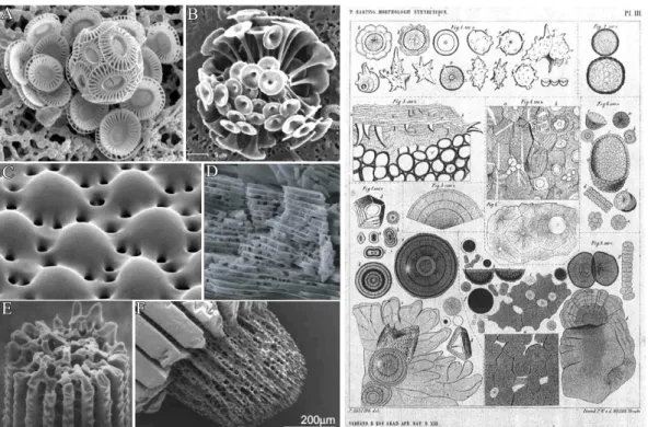
The principle how nature creates the hard parts in living organism has for long been a fas- cinating topic and also raised a still growing research field. Such elaborate architectures are often hallmarked by sinuously shaped surfaces which hardly reflect the geometric con- straints dictated by the crystallographic phase of the mineral. This is in contrast to classic abiotic crystals, where the final crystal shape is defined by the arrangement of atoms within the structure. First pioneering works in the end of the19th century by Pieter Harting were dedicated to elucidate natures strategy to create such astonishing forms. It soon became clear that the formation of such complex functional structures is linked to the intimate interaction between organic and inorganic matter.1 His findings were based on the precipitation of so called "calcospherites" which resemble certain biominerals and are yielded by combining inorganic salts with biological (organic) matter2(see Figure 1.1 right).
On that basis, numerous investigations were dedicated to unravel the design principles of certain biominerals (cf. Figure 1.1 left) and, at the same time, shed light on mechanistic details for the formation of such elaborate organic-inorganic composite materials. Up to now a vast amount of biominerals has been identified, and all of them perform, due to their unique properties, several functions ranging from skeletal support and protection, to grav- ity, magnetic field perception,8,9or serving as specialized photosensitive organs.10,11Among identified biominerals, calcium carbonate is by far the most often utilized material and oc- curs in different polymorphs such as calcite (being the thermodynamic stable form at ambient conditions), aragonite, vaterite as well as the amorphous calcium carbonate (ACC) besides monohydrocalcite.12
In this context, nacre (also referred to as "Mother of Pearls") is an interesting biomaterial as
C
E F
D
Figure 1.1. Left: SEM images of some exemplary biominerals. (A-B) Calcitic coccoliths of the marine calcareous alga Discosphaera tubifera and Emiliana Huxleyi3,4. (C) Photore- ceptors in the light-sensitive brittlestar Ophiocoma wendtii comprised of calcite.4) (D) SEM close-up view of nacre from the bivalve A. rigida.5) (E-F) Tips of a sea urchin spine Paracen- trotus lividus.6,7 Right: Original drawings of Harting showing the yielded "calcospherites"
upon precipitation of inorganic salts in the presence of organic matter.2
it attracts the attention of scientists from a wide range of research fields due to its outstand- ing remarkable mechanical properties (ca. 3000 times tougher than pure aragonite13). Such features are explained by the complex ordering of 3-D calcium carbonate plates in a matrix of organic macromolecules, which is also described as a "brick and mortar" arrangement of organic and inorganic components (see Figure 1.1D).
Beneath the structural design further central aspects in the field of biomineralization are the control over nucleation, growth, selection of polymorph and the shape of the resulting composite mineral. These manifold requirements are accomplished in natural crystallization processes via a synergistic interplay between the mineral phase and the surrounding organic matrix. The latter can be treated as an additive which controls the crystallization events over multiple length scales beginning from the Ångström level, (where the arrangement of atoms decides which polymorph of the mineral is preferred) up to the mesoscopic scale in which the manner of integration and alignment of building units defines the final appearance of the
General Background
A
C D
B
Figure 1.2. (A) SEM cross section of a naturally ocurring skelatal plate of a sea urchin spine and (B) the corresponding polymeric membrane replica.14(C) Polycrystalline calcite crystals precipitated in the porous polymer membrane together with a porous calcite single crystal, yield with the same method.15
biomineral.
Among the additives which are used for the control in biomineralization processes one can discriminate between two principal types. The first one is comprised of water insoluble biopolymers like chitin or collagen and is utilized by organism for the construction of scaf- folds for crystallization.16 In such templating approaches, mineral deposition occurs in spe- cialised micro-environments like vesicles or even larger compartments separated from the external aqueous neighborhood, by membraneous envelopes, in order to regulate several parameters like flux of ions, saturation state or pH of the inner region.6,8,13 Additionally, such microreactors are capable to promote crystallization by heterogenous nucleation pro- viding lower activation energy barriers than homogeneous nucleation.16 Hereby, immobi- lized organic frameworks act as structured substrates offering preferential sites for oriented nucleation17and, beyond that, molding the mature mineral within the walls of the compart- mentalized reaction volume providing global control of the morphology.8
C
E
G H
F D
Figure 1.3. Gallery of various minerals, precipitated in the presence of soluble organic (polymeric) molecules. (A) "Microtrumpet" consisting of self-assembled calcite microcrys- tals formed under the influence of 1,3-diamino-2-hydroxypropane-N,N,N’,N’-tetraacetate.3 (B) Ordered chains of BaCrO4 nanoparticles grown in a surfactant based environment (reproduced from18). (C) Silica particles with helical morphologies synthesized with a pyridinium-based cationic surfactant.19(D) Polycrystalline CaCO3 (vaterite) helices precip- itated in the presence of poly(aspartate).20(E) Nanofibres composed of BaCO3with double- stranded and cylindrical helical morphologies formed in a phosphonated block co-polymer controlled mineralization process.21 (F) Nanocrystalline BaCO3 superstructures obtained in the presence of poly(ethyleneoxide)-block-eicosa aspartate.22 (G) Neuronlike calcium phos- phate/polymer (PEO-b-PMAA-C12) mesostructures.23 (H) Spiral superstructures of BaCO3 crystals prepared in a PNIPAM-b-PLGA polymer micelle solution.24
On the other hand, soluble organic (macro)molecules, also referred as so-called soluble or
General Background functional matrix allow for the control over the ongoing crystallization within the micro- environments.16 This occurs primarily via face-specific adsorption on the growing single crystal or also via stabilization of small building units which self-assemble to polycrystalline architectures later on.
The synergistic interplay between the multiple crystallization modifiers in the course of biomineral formation has inspired scientists from various fields to develop novel synthetic strategies for the creation of novel "biomimetic" materials with desired properties and special morphologies.16,25One one of these concepts for the in vitro synthesis of biomimetic material represents a "top down" approach where the organic matrix (or its polymeric replica), is uti- lized as 3D template in analogy of direct molding strategies in biological systems (see Figure 1.2 A-B). Indeed, macroporous calcite single crystals with the same morphology as skele- tal plates of sea urchins were successfully produced via direct precipitation in a sponge-like polymer membrane (Figure 1.2 C-D).15,26Alternatively, ACC can also be utilized as precur- sor material in the same approach for the generation of calcite crystals with extraordinary morphologies. Thereby it is advantageous that the amorphous precipitate can be suitably molded within a preferred geometry to yield crystalline matter with identical shape.27–29Fur- ther experiments revealed additionally that this strategy is not limited to calcium carbonate, since similar sponge-like structures reminiscent of sea urchin skeletal plates could be yielded with SrSO4, PbSO4, CuSO4·5 H2O,30gold, titanium,31and copper,32respectively.
Truly, a certain drawback in this "top down" strategy is the complicate and time consum- ing preparation of suitable templates, making a large scale industrial production impossible.
Thus, "bottom up" approaches, where formation of elaborate inorganic matter is based on concerted self-assembly of nanosized building units, are desired, using water soluble organic additives which fulfill the role of the functional matrix in biomineralization processes. There- fore, scientists have screened the influence of a vast amount of additives on the crystallization processes of certain minerals, ranging from simple ions33–35or small organic molecules like citric acid36,37 over ordinary polymers and polyelectrolytes20,38–40and surfactants18,19,41,42 to complex block copolymers (cf. Figure 1.3).21,22,43–47
The morphological complexity achieved, by introducing (organic) mineralization additives,
formation where growth occurs via attachment of single ions or molecules onto crystal faces of already existing nuclei (pathway (a) in Figure 1.5). The latter can either result from het- erogeneous nucleation on foreign surfaces such as dispersed impurities (e. g. dust particles), crystal seeds, or tube walls. This process is energetically favored, as the free energy to cre- ate a new interface is reduced. On the other side, nuclei can alternatively be formed in the bulk volume via homogeneous nucleation from supersaturated solutions. This is mainly a stochastic event and occurs via association of ion clusters. However, the question whether individual clusters grow or not is dependent on two opposing effects in the overall Gibbs free energy of cluster formation∆GNucl(Figure 1.4). The latter is typically related to the positive term of the surface energy∆GSurf (being proportional to the square of the spherical cluster radiusr2) and to the negative contribution, resulting from a volume term that scales withr3 which is due to the gain in bulk energy∆GBulk since a new crystal lattice is formed. These two effects become balanced when the nucleus reaches a certain, so-called critical cluster size, r∗. Enlargement of the nucleus above r∗ consequently leads to a gain in the net free energy as the volume term overbalances the surface energy term (cf. Figure 1.4).
According to the classical model, growth occurs via ion attachment and replication of the unit-cell, yielding crystals with defined geometrical outer shapes, smooth surfaces and fixed angles. Such simplistic descriptions for crystal formation might be valid for geological min- erals, but classic theories cannot explain the presence of the typical sinuous shapes in biomin- erals (which can also be single crystalline). Instead, mineralization can also proceed along so called "non-classical crystallization" pathways, starting from pre-formed nanoparticles which serve as building blocks for the final architectures.50–53 Figure 1.5 (pathway b and c) gives an schematical overview of processes involved. In the absence of additives (pathway b), nanocrystalline building blocks can lock in and fuse to form an iso-oriented single crystal.
This phenomenon is also called oriented attachment.54Alternatively, when suitable additive species are present they can cover the surface of primary building units (being of either crys- talline or amorphous nature). In this way, fusion to single crystals (as described in pathway (a) and (b) in Figure 1.5) is at first prevented as the adsorbed polymers dictate the mode of interaction between individual nanoscale units. The stabilized, primary nanoparticles are
General Background
ΔG
ΔG*Nucl
ΔGNucl ΔGSurf(~r )2
ΔGBulk(~r )3
r* r
Figure 1.4. Classical nucleation theory: plot of the Gibbs free energy ∆G for the for- mation of nucleation cluster from supersaturated solutions in dependence of cluster radius.
The balance between the cost in energy for creating new surfaces∆GSurf and the gain due to attractive forces in the emerging lattice (∆GBulk) provoke a maximum in the net curve (∆GNucl), which defines the critical cluster size r∗ and the corresponding activation energy for nucleation∆GNucl∗.
Figure 1.5. Schematic representation of classical (a) and non-classical (b) crystallization.
Pathway (c) highlights the stabilization of primary nanoparticles with additives like polymers or surfactants which finally yields mesocrystals via concerted self-assembly of individual nano-units.49,50
Figure 1.6. (a-c) SEM images of helical BaCO3 fibers formed in solutions containing phos- phonated block copolymers.(d-g) Mechanism explaining the helical arrangement. Face se- lective adsorption of the polymer on (110) planes and sterical hindrance (d) favors particle aggregation in a staggered manner over other, energetically less favorable, arrangements (g).
Further growth of individual particles occurs via epitaxial attachment to the uncovered lateral faces (020) and (011). This leads to a situation where incoming building units differentiate between favorable and unfavorable sites for attachment. (b) This eventually induces a helical winding of the whole aggregate, yielding either right-or left-handed superstructures.55
subsequently capable to self-assemble to so called mesocrystals which is an abbreviation for
"mesoscopically structured crystals" (pathway c, Figure 1.5). Similarly additive covered hy- brid particles can aggregate in a manner where they are perfectly aligned in a common crys- tallographic fashion, giving them properties of "ordinary" single crystals in terms of X-ray scattering, despite consisting of multiple individual units (Figure 1.5, pathway b). It seems logical that such additive-mediated crystallization routes enable new synthesis strategies for pseudo-single crystals with remarkable morphologies and unique properties. However, the mesoscale arrangement of previously stabilized precursor units can also produce polycrys- talline superstructures, dependent on several parameters like specific interactions between
General Background the adsorbed layers or re-alignment phenomena during aggregation in the presence of spatial constraints.52 An interesting situation arises when self-aggregation of nanoparticles is pro- voked by polymeric additives that selectively adsorb on distinct faces of anisotropic (crys- talline) particles. This mechanism is nicely exemplified in Figure 1.6, where crystallization of BaCO3 occurred in the presence of sterically demanding racemic block copolymers.55 In this case, selective adsorption of the stiff polymer onto (110) faces of witherite leads to re- stricted growth along this direction and to stabilization of the involved face at the same time (Figure 1.6C). This causes staggered arrangement of the carbonate crystals and allows in- coming building blocks to discriminate between the lateral faces during aggregation, finally yielding (racemic) helical superstructures (Figure 1.6A).
2 Silica-carbonate biomorphs
2.1 Aims and purpose of the thesis
2.1.1 Introduction
The summarized findings presented above in the field of bio-mimetic material science strongly suggest the presence of elaborate organic (macro)molecules and/or supramolecular tem- plates for the synthesis of polycrystalline carbonate structures. In contrast, even simple purely inorganic additives like metal ion cations such as Mg2+ or Sr2+ were found to in- fluence the polymorph selection during CaCO3 crystallization,36,56,57 but complex shapes, as found in the presence of organic additives, remained absent. This is drastically dif- ferent when using silica as inorganic additive. Silica itself is a highly abundant material, occurring in Nature as a solid mineral or in dissolved form. In the latter case, the aque- ous chemistry is quite complex involving an interplay between dissolution/precipitation, adsorption/desorption and complexation processes. In this context, Juan Manuel García- Ruiz observed during his doctoral thesis in the late 1970t’s very spectacular alkaline-earth carbonate-based crystal architectures, when growth was carried out in silica gels at high pH (Figure 2.1).58–60Indeed, he observed aggregates in purely abiotic crystallization media with similar complex morphologies as those displayed by living organisms in biomineralization processes (cf. Figure 2.1). In order to express their morphological resemblance to cer- tain biological materials, these structures are termed "silica-carbonate biomorphs". Findings were at first unexpected as the formation of curved shapes far away from any crystallo- graphic symmetry was exclusively ascribed to living nature. However, it was demonstrated in later studies that morphology alone can be misleading when discussing about biogenicity in general, i.e. the biological origin of putative precambrian microfossils (Figure 2.2 ).62–64
Aims and purpose of the thesis
A
B
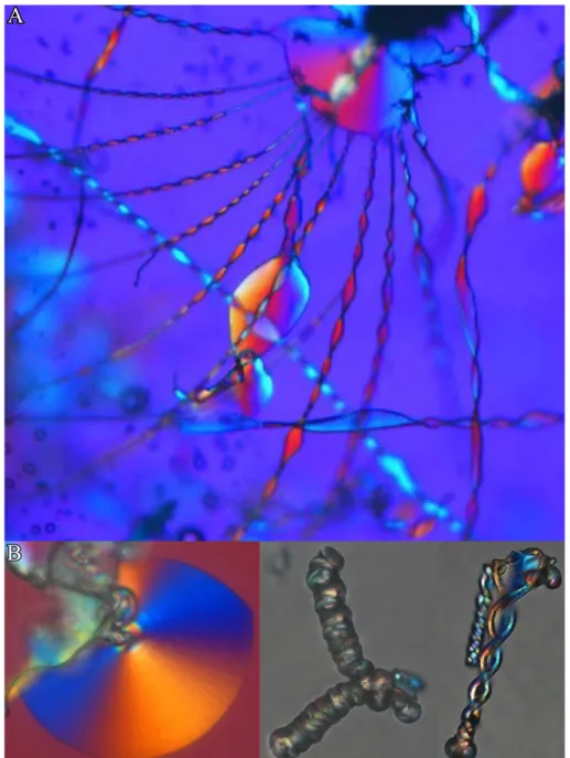
Figure 2.1. Polarized optical micrographs of complex BaCO3 architectures, precipitated in silica containing matrices at elevated pH (panel (A) is reproduced from61).
The similarity of silica-carbonate biomorphs to certain biominerals is not only restricted to non-crystallographic shapes. They also show hierarchical ordering of participating building units and continuous intergrowth of components involved (silica and carbonate) over mul- tiple length scales. Inspired by these findings, silica biomorphs are an interesting model system for laboratory scale studies for the design of novel and advanced materials following the bottom-up strategy with simple (and cheap) components.
B
C D
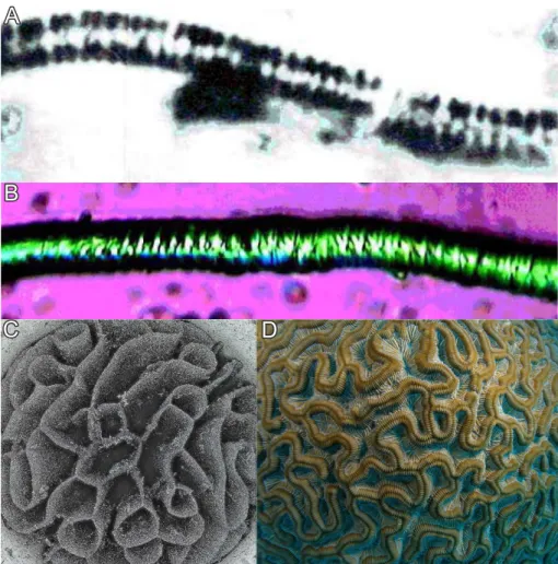
Figure 2.2. Biomimetic but abiotic: Comparison of a Precambrian microfossil (A) and a worm-like "biomorph"64 (B). (C) A coral-like silica biomorph and its living counterpart65 (D).
2.1.2 Preparation methods
Another feature of silica biomorph, which is interesting for material scientists in general, is their quite simple preparation. In early works, their synthesis was described utilizing an U-shaped cassette, containing a silica gel body with a pH of about 10 - 10.5. Precipita- tion of the carbonate architectures was induced via counter-diffusion of separate solutions of alkaline-earth metal salts (typically Ca, Ba, or Sr) and dissolved sodium carbonate.58–60 Alternatively, an ordinary beaker can be used, half filled with silica gel and containing pre- dissolved atmospheric carbon dioxide or sodium carbonate. In this case, crystallization is induced by covering the gel with concentrated solutions of barium-, strontium-, or calcium chloride (0.5-1 M) which diffuse into the gel.58–60 During the unidirectional in-diffusion process, the pH is locally decreased and precipitation of biomorphic aggregates occurs at
Aims and purpose of the thesis different locations throughout the gel matrix. The yield (i.e. the size and number) of silica- biomorphs is thereby a function of the respective relative distance from the solution-gel inter- face. In contrast, biomorphs can also be grown from stagnant alkaline solutions containing equal volumes of dissolved silica and solutions of barium and/or strontium salts.62,66,67 The reactive volume is in this case exposed to the atmosphere and gradual absorption of CO2 by the system at the given alkaline conditions (pH ranges roughly from 10.2 - 11.1, see chapter 5) induces crystallization of astonishing carbonate architectures selectively on the solution/air and solution/tube wall interfaces via heterogeneous nucleation.68 This prepara- tion method was predominatly applied in the respective experiments in the present thesis. As silica source, commercially available water glass can be used or, alternatively, tetraethoxysi- lane (TEOS), as recently revealed by Voinescu et al.69 The influence of concentrations of components on the resulting morphologies was extensively studied in recent works,70,71but in typical standard experiments the "optimal" concentrations for well developed biomorphs turned out to be 5 mM Ba2+and 8.4 mM SiO2.
A B
C
Figure 2.3. Different types of morphologies observed at standard conditions in solution experiments (c(Ba2+) = 5 mM, c (SiO2) = 8.9 mM, initial pH = 11.0) Scalebars are 20µm.
A further parameter that has drastic influence on the morphological evolution of aggregates is the particular pH at which crystallization is carried out, as it strongly influences the chemistry and solubility of the silica and carbonate species in solution. In gel experiments the optimal pH window for the production of silica biomorphs ranges, according to Melero-García et
tail in chapter 5, but earlier works report that well developed biomorphs only occur above pH values of 10. Instead, at lower pH regimes exclusively cauliflower-like dendrites were observed.58,66,73Thus, the pH in a typical standard experiment performed in the context of this thesis was initially adjusted to 11.0.
Those experiments yielded mainly three different types of morphologies, namely nearly flat leaf-like objects (Figure 2.3A), worm-like structures (Figure 2.3B) and twisted helicoids (Figure 2.3C).
2.1.3 Microstructure and composition
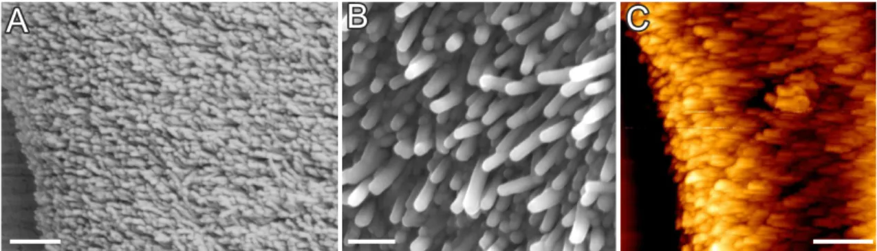
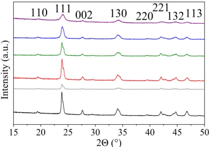
A closer inspection of the structural details of the as-formed architectures reveals a textured, polycrystalline core being composed of myriads of nanocrystals (Figure 2.4) with a typical size of 200-400 nm in length and about 50 nm in width.61,62,67,74–76Individual rod-like units consist of pseudohexagonal BaCO3(witherite) crystals. The hexagonal columnar habit is a common phenomenon among the aragonite-type carbonates and is due to the cyclic twinning on the{110}planes.77
A B C
Figure 2.4. (A-B) FESEM and (C) AFM close-up views on the microstructure of silica- carbonate biomorphs, revealing myriads of nanoscale BaCO3-rods.
Further studies concerning the arrangement of building units within the aggregates show that they are not randomly packed, but rather follow a distinct long-range order on the mesoscale.61–63,67This is achieved, as individual crystallites are nearly parallel aligned with respect to the long axis of the rods (which is their crystallographic c-axis, Figure 2.5A). How- ever, on the mesoscopic scale, individual building units exhibit a slight, mutual misalignment to one another which is approximately kept constant throughout the whole aggregate such that all the crystalline nano-units describe an orientational field with constantly varying vec-
Aims and purpose of the thesis
A
D E
B C
Figure 2.5. (A) TEM micrograph, visualizing the parallel alignment of individual nanocrys- tallites (adopted from,61scalebar is 30 nm). (B) Optical micrograph of a circular sheet between crossed polarizers, showing the typical Maltese-cross extinction pattern, typical objects with anisotropic building units. (C) TEM image of a sample prepared by thin- sectioning biomorphs with leaf-like morphologies. Arrows indicate preferred orientation of the crystallites.61(D-E) Quantitative birefringence optical micrographs (Abrio) of sheets.
The difference in color in the image means different orientations of the subunits, marked with red arrows.
tor (Figure 2.5C). These trends can be analyzed in polarized optical microscopy images of flat sheet-like architectures between crossed polarizers, where the typical Maltese-cross ex- tinction pattern is visible (Figure 2.5B). These color textures in the optical micrographs indi- cate the radial orientation of nanocrystallites outwards starting from a common center in the sheet. The inner orientation of the subunits within the sheet-like object can be furthermore analyzed with a quantitative birefringence imaging microscopy technique78(Abrio∗). In this case, the varying orientation field of the grains can be nicely visualized as different colors
∗Abrio imaging system, Cri, Inc. The system measures the retardance magnitude at every pixel of a charge coupled device image.79This means that at every pixel with the same color index has the sub-structure the same orientation.
can be quantified according to the color wheel (inset in Figure 2.5D-E).
t = 0 sec t = 30 sec t = 60 sec
Carbonate core
Silica skin
Colloidal silica membrane
Figure 2.6. Top: FESEM images of fractured silica biomorphs, revealing the dual composite character with an outer silica skin sheathing the inner carbonate texture (images reproduced from61). Scalebars are 2 µm (left) and 500 nm (right). Below: Sequence of optical mi- croscopy images showing the gradual dissolution of the inner carbonate phase from a worm- like structure with dilute acid. Such a treatment leaves a hollow silica membrane with the original worm-like morphology.
Apart from the ordering of the nanoparticles, silica biomorphs are also dual composite mate- rials as they are comprised of carbonate and co-precipitated amorphous silica.58–64,66–69,73,75,76,80,81
In most cases the entire carbonate part of mature architectures is enveloped by a continu- ous skin of agglomerated colloidal silica spheres which can be nicely visualized in images of fractured aggregates (Figure 2.6). The presence of external silica layers can either be demonstrated by EDX spectroscopy, or by immersion of selected aggregates into dilute acid, leading to selective dissolution of the carbonate core (Figure 2.6). The remaining silica parts (the so-called "ghost") still exhibit the original morphology of the previously intact biomorph architecture. These experiments can be nicely followed in-situ with optical microscopy, as
Aims and purpose of the thesis shown in the time-laps sequence in Figure 2.6 (below).62,63,67,76,82–84 Likewise, the silica part of the biomorphs can also be removed selectivley with dilute NaOH, leaving the intact car- bonate ultrastructure.61,67 It was furthermore reported that the dual composite character of silica biomorphs is not only restricted to the macroscopic length scale. Also single car- bonate rods could be surrounded by siliceous skins, stabilizing them against ripening.74,85 This suggests mutual intergrowth of components over multiple length scales, as observed in many biominerals, and supports furthermore the resemblance of these unique aggregates to products from biological mineralization.
2.1.4 Formation mechanism
Beneath studies concerning the compositional and structural details, recent investigations related to silica biomorphs were focused on morphogenetic scenarios on the nano- and mi- crometer level, and especially on the physical origin for the formation of these fascinating architectures, since all these aspects remained obscure for long time.61,62,86,87
A) B)
Figure 2.7. A) optical81and B) FESEM micrographs61 showing primary nucleated rod-like barium carbonate crystals that start to split at their tips due to the poisoning influence of silica species. Scale bars in B) are 5µm.
Stage 1: Fractal Branching
In general, morphogenesis can be subdivided into two fundamental growth modes which are run through successively depending on the temporally changing conditions in the system.
and growth of elongated BaCO3microcrystals (Figure 2.7) as soon as the bulk volume gets supersaturated, i.e. when enough CO2 has entered into the system in solution experiments.
Further growth of the single-crystalline core leads to self-similar splitting of the rod-like crystal at its tips (Figure 2.7).66 These observations are caused by the poisoning influence
A
A B C
1µm 5µm 5µm
D E F
Figure 2.8. Continuous fractal branching in the presence of silica impurities. Initially rod- like witherite crystals successively develop into dumbbell-like aggregates (A-B), yielding closed, spherulithic (C) or even spacious, cauliflower-like architectures (D-F). Scalebars are 1µm (A) and 2µm (B-C). (Panels (D-F) adopted from81)
of partially condensed silica species which act as non-absorbable polymeric impurities, that are pushed ahead of the growth front, generating the formation of misoriented (with respect to the crystalline lattice) two-dimensional "islands".89 Such a growth mechanism yields at first tilted projections which emanate at non-crystallographic angles, leading to dumbbell- shaped particles which evolve, upon continuous bifurcation, into rather closed spherulithic structures or spacious cauliflower-like architectures (cf. Figure 2.8 E-F), depending on the intensity of branching.81The described morphogenetic scenario was observed in all the stud- ies conducted in the context of this thesis (see chapter 5 and 7), but the ultimate experimental evidence that exclusively the silica species is responsible for the ramification of the initial carbonate seed is given in chapter 3. Similar growth mechanisms are also well-documented for fluorapatite crystals, precipitated in gelatin (Figure 2.9).88,90,91 Here the crystals passed
Aims and purpose of the thesis
Figure 2.9. Fluorapatite architectures, grown in a gelatin matrix, with dumbbell- and notched-sphere-like morphologies (right), and schematic drawings highlighting their forma- tion mechanism by self-similar branching.88
the same morphological transformation from rod-to-dumbbell-to-sphere", due to successive splitting of the parental crystal seed.
Stage 2: Chemically Coupled Co-Precipitation
The presented first stage of biomorph morphogenesis basically occurs roughly within the first 4 h after mixing of reagents.75 After this period, the silica additive has blocked any active sites for attachment of carbonate units onto the highly branched aggregate, prevent- ing further growth of the fractal architectures. This causes high supersaturation levels of the carbonate species on a local scale, and, as a consequence, myriads of nanocrystals nucleate in a three-dimensional fashion across the whole surface of the locked spherulite.
The reason for the spontaneous stabilization and continuous production of nanosized crys- tallites is principally based on reversed solubility trends of carbonate and silica with pH (silica is soluble at high pH and carbonate at low pH, see Figure 5.14).81,86 At the given alkaline conditions (pH 9-11) where growth typically occurs, there exist substantially frac- tions of carbonate and bicarbonate ions in equilibrium. Therefore, nucleation of BaCO3 strongly influences the equilibrium as CO32 – ions are continuously withdrawn according to:
HCO3– −−↽−−⇀CO32 – +H+ Ba
2+
−−−→BaCO3. Consequently HCO3– ions in the near vicinity will dissociate and release a proton in order to balance the short state of local disequilibrium.
1. Nucleation of BaCO3 local pH Gradient 2. Generation of a
Si(OH) O3 -+H+ Si(OH)4 Si(OH)4 SiO2
3.Precipitation around growing particles
pH pH
HCO3- CO +32- H+Ba2+BaCO3
Stabilization of crystalites New event of nucleation
HCO3-
-H O2
2-SiO-
+ BaCO3 CO32-
SiO2 -SiO OSi-
pH pH
B)
Figure 2.10. The proposed formation model for the continuous production of nanosized crystals, based on the chemically coupled precipitation of carbonate and silicate. (A) During growth of rod-like barium carbonate crystals (red) the pH decreases locally relative to the bulk (gradient highlighted as green shadow) as bicarbonate ions dissociate. This in turn triggers polymerization of silicate which will re-increase the local pH on the growth front, thus raising the supersaturation of BaCO3in the close vicinity subsequently causing a novel event of carbonate nucleation. (B) Overall, a loop process is generated in which the involved components alternately precipitate. In this way, building units for the construction of silica biomorphs are continuously produced.
However this provokes a local pH gradient relative to the bulk around the as-nucleated car- bonate particles, (Figure 2.10A, green shaded area around growing barium carbonate par-
Aims and purpose of the thesis ticles) which in turn has drastic influence on the solubility of the silica species (cf. Figure 5.14) nearby the just nucleated carbonate nanoparticles. As a result, silica polymerizes lo- cally around the surface of the developing crystals, forming coating layers and thus pre- venting particles from further growth beyond the nanometer scale. Targeted precipitation of SiO2 in the vicinity of the carbonate units is in principle achieved by protonation of acidic silanol groups (caused by previously dissociated protons) which become subject to con- densation reactions, inducing polymerization of silica according to the following sequence:
2−Si−O– +2 H+ −−↽−−⇀ -Si-O-Si- −−−→−H2O SiO2↓ (see Figure 2.10). In contrary, these reactions will re-increase the local pH (relative to the bulk) again , thus re-shifting the car- bonate/bicarbonate equilibrium towards higher fractions of CO32 –. This also elevates the carbonate supersaturation in close vicinity of freshly generated carbonate/silicate nanopar- ticles, therefore triggering a novel event of barium carbonate nucleation (Figure 2.10A).
In this way, an autocatalytic loop process is generated where involved components are al- ternately mineralized, leading to continuous production of uniform building units, which arrange spontaneously, by a still unknown process, to elaborate architectures with sinuously shaped surfaces. The presented mechanism is summarized in Figure 2.10B.
With regard to the envisaged formation model, the particular bulk pH in the growth media plays an essential roles during morphogenesis, as it strongly influences the speciation of in- volved components. To induce the dynamic interplay between carbonate and silica, certain bulk pH dependent requirements have to be fulfilled, as recently demonstrated for morpho- genesis in silica-gel setups.72 This issue will be furthermore discussed in the framework of the thesis for growth of silica biomorphs from stagnant solutions (chapter 5).
Experimental indications for the theoretical formation model
Despite of the nuerous studies performed up to now the presented scenario for the continu- ous production of silica stabilized carbonate rods has still mainly a theoretical background.
The proposed model implicates however an oscillatory local pH at the growth front, as this parameter is coupled with the alternating mineralization of the two components. Such pe- riodic pH variations are though reduced to the region of active growth which extends only over some microns, as deduced from experiments in stirred solutions.87A direct experimen- tal evidence for the proposed model of autocatalytic growth lies in the in-situ recording of
was to measure the pH in the close vicinity of just-growing biomorphs, utilizing special dyes in combination with a UV-Vis microscope. First experiments using mixtures of UV-active pH indicators did unfortunately not yield the expected results, which was primary based on the insufficient spacial resolution of the instrumental setup.
Apart from that, further attempts to explain the spontaneous stabilization of carbonate nano- rods were put forward in terms of crystallographic arguments by Aquilano et al.85 In this work the miniaturization of carbonate units is ascribed to epitaxially matching of α-quartz on witherite surfaces. Thus adsorption of silicate (which exists in case of biomorphs rather in the amorphous form) onto BaCO3 faces via attachment of single chains or formation of 2D islands is energetically favored. Such interactions are also certainly important for the generation of extended silica sheaths around individual building units.
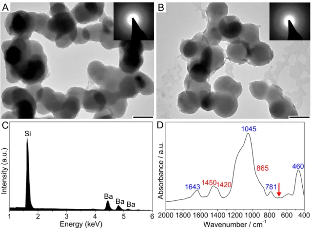
In the scope of the proposed pH oscillations, recent studies following a different approach, demonstrated that specific, pH induced interactions between carbonate and silicate can in fact trigger local SiO2 precipitation around growing ACC particles at alkaline conditions, (Figure 2.11a) strongly influencing the crystallization process.92 In contrast to the prepara- tion methods presented for biomorphs, the experiments were carried out at higher super- saturation via direct mixing of solutions (CaCl2 and dissolved SiO2 containing carbonate).
Corresponding TEM images and micro-EDX spectra (Figure 2.11) of early stage particles clearly evidence that ACC nanograins become coated by extended silica layers upon pH- coupled co-precipitation. The thickness of the skins could be furthermore tuned by varying the silica concentration in the reactive solutions. Results deduced from this investigation can be regarded as fundamental support for the proposed growth mechanism, as local pH gradients provoked by homogenous nucleation of carbonate particles leads to silica poly- merization all over their surface. Those results gave additionally the motivation for a study conducted in this thesis following a similar procedure, but replacing calcium by barium in order to investigate if similar tendencies are apparent at the given high (and defined) carbon- ate supersaturation. Results of these experiments are given in chapter 3.
In view of the still quite hypothetical model for the formation of silica biomorphs, exter- nal silica skins, as clearly detected in case of homogeneous nucleated ACC particles from
Aims and purpose of the thesis
Figure 2.11. Amorphous CaCO3 nanoparticles with silica skins yielded via direct mixing of CaCl2 and SiO2 containing carbonate solutions at high alkaline pH. EDX-microanalysis of the two different layers reveals that the core of the particles is enriched in CaCO3 while the outer layer mainly consists of silica. (adopted from92)
silica containing, should also be detectable for nanorods constituting biomorphs. However published data concerning this issue are ambiguous, as silicon counts are present when per- forming micro-EDX studies on individual building units of biomorphs but pronounced silica skins could not be verified on TEM micrographs (see also Figure 2.5A).61,75 Further hints were given by Terada et al. in a recent publication where distinct silica coatings around the building units of silica biomorphs grown in gel were postulated. However final evidences supporting this notion were not given.82
In order to shed further light on the detailed role of silica during the generation of nanoscale barium carbonate, an experimental procedure was utilized in this thesis which was aimed on the targeted production of BaCO3 nanorods (in the presence of silica), at conditions in
ter 4). This was achieved with the help of a titration based assay in combination with an ion selective electrode (ISE) sensitive for Ba2+. In a recent work by Gebauer et al.,93 this method was employed to get fundamental insights into the early stages of CaCO3 precipi- tation using an ion selective electrode (ISE) sensitive for Ca2+. Presented findings lead to novel views on the nucleation of calcium carbonate in general, as so called prenucleation clusters were detected, being a thermodynamically stable phase existing in equilibrium with dissolved ions occurring prior to the formation of a solid phase. The applied experimental technique also enables detailed views on the influence of certain additives during different stages of calcium carbonate crystallization.94–97 In this case, interactions between involved components are discussed on basis of certain characteristics of the temporal development of the free calcium concentration during addition of CaCl2to a carbonate buffer, containing the respective additive at constant pH. Exemplary curves for the time dependent development of
Figure 2.12. Temporal development of the amount of free Ca2+ions, monitored during con- tinuous addition of CaCl2into a carbonate buffer in the presence and in absence of additives.
Typical characteristics of these plots were used to describe the role of the respective addi- tive on the nucleation of calcium carbonate. Stage 1: Slope of the linear increase during the prenucleation stage indicates the fraction of Ca2+bound in clusters and thus the apparent sta- bility of prenucleation clusters. Stage 2: Time of nucleation which is usually retarded in the presence of additives. Stage 3: Level of free calcium in the postnucleation stage, therefore representing the solubility of the initially precipitated phase.97
Aims and purpose of the thesis the free calcium potentials in the absence and in the presence of additives (in this case amino acids97) are presented in Figure 2.12. At first, a linear increase of free Ca concentration is visible, since calcium chloride is continuously added to the buffer solution. In this regime the concentration of free Ca2+ions was always found to be somewhat lower than the dosed amount, being a result of equimolar binding of Ca2+ and CO32 – ions in stable prenucleation cluster.93,98When the curve reaches its maximum, a solid phase nucleates and consequently the amount of free calcium drops to a level that corresponds to the solubility of the pre- cipitated CaCO3 phase. This plateau remains virtually unchanged upon further addition of calcium which is related to the presence of two different phases in equilibrium. As depicted in Figure 2.12, the curve exhibits certain characteristics which allows to interpret the influ- ences of the additive on the nucleation: (1) Slope in the linear part of the titration curve reflecting the stability of prenucleation clusters. (2) Point of nucleation, visible as maximum of the peak and (3) Level of solubility (ion product), in most cases observed immediately af- ter nucleation when the solid phase is in equilibrium with the surrounding solution. All these parameters were found to be strongly influenced by additives in case of calcium carbon- ate precipitation.94–97 However, a detailed study on silica-related influences on all of these parameters during precipitation of BaCO3 is given in chapter 4.
2.1.5 Phenomenological description of morphology development
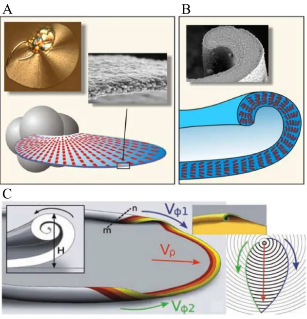
While continuous production of nanoscale building units is explained by a chemically cou- pled co-precipitation mechanism, morphogenesis of characteristic architectures on macro- scopic length scales depends on even more delicate factors, as recently proposed by García- Ruiz et al.81 Virtually all complex curved architectures displayed by silica biomorphs, ini- tially arise from globular units, yielded during the first stage of morphogenesis via contin- uous branching of parental crystal seeds. In the second stage, quasi-2D laminar segments with nearly radial symmetry, being composed of myriads of nanosized carbonate crystallites, start to sprout out from the globular units (Figure 2.13A). Recent investigations with time- lapse video microscopy revealed that the radial growth velocities (Vρ, see Figure 2.13C) are equal in all directions (typically some microns per minute) which is the reason why the grow- ing aggregate adopts the shape of a circular disk (cf. Figure 2.5).61,75,81,86 Interestingly, the main part of the evolving sheets develop scrolled margins, either pointing down- or upwards
C
Figure 2.13. Illustration of the macroscopic growth behavior of silica biomorphs. (A) Af- ter formation of closed spherulites (indicated as grey globules), thin, polycrystalline leaf- like segments start to sprout out which are composed of myriads of nanorods, generated by chemically coupled co-precipitation. (B) Continuous laminar growth proceeds until curva- ture is induced, as the sheet becomes scrolled at some point around its perimeter. (C) After formation of scrolled margins, growth in radial direction (Vρ) arrests, and curls propagate tangentially along the rim of the sheet with azimuthal growth velocitiesVϕ1andVϕ2 usually yielding cardoid architectures. The relative height H and the relative handedness of two dis- tinct curls thereby determine the occurrence of more twisted morphologies like helicoids or worms.61,81,86
at arbitrary positions on the rim (Figure 2.13B). Therefore, these singular curling events in- troduce curvature to the system and serve as starting points for the morphogenesis of highly twisted shapes.81Such curls were proposed to propagate along the perimeter of the leaf-like
Aims and purpose of the thesis
Figure 2.14. Morphogenesis of a helix. (A) Sequence of optical micrographs in which curled segments with equal handedness gradually approach from different directions (the upper curl bent towards the camera, lower curl bent downwards) and meet at the cusp. Both segments intertwine and produce a regular helix upon further growth along the radial direction (indi- cated by the red arrow). (B) Scheme showing the front-view of the same scenario where two like-handed curls with the same height H and azimuthal growth velocitiesVϕ1andVϕ2
lock-in to produce a helicoid.81(images reproduced from61)
morphologies in a manner reminiscent of a surfing wave (Figure 2.13C).61,81 At this point, growth arrests along the radial pathway and a new growth front is generated that propagates orthogonally to the previous circular direction. As in case of the radial growth front, the velocity of tangential propagation (Vϕ1 and Vϕ2, see Figure 2.13C) is also approximately constant, nevertheless the absolute values ofVϕ2 andVϕ2 were found to be slightly higher thanVρ.75,81Therefore the initially circular shape becomes distorted and the leaf-like objects form one or more cusps at positions where two curled segments arrive from opposite direc-
objects with cardioid shapes. The question whether the final aggregate will adopt a twisted shape, as displayed by helicoidal or worm-like architectures is strongly related to the relative velocities of azimuthal and radial growth and to the handedness and height of the curled seg- ments, as described in detail by García-Ruiz et al.81 A scenario, that exemplarily describes the formation of a helix is presented in Figure 2.14. The sequence shows a case, where two curled segments with equal directionality (one bent downwards the other upwards) and height approach at a cusp from opposite sides of the sheet segment. When the two curls collide, they will intertwine and wind around each other (cf. Figure 2.14B). Upon further growth in radial direction, the induced twist is retained, and due to continuous winding (dic- tated by the curled rims) a nearly perfect helicoid is formed.61,81In contrast, when the height difference between two curls is drastically pronounced and on of them grows faster, a situ- ation arises in which the higher curled segment prefers curling around itself, resulting in a worm-like morphology.81. However, a still open question in this context is the origin of cur- vature, i.e. why do initially flat sheets start to curl at a certain point along the rim? Possible answers to this question are given in chapter 6.
2.1.6 Biomorphs with SrCO
3and CaCO
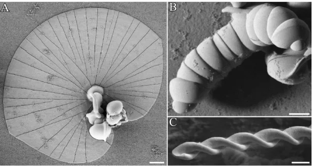
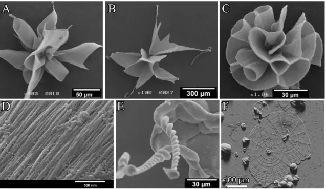
3The stunning crystal architectures presented in the sections above are, however, not only limited to BaCO3. Replacing this metal ion by strontium yields widely identical biomor- phic objects to those observed with barium carbonate (witherite).59 Indeed, Terada et al.82 isolated petal-like objects (Figure 2.15A-C) when growth of strontianite biomorphs was per- formed in silica gels at high pH. Upon further growth, complex wounded helical ribbons formed sporadically at the tips of the petals (Figure 2.15E).82Close-up views on the aggre- gates revealed, in contrast to witherite biomorphs, rather fibrous subunits which are individ- ually sheathed by siliceous skins.82 As in case of silica-gel setups, crystallization of silica- strontianite biomorphs from solution results in aggregates with morphologies as observed with BaCO3 (see Figure 2.15F, and chapter 7). These findings imply that only carbonate minerals with aragonite-type lattices are able to produce biomorphs when crystallization is carried out in silica gels or sols. This requirement is fulfilled for strontianite and witherite (or- thorhombic lattice, space group Pmcn) at room temperature. However, CaCO3, the mineral
Aims and purpose of the thesis
100 µm
A
D E F
B C
Figure 2.15. Silica biomorphs composed of SrCO3 (strontianite). (A-E) Morphological variety observed in gel setups at pH 10.5. (A-B) Petal-like products from which helicoids (E) emerge upon further growth. (C) Close-up view revealing the texture of observed crystal architectures. (F) Leaf-like objects grown in direct contact with the substrate observed when synthesis is carried out in solutions. (A-E reproduced from82)
of utmost interest among the alkaline-earth carbonate series, forms predominantly rhombo- hedral calcite crystals at ambient conditions (due to the smaller size of the cation). Thus, corresponding precipitates in silica rich environments did not yield biomorphs in the true sense, since characteristic morphologies like helicoids or worms remained absent. Instead, biomimetic aggregates with finger-like structures60 or bundles reminiscent of natural sheaf of wheat99,100 (Figure 2.16) or even spherulithic single crystals displaying unusual three- fold symmetry were obtained.101 In contrast to aggregates observed in case of strontianite and witherite, morphogenesis of the calcite architectures is not explained by coupled co- precipitation of silica and carbonate, but rather via selective adsorption of silicate species on specific crystallographic planes causing changes in the relative growth rates of distinct calcite faces.99–101In this way, the rhombohedral crystal habit becomes elongated along the c-axis and develops subunits with a three-pointed star-like appearance (three wings arranged at mutual angles of∼120◦, see inset of Figure 2.16C).61,70,101
In opposite, CaCO3 biomorphs with mainly coral-like appearance precipitated, when the
C
Figure 2.16. (A-B) Radially arranged sheaf of wheat bundles observed when CaCO3crystal- lization is carried out in silica gels at alkaline pH (reproduced from99. (C) Complex calcite architectures with unusual threefold symmetry observed at high carbonate supersaturation due to selective adsorption of silicate species on certain planes of the calcite lattice.101
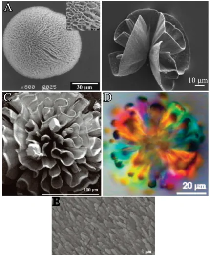
conditions were adjusted in order to shift the CaCO3polymorph selection in the direction of aragonite, indicating that the mineralogy (i.e. orthorhombic lattice) is an important parameter for the production of sinuously shaped carbonate architectures in silica matrices.61One strat- egy in this context is to increase the synthesis temperature to80◦Csince the aragonite modi- fication becomes kinetically favored over the calcite polymorph at these conditions. This was successfully accomplished by Voinescu et al.,84 and hierarchically structured morphologies reminiscent of natural coralline shapes or architectures with flower-like appearances were isolated from diluted silica sols (Figure 2.17B-D).84 Similar aggregates (cf. Figure 2.17A) were observed by Imai et al. in silica gels where growth was induced utilizing aragonite seed crystals at ambient conditions.83 Likewise, preliminary work, perfromed by Bittarello et al., demonstrated the spontaneous presence of aragonite biomorphs in silica gels at specific con-
Aims and purpose of the thesis
A
C D
Figure 2.17. CaCO3biomorphs. (A-D) FESEM and polarized optical micrographs of arag- onite aggregates with coral-like morphologies synthesized in (A) silica gel at pH 10.583 or (B-D) in diluted silica sols84 at 80◦C. (E) Hierarchical ordering of aragonite subunits of coral-like morphologies.84
ditions in terms of pH and reagents concentration and location in the gel.85A potential novel strategy to produce CaCO3biomorphs at room temperature via a simple one-pot route with- out the need of external aragonite triggers (like seed crystals) was to work with mixtures of calcium and barium or strontium. Findings of this study are given in chapter 7.
3 Bottom-Up Self-Assembly of Amorphous Core-Shell-Shell
Nanoparticles and Biomimetic Crystal Forms in Inorganic Silica-Carbonate Systems
3.1 Abstract
Mineralization of alkaline-earth carbonates in silica-rich media at high pH leads to fasci- nating crystal morphologies that strongly resemble products from biomineralization, despite the absence of any organic matter. Recent work has demonstrated that elaborate CaCO3 structures can be grown in such systems even at high supersaturation, as nanoparticles of amorphous calcium carbonate (ACC) were spontaneously coated by skins of silica, which decelerated their re-dissolution and thus served as storage depots continuously supplying growth units for the formation of crystalline calcite. In this chapter barium carbonate was precipitated under similar conditions and surprisingly different behavior was detected. At low silica concentrations, there was no evidence for an amorphous carbonate precursor phase and crystallization occurred immediately, resulting in elongated crystals that showed progressive self-similar branching and bifurcation due to the poisoning influence of silicate oligomers on the growth process. Above a certain threshold in the silica content, rapid crys- tallization was in turn prevented and amorphous nanoparticles were stabilized in solution.