Synthesis and Characterization of Ag
2MnSnS
4, a New Diamond- like Semiconductor
Daniel Friedrich,[a]Sebastian Greil,[a]Theresa Block,[b]Lukas Heletta,[b]Rainer Pöttgen,[b]and Arno Pfitzner*[a]
Dedicated to Prof. Wolfgang Bensch on the Occasion of his 65th Birthday
Abstract. Phase pure Ag2MnSnS4was synthesized from the elements using standard high-temperature solid-state methods. Its crystal struc- ture was solved from single-crystal X-ray diffraction data collected from a pseudo-merohedrally twinned crystal as well as by Rietveld refinement of X-ray powder diffraction data. Ag2MnSnS4crystallizes in the monoclinic space groupPc(no. 7) with the unit cell parameters a = 6.651(1), b = 6.943(1), c = 10.536(2) Å,β = 129.15(1)°, V = 337.3(1) Å3, andZ= 2. The tetrahedraly compound crystallizes in a superstructure of the wurtzite type and the tetrahedra volumes are in good agreement with the model for diamond-related compounds de-
Introduction
The first tetrahedral compound in literature is stannite (Cu2FeSnS4),[1]whose name also describes a structure-type of quaternary tetrahedral compounds that are derived from sphal- erite. Starting in 1970 several more of these quaternary com- pounds were synthesized. Their structures were either related to the sphalerite structure-type or could be derived from the wurtzite structure-type.[2–4] The latter materials are conse- quently called wurtz-stannite type compounds. Usually, these compounds exhibit semiconducting properties and may be used as diamond-like semiconductors (DLS).[5–6] In recent years, this class of compounds received a lot of attention as these materials exhibit a broad range of interesting physico- chemical properties depending on the chemical composition of the solids. Their optical and electrical properties make them very promising photovoltaic materials. Especially Cu(In,Ga)(S,Se)2 is in the focus of photovoltaics due to its high efficiency.[7–8]The quaternary compound Cu2ZnSnS4[9]is also investigated as photovoltaic material. Steinhagen sug- gested nanocrystals of Cu2ZnSnS4 to use them as a cheaper route to photovoltaics,[10]andScraggfound that the bandgap
* Prof. Dr. Arno Pfitzner Fax: +49 941 943 4551
E-Mail: arno.pfitzner@chemie.uni-regensburg.de [a] Institut für Anorganische Chemie
Universität Regensburg Universitätsstraße 31 93040 Regensburg, Germany
[b] Institut für Anorganische und Analytische Chemie Westfälische Wilhelms-Universität Münster Corrensstraße 30
48149 Münster, Germany
Supporting information for this article is available on the WWW under http://dx.doi.org/10.1002/zaac.201800142or from the au- thor.
rived from the wurtzite structure type. The red semiconductor Ag2MnSnS4has an optical bandgap ofEg= 2.0 eV and is stable up to its peritectic decomposition temperature of approximately 700 °C.
Ag2MnSnS4is a Curie–Weiss paramagnet with an experimental mag- netic moment ofμexp= 5.4(1)μBper manganese atom. Antiferromag- netic ordering is detected at a Néel temperature ofTN= 8.8(1) K.119Sn Mößbauer spectra at 78 K underline the single tetrahedrally coordi- nated SnIVsite (δ= 1.34(1) mm·s–1). The 6 K spectrum (magnetically ordered state) reveals a small transferred magnetic hyperfine field of 1.02(1) T.
(1.49 eV) of Cu2ZnSnS4is quite close to the optimum for the usage in solar cells.[11] Non-centrosymmetric, diamond-like semiconductors are also very interesting materials in the field of non-linear optics.[12,13]Interesting examples in this field are for example GaAs, GaP, AgGaQ2(Q= S, Se),[13,14]and quater- nary compounds like Li2MnSnSe4[15]or Li2CdGeS4.[16]
Recently, tetrahedral networks deviating from this diamond- like topology (LiGaGe2Se6, Li2In2MQ6withM= Si, Ge,Q= S, Se) were also reported to have interesting non-linear optical properties.[17,18]Furthermore, materials containing Mn2+, Fe2+
or Co2+ are of special interest due to their magnetic proper- ties.[19–22] Nénert discussed the potential of Cu2MIIMIVS4 as multiferroics,[22]as such materials show coupling of both their magnetic and their electronic properties. The authors found that several of these compounds are possible multiferroics with high polarization due to their symmetry and structural consid- erations. In detail Cu2MnGeS4and Cu2MnSnS4were investi- gated and both compounds show a linear magnetoelectric ef- fect.[6]The same arguments should hold true for Ag2MnSnS4, since its crystal structure is quite similar to that of Cu2MnGeS4. The crystal structures of such diamond-like tetra- hedral compounds can usually be derived from the two dif- ferent polymorphs of zinc sulfide. Normal-valent tetrahedral compounds are also called adamantane compounds.[20]Devia- tions of an average of four valence electrons per atom will cause structural distortions as for example shown for Cu3TeS3Cl. This compound has an excess of two valence elec- trons, which are located in a lone electron pair at the Te4+
cation.[23] Possible combinations of elements for such ada- mantane compounds can be derived via cross-substitution as shown byParthé.[24]
It has been shown that a superstructure related either to the wurtzite-type or to the sphalerite-type results in ternary and
multinary materials of this class depending on the volume dif- ferences of the constituting tetrahedra [MQ4].[25,26]According to this model the volume differences of the tetrahedra [MQ4] have a strong impact on the structure type. For small volume differences a sphalerite-type related structure will occur, and wurtzite-type related structures are preferred in case of differ- ences greater than a critical value. A different model suggests wurtzite-type related superstructures for silver compounds and sphalerite-type related superstructures for copper compounds, thus referring to the smaller ionic radius of copper.[27]It also suggests that the wurtzite structure-type should be preferred at higher temperatures. This trend describes some compounds quite well, though it is not as quantitative as the model pre- sented above.[27] A third model[28] refers to the c/a-ratio of tetrahedral compounds. At room temperature there is an ideal ratio of 1.633 for wurtzite-type structures. Those compounds having a smaller ratio prefer a wurtzite-type structure, while those having a larger ratio will consequently prefer the sphaler- ite-type structure. This ratio decreases at higher temperatures and therefore describes why wurtzite-type modifications tend to be more stable at higher temperatures. Further articles have been published which discuss the volumes of tetrahedral com- pounds and their influence on the structure type.[25–32]
Herein, we present the synthesis and crystal structure deter- mination of the adamantane compound Ag2MnSnS4, which has been investigated earlier.[21,54]However, only its antiferromag- netic properties and an erroneous orthorhombic metric were reported at that time. It has recently been mentioned also by Lekse, but no reasonable structure model is known to date.[33]
Very recently a structure determination was reported assuming orthorhombic symmetry and neglecting the monoclinic sym- metry of the title compound thus leading to quite significantly enlargedR-values.[34]
Results and Discussion
Crystal Structure Determination of Ag2MnSnS4
The crystal structure of Ag2MnSnS4was solved from single- crystal X-ray diffraction data collected at 20 °C. Ag2MnSnS4 crystallizes in the monoclinic space groupPc(no. 7) witha= 6.651(1),b= 6.943(1), c= 10.536(2) Å, β= 129.15(1)°,V= 337.3(1) Å3, and Z = 2. An initial structure solution in the orthorhombic space groupPmn21resulted in unusually highR values (see also reference[34]). Therefore, a symmetry re- duction was considered, i.e., monoclinic symmetry with pseudo-merohedral twinning similar to Li2ZnSnS4.[35] This symmetry reduction leads to the monoclinic space group Pn (no. 7), a non-standard setting of Pc. The resultingR1 value dropped significantly fromRortho= 0.0490 toRmono= 0.0348.
An additional twofold-axis was introduced to take the pseudo- symmetry into account. The corresponding twin law (in Pn), 1 0 0, 0 –1 0, 0 0 –1, dropped the finalRvalues toR1(all data)
= 0.0283 andwR2(all data) = 0.0518. The non-standard setting Pn, however, was only used to emphasize the symmetry rela- tions to the orthorhombic supergroupPmn21. For the following discussions, the structure of Ag2MnSnS4 is presented in the
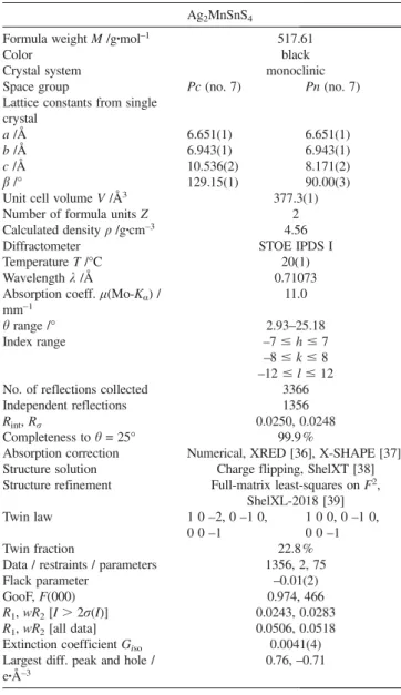
standard settingPc. The final crystallographic data and experi- mental details of the data collection are listed in Table 1. The atomic coordinates, equivalent displacement parameters, and anisotropic displacement parameters are listed in Table 2 and Table 3. Table 4 shows selected bond lengths and angles.
Table 1.Crystallographic data for Ag2MnSnS4. Ag2MnSnS4
Formula weightM/g·mol–1 517.61
Color black
Crystal system monoclinic
Space group Pc(no. 7) Pn(no. 7)
Lattice constants from single crystal
a/Å 6.651(1) 6.651(1)
b/Å 6.943(1) 6.943(1)
c/Å 10.536(2) 8.171(2)
β/° 129.15(1) 90.00(3)
Unit cell volumeV/Å3 377.3(1)
Number of formula unitsZ 2
Calculated densityρ/g·cm–3 4.56
Diffractometer STOE IPDS I
TemperatureT/°C 20(1)
Wavelengthλ/Å 0.71073
Absorption coeff.μ(Mo-Kα) / 11.0 mm–1
θrange /° 2.93–25.18
Index range –7ⱕhⱕ7
–8ⱕkⱕ8 –12ⱕlⱕ12 No. of reflections collected 3366
Independent reflections 1356
Rint,Rσ 0.0250, 0.0248
Completeness toθ= 25° 99.9 %
Absorption correction Numerical, XRED [36], X-SHAPE [37]
Structure solution Charge flipping, ShelXT [38]
Structure refinement Full-matrix least-squares onF2, ShelXL-2018 [39]
Twin law 1 0 –2, 0 –1 0, 1 0 0, 0 –1 0,
0 0 –1 0 0 –1
Twin fraction 22.8 %
Data / restraints / parameters 1356, 2, 75
Flack parameter –0.01(2)
GooF,F(000) 0.974, 466
R1,wR2[I⬎2σ(I)] 0.0243, 0.0283 R1,wR2[all data] 0.0506, 0.0518 Extinction coefficientGiso 0.0041(4) Largest diff. peak and hole / 0.76, –0.71 e·Å–3
The competition between the space groupsPmn21 andPn has already been described byParthéin 1969.[2] The authors mentioned that for Cu2CdGeS4, also an adamantane com- pound, it is difficult to determine the right space group.
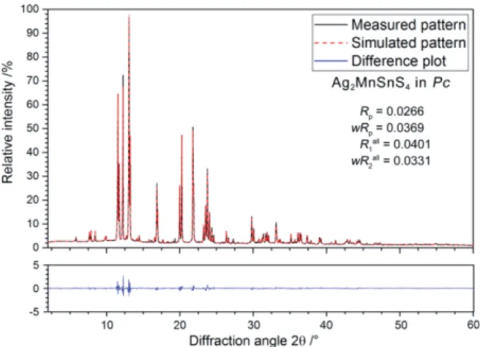
Furthermore, they already pointed out that the diffraction pat- terns are identical for both space groups. This also holds true for Ag2MnSnS4. The X-ray powder diffraction patterns for the orthorhombic and for the monoclinic structure models there- fore cannot be distinguished. The symmetry relations between Pmn21andPn, as well as the relations to the wurtzite structure type are shown in aBärnighausen-tree (see Figure 1).[40]
Consequently, theRvalues for the pattern fit of the Rietveld refinement of Ag2MnSnS4 were practically identical for the space groups Pmn21 and Pc. However, similar to the single-
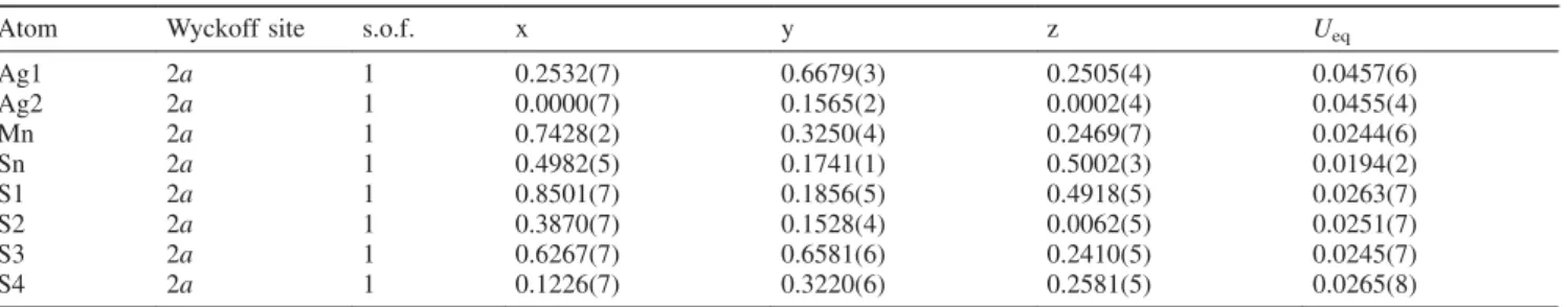
Table 2.Atomic coordinates and isotropic displacement parametersUeqa)/Å2for Ag2MnSnS4(20 °C) in the space groupPc.
Atom Wyckoff site s.o.f. x y z Ueq
Ag1 2a 1 0.2532(7) 0.6679(3) 0.2505(4) 0.0457(6)
Ag2 2a 1 0.0000(7) 0.1565(2) 0.0002(4) 0.0455(4)
Mn 2a 1 0.7428(2) 0.3250(4) 0.2469(7) 0.0244(6)
Sn 2a 1 0.4982(5) 0.1741(1) 0.5002(3) 0.0194(2)
S1 2a 1 0.8501(7) 0.1856(5) 0.4918(5) 0.0263(7)
S2 2a 1 0.3870(7) 0.1528(4) 0.0062(5) 0.0251(7)
S3 2a 1 0.6267(7) 0.6581(6) 0.2410(5) 0.0245(7)
S4 2a 1 0.1226(7) 0.3220(6) 0.2581(5) 0.0265(8)
a)Ueqis defined as one third of the trace of the orthogonalizedUijtensor.
Table 3.Anisotropic displacement parametersUij/Å2for Ag2MnSnS4(20 °C) in the space groupPc.
Atom U11 U22 U33 U23 U13 U12
Ag1 0.042(1) 0.047(1) 0.046(1) 0.008(1) 0.027(1) 0.009(1)
Ag2 0.045(1) 0.047(1) 0.047(1) –0.002(1) 0.030(1) 0.000(1)
Mn 0.025(1) 0.024(1) 0.024(1) –0.007(2) 0.016(1) –0.007(2)
Sn 0.018(1) 0.020(1) 0.020(1) 0.000(1) 0.012(1) 0.001(1)
S1 0.023(1) 0.032(1) 0.027(2) 0.003(2) 0.017(1) 0.004(2)
S2 0.025(2) 0.021(1) 0.026(2) 0.002(2) 0.015(1) –0.001(2)
S3 0.029(2) 0.022(1) 0.025(2) 0.004(2) 0.018(1) 0.005(2)
S4 0.023(2) 0.029(2) 0.027(2) 0.001(2) 0.016(1) 0.002(2)
Table 4. Selected interatomic distances /Å and angles /° for Ag2MnSnS4(20 °C).
Distance /Å Angle /°
Ag1–S1i 2.534(6) S1i–Ag1–S3 111.3(2)
Ag1–S3 2.550(5) S1i–Ag1–S2ii 111.4(2) Ag1–S2ii 2.564(5) S3–Ag1–S2ii 110.7(2)
Ag1–S4 2.572(5) S1i–Ag1–S4 105.1(2)
S3–Ag1–S4 109.4(2)
S2ii–Ag1–S4 108.7(2)
Ag2–S2 2.534(6) S2–Ag2–S1iii 111.1(1)
Ag2–S1iii 2.556(5) S2–Ag2–S4 109.9(2)
Ag2–S4 2.563(5) S1iii–Ag2–S4 108.0(2)
Ag2–S3i 2.592(5) S2–Ag2–S3i 108.3(2)
S1iii–Ag2–S3i 109.4(2) S4–Ag2–S3i 110.3(1)
Sn–S1 2.399(4) S1–Mn–S2 110.9(2)
Sn–S4 2.399(4) S1–Mn–S3 106.6(3)
Sn–S2v 2.401(4) S2–Mn–S3 111.0(3)
Sn–S3ii 2.405(4) S1–Mn–S4iv 110.2(3)
S2–Mn–S4iv 110.0(3) S3–Mn–S4iv 108.1(2)
Mn–S1 2.404(7) S1–Sn–S4 109.4(2)
Mn–S2 2.425(6) S1–Sn–S2v 110.9(1)
Mn–S3 2.427(6) S4–Sn–S2v 107.5(2)
Mn–S4iv 2.454(8) S1–Sn–S3ii 108.5(2)
S4–Sn–S3ii 110.6(1) S2v–Sn–S3ii 109.9(2) Symmetry codes used to generate equivalent atoms: (i) x–1, –y+1, z–1/2; (ii)x, –y+1,z+ 1/2; (iii)x–1, –y,z–1/2; (iv)x+ 1,y,z; (v)x, –y,z+ 1/2.
crystal analysis, significantly better structural R-values were obtained for the monoclinic structure model. Figure 2 shows a plot of the refined diffraction pattern of Ag2MnSnS4. Further details on the measurement and refinement, as well as the re- fined atomic coordinates and isotropic displacement param- eters can be found in the Tables S1 and S2 (Supporting Infor- mation).
Figure 1. Bärnighausen-tree[40] showing the structure relations be- tween the wurtzite structure and Ag2MnSnS4inPn andPc, respec- tively.
Figure 2. Rietveld refinement of the X-ray diffraction pattern of Ag2MnSnS4(λ= 0.70930 Å,T= 20 °C, 0.3 mm glass capillary, 3885 data points) in the space groupPcincluding the difference plot.
An EDX analysis of the powdered, phase-pure substance used for the Rietveld refinement confirmed the sum formula of the compound Ag2MnSnS4(see Figure S1, Table S3, Sup- porting Information).
Thermal Analysis
The possibility of a polymorphic phase transition of Ag2MnSnS4 fromPnto Pmn21 was studied using differential thermal analysis (DTA). The analysis revealed several thermal effects in the two heating- and cooling cycles (see Figure S2, Supporting Information). After the experiment, the sample color had changed from reddish brown to metallic gray. A phase analysis using X-ray diffraction after this analysis re- vealed a mixture of Ag8SnS6, MnS, and Ag2MnSnS4 as the minority phase, respectively. The high-temperature behavior was further confirmed by high- and low-temperature X-ray powder diffraction experiments (see Figure S3, Supporting In- formation). This analysis confirmed that Ag2MnSnS4 indeed decomposes at 700 °C and MnS remains as the sole detectable crystalline phase. Upon subsequent cooling below 520 °C, Ag8SnS6 also started to crystallize in the sample, confirming the results of the thermal analysis. Ag2MnSnS4therefore has a peritectic decomposition temperature of approximately 700 °C.
Interestingly, a slight shift of several reflections of Ag2MnSnS4 can be observed when the compound is heated to temperatures above 400 °C. This could indicate a phase- transition to another polymorph with higher symmetry (e.g.
orthorhombic symmetry), as observed for example in the par- ent compound Li2MnSnS4.[41] However, as the diffraction pattern only slightly changes compared to the ambient tem- perature modification, a Rietveld refinement of this phase could not confirm this higher symmetry. Thus, phase transi- tions to a sphalerite-type related phase as discussed in refer- ence[34]can be ruled out based on our high-temperature dif- fraction studies.
Crystal Structure Description of Ag2MnSnS4
Ag2MnSnS4is a normal valent adamantane structure accord- ing to the definition given byParthé.[24]It crystallizes isopoin-
tal to other adamantane compounds like Li2ZnSnS4,[35]
Li2FeGeS4,[42,43] Li2FeSnS4,[42,43] Ag2FeSiS4[42] or Li2MnSnS4,[41]with switched positions of the anions and cat- ions. Ag2MnSnS4contains two silver, one manganese, one tin, and four sulfur sites (Figure 3). Each sulfide anion (S1, S2, S3, S4) is located in a tetrahedral surrounding by Ag1, Ag2, Mn, and Sn. The Ag+, Mn2+, and Sn4+cations are tetrahedrally co- ordinated by one of each of the independent sulfide anions (S1, S2, S3, S4).
Figure 3.Section of the crystal structure of Ag2MnSnS4, showing the diamond-like arrangement of the atoms.
The crystal structure of Ag2MnSnS4 can be derived from the wurtzite type via cross-substitution by replacing the zinc cations with stoichiometric amounts of silver, manganese, and tin.[24]
The average volume differences were calculated according to the method ofBernertby comparison of the individual tetra- hedra volumes with the average volume.[31]The values of the tetrahedra volumes are 8.54(5) Å3 for Ag1, 8.62(5) Å3 for Ag2, 7.33(5) Å3 for Mn, and 7.10(4) Å3 for Sn, which result in an average tetrahedral volume of 7.90 Å3.[44]The individual values deviate from the average tetrahedral volume by 7.5 % for Ag1, 8.4 % for Ag2, 7.7 % for Mn, and 11.2 % for Sn.
From these values, an average volume difference of 8.7 % is calculated for Ag2MnSnS4. This value lies in the region for hexagonal closest packed atoms. Therefore, the wurtzite type superstructure of Ag2MnSnS4 fits perfectly to the model pre- sented by Bernert.[31] The volumes of the tetrahedra around S1, S2, S3, and S4 are almost identical with values of 7.70(5) Å3 for S1, 7.83(5) Å3 for S2, 7.93(5) Å3 for S3, and 7.97(5) Å3 for S4. This observation of similar volumes of the tetrahedra around the sulfide anions compared to the cation coordination polyhedra also holds true for the adamantane compounds mentioned above.[35,41–43]
Optical Properties
The optical bandgap of the dark red powdered substance was determined by UV/Vis diffuse reflectance spectroscopy (Figure 4). The absorption data was calculated using a modi- fied Kubelka-Munk function.[45,46] Extrapolation of the linear part onto the baseline revealed an optical bandgap of 2.0 eV, which is in good agreement with the dark red color of the
powdered substance and literature data. This value is slightly smaller than the optical bandgap of ca. 2.3 eV found in the analogous lithium compound Li2MnSnS4.[41]
Figure 4. Diffuse reflectance spectrum of Ag2MnSnS4. The optical bandgap was determined by extrapolation of the linear part of the modified Kubelka-Munk function onto the baseline, which is indicated by the dashed lines.
Raman Spectroscopy
The Raman spectrum of Ag2MnSnS4 (see Figure 5) shows one very prominent vibration with a Raman shift of 337 cm–1. This vibration can be attributed to the Sn–S stretching vi- brations[47] in Ag2MnSnS4, while the less intense vibrations result from Mn–S and Ag–S stretching vibrations as well as deformation and lattice vibrations.
Figure 5.Raman spectrum of Ag2MnSnS4(T= 25 °C,λ= 532 nm).
Magnetic Susceptibility Data
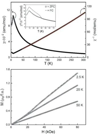
Figure 6 (top) shows the temperature dependence of the re- ciprocal magnetic susceptibility taken at a magnetic flux den- sity of 10 kOe. The data was fitted according to a modified Curie–Weiss law in the temperature range from 50–310 K, leading to an experimental magnetic moment of μexp = 5.4(1)μB per manganese atom, a weak temperature indepen- dent contribution ofχ0= –0.0016(1) emu·mol–1(a diamagnetic
impurity phase) and a Weiss constant ofθP= –23.9(2) K. The experimental magnetic moment is slightly lower that the theo- retical value of 5.92μBfor a Mn2+cation in high-spin state. If one considers the intrinsic diamagnetic contribution of –290⫻10–6emu·mol–1for (2Ag+)Mn2+Sn4+(4S2–) (data taken fromLueken[48]), the similar fit results in a temperature inde- pendent contribution of χ0 = –0.0013(1) emu·mol–1 without changing the value of the experimental magnetic moment, un- derlining the influence of the diamagnetic impurity phase and thus the reduced moment.
Figure 6.(top) Temperature dependence of the susceptibility measured with an applied field of 10 kOe in the temperature range of 3–300 K in ZFC mode. Fit of the inverse magnetic Susceptibility with a modi- fied version of the Curie–Weiss law depicted in red; (inset) magnetic susceptibility measured in ZFC/FC mode with a magnetic flux density of 100 Oe; (bottom) magnetization isotherms measured in the magnetic field range of 0–80 kOe at 2.5, 20, and 50 K.
An additional measurement was performed in zero-field- cooled / field-cooled mode at 100 Oe (inset of Figure 6, top).
The low-field data clearly manifest antiferromagnetic ordering of the manganese spins below a Néel temperature of TN = 8.8(1) K. A second anomaly is detected atTC= 40.2 K that can be ascribed to a yet unknown ferromagnetic impurity phase of very low quantity. The anomaly is already suppressed in the 10 kOe measurement. The magnetization behavior of Ag2MnSnS4 is shown in Figure 6 (bottom). At 20 and 50 K (well above the magnetic ordering temperature) we observe a linear increase of the magnetization as is usual for a paramag- netic material. The increase is at first also linear in the 2.5 K isotherm but at a critical field of HC= 16(1) kOe we observe
a steeper increase. This metamagnetic step (antiparallel-to-par- allel spin realignment) further manifests the antiferromagnetic ground state. The magnetization at 2.5 K and 80 kOe isμsm= 1.5(1)μB/ f.u.
119Sn Mössbauer Spectroscopy
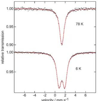
The119Sn Mössbauer spectra of Ag2MnSnS4at 6 and 78 K are presented in Figure 7 along with transmission integral fits.
The 78 K spectrum shows a single signal at an isomer shift of δ = 1.34(1) mm·s–1in close agreement with the crystal struc- ture. The isomer shift nicely fits into the family of thiostanna- tes(IV). A larger compilation of isomer shifts by Lippens[49]
shows a quite narrow range from 1.1 to 1.4 mm·s–1. The small deviation of the SnS4 tetrahedra from ideal symmetry is re- flected in a weak quadrupole splitting parameter of ΔEQ = 0.27(3) mm·s–1. The experimental line width of 0.93(2) mm·s–1 is in the usual range. Below the Néel temperature the signal becomes slightly broadened, resulting from a small transferred hyperfine field ofBhf = 1.02(1) T. Since the transferred field and the quadrupole splitting are small, independent refinement of these parameters was not possible (too large correlation).
For fitting of the 6 K data the quadrupole splitting parameter was fixed atΔEQ= 0 mm·s–1.
Figure 7. Experimental (black dots) and simulated (solid red lines)
119Sn Mössbauer spectra of Ag2MnSnS4at 6 and 78 K.
Conclusions
Ag2MnSnS4, a new diamond-like semiconductor was ob- tained from the elements by high-temperature synthesis in evacuated quartz glass ampoules. The compound is a red dia- mond-like semiconductor with an optical bandgap of ca.
2.0 eV. X-ray diffraction of phase-pure samples showed a (pseudo-)orthorhombic metric. The crystal structure of Ag2MnSnS4is in fact monoclinic and the structure was solved
from a pseudo-merohedrally twinned crystal. In order to con- firm the monoclinic structure model, a Rietveld refinement of the X-ray diffraction pattern was performed. Both methods converged with significantly betterRvalues for the monoclinic structure model as compared to the orthorhombic symmetry.
The crystal structure of the title compound can be derived from the wurtzite structure type, which is in good accordance with the tetrahedral volume concept.[25–30]Ag2MnSnS4is stable up to 700 °C and apparently undergoes a reversible phase-transi- tion above 400 °C. Further details could not be resolved yet, but a transformation to a sphalerite-type modification can cer- tainly be excluded.
Experimental Section
Synthesis of Ag2MnSnS4:Ag2MnSnS4was prepared by heating stoi- chiometric amounts of silver (Ag, Chempur 99.9 %), manganese (Mn, Chempur 99.95 %), tin (Sn, Chempur 99+ %), and sulfur (S, Chempur 99.999 %) in evacuated quartz glass ampoules. After an initial heating process of 1 d at 400 °C, which is necessary to avoid a too high sulfur pressure, the ampoule was then heated to 800 °C for 1 d to ensure a complete reaction of the elements. Finally, the reaction mixtures were annealed at 500 °C for 8 d, homogenized by grinding, and sub- sequently annealed at 500 °C for several days.
Single-crystal X-ray Diffraction: Single-crystal X-ray diffraction data were collected with a STOE IPDS I using Mo-Kαradiation (λ= 0.71073 Å) radiation at ambient conditions. The diffraction data was corrected by a numerical absorption correction using X-SHAPE[37]and X-RED.[36]The resulting data had a completeness of 99.9 % within 25° θ. The crystal structure was solved by charge flipping methods using ShelXT[38]and refined onF2with ShelXL using full-matrix le- ast-squares methods.[39] The experimental parameters and details of the structure solution and refinement are summarized in Table 1.
X-ray Powder Diffraction: The X-ray powder pattern used for the Rietveld refinement was collected with a STOE Stadi P diffractometer, equipped with a Dectris Mythen 1 K detector, using monochromatized Mo-Kα1radiation (λ= 0.70930 Å). The powdered sample was mea- sured in Debye–Scherrer setup in a glass capillary (diameter 0.3 mm) in order to minimize preferred orientation effects on the reflection in- tensities. The Rietveld refinement was performed using Jana2006.[50]
A manual background combined with 5 Legendre polynoms was used.
The reflection profiles were described using a fundamental parameter approach and Pseudo Voigt functions, refining the parameters CSizeG and StrainL. The orthorhombic and monoclinic structure models ob- tained from the single-crystal measurements were used as starting models, however, the same models could also be obtained using charge flipping methods using SUPERFLIP.[51]The atomic coordinates and isotropic displacement parameters were refined without any restraints and converged straight forward.
In situ high-temperature measurements were performed using mono- chromatized Mo-Kα1radiation (λ= 0.70930 Å) and a STOE capillary furnace 0.65. The furnace temperature was controlled by a Eurotherm 24.16 controller (ΔT = ⫾1 °C). For the low-temperature measure- ments, an Oxford Cryosystems Cryostream 700 was used. The pow- dered sample was flame sealed in quartz glass capillaries (diameter 0.3 mm) for the temperature dependent measurements. Due to the fur- nace architecture, the capillary had to be placed in another 0.5 mm quartz glass capillary for the high-temperature measurements.
The diffraction data was collected in the range from 5.05° ⱕ2θ ⱕ 23.92° with an irradiation time of 300 s in the temperature region from –150 °C to 25 °C and 25 °C to 850 °C in steps of 10 K (heating-/cool- ing rate 5 K·min–1, 10 min annealing prior to each measurement). The STOE WinXPOW program package was used for the data collection and analysis.[52]
Further details of the crystal structure investigations may be obtained from the Fachinformationszentrum Karlsruhe, 76344 Eggenstein- Leopoldshafen, Germany (Fax: +49-7247-808-666; E-Mail:
crysdata@fiz-karlsruhe.de, http://www.fiz-karlsruhe.de/request for de- posited data.html) on quoting the depository number CSD-434389.
SEM/EDX Analysis:Quantitative EDX analysis was performed with a Zeiss EVO MA 15 SEM equipped with a Bruker Quantax EDX system with an X Flash Detector 630 M. A powdered sample of Ag2MnSnS4(sputtered with an Au/Pd alloy) was analysed at three different positions with a voltage of 30 keV and an irradiation time of 120 s.
Thermal Analysis: The Differential thermal analysis (DTA) of Ag2MnSnS4was performed with a SETARAM TG-DTA 92 16.18 in an evacuated quartz glass ampoule using Al2O3as reference material.
During the measurement two heating- and cooling-cycles were col- lected in the temperature range of 25–1000 °C with a heating- and cooling rate of 10 K·min–1.
Raman Spectroscopy: The Raman spectrum was recorded with a Thermoscientific DXRTMSmartRaman spectrometer (excitation wave- lengthλ= 532 nm) in the range of 50–500 cm–1with a resolution of 0.5 cm–1.
UV/Vis Spectroscopy: Diffuse reflectance measurements were per- formed with a Bruins Omega 20 UV/Vis spectrometer using BaSO4as a white standard (100 % reflectance). The absorption spectrum was calculated using a modified Kubelka-Munk function.[45,46]
Magnetic Characterization:The investigation of the magnetic prop- erties was carried out using a Quantum Design Physical Property Mea- surement System (PPMS) equipped with a Vibrating Sample Magne- tometer (VSM). 39.413 mg of the sample were fixed in a polypropyl- ene capsule and attached to a brass sample holder of the VSM. The measurements were performed in the temperature range of 2.5–310 K applying magnetic fields of up to 80 kOe.
Mössbauer Spectroscopy: A Ca119mSnO3 source was used for the Mössbauer spectroscopic experiments, which were carried out with a Continuous Flow Cryostat System (Janis Research Co LLC.) at 6 and 78 K. The Mössbauer source was kept at room temperature. The sam- ple was enclosed in small PMMA containers at an optimized thickness.
Fitting of the data was done by using the Normos-90 program pack- age.[53]
Supporting Information(see footnote on the first page of this article):
Table S1, S2 show details of the Rietveld refinement of Ag2MnSnS4, the atomic positions, and isotropic displacement parameters obtained from the Rietveld refinement. Figure S1 and Table S3 show the SEM/
EDX analysis of Ag2MnSnS4. Figure S2 shows the thermal analysis (DTA) of Ag2MnSnS4. Figure S3 shows the low- and high-temperature X-ray diffraction patterns of Ag2MnSnS4 in the range of -150 to 850 °C.
Acknowledgements
The authors would like to thankProf. Dr. Manfred Scheer(University of Regensburg) for the Raman spectroscopic measurements.
Keywords: Diamond-like semiconductor; Magnetic proper- ties; Mößbauer spectroscopy; Non-ambient X-ray powder dif- fraction; Rietveld refinement
References
[1] L. O. Brockway,Z. Kristallogr.1934,89, 434–441.
[2] E. Parthé, K. Yvon, R. H. Deitch,Acta Crystallogr., Sect. B1969, 25, 1164–1174.
[3] W. Schäfer, R. Nitsche,Z. Kristallogr.1977,45, 356–370.
[4] S. Harada,Mater. Res. Bull.1973,8, 1361–1369.
[5] N. A. Goryunova, J. C. Anderson (Eds.),The Chemistry of Dia- mond-Like Semiconductors, Chapman and Hall, London1966,19, 120.
[6] J. W. Lekse, M. A. Meghann, K. L. McNerny, J. Yeon, P. S. Hala- syamani, J. A. Aitken,Inorg. Chem.2009,48, 7516–7518.
[7] S. Wagner, P. M. Bridenbaugh,J. Cryst. Growth1977,39, 151–
159.
[8] S. Siebentritt,Thin Solid Films2002,403,1–8.
[9] L. Guen, W. S. Glaunsinger, A. Wold,Naturwissenschaften1965, 52, 426–426.
[10] C. Steinhagen, M. G. Panthani, V. Akhavan, B. Goodfellow, B.
Koo, B. A. Korgel,J. Am. Chem. Soc.2009,131, 12554–12555.
[11] J. J. Scragg, P. J. Dale, L. M. Peter, G. Zoppi, I. Forbes, Phys.
Status Solidi2008,245, 1772–1778.
[12] L. K. Samanta, G. C. Bhar, Phys. Status Solidi 1977, 41, 331–
337.
[13] D. A. Roberts,Appl. Optics.1996,24, 4677–4688.
[14] G. Catella, D. Burlage,Mater. Res. Bull.1998,33, 28–36.
[15] X. Li, C. Li, M. Zhou, Y. Wu, J. Yao,Chem. Asian J.2017,24, 3172–3177.
[16] J. A. Brant, D. J. Clark, Y. S. Kim, J. I. Jang, J.-H. Zhang, J. A.
Aitken,Chem. Mater.2014,26, 3045–3048.
[17] D. Mei, W. Yin, K. Feng, Z. Lin, L. Bai, J. Yao, Y. Wu,Inorg.
Chem.2012,51, 1035–1040.
[18] W. Yin, K. Feng, W. Hao, J. Yao, Y. Wu,Inorg. Chem.2012,51, 5839–5843.
[19] E. Honig, H. S. Shen, G. Q. Yao, K. Doverspike, R. Kershaw, K.
Dwight, A. Wold,Mater. Res. Bull.1988,23, 307–312.
[20] L. Guen, W. S. Glaunsinger, A. Wold,Mater. Res. Bull.1979,79, 463–467.
[21] A. M. Lamarche, A. Willsher, L. Chen, G. Lamarche, J. C. Wool- ley,J. Solid State Chem.1991,94, 313–318.
[22] G. Nénert, T. T. M. Palstra,J. Phys.: Condens. Matter2009,21, 176002.
[23] a) A. Pfitzner,Inorg. Chem.1998,37, 5164–5167; b) A. Pfitzner, S. Reiser, T. Nilges, W. Kockelmann,J. Solid State Chem.1999, 147, 170–176.
[24] E. Parthé inCrystal Structures of Intermetallic Compounds(Eds.:
J. H. Westbrook, R. L. Fleischer), J. Wiley and Sons, New York, 2000, vol. 1, 117–137.
[25] A. Pfitzner, S. Reiser,Z. Kristallogr.2002,217, 51–54.
[26] T. Bernert, A. Pfitzner,Z. Anorg. Allg. Chem.2002,628, 2161.
[27] M. Robbins, M. A. Miksovsky,J. Solid State Chem.1972,5, 462–
466.
[28] M. E. Fleet,Mater. Res. Bull.1976,11, 11179–11183.
[29] A. Pfitzner, T. Bernert,Z. Kristallogr.2004,219, 20–26.
[30] T. Bernert, A. Pfitzner,Z. Anorg. Allg. Chem.2004,630, 1711.
[31] T. Bernert, A. Pfitzner,Z. Kristallogr.2005,220, 968–972.
[32] T. Bernert, A. Pfitzner,Z. Anorg. Allg. Chem.2006,632, 1213–
1218.
[33] J. W. Lekse, PhD Thesis, Duquesne University2009.
[34] G. E. Delgado, N. Sierralta, M. Quintero, E. Quintero, E. Moreno, J. A. Flores-Cruz, C. Rincón,Rev. Mex. Fís.2018,64, 216–221.
[35] J. W. Lekse, B. M. Leverett, C. H. Lake, J. A. Aitken, J. Solid State Chem.2008,181, 3217–3222.
[36] X-RED32, STOE & Cie GmbH,2004.
[37] X-Shape, STOE & Cie GmbH,1999.
[38] G. M. Sheldrick,Acta Crystallogr., Sect. A2015,71, 3–8.
[39] G. M. Sheldrick,Acta Crystallogr., Sect. C2015,71, 1–2.
[40] H. Bärnighausen,MATCH Commun. Math. Chem.1980,9, 139–
175.
[41] K. P. Devlin, A. J. Glaid, J. A. Brant, J.-H. Zhang, M. N. Smec, D. J. Clark, Y. S. Kim, J. I. Jang, K. R. Daley, M. A. Moreau, J. D. Madura, J. A. Aitken,J. Solid State Chem.2015,231, 256–
266.
[42] C. D. Brunetta, J. A. Brant, K. A. Rosmus, K. M. Henline, E. K. J. H. MacNeil, J. A. Aitken,J. Alloys Compd. 2013, 574, 495–503.
[43] S. Azam, S. A. Khan, S. Goumri-Said,Solid State Sci.2017,72, 71–79.
[44] T. Balic Zunic, I. Vickovic,J. Appl. Crystallogr.1996,29, 305–
306.
[45] P. Kubelka, F. Munk,Z. Techn. Physik1931, 593.
[46] H. Kisch,Angew. Chem. Int. Ed.2013,52, 812–847.
[47] A. J. Smith, P. E. Meek, W. Y. Liang,J. Phys. C: Solid State Phys.
1977,10, 1321–1333.
[48] H. Lueken,Magnetochemie, Teubner, Stuttgart,1999.
[49] P. E. Lippens,Phys. Rev. B1999,60, 4576–4586.
[50] V. Petricek, M. Dusek, L. Palatinus, Z. Kristallogr. 2014, 229, 345–352.
[51] L. Palatinus, G. Chapuis,J. Appl. Crystallogr.2007,40, 789–790.
[52] STOE WinXPOW, Version 3.10, STOE & Cie GmbH, Darmstadt 2016.
[53] R. A. Brand, Normos Mössbauer Fitting Program, University of Duisburg, Duisburg, Germany2002.
[54] F. Keutsch, D. Topa, R. Fredrickson, E. Makovicky, W. Paar, Min- eralogical Magazine 2018, 1-19, DOI: 10.1180/mgm.2018.139, Accepted Article.
Received: April 12, 2018 Published online:䊏
D. Friedrich, S. Greil, T. Block, L. Heletta, R. Pöttgen, A. Pfitzner* ... 1–9 Synthesis and Characterization of Ag2MnSnS4, a New Dia- mond-like Semiconductor