E-Mail karger@karger.com
Original Paper
Pathobiology 2016;83:165–169 DOI: 10.1159/000443311
The Microphthalmia-Associated Transcription Factor p.E318K Mutation Does Not Play a
Major Role in Sporadic Renal Cell Tumors from Caucasian Patients
Christine G. Stoehr
aBernhard Walter
bStefan Denzinger
cPaola Ghiorzo
fRichard A. Sturm
gRaoul Hinze
dHolger Moch
hKerstin Junker
eArndt Hartmann
aRobert Stoehr
aa Institute of Pathology and b Department of Urology, University Hospital Erlangen, Friedrich Alexander University Erlangen-Nuremberg, Erlangen , c Department of Urology, University of Regensburg, Caritas St. Josef Medical Center, Regensburg , d Institute of Pathology, HELIOS Hospital Schwerin, Schwerin , and e Department of Urology and Pediatric Urology, Saarland University Medical Center, Homburg/Saar , Germany; f Department of Internal and Medical Specialties, University of Genoa, Genoa, Italy; g Institute for Molecular Bioscience, University of Brisbane, Brisbane, Qld., Australia; h Institute of Surgical Pathology, University Hospital Zurich, Zurich , Switzerland
prehensive data on sporadic renal cell tumors are missing.
We therefore tested a large cohort of sporadic renal tumors for MITF p.E318K mutation status. Methods: Genomic DNA was extracted from 426 formalin-fixed, paraffin-embedded sporadic renal tumors that had been graded according to the 2004 WHO classification of renal tumors and staged ac- cording to the 2002 TNM classification. The tumor cohort was enriched with papillary and chromophobe RCC, and also contained benign oncocytomas. DNA was tested for MITF p.E318K by pyrosequencing. Results: Of 403 analyzable tu- mors, 402 renal tumors were wild-type ones, and only 1 case showed the MITF p.E318K mutation. This tumor was a clear- cell RCC (pT3b N0 M0 G3 according to the TNM classification 2002). The affected patient was male, 61 years old, and had no known coexisting malignancies. Conclusion: The MITF p.E318K mutation does not appear to play a major role in sporadic RCC carcinogenesis, but is possibly restricted to a rare subpopulation of inherited RCC. © 2016 S. Karger AG, Basel
Key Words
Microphthalmia-associated transcription factor · Mutation · Renal cell carcinoma · Pyrosequencing
Abstract
Objective: The transcription factor MITF (microphthalmia- associated transcription factor) is known to induce expres- sion of hypoxia-inducible factor (HIF1-α), which is involved in renal carcinogenesis. The MITF p.E318K mutation leads to deficient SUMOylation of MITF, resulting in enhanced activa- tion of its target genes. A case-control study on melanoma patients who coincidentally were affected by renal cell car- cinoma (RCC) has revealed an elevated risk for mutation car- riers to be affected by one or both of these malignancies, suggesting a possible role for MITF p.E318K in renal carcino- genesis. The same study described an MITF mutation fre- quency of 1.5% in a small cohort of sporadic RCC, but com-
Received: September 8, 2015
Accepted after revision: December 14, 2015 Published online: March 22, 2016
PD Dr. Dr. Robert Stoehr, PhD
Institute of Pathology, University Hospital Erlangen Krankenhausstrasse 8–10
DE–91054 Erlangen (Germany) E-Mail christine.stoehr @ uk-erlangen.de © 2016 S. Karger AG, Basel
1015–2008/16/0834–0165$39.50/0 www.karger.com/pat
Introduction
Cellular functions and intracellular communication are controlled and regulated both at the DNA and post- translational levels. Posttranslational modifications (e.g.
phosphorylation, ubiquitination, and acetylation) of proteins are essential for triggering signal cascades and for maintaining fundamental developmental processes.
Dysregulation of posttranslational modification might lead to dramatic malfunction of the cell, and is known from most malignancies. However, as recently demon- strated, a deregulated posttranslational modification net- work may act as a therapeutic target (e.g. in pancreatic cancer), and is a major focus of ongoing studies in pros- tate cancer [1, 2] .
Reversible covalent conjugation of SUMO (small ubiquitin-related modifier) is a posttranslational modi- fication that can modulate the function (e.g. activity, cellular localization, and stability) of a large spectrum of proteins [3] . SUMOylation frequency of a target protein is increased by the presence of the consensus motif ΨKXE, but proteins without this motif might also be prone to this modification [4] . Of important functional relevance is a connection between the SUMOylation and ubiquitina- tion networks, as SUMOylation can lead to ubiquitin- dependent degradation by the proteasome [5] .
The MITF (microphthalmia-associated transcription factor) gene encodes for a basic-helix-loop-helix leucine zipper transcription factor and is part of the Myc super- gene family. The MITF gene contains two SUMOylation sites that can regulate SUMOylation and transcriptional activity of the MITF protein [6] .
Recently, a germline missense mutation (p.E318K) in the MITF gene was described, and a predisposition to fa- milial and sporadic melanoma in carriers of this specific change was documented. The affected codon is located within a SUMOylation consensus motif, and the muta- tion interferes with MITF SUMOylation. In vitro studies revealed both enhanced binding of mutated MITF to the promoter of the hypoxia-inducible factor (HIF1-α) gene, which is a target gene of the MITF transcription factor, and increased activity of the HIF1-α promoter caused by this binding [7, 8] . Increased expression of HIF1-α is cor- related with the development of clear-cell renal cell carci- nomas (RCC), and overexpression of HIF1-α has been found in most clear-cell RCC cases analyzed to date [9] . Most interesting, a significantly increased frequency of RCC compared to normal controls was detected in mela- noma patients with the p.E318K germline mutation in MITF [7, 10] . These findings suggest MITF as a possible
connection point between melanoma and RCC. In addi- tion, mutation analysis of a small cohort of sporadic RCC cases showed a MITF p.E318K mutation frequency of 1.5% [7] . Currently, the role of the MITF mutation in RCC development remains unclear, as comprehensive mutation data from sporadic RCC cases are missing.
Therefore, the aim of our study was to screen a large co- hort of Caucasian sporadic renal tumor cases for the MITF p.E318K mutation.
Materials and Methods Patients and Tissue Samples
Overall, 426 formalin-fixed, paraffin-embedded archival spo- radic renal tumors were used for the study. The cases were classi- fied according to the 2004 WHO classification of renal tumors and staged according to the 2002 TNM classification [11, 12] . Charac- teristics of the study cohort are shown in table 1 . Prior institution- al review board approval (University Hospital Erlangen, Germa- ny) was obtained for the study.
Table 1. Patient and tumor characteristics (n = 426) Gender, n
Male 217
Female 131
Unknown 78
Age, years
Range 19–91
Median 65
Mean 63.04
Tumor type, n
Clear-cell RCC 175
Papillary RCC 77
Chromophobe RCC 97
Renal oncocytoma 58
Others 19
2002 TNM classification, n
pT1a 129
pT1b 80
pT2 58
pT3a 41
pT3b 31
Unknown 29
2002 TNM stage, n
Stage I 160
Stage II 32
Stage III 47
Stage IV 21
Unknown 108
MITF p.E318K Mutation in Sporadic RCC
Pathobiology 2016;83:165–169
DOI: 10.1159/000443311 167
Tissue Microdissection and DNA Isolation
DNA was extracted from renal tumor samples from serial sec- tions after manual microdissection as described previously [13] . In brief, 5-μm-thick serial sections of the tumor tissue were de- waxed and stained with 0.1% methylene blue for 15 s. Using an inverted microscope, the tumor tissue (identified through match- ing with a marked hematoxylin and eosin-stained section re- viewed by an experienced surgical pathologist) was scraped off with a sterile needle to obtain a purity of the cells of at least 85%.
Isolation of genomic DNA of the microdissected tumor tissue was performed using the High Pure PCR Template Preparation Kit (Roche, Mannheim, Germany) according to the manufacturer’s instructions.
Sequence Analysis of Codon 318 of MITF
For mutation analysis, the hot spot region of the MITF gene containing codon 318 was amplified using the multiplex PCR-kit according to manufacturer’s instructions (Qiagen, Hilden, Ger- many) and the following primers: forward: 5 ′ -TTATTCCATC- CACGGGTCTC-3 ′ , and reverse: 5 ′ -biotin GAGGTCTTGGCTG- CAGTTCT-3 ′ . The cycling conditions were as follows: a single cycle of denaturation at 94 ° C for 5 min, 45 cycles at 94 ° C for 1 min, 58.3 ° C for 1 min, and 72 ° C for 1 min, and a final 10-min extension at 72 ° C. For pyrosequencing (PyroMark Q24; Qiagen) single-stranded DNA was prepared from 25 ml of biotinylated PCR product with streptavidin-coated sepharose and 0.5 m M of the sequencing primer 5 ′ -AATCGGATCATCAAGC-3 ′ using the PSQ Vacuum Prep Tool (Qiagen). Pyrosequencing assay setup was selected in ‘Sequence to Analyze’ with the sequence AAG/A AAC CCG TT. The following dispensation order was used: GAA GAC GT.
Results
Sequencing data were available from 403/426 (94.6%) cases. In 23 cases, no PCR products were obtained due to strong fragmentation of the DNA after formalin fixation.
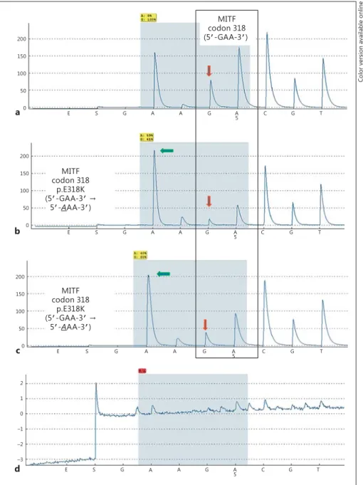
Of the 403 analyzable cases, 402 (99.8%) showed wild- type MITF p.E318. Only 1 (0.2%) case showed the MITF p.E318K mutation ( fig. 1 ). This case was a clear-cell RCC (pT3b N0 M0 G3 according to the TNM classification 2002). The affected patient was male, 61 years old, and had no known coexisting malignancies.
Discussion
In this study we analyzed the p.E318K mutation of MITF in a large cohort of sporadic renal cell tumors.
Only one of the cases investigated showed the mutation known as a germline alteration from patients with famil- ial melanoma and/or RCC. The resulting allele frequency of the mutation in our cohort was 0.12%. The low fre- quency detected might hint to technical problems or
contaminated material, but positive and negative con- trols showed the expected results. Moreover, this muta- tion frequency was comparable to those of the general populations of France (frequency = 0.3%), Australia (fre- quency = 0.72%), Poland (0.09%), and the UK (frequen- cy = 0.85%) [8, 14, 15] . Furthermore, Guo et al. [16] per- formed exome sequencing on 10 clear-cell RCC and screened >1,000 genes (including MITF) in an addition- al 88 clear-cell RCC cases, but could not detect an MITF mutation. Furthermore, the TCGA Research Network published an extensive molecular profiling of 417 clear- cell RCC samples. Overall, MITF alterations were report- ed in 2.6% of the cases with 9/11 mutated tumors show- ing deep deletions and only 2/11 mutated tumors dis- playing point mutations (p.T110M, p.G165D). The p.E318K mutation could not be detected at all [17] . Through cBioPortal [18, 19] , provisional sequencing data from an additional 161 papillary RCC cases ana- lyzed by the TCGA Research Network are available. In this cohort, 3/161 (2.6%) cases showed truncating (p.
K118 * , p.K198fs) or missense (p.A205V) mutations within the MITF gene. Again, the p.E318K mutation al- lele was not found. Thus, combining all available data, the overall MITF mutation frequency is approximately 1.4% (15/1,079 cases with mutation).
Altogether, these data support our results on MITF p.E318K frequency in sporadic RCC and therefore argue for their validity. In conclusion, the MITF p.E318K muta- tion only plays a minor role in the development of spo- radic RCC.
Despite the rarity of MITF mutations, they might trig- ger RCC development in specific cases, as mutated MITF displays enhanced binding to the HIF1-α promoter and has been shown to increase HIF1-α promoter activity.
Moreover, MITF also acts as a transcription factor and is able to directly control MET transcription [20] . Activat- ing mutations in the MET proto-oncogene are especially frequent in papillary type 1 RCC (about 15%) [21] . These mutations lead to activation of several pathways resulting in, for example, tumor cell proliferation, invasion, and angiogenesis. Mutations in MITF interrupting its con- trolled degradation might constantly increase MET tran- scription and result in similar effects as mutational MET activation. It is conceivable that this effect, in combina- tion with additional harmful events, makes a cell prone to malignant transformation. This is supported by the re- cent description of an actin gamma 1 (ACTG1)-MITF fu- sion in a case of papillary RCC [22] . In vitro studies of this fusion protein revealed an induction of, for example, HIF1-α and MET transcription in HEK293T cells, and an
increased colony-forming ability in NIH3T3 cells. These data gave the first evidence for a similar tumorigenic po- tential of MITF fusion proteins as is attributed to TFE3 fusions.
In summary, the MITF p.E318K mutation does not seem to play a major role in sporadic RCC development in Caucasians, but is possibly restricted to a rare subpop- ulation of inherited RCC. Nonetheless, in general, MITF alterations might contribute to the development of spo- radic RCC as they affect targets already known to be in- volved in RCC pathobiology.
Acknowledgement
The authors thank Verena Popp, Karina Kalb, Claudia Knoll, and Nina Oks for excellent technical assistance.
Disclosure Statement
The authors have no conflict of interest.
codon 318MITF (5-GAA-3)
codon 318MITF p.E318K (5-GAA-3ମ
5-AAA-3)
codon 318MITF p.E318K (5-GAA-3 ମ
5-AAA-3)
200 150 100 50 0
200 150 100 50 0
200 150 100 50 0
2 1 0 –1 –2 –3
E S G A A G A C G T
5
E S G A A G A C G T
5
E S G A A G A C G T
5
E S G A A G A C G T
5
a
b
c
d
Fig. 1. Representative examples for pyro- sequencing analysis (pyrogram) of codon 318 (5 ′ -GAA-3 ′ ) of the MITF gene. a RCC showing a wild-type sequence. b Positive control for the MITF p.E318K (5 ′ -GAA-3 ′
→ 5 ′ - A AA-3 ′ ) mutation from a melanoma case with the germline mutation. The downward-pointing arrow indicates the reduction of the wild-type ‘G’ signal and the increase in the mutant ‘A’ signal. The
‘G’-to-‘A’ mutation led to a stretch of 5× ‘A’
in a row interrupted by the ‘G’ signal (downward-pointing arrow) from the wild-type background. c Clear-cell RCC with the MITF p.E318K mutation. The py- rogram is identical to that of the positive control. d Negative control (H 2 O).
Color version available online
MITF p.E318K Mutation in Sporadic RCC
Pathobiology 2016;83:165–169
DOI: 10.1159/000443311 169
References
1 Chen Z, Lu W: Roles of ubiquitination and SUMOylation on prostate cancer: mecha- nisms and clinical implications. Int J Mol Sci 2015; 16: 4560–4580.
2 Damaskos C, Karatzas T, Nikolidakis L, Kostakis ID, Karamaroudis S, Boutsikos G, et al: Histone deacetylase (HDAC) inhibitors:
current evidence for therapeutic activities in pancreatic cancer. Anticancer Res 2015; 35:
3129–3135.
3 Sriramachandran AM, Dohmen RJ: SUMO- targeted ubiquitin ligases. Biochim Biophys Acta 2014; 1843: 75–85.
4 Yang XJ, Chiang CM: SUMOylation in gene regulation, human disease, and therapeutic action. F1000Prime Rep 2013; 5: 45.
5 Miteva M, Keusekotten K, Hofmann K, Praef- cke GJ, Dohmen RJ: SUMOylation as a signal for polyubiquitylation and proteasomal deg- radation. Subcell Biochem 2010; 54: 195–214.
6 Paillerets BB, Lesueur F, Bertolotto C: A germline oncogenic MITF mutation and tu- mor susceptibility. Eur J Cell Biol 2014; 93:
71–75.
7 Bertolotto C, Lesueur F, Giuliano S, Strub T, de Lichy M, Bille K, et al: A SUMOylation- defective MITF germline mutation predis- poses to melanoma and renal carcinoma. Na- ture 2011; 480: 94–98.
8 Yokoyama S, Woods SL, Boyle GM, Aoude LG, MacGregor S, Zismann V, et al: A novel recurrent mutation in MITF predisposes to familial and sporadic melanoma. Nature 2011; 480: 99–103.
9 Gudas LJ, Fu L, Minton DR, Mongan NP, Na- nus DM: The role of HIF1α in renal cell car- cinoma tumorigenesis. J Mol Med 2014; 92:
825–836.
10 Ghiorzo P, Pastorino L, Queirolo P, Bruno W, Tibiletti MG, Nasti S: Prevalence of the E318K MITF germline mutation in Italian melano- ma patients: associations with histological subtypes and family cancer history. Pigment Cell Melanoma Res 2013; 26: 259–262.
11 Eble JN SG, Epstein JI, et al: Tumours of the Urinary System and Male Genital Organs.
Lyon, IARC Press, 2004.
12 Sobin LH, Wittekind C (eds): TNM Classifi- cation of Malignant Tumors, ed 6. New York, John Wiley & Sons, 2002.
13 Stoehr R, Taubert H, Zinnall U, Giedl J, Gaisa NT, Burger M, et al: Frequency of TERT pro- moter mutations in prostate cancer. Pathobi- ology 2015; 82: 53–57.
14 Sturm RA, Fox C, McClenahan P, Jagirdar K, Ibarrola-Villava M, Banan P, et al: Phenotyp- ic characterization of nevus and tumor pat- terns in MITF E318K mutation carrier mela- noma patients. J Invest Dermatol 2014; 134:
141–149.
15 Gromowski T, Masojc B, Scott RJ, Cybulski C, Gorski B, Kluzniak W, et al: Prevalence of the E318K and V320I MITF germline mutations in Polish cancer patients and multiorgan can- cer risk-a population-based study. Cancer Genet 2014; 207: 128–132.
16 Guo G, Gui Y, Gao S, Tang A, Hu X, Huang Y, et al: Frequent mutations of genes encoding ubiquitin-mediated proteolysis pathway components in clear cell renal cell carcinoma.
Nat Genet 2011; 44: 17–19.
17 Cancer Genome Atlas Research Network:
Comprehensive molecular characterization of clear cell renal cell carcinoma. Nature 2013;
499: 43–49.
18 Cerami E, Gao J, Dogrusoz U, Gross BE, Sum- er SO, Aksoy BA, et al: The cBio cancer ge- nomics portal: an open platform for exploring multidimensional cancer genomics data.
Cancer Discov 2012; 2: 401–404.
19 Gao J, Aksoy BA, Dogrusoz U, Dresdner G, Gross B, Sumer SO, et al: Integrative analysis of complex cancer genomics and clinical pro- files using the cBioPortal. Sci Signal 2013;
6:pl1.
20 Beuret L, Flori E, Denoyelle C, Bille K, Busca R, Picardo M, et al: Up-regulation of MET expression by alpha-melanocyte-stimulating hormone and MITF allows hepatocyte growth factor to protect melanocytes and melanoma cells from apoptosis. J Biol Chem 2007; 282:
14140–14147.
21 Massari F, Ciccarese C, Santoni M, Brunelli M, Piva F, Modena A, et al: Metabolic altera- tions in renal cell carcinoma. Cancer Treat Rev 2015; 41: 767–776.
22 Durinck S, Stawiski EW, Pavia-Jimenez A, Modrusan Z, Kapur P, Jaiswal BS, et al: Spec- trum of diverse genomic alterations define non-clear cell renal carcinoma subtypes. Nat Genet 2015; 47: 13–21.