Contribution to the genus Diploneis (Bacillariophyta): Twelve species from Holarctic freshwater habitats proposed as new to science
Horst L
ange–B
ertaLot1& André F
uhrmann21 Institute for Ecology, Evolution, Diversity, Goethe–University Frankfurt, Biologicum, Max–von–Laue–Str. 13, 60439 Frankfurt am Main
2 Fachbereich 8, Goethe–Universität Frankfurt, Grüneburgplatz 1, 60629 Frankfurt am Main; Corresponding author e–mail: fuhrmann@em.uni–frankfurt.de
Abstract: Numerous questionable extant populations of Diploneis with cribrate areolae are morphologically compared (in LM and SEM) to resembling established taxa. In most of these examinations the types are available and documented in the literature by several authors during the last forty years. All taxa in question belong ecologically to the minority of Diploneis that live exclusively in freshwater. As main result twelve species from Europe and one from Florida, U.S.A., are selected and proposed as new to science. These are: D.
abscondita, D. calcilacustris, D. hinziae, D. lusatica, D. modicahassiaca, D. oblongellopsis, D. praetermissa, D. puellafallax, D. tirolensis, all from Central Europe (Austria, Germany, Switzerland); D. nanofontanella and D. tundra from European Arctic Russia; and D. parahinziae from Florida. After critical examination, D. arctica stat. nov., D. ladogensis stat. nov., and D. dilatata stat. nov. are proposed to be transferred from infraspecific to species rank.
Key words: Autecology, Bacillariophyta, biogeography, cribra, diatoms, Diploneis, Europe, freshwater, ultrastructure
I
ntroductIonThe large majority of known Diploneis taxa live in marine habitats, many of them being cosmopolitan, whereas comparatively few inhabit freshwater habi- tats. The latter are restricted to distinct biogeographi- cal plant realms such as the Holarctic, the Paleo– and Neotropics. Over many decades of the 20th century the European watercourses have suffered from strong eutrophication and organic pollution. Since Diploneis is rather intolerant to such conditions the huge rivers in Germany, Poland and France have been widely devoid of Diploneis species. All populations described and discussed in this paper originate from stagnant oligo–
to slightly eutrophic waters –– with the exception of one pond–effluent.
hustedt (1930) distinguishes no more than ten taxa for Central Europe. The last comprehensive treatise on Diploneis has been hustedt (1937), inclu- ding fifteen taxa which inhabit freshwater of North and Central Europe. His conceptions of freshwater species have been perpetuated since then, i.e. over a period of more than seventy years without thorough actualisa- tion. CLeve–euLer (1953) has added some new taxa from Scandinavia but mainly extending extant species
concepts. PatriCk & reimer (1966) report fourteen for North America, including many brackish water taxa.
More recently germain (1981) counts eight taxa in South–West France. krammer & Lange–BertaLot
(1986) list seventeen freshwater and seven brackish water taxa in almost entire Europe. In the Red List of endangered species in Germany and neighbouring countries from 1996 (Lange–BertaLot 1996) this number had not increased. In their recent atlas of dia- toms, covering numerous large and small watercourses in the rather large monitoring area of the Rhône–Al- pes region in France, Bey & eCtor (2013) end up with only five established plus two non–identified species of Diploneis. Their studies, mainly carried out be- tween 2007 and 2011, covered 157 sampling stations and pertain not only to the Rhône catchment area but also cover part of the Loire river area with its tributa- ries from the Eastern Central Massif mountains. Even though the geological and ecological variability of the investigated sites is thus very high, it yields a surprisin- gly low number of Diploneis taxa. Moreover, it should be mentioned that Bey & eCtor take into considerati- on new taxonomical results concerning the genus that have become available only after the appearance of the comprehensive “Süßwasser–Flora” (krammer &
Dedicated to Friedel Hinz
Lange–BertaLot 1986). Since then new freshwater taxa of Diploneis had not only be described from South America, Asia, and the paleotropical Pacific island of New Caledonia, but likewise from Europe. New (since krammer & Lange–BertaLot 1986) taxa from Europe are:
Diploneis ovalis ssp. arctica Lange–BertaLot in Lange–BertaLot & genkaL 1999,
D. krammeri Lange–BertaLot & reiChardt 2000, D. fontium reiChardt & Lange–BertaLot 2004, D. fontanella Lange–BertaLot in Werum &
Lange–BertaLot 2004,
D. separanda Lange–BertaLot in Werum &
Lange–BertaLot 2004.
We may mention that a noteworthy location for en- demic Diploneis species is Lake Ohrid (Macedonia/
Albania). From fifteen species recently presented in Jovanovska et al. (2013), five are described as new.
As explained in more detail in the Discussion (see below), freshwater Diploneis fall into few con- spicuous groups that combine certain morphological features of the valve, particularly concerning their pa- tterns of areolae. The group under study here compris- es species that exhibit cribrate areolae of various size, arranged in uniseriate or biseriate, rarely pluriseriate striae. Also included here are species where striae gra- dually switch over from a uniseriate to a bi– or plurise- riate pattern. As far as we know, this group happens to be the by far most populated by freshwater taxa of the genus Diploneis in Europe and elsewhere.
Dedication. Friedel Hinz has served the diatom community for many decades as curator of one of the world’s most important diatom collections, the Fried- rich Hustedt–Archives in Bremerhaven. As in count- less other cases over the years, she has helped the authors of the present study by providing photographs of the taxon that Hustedt took to be Diploneis parma, one of the starting points for this investigation. We de- dicate the newly described taxon, Diploneis hinziae, to her on the occasion of her retirement.
M
aterIalsandM
ethodsThese observations on taxonomically critical Diploneis po- pulations have been carried out during fifteen years since 1999. Samples have been taken sporadically over a longer period from 1982 up to 2011. Colleagues, friends and former students have collected from mainly stagnant freshwater in various regions of Germany, Austria, Switzerland, Europe- an Russia and Florida, U.S.A. Those who provided us with samples, slides or ecological data, as far as available, are mentioned under the typifications of the new taxa. Usually the samples were given to us with the intent of asking for identification of critical diatom taxa in general rather than for the purpose of investigating specifically the genus Diploneis.
However, in the course of time, data of questionable Diplo-
neis specimens in slides were recorded or marked with co- ordinates in different light microscopes. The following light microscopes were used: Leitz Ortholux, Leitz Dialux, Leitz Diaplan, Zeiss Standard Universal, Nikon Eclipse E600. All micrographs were taken with apochromatic lenses of apertu- re no less than 1.3. Brightfield illumination, film (Agfa Ortho Professional), and for more recent work digital photographic equipment was used throughout. SEM studies were carried out with a Hitachi S–4500 at the Goethe–Universität Frank- furt am Main, operated at 10kV by Manfred Ruppel. For the purpose samples were fixed on aluminium stubs and sputter–
coated with 40 nm gold.
As samples, including slides, reached us from va- rious sources, a number of standard cleaning methods were employed. Most samples were cleaned by oxidisation in hot sulphuric acid or in hot hydrogen peroxide with potassium permanganate added. In some cases a pre–cleaning in HCl was necessary to remove calcareous matter. All material for LM was mounted in Naphrax with a refractive index of ca.
1.7.
r
esultsIn this section we describe species proposed as new to science and new combinations of established taxa.
Although some of these taxa are morphologically clo- sely related, such as D. hinziae and D. parahinziae, we present them, for the reader’s convenience, in strict al- phabetical order.
Diploneis abscondita lange–Bertalot et g. hof-
Mann sp. nov. (Figs 1–7 LM, 106–108 SEM external view)
Diagnosis
Valvae ellipticae ad lineari–ellipticae cum facie ar- cuata in limbos apicibus late rotundatis. Longitudo 18–40 µm, latitudo 8–15 µm. Ratio longitudo/latitudo 1.8–2.7. Raphe filiformis recta poris centralibus par- vis vix dilatatis aliquid distanter sitis extremis termi- nalibus deflexis unilateraliter usque ad limbum valvae.
Area axialis anguste lanceolata. Area centralis parva vel moderate dilatata semper distincte separata ab area axiali fere elliptica ad instar. Appendices in forma litte- rae H vix formatae. Canales axiales interni cum serie- bus compositis pluribus areolis fere indistinctis aream rhombico–lanceolatam formantes. Striae transapicales regulares radiantes omnino, 13–15 in 10 µm uniseria- tae sed transientes biseriatae cum areolis oppositis pro- pe margines faciei valvae et ad limbos. Numerus areo- larum 13–18 in 10 µm. Aspectus ultramicroscopicus externus vide Figs 106–108. Vide particulariter raphem cum extremis terminalibus longius deflexis in limbos valvae. D. hinziae differt proprie canalibus axialibus angustioribus latitudine superiori (10–23 µm) ratione longitudo/latitudo minori (1.3–1.6) et area centrali mi- nus expansa.
Holotype (designated here): Slide G. Hofmann no.
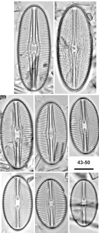
Figs 1–24. LM: Diploneis abscondita sp.nov., Marbe pond effluent, Saxony, Germany, (6) designated holotype. Striae turning from uni– to biseriate near the valve margins; areolae opposed, not alternating; (8–24) D. calcilacustris sp.nov., Lake Mainflingen, near Hanau, Hassia, Germany (Figs 11, 13, 15, 18, 20, 21), Lake Bohinj, Julian Alps, Slovenia (Figs 8–10, 12, 14, 16, 17, 19, 22–24), (10) designated holotype.
Scale bar 10 µm.
5702 in Coll. Lange–Bertalot, Naturmuseum Sencken- berg, Frankfurt (FR); material no. 414650. Represen- ted by Fig. 3.
Type locality: Marbegraben, brook in Staßfurt–
Glöthe, Sachsen–Anhalt, Germany, effluent from pond Ruschenschacht (former mining excavation); leg. M.
Werum 16.7.2010.
Etymology: The Latin adjective abscondita means hi- dden and refers to the so far overlooked occurrence.
Description
Valves elliptical to linear–elliptical with bluntly roun- ded ends. Only central parts of the valve face are almost flat, whereas marginal parts become arcuate as they gently merge into the poorly differentiated mantle. Length 18–40 µm, breadth 8–15 µm. Length–
to–breadth ratio 1.8–2.7. Raphe filiform, straight, with slightly expanded central endings which are modera- tely widely spaced; longer terminal fissures bent onto the same side of the valve mantle. Axial area narrow but expanded lanceolate towards the centre. Central area rather small with variable outlines, often apica- lly elliptical, but clearly separated in all cell–cycle stages. H–shaped appendices barely developed. Axial internal canals are marked outside by transapical striae composed of three or four areolae, altogether forming a conspicuous rhombical–lanceolate area being soft- ly arched around the central nodule. Adjacent regular striae radiate throughout, 13–15 in 10 µm, uniseriate but becoming biseriate near the valve margins onto the mantle. The areolae are opposed, not alternating, appearing here as single puncta in LM (cf. SEM Figs 106–107). Number of areolae in 10 µm is 13–18. SEM external view see Figs 106–108. Note in particular the raphe central and distal endings and the arrangement of cribrate areolae gradually cleaved with opposed parts.
Differential description
Taxa more or less similar as to outlines and size dimen- sions possess different fine structure patterns.
Distribution and ecology
As yet critically observed only at the type location.
This is a moderately eutrophic “hard–water” brook (slowly flowing, width 1 m, depth 10–30 cm) with a species–rich diatom assemblage typical of anthro- pogenically unpolluted and electrolyte–rich, not yet brackish water (sulphate ions 800–2000 mg.l–1, con- ductivity 1500–2500 µS.cm–1).
Diploneis arctica (lange–Bertalot) lange–Berta-
lot et A. fuhrMann stat. nov. (Figs 41–42)
Basionym: Diploneis ovalis ssp. arctica Lange–BertaLot in Lange– BertaLot & genkaL 1999, Iconographia Diatomologica 6, p. 43, figs 43: 1–3, 44a: 1–5.
A critical examination of the distinctive characters se- parating ssp. arctica from the nominate subspecies D.
ovalis, as emended in Lange–BertaLot & reiChardt
2000, makes a separate species rank more appropriate
rather than assessing the two taxa as conspecific. This receives support when comparing microphotographic records in as yet unpublished work by E. Pinseel ba- sed on samples from Spitsbergen. The similarity with respect to the broadly elliptical outline and the shape of the large central area may have been overestima- ted while the significantly higher density of striae and areolae of the Arctic taxon has been downplayed. We also note that when the Arctic taxon was established in 1999, the type material of D. ovalis (hiLse) raBenhorst had not yet been available for SEM investigation.
Striae 17–19 in D. arctica (versus 11–15) in 10 µm, areolae 15–21 (not 12–15) in 10 µm. Moreover, the shape of the domed valves and the detailed pattern of areolation are clearly distinct between the two taxa;
cf. Lange–BertaLot & genkaL (1999, plates 43–44b) and Lange–BertaLot & reiChardt (2000, plates 1–3).
As to Diploneis ovalis (hiLse) CLeve ssp. ovalis sensu Lange–BertaLot & genkaL 1999, figs 44b: 1–3 in par- ticular, conspecificity with the lectotypified population of Navicula ovalis in raBenhorst, Algen Europas, Ex- siccat no. 1025, appears unlikely.
Diploneis calcilacustris lange–Bertalot et a. fuhr-
Mann sp. nov. (Figs 8–24 LM, 109–111 SEM external and internal views)
Synonyms: Diploneis ovalis sensu Foged 1976, fig. 2: 13; Diploneis parma sensu Lange–BertaLot & metzeLtin 1996 pro parte, i.e. fig.
85: 5; Diploneis parma sensu hoFmann et al. 2013, fig. 65: 10; Di- ploneis parma sensu krammer et Lange–BertaLot as documented by Bey & eCtor 2013, p. 415, figs 1–19.
Diagnosis
Valvae ellipticae ad lineari–ellipticae apicibus cunea- tim rotundatis. Longitudo 20–45 µm, latitudo 12–17 µm. Ratio longitudo/latitudo 1.6–2.7. Raphe filiformis recta poris centralibus distincte dilatatis sitis in area axiali angusta lineari. Area centralis parva elliptica cir- citer 1/4–1/5 latitudinis valvae extendens. Appendices areae centralis minime formatae. Depressiones ad ra- phem utrimque non vel leviter aspectabiles microsco- pio photonico. Canales apicales interni angusti cum una serie punctorum separata a striis transapicalibus.
Istae striae distincte radiales omnino, 10–12 in 10 µm. Areolae 12–15 in 10 µm uniseriatae in partibus adaxialibus et biseriatae prope margines valvarum aut oppositae vel alternantes. Aspectus ultramicroscopicus externus internusque vide Figs 109–111.
Holotype (designated here): Slide Seen 224 in Coll.
Lange–Bertalot, Naturmuseum Senckenberg, Frank- furt (FR), represented by Fig. 10.
Type locality: Lake Bohinj, a calciumcarbonate–rich lake in the Julian Alps, Slovenia.
Etymology: The Latin epithet combines reference to the element calcium (calx) with the adjective lacustris (from lacus = lake). This species can be observed al- most exclusively in carbonate–rich lakes.
Description
Outlines strictly elliptical to oblong–elliptical, more or less cuneately rounded ends. Length 20–40 µm, breadth 12–16 µm, length–to–breadth ratio 1.6–2.7.
Raphe filiform, straight with distinctly expanded cen- tral ends and shortly curved distal ends. Axial area narrow. Central area elliptical, expanded transapically 1/4 to 1/5 of the valve breadth. H–shaped appendices (so–called horns in the literature) weakly developed.
Internal depressions parallel to the raphe slit, visible in LM view by focusing. Apical internal canals mar- ked by a single or two apertures to the valve outside, closely adjacent to the transapical striae, being mode- rately radiate throughout or somewhat stronger radiate close to the apices, 10–12 in 10 µm. Areolae 13–15 in 10 µm, appearing uniseriate with exception of a few ones close to the margins where they become biseriate in each stria. SEM external and internal view see Figs 109–111.
Differential description
D. hinziae sp. nov., living in electrolyte–poor waters of the Alps and in the Arctic/sub–Arctic regions, has almost the same cell sizes, striation and areolation but differs in its characteristic outlines, being more broadly elliptical with bluntly rounded apices. The central ends are more widely spaced in specimens of comparable size. The extension of the central area of D. hinziae displays less transapical asymmetry, the secondary side of the valve being distinctly more curved in D. calci- lacustris. The striae are less radiate proximally, each exhibiting more areolae due to the broader valve shape.
D. parahinziae from subtropical Florida, U.S.A., found in acidic water, differs likewise in having broader li- near–elliptical valve outlines with more broadly roun- ded ends, a higher striae– and areolae density, the latter being 15–20 (vs. 13–15) in 10 µm. Areolae of the val- ve face are for the larger part cleaved and arranged in
an alternately biseriate pattern. The canal areolae are smaller proximally.
D. elliptica (kützing) CLeve (Fig. 125, as as- sociated with the type population) is mainly distingu- ished by an approximately equal striae– and areolae density of 8–10 in 10 µm; see likewise type specimens in Lange–BertaLot & reiChardt 2000. Areolae are not divided. Central area is more broadly extended in comparable stages of both taxa. In internal SEM view D. elliptica shows broader virgae between narrower aveoli in contrast to D. calcilacustris with its equally broad virgae and aveoli; compare Fig. 111 with fig. 84:
10 in zeLazna–WieCzorek (2011).
Distribution and ecology
Infrequent but moderately abundant in places. These are oligo– to mesotrophic, generally oligosaprobic, calciumcarbonate–rich lakes, fountains, or springs in Europe.
Taxonomical comments
This taxon has been recorded and repeatedly documen- ted photographically under various established names of Diploneis; see under Synonyms above. It is some- times associated with rather resembling populations (morphodemes) which differ mainly by higher striae and areolae densities: 12–17 (vs. 10–12) striae and 15–
20 (vs. 13–15) areolae in 10 µm. They will be subject to investigation in a separate publication.
Diploneis dilatata (M. Peragallo) lange–Bertalot et A. fuhrMann stat. nov. (Fig. 124)
Basionym: Navicula smithii var. dilatata M. PeragaLLo in temPère
& PeragaLLo 1908, Diatomées du Monde Entier, 2nd. ed., p. 56, no.
103–104.
Synonyms: Diploneis smithii var. dilatata (M. PeragaLLo) terry
1908, “Additional lists of Connecticut diatoms”, Rhodora 10, p.
182.; Diploneis smithii var. dilatata (M. PeragaLLo) Boyer 1927, Proc. Nat. Acad. Sci. Philadelphia 79(2), Suppl., p. 355.
Figs 25–42. LM: Diploneis modicahassiaca sp.nov., Mainflingen, (29) designated holotype; (34–40) D. praetermissa sp.nov., Weissensee, Tyrol, Austria, (39) designated holotype; (41–42) D. arctica stat.nov., Spitsbergen. Scale bar 10 µm.
PatriCk & reimer (1966, p. 411, fig. 38: 3) give a line drawing of a specimen from the type population in freshwater, Fall Mountain, Bristol, Hartford Co., Co- nnecticut. For a recent microphotograph, taken by M.
Potapova, from the lectotype–slide, see Fig. 124.
The outlines, longitudinal canals, and the cent- ral area with H–shaped appendices clearly differ from Diploneis smithii (Brébisson ex W. Smith) Cleve 1894 var. smithii. From the present taxonomical point of view conspecificity must be excluded. Moreover, the
“second hand” concept of Diploneis smithii var. dila- tata illustrated in krammer & Lange–BertaLot (1986, fig. 112: 4) is obviously incorrect: there can be little doubt that these specimens from the Baltic Sea repre- sent another species that morphologically clearly dif- fers from Peragallo’s freshwater taxon and thus needs a revision.
Diploneis hinziae lange–Bertalot et A. fuhrMann
sp. nov. (Figs 43–50 LM, 113ab–114 SEM external and internal views)
Synonyms: D. parma Cleve sensu hustedt 1937; fig. 1066 (spe- cimen on the right–hand–side); D. parma Cleve sensu krammer &
Lange–BertaLot 1986; figs 109: 1–4.
Diagnosis differens versus Diploneis parma cleve
1891Valvae late ellipticae quoad specimina minora et late lineari–ellipticae quoad specimina maiora (numquam latius ellipticae ad leviter rhombico–ellipticae). Apices latissime rotundati (non cuneatim rotundati). Longi- tudo valvarum populationis typici 23–50 µm, latitu- do 15–18 µm. Ratio longitudo/latitudo 1.5–2.8 (nec 1.3–1.6). Raphe filiformis extremis centralibus modice distanter sitis inter se et extremis terminalibus curte unilateraliter deflexis. Area axialis angusta, linearis cum depressionibus internis utrimque ad raphem an- gustis sed dilatis proximaliter ad extrema centralia raphis. Area centralis parum dilatata ita semper parva (nec distincte expansa) circiter elliptica. Appendices areae centralis “cornua” dicta vix vel plerumque le- viter formatae. Canales apicales interni fere angusti zonam anguste lanceolatam circum aream centralem facientes cum una vel duobus seriebus punctorum se- paratis a striis transapicalibus regularibus. Istae striae leviter tum modice radiales in mediis partibus valvae, 11–12.5 in 10 µm quoad populationem typicam com- positae areolis duplicibus in partibus marginalibus sed singulis in partibus adaxialibus, 14–15 in 10 µm quoad populationem typicam.
Holotype (designated here): Slide O 4/57 in Coll.
Hustedt, AWI, Bremerhaven, Fig. 45 representing ho- lotype specimen.
Type locality: Unterer Grialetschsee, near Davos, Switzerland, 2500 m a.s.l. (Photographs taken by Frie- del Hinz.)
Etymology: We dedicate this taxon to Friedel Hinz, long–time curator of the Friedrich Hustedt–Archives in Bremerhaven.
Description
Valves of smaller specimens are broadly elliptical, the larger broadly linear–elliptical, all with very bluntly rounded apices. Length of type specimens 23–50 µm, breadth 15–18 µm. Length–to–breadth ratio 1.5–2.8.
Raphe filiform, straight, moderately expanded towards the central ends which are slightly deflected like the short terminal fissures to the same side of the valve.
Narrow apical depressions are flanking the internal ra- phe slit. The axial area as a whole is (very) narrow, almost linear; so–called H–shaped appendices or horns are very weakly developed. The central area is small and elliptical. The apical zone above the internal lon- gitudinal canals is narrow, slightly arched around the central nodule, marked by a single or double row of puncta. Regular transapical striae, 11–12.5 in 10 µm, are weakly radiate or subparallel at the valve centre, becoming moderately more strongly radiate near the apices. Areolae appear simple closer to the raphe ster- num, becoming doubled in the marginal parts, 14–15 in 10 µm. SEM, external and internal view, see Figs 113ab–114. All areolae are cribrate, other features as seen in LM view are confirmed.
Differential description versus Diploneis parma cleve 1891 sensu stricto
Cleve’s taxon is distinguished by a consistently broa- der, almost rhombic–elliptical valve with slightly cu- neately rounded ends. Its length–to–breadth ratio is 1.3–1.6 versus 1.5–2.8 for D. hinziae. Moreover, the areola density is higher on average, approximately 20 in 10 µm. For detailed analyses of the type population and others, including fine structures in SEM and TEM, see idei & koBayasi (1988).
Distribution and ecology
Evidently very uncommon in Central Europe but mo- derately abundant at the type location where it is as- sociated with predominantly acidophilous diatom taxa under low alkalinity and conductivity conditions. From Finland documented by krammer & Lange–BertaLot
(1986, figs 109: 3–4); from Spitsbergen (Svalbard) do- cumented with microphotographs by E. Pinseel (Uni- versity of Ghent). Distribution may be “nordic–alpine”.
Records under the name Diploneis parma from Europe belong neither to the true D. parma nor to D. hinziae but to an as yet undescribed taxon. See e.g. BLanCo et al. (2010, figs 50: 30–33).
Comments
D. hinziae has been mistaken for D. parma by several authors in the past. As one of the first authors, Hustedt (1937; 1943, p. 150) has enlarged the original concept of D. parma inappropriately. His fig. 1066 (1937, p.
674) illustrates two heterogeneous specimens, the left one representing a specimen from Sweden. That speci- men is not from the lectotype material (Lake Lojo, Fin- land), selected later on by idei & koBayasi (1988), but
109: 5 may not fit the given scale of x1500 but x1000;
figs 109: 6–7 may belong to another taxon together with “D.parma” sensu Lange–BertaLot & metzeLtin
(1996, figs 85: 6–7).
Diploneis ladogensis P.T. cleve
Basionym: Diploneis elliptica var. ladogensis P.t. CLeve 1891, The diatoms of Finland, Acta Soc. Fauna et Flora Fennica 8(2), p. 43, fig. 2: 9.
Comparing critically the photographic illustrations of this taxon in the literature, there is no doubt that con- specificity has to be excluded. See e.g. Lange–Ber-
taLot & reiChardt (2000), figs 7: 1–3, representing stages from the type population of Navicula elliptica Kützing (slide B.M. 18739). On the other hand, for D.
elliptica var. ladogensis see CLeve–euLer (1953, figs 646A: b,c); krammer & Lange–BertaLot (1986, figs 108: 3,5); Foged (1981, fig. 14: 8).
Independently of the present authors, Jovanovs-
ka, Levkov & edLund (forthcoming 2015) also come to the conclusion that the taxon, as observed in Lake Hövsgöl, Mongolia, ought to be elevated to species rank; cf. the detailed discussion in their article.
Diploneis lusatica lange–Bertalot et G. hofMann
sp. nov. (Figs 51–57 LM, 115–118 SEM external and internal views)
Diagnosis differens versus Diploneis subovalis P.T.
cleve 1894
Valvae late ellipticae ad plerumque aliquid rhombico–
ellipticae (nec ellipticae ad leviter lineari–ellipticae) apicibus fere cuneatim late rotundatis. Facies valva- rum paulatim in limbos curvata. Longitudo 20–50 µm, latitudo 13–23 µm. Ratio longitudo/latitudo 1.4–2.2.
Raphe paene recta filiformis extremis centralibus ter- minalibusque fere curte ad idem latus valvae deflexis poris centralibus parum dilatatis. Area axialis et de- pressiones axiales parallelae prope raphem non diffe- rentes. Area centralis generaliter anguste elliptica cum area axiali continuum formans sed in media parte ple- rumque plus minusve constricta, ita paene rectangulata apparens (differt ab area fere lata iconotypi ex Nova Zealandia). Zona apicalis lanceolata supra canales in- ternos cum striis transapicalibus curtis compositis pau- cis areolis irregulariter sed semper uniseriatis. Striae transapicales regulares omnes radiatae biseriatae 8–10 in 10 µm. Areolae duplicatae alternantes 18–20 in 10 µm. –– Aspectus ultramicroscopicus externus inter- nusque vide Figs 115–118.
Diploneis pseudovalis hustedt 1930 proprie di- ffert quoad lineamentum plerumque lineari–ellipticum et dimensiones inferiores, 16–31 µm longas, 8–14 µm latas etiam habitat in aquis subsalsis. Diploneis smithii (BréBisson) CLeve habitat cum populationibus diver- sissimis et certe heterospecificis in aquis marinis vel subsalsis differt proprie structura speciosa supra cana- les internas atque area centrali vix dilatata et numquam constricta in medio quamquam individua dicta Diplo-
Figs 43–50. LM: Diploneis hinziae sp.nov., Grialetschsee, near Da- vos, Switzerland, (45) designated holotype. Scale bar 10 µm.
from Rosslängen, Smaland in Sweden, another syntype among three locations mentioned by CLeve (1891). The difference to the specimen on the right in fig. 1066, ori- ginating from “Unterer Grialetsch See” in Switzerland, is quite obvious. However, Hustedt acknowledged only minor differences and treated both as representatives of a single taxon. Krammer & Lange–BertaLot (1986, figs 109: 1–2) uncritically followed Hustedt’s ambigu- ous concept. Thus, it appears that not all of the spe- cimens identified erroneously in krammer & Lange– BertaLot (1986) as D. parma belong to D. hinziae; fig.
neis smithii var. dilatata (PeragaLLo) terry 1908 pro parte (vide WitkoWski et al. 2000, figs 90: 5–6) vero similes apparentia.
Holotype (designated here): Slide S–2519, Seen no.
225 (G.H. 2762) in Coll. Lange–Bertalot, Naturmuse- um Senckenberg, Frankfurt (FR), represented by Fig.
53.Type locality: Water reservoir of Spremberg, Lausitz region, Saxony, eastern Germany; leg. 4.8.2008.
Etymology: The name of the German region Lausitz (in Slavic languages “Luciza”) was in Roman times Lusatia (adj. lusatica). It refers to the type locality in the wetlands between the river Görlitzer Neiße and the upper part of the river Spree.
Description
Valves broadly elliptical to almost rhombic–elliptical with somewhat cuneately, bluntly rounded ends. Valve face flat in the central part, whereas the large marginal part is domed and merged gradually into the mantle.
Length 20–50 µm, breadth 13–23 µm. Length–to–
breadth ratio 1.4–2.2. Raphe filiform and almost strai- ght, both central and terminal ends shortly deflected to the same side of the valve. Central pores inconspi- cuous. Axial area with apical depressions flanking the raphe slit internally, narrow, slightly expanded towards the central area which appears more or less constricted in the middle by few transapical striae, whence almost rectangular in shape in most specimens. The apical zone with underlying longitudinal canals is marked by short striae composed of few puncta. Regular transapi- cal biseriate striae, 8–10 in 10 µm, radiate throughout, each composed of alternating double areolae, 18–20 in 10 µm. –– SEM external and internal views, see Figs 115–118.
Differential description
Diploneis pseudovalis hustedt 1930 differs mainly by the linear–elliptical outline of most specimens, smaller dimensions on average, 16–31 µm long, 8–14 µm broad, and autecology, living in saline springs. Di- ploneis subovalis CLeve 1894, at least the type from freshwater in New Zealand, differs mainly in its slight- ly linear–elliptical outline (instead of rhombic–ellip- tical), and its larger, elliptical (instead of constricted to rectangular) central area. Diploneis smithii (BréBi-
sson) CLeve 1894 certainly represents a heterospecific complex of taxa. The nominate variety is significant- ly larger celled with a less expanded central area and living in marine and brackish habitats. However, Di- ploneis smithii var. dilatata (PeragaLLo) terry 1908 in the sense of several authors (e.g. WitkoWski et al.
2000, figs 90: 5–6) is quite similar. Nevertheless, any conspecificity with the nominate variety can be exclu- ded and in the rank of a variety Peragallo’s taxon has no priority.
Distribution and ecology
Very abundant at the type locality, a large artificial lake, eutrophic but oligosaprobic freshwater with sli- ghtly elevated conductivity, 800–900 µS.cm–1. Asso- ciated taxa are various Planothidium species and, in particular, the commonly infrequent Navicula oppug- nata, together with Navicula gregaria, N. germainii, N. viridula, N. amphiceropsis (see Lange–BertaLot
2001). Remarkable is the high content of sulphate, ca.
300 mg.l–1, due to mining activities upstream.
Diploneis modicahassiaca lange–Bertalot et A.
fuhrMann sp. nov. (Figs 25–33 LM, 112a–b SEM external view)
Diagnosis
Valvae omnes stricte lineari–ellipticae apicibus late ro- tundatis. Longitudo 12–16 µm, latitudo 5–6.6 µm. Ra- tio longitudo/latitudo 2.4–2.7. Raphe filiformis distinc- te recta apparens. Area axialis anguste linearis, circiter 0.7–1.0 µm, interrupta nodulo centrali, area centralis circum nodulum non formata. Area canalium zonam comparate angustissime linearem formans punctis in- distincte apicaliter ordinatis signata. Striae transapi- cales in media parte valvae parum radiantes tum leviter ad modice denique fortius radiantes usque sub apices, 16–18 in 10 µm. Aspectus ultramicroscopicus externus vide Figs 112a–b. A Diploneis modica hustedt 1945 differt areolis triseriatis in partibus marginalibus faciei et limbis valvae (aspectus microscopio electronico).
Satis differt ab alteris speciebus plus minusve compa- rabilibus complexu omnium signorum typicorum par- ticulariter a Diploneis puella (sChumann) CLeve quoad protologium etiam D. puella sensu MöLder & tynni
aequaliter sensu ehrLiCh etiam quoad conceptiones differentes auctorum diversorum (vide infra).
Holotype (designated here): Slide G.H. S–3351 in Coll. Lange–Bertalot, Naturmuseum Senckenberg Frankfurt a.M. (FR), represented by Fig. 29.
Type locality: “Mainflinger See”, a large pond in a gravel pit, on stones. Mainflingen, near Seligenstadt, Germany, Transect no. 2; leg. G. Hofmann 2.8.2014.
Etymology: The Latin adjective modicahassiaca re- fers to the type location in the German state of Hesse, modica in Latin means unpretentious.
Description
Valves strictly linear–elliptical with broadly rounded apices. Length 12–16 µm, breadth 5–6.6 µm. Length–
to–breadth ratio 2.4–2.7. Raphe filiform, straight with inconspicuous central and distal ends. Depressions parallel to the raphe are barely recognizable. Axial area very narrow, linear. A central area is not separa- ted, except for the approximately square or sometimes very weakly constricted central nodule. Zone of the longitudinal canal system is by comparison extremely narrow; canal perforation is hard to differentiate from the transapical striae. Striae weakly radiate proxima-
lly, becoming moderately radiate towards and strongly radiate near the apices; 16–18 in 10 µm. Areolae are not discernible. SEM external view see Figs 112a–b.
Raphe slit almost straight but with shortly bent, not de- pressed central ends and slightly deflected distal ends.
Axial area ca. 0.7–1.0 µm broad, defined by single or obliquely paired small cribra consisting of four or five poroids. The adjacent striae are biseriate, becoming broader and distinctly triseriate closer to the valve face margins and onto the mantle. Each cribrum is com- posed of mostly four, sometimes five poroids; some cribra appear to be reduced to less than four poroids.
The density of the areolae, pluriseriate and arranged in opposition, is very high compared to other cribrate Diploneis taxa except for D. modica, approximately 50 in 10 µm.
Distribution and ecology
As yet only observed from the type location. This is a gravel pit filled with ground water after exploitation. It is situated in the River Main valley close to Seligen- stadt, not far from Frankfurt. The taxon is quite rare in the location and was found in a diatom assemblage of more than 100 taxa in a single sample, including several other Diploneis species, i.e. D. calcilacustris, D. elliptica, D. petersenii, D. minuta. The water is mo- derately alkaline with a medium conductivity, oligosa- probic and eutrophic.
Taxonomical comments
A somewhat resembling, undescribed population has recently been observed by us in Lake Baikal, Siberia (unpublished data). What makes D. modicahassiaca (as well as D. modica, but see below) so particular is the combination of, first, extremely narrow apical canals with, second, the small size of the cribra, each composed of two to four, rarely five poroids, and arran- ged triseriately (biseriately in D. modica) in the sub- marginal parts of the valve face and mantle. Moreover, the density of the striae areolae (as opposed to the canal areolae), ca. 50 in 10 µm, is much higher than in any other known Diploneis species with cribrate areolae, including the ones here described as new. We also po- int out that the comparatively small cell size with a len- gth–to–breadth ratio of 2.4–2.7 already distinguishes D.modicahassiaca from D. puella both in the original sense of Schumann as well as according to the con- cepts of other authors.
The most resembling taxon is D. modica hus-
tedt 1945, described from the “Ancient Lakes” Ohrid and Prespa, The Balkans, now belonging to Albania and Macedonia. krammer & Lange–BertaLot (1986) report and illustrate few specimens from two other lo- cations in Austria, Namlos in Tyrol, and Lunz in the north–eastern Alps, both with questionable identifica- tion. Levkov et al. (2007), plate 124, contains the first SEM images from Lake Ohrid, but neither these nor their LM images represent D. modica, as they observe
in their later work on Diploneis in Lake Ohrid (2013, p.
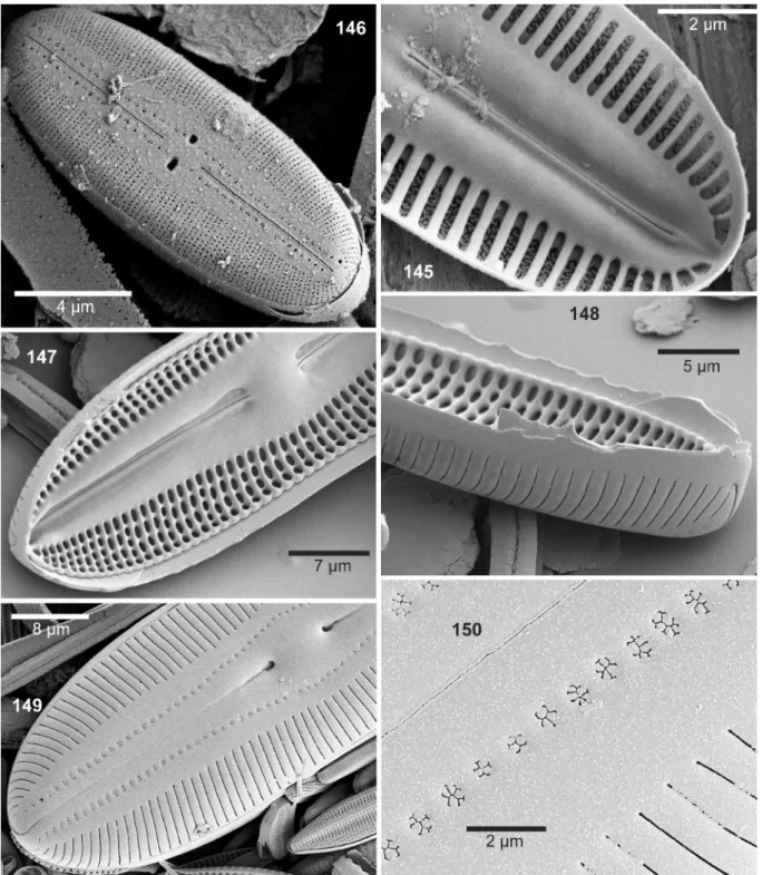
258). The latter publication includes a diagnosis with four LM and two SEM photographs of the true D. mo- dica from Lake Ohrid. The metric data conform appro- ximately to D. modicahassiaca but the remark that “the striae are biseriate, and each alveolus opens externa- lly in groups (3–4) of small and rounded pores, 50 in 10 µm” suggests that a trifurcation of alveoli towards the margin of the valve, characteristic for D. modica- hassiaca, is absent. This is confirmed by an inspecti- on of fig. 150 where such triseriate arrangement be- tween broader virgae is not visible; cf. by contrast Figs 112a–b showing D. modicahassiaca. D. modica has as yet never been observed in Germany or neighbouring countries, except for a doubtful identification from the Austrian Alps.
Diploneis nanofontanella lange–Bertalot et A. fuhrMann sp. nov. (Figs 58–63 LM, 64 SEM in- ternal view)
Diagnosis differens versus Diploneis fontanella lange–Bertalot in WeruM & lange–Bertalot 2004Synonym: Diploneis (? nov.) spec. in Lange–BertaLot & genkaL
1999, figs 43: 5–10.
Valvae oblonge–ellipticae ad lineari–ellipticae apici- bus obtuse rotundatis. Longitudo 14–24 (non 16–30) µm, latitudo 5.5–6 (non 7–8.5) µm. Ratio longitudo/
latitudo 3.3–4 (2.0–3.6 paulo differt). Raphe leviter lateralis recta (non differt). Area axialis angusta sed dilatata ad mediam versus (non differt). Area centralis comparate ampla, 2–2.5 µm lata, distincte separata ab area axiali, paene dimidium = 1/2 latitudinis valvae ex- tendens (non tertium = 1/3). Appendices areae circula- ris et canales axiales non differunt. Striae transapicales fortius radiantes usque ad apices 19–22 (nec 16–18) in 10 µm. Aspectus ultramicroscopicus internus vide Fig. 64. Systema sternum cum canalibus axialibus di- midium latitudinis valvae extendens (vide area axialis, supra).
Holotype (designated here): Slide RUS–30 in Coll.
Lange–Bertalot, Naturmuseum Senckenberg, Frank- furt (FR). Holotype shown in Fig. 60. Type material M5 in Coll. S. Genkal, Papanin Institute for Biology of Inland Waters, Russian Academy of Sciences, Borok.
Type locality: Mestnyi Island, Yugorski–Shar Strait, Arctic Ocean, among bryophytes in a small river; leg.
N.V. Vekhov, Sept. 1995.
Etymology: The epithet refers to the smaller size di- mensions in comparison with the similar D. fontanella.
Lat. nanus = dwarf.
Differential description versus Diploneis fontanella lange–Bertalot in WeruM & lange–Bertalot
2004Valves oblong–elliptical to linear–elliptical with blunt- ly rounded ends. Length 14–24 (versus 16–30) µm,
breadth 5.5–6 (not 7–8.5) µm. Length–to–breadth ra- tio 3.3–4 (versus 2.0–3.6, is weakly different). Raphe appears slightly lateral, but this may be due to the in- ternal depressions flanking the raphe sternum. Axial area narrow distally, expanding towards the middle of the valve (not different). Central area conspicuous- ly large with respect to the narrowness of the valves, extending to about one–half of the valve breadth (not ca. one–third). Indistinct appendices of the central area and axial canals not different. Striae radiate throughout, 19–22 (not 16–18) in 10 µm. Areolae uniseriate, about 25 in 10 µm (not different). –– SEM internal view, see Fig. 64, cf. Also another SEM internal view in Lange– BertaLot & genkaL 1999, fig. 43: 10. The closing membranes covering the entire alveoli are uncorroded here, being distinctly narrower than the virgae. The ca- nal system, together with the central nodule, extends to one–half of the valve breadth, as does the central area outside. The central part is clearly not set off due to al- most evenly shortened striae (different from the shape in D. fontanella). The narrow depressions (furrows) on either side of the raphe sternum diverge at the central ends of the raphe slit (as opposed to being almost pa- rallel in D. fontanella, cf. Werum & Lange–BertaLot 2004, fig. 74: 15).
Diploneis separanda Lange–Bertalot in Werum
& Lange–BertaLot 2004 differs mainly by a consis- tently smaller central area, broader valves, 6–7 µm, and a higher areola density, approximately 30–35 in 10 µm.
Distribution and ecology
As yet only known from the type locality, a creek with pristine water which may be under the influence of strong winds from the Arctic Ocean. The species–rich diatom assemblage consists of oligotraphentic, acido- philous taxa associated with several alkaliphilous taxa tolerating a moderately high conductivity.
Diploneis oblongellopsis lange–Bertalot et a. fuhrMann sp. nov. (Figs 65–74 LM, 119–123 SEM external and internal views)
Synonyms: Diploneis oblongella (nägeLi) CLeve Euler sensu Lange–BertaLot & metzeLtin 1996, figs 85: 8–9; Diploneis oblon- gella sensu krammer & Lange–BertaLot 1986 pro parte, fig. 108:
10; Diploneis spec. Nr. 1 sensu metzeLtin & WitkoWski 1996, fig.
12: 7.
Diagnosis
Valvae ellipticae vel lineari–ellipticae apicibus late rotundatis. Longitudo 11.6–25 µm, latitudo 5.7–7.8 µm. Raphe filiformis recta extremis centralibus termi- nalibusque fere inconspicuis. Area axialis angusta con- spicue linearis. Area centralis parva paene vacans vel plerumque quoad individua maiora apicaliter elliptica usque ad circiter quartum latitudinis valvae extendens.
Zona canalium comparate angustissima distincte ar- cuata circum nodulum centralem. Striae transapicales fortius radiantes omnino, 20–24 in 10 µm. Areolae unaquisque discernendae omnes semper uniseriatae,
25–30 in 10 µm. Aspectus ultramicroscopicus exter- nus internusque vide Figs 119–123. Extrema centra- lia externa curte uncinata. Extrema terminalia aliquid variabiliter formata sed omnia fere curte ad latus idem.
Areolae striarum ubique stricte uniseriatae, 25–30 in 10 µm confirmatae omnes occlusae externe cribris cir- cularibus compositis 7–20 poris. Ita differt particula- riter a D. praetermissa sed etiam omnibus speciebus generis cum cellulis parvis hic comparatis.
Figs 51–57. LM: Diploneis lusatica sp.nov., Spremberg water re- servoir, Saxony, Germany, (53) designated holotype. Scale bar 10 µm.
Etymology: The Latin–Greek epithet means “with the appearance of” D. oblongella auct. nonnull. It refers to Navicula oblongella Nägeli ex Kützing 1849 with the intention to remember in the future that ambiguous name (see comments).
Description
Valves elliptical to linear–elliptical with broadly rounded apices. Length 11.6–25 µm, breadth 5.7–7.8 µm. Raphe filiform, straight without recognizable pe- culiarities at central and distal ends. Axial area very narrow, strictly linear throughout, i.e. not extended proximally or narrowed distally. Central area barely present in smallest specimens, becoming larger and progressively set off from the axial area with elon- gation of cell cycle stages, shaped apically elliptical, extended to about a quarter of the valve breadth. H–
shaped appendices absent. Axial canals very delicate, marked on either side by an apical line of single puncta which curve around the central nodule. These more strongly contrasted puncta are barely set off from the uniseriate areolae of the striae, 20–24 in 10 µm, which are conspicuously radiate throughout. The areolae are constantly more densely spaced than the striae and can still be resolved in appropriate focus without oblique lighting, 25–30 in 10 µm. SEM external view see Figs 119–120, 122–123. The relief of striae and virgae on valve face and mantle is extremely shallow irrespec- tive of the amount of tilting. Thus the valve surface appears almost smooth, if the cribrate areolae are un- corroded (Figs 123). With higher magnification, more than ×10000 (Fig 120), all cribra appear circular, con- sisting of ca. 7–20 single pores, gradually growing in diameter and number from the zone of canals towards and onto the mantle. After corrosion (Fig. 122) the are- olae perforating the underlying canals are more con- trasted, whence their position becomes easier recog- nizable, lying in a single apical line on either side of the sternum, weakly set off from the stria areolae. Slits of both raphe branches with unilaterally, shortly bent central and distal ends. The central ends are enclosed in short delicate raphe ribs and depressed into the valve surface. Internal view see Fig 121 after corrosion of all finer structures including occlusion membranes on the alveoli. If uncorroded, it may be very difficult to distin- guish D. oblongellopsis from D. praetermissa since the uni– vs. biseriate areolae are then masked.
Differential description vs. D. praetermissa sp. nov.
and other similar small–celled taxa
Unless both taxa can directly be compared in the same slide D. praetermissa (see below) is rather difficult to distinguish in LM view but very easy in SEM, mainly by regularly biseriate alternating areolae in consider- ably higher density. Moreover the profile of striae/vir- gae is higher; cribra in the canal zone are larger; distal raphe fissures are considerably longer. Such differen- tiating features concern likewise populations which
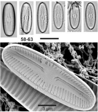
Figs 58–63. LM: Diploneis nanofontanella sp.nov., Mestnyi Island, Russian Arctic, (60) designated holotype. Scale bar 10 µm.
Fig. 64. SEM internal view: D. nanofontanella sp. nov., Mestnyi.
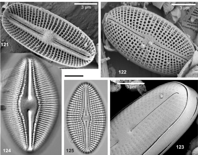
Figs 65–74. LM: Diploneis oblongellopsis sp.nov., Lake Laach, Ei- fel Mountains, Germany, (Figs 65, 71–72), Brunnsee, Upper Bavaria (Figs 66–70, 73–74), (65) designated holotype. Scale bar 10 µm.
Holotype (designated here): Slide Seen/251 in Coll.
Lange–Bertalot, Naturmuseum Senckenberg, Frank- furt (FR), represented by Fig. 65.
Type locality: Lake Laach, close to Maria Laach monastery, Eifel Mountains, West Germany. Sample LS 7, sediment 1–20 cm, subfossil, likely ca. 100 years old (leg. Simone Illig, 2010.)
are recorded under the problematic (because ambigu- ous) name D. oblongella; see the comments below on D. praetermissa. D. modica hustedt has subparallel rather than more strongly radiate striae proximally and biseriate areolae just as D. fontanella or D. separanda (see elsewhere).
Distribution and ecology
We identified D. oblongellopsis in various strictly oli- gotrophic lakes and a slightly eutrophic one in central Europe, all rich in calcium carbonate content; a single specimen on the Arctic Bear Island (Svalbard); indivi- dual–rich on Gotland Island, the Baltic Sea, Sweden.
The taxon should be wider distributed but is not easily separated from resembling taxa in LM. Quite often as- sociated with D. separanda and D. praetermissa nov.
sp.
Taxonomical comments
The current and historical concepts of D. oblongella are unsatisfactory because ambiguous. What are we to do with such a “catch all” taxon? Because there is no illustration of the basionym Navicula oblongella nä-
geLi ex kützing it is not possible to designate a lecto- type with ensuing epitypification. We would have to wait until an appropriate specimen may be found in Nägeli’s original sample from Zurich, Switzerland. In a later second hand “type slide” (prepared from ori- ginal type material) we could not find any specimen that would answer to the extremely poor protologue given by kützing 1849 (see Werum & Lange–Berta-
Lot 2014, p. 145). The first to present an illustration of Nägeli’s taxon was Grunow in van heurCk (1880–85).
The origin of the depicted specimen is unknown. It is 13.5 µm long, 6 µm broad, ca. 18.5 striae with rough- ly 25 puncta in 10 µm. However, even Grunow may have had no well–defined concept. In the text part of van heurCk (1885) where both authors are responsi- ble with regard to taxonomical decisions, Grunow ends up with a diagnosis: 20 µm long, 7.5 µm broad, ca. 16 striae in 10 µm with reference to Type de Synopsis no.
108, Oxford, England. PatriCk & reimer (1966, fig.
38: 8) made a line drawing of a specimen from CLeve
& möLLer exsiccate no. 139 which likewise has been authorized by Grunow: it is 30 µm long, 11 µm broad, 14–15 striae and ca. 18 puncta in 10 µm. Clearly this is too wide a spectrum for one and the same species.
Even wider is the concept of PatriCk & reimer, as expressed in their diagnosis: 10–100 µm long, 6–35 µm broad, 10–19 striae and 13–20 areolae in 10 µm –– all these data concerning D. oblongella, including an allegedly younger homonym, D. ovalis (hiLse) P.T.
CLeve. From a modern taxonomical point of view, such a concept is untenable. As it happens, the basionym of D. ovalis (hiLse) P.T. CLeve, Pinnularia ovalis hiLse, has proved to be a different, large–celled taxon with a different structural pattern (Lange–BertaLot & rei-
Chardt 2000). The consequence of such an extremely
extended concept is displayed in Foged (1977, p. 50, figs 17: 1–6) where all specimens are taken to belong to the same species: figs 1–3 and 6 are identified as D.
ovalis var. ovalis, figs 4 and 5 are var. oblongella. From the present, more critical point of view, four distinct species are shown: Fig. 1 is D. krammeri, a taxon fre- quently mistaken for D. ovalis in the past (see Lange– BertaLot & reiChardt 2000); figs 2,3,6 are closely related to D. elliptica (see photographs of type speci- mens in Lange–BertaLot & reiChardt 2000); fig. 4 is D. fontanella; fig. 5 belongs to D. oblongellopsis, the stria density being counted with 23–24 (not erroneous- ly 16–17) in 10 µm.
Diploneis parahinziae lange–Bertalot, a. fuhr-
Mann et h. rIngel sp. nov. (Figs 86–92 LM, 126–129 SEM external and internal views)
Diagnosis differens versus Diploneis hinziae sp. nov.
(vide supra)
Differt proprie densitate superiori striarum 12.5–14 (nec 11–12.5) in 10 µm. Areolae 15–20 in 10 µm pa- rum differunt sed extra zonam canalium regulariter bi- seriatae et prope marginem limbi triseriatae ordinatae (nec uniseriatae et in limbo biseriatae). Valvae omnes praesentes in loco typico 17–26 µm longae (nec usque ad 50) et 11–14 µm latae (nec usque ad 18).
Holotype (designated here): Slide Am–N–130 in Coll. Lange–Bertalot, Naturmuseum Senckenberg, Frankfurt (FR), represented by Fig. 87.
Type locality: Everglades National Park (Florida, U.S.A.), 20 miles west of Miami, boat landing close to National Road No. 41, epiphytic on Lemnaceae, leg.
Hubert Ringel, 17.11.2010.
Etymology: The prefix para in classical Greek means:
beside of (a similar taxon; here: D. hinziae, see above).
Description
Valves elliptical to weakly linear–elliptical, ends
Figs 75–85. LM: Diploneis tirolensis sp.nov., Weissensee, (78) de- signated holotype. Scale bar 10 µm.
bluntly rounded. Length 17–26 µm, breadth 11–14 µm. Length–to–breadth ratio 1.5–1.9. Raphe filiform, appearing almost straight or (mostly) deflected towards expanded central ends; distal ends indistinct. Axial area narrow with a parallel dark line on either side of the raphe indicating internal depressions along the ster- num (which become visible by focusing). Central area elliptical, small, 1/6 to 1/4 of the valve breadth. H–sha- ped appendices are not developed. Sector of internal canals very narrow with canal areolae appearing barely set off from the other areolae. Striae weakly radiate in proximal parts, becoming rather strongly radiate close to the ends, 12.5–14 in 10 µm. Areolae appear uniseria- te, becoming indistinctly pluriseriate in marginal parts, 15–20 in 10 µm.
SEM external view see Figs 126 with intact cribra and Fig. 127 with corroded cribra. Raphe with short, crochet–hook shaped central ends and longer distal fissures which are bent towards the mantle, all to the same side. The valve surface is not complete- ly domed but flattened in the central part. One or two single areolae with circular cribra are arranged above the underlying canal system, somewhat separated from the biseriate areolae at first in apical juxtaposition, then becoming gradually alternating. Finally, on the valve mantle, areolae become smaller and triseriate between narrow virgae. Close to the margin a small open pore in the middle between two virgae becomes visible, main- ly in distal parts (Fig. 128). Internal view see Fig. 129.
Raphe canal system and central nodule do not differ from many other freshwater Diploneis species, par- ticularly not from D. hinziae. This concerns likewise uncorroded occluding membranes covering the entire alveoli and masking the arrangement of areolae. Vir- gae and striae are about equally broad –– in contrast to the proportions in external valve view. An internal view with corroded occlusion membranes could not be found.
Differential description vs. D. hinziae sp. nov. (see above) LM view
Largest cell cycle stages of the population from Florida (others are as yet not known) do not exceed a valve len- gth of 26 (vs. 23–50) µm and a valve breadth of 14 (vs.
15–18) µm. Stria density is 12.5–14 (vs. 11–12.5) in 10 µm, areola density 15–20 in 10 µm. SEM view: The biseriate striae become triseriate in the valve mantle, whereas striae continue clearly biseriate throughout in D. hinziae (see LM and SEM Fig. 114 of internal view.) Distribution and ecology
As yet only known from the Everglades, Florida. This is a border area between two biogeographic realms, the southern Holarctic and the Neo–Subtropics. The dia- tom assemblage in the type locality is dominated by acidophilous or acidobiontic taxa, including various Eunotia spp. indicating dystrophic conditions.
Taxonomical comments
This Florida population is ranked as a species inde- pendent of the resembling D. hinziae based on several distinguishing morphological characteristics. In addi- tion, the autecology, living in dystrophic water, and the biogeographic occurrence in a subtropical border region contribute to justifying this decision. For a po- ssibly different taxonomical ranking in the future more sample populations of both taxa will be needed in order to carry out molecular genetic or population biological investigations (e.g. testing for cross–breeding).
Diploneis praetermissa lange–Bertalot et A. fuhr-
Mann sp. nov. (Figs 34–40 LM, 134ab–138 SEM ex- ternal and internal views)
Diagnosis differens versus Diploneis separanda lange–Bertalot in WeruM & lange–Bertalot 2004Lineamenta valvarum simillima et dimensiones similli- mae, ellipticae ad lineari–ellipticae apicibus late rotun- datis. Longitudo 8–25 µm, latitudo 5.5–7.8 µm. Raphe linearis recta extremis centralibus curtissime curvatis et extremis terminalibus comparate longe uncinatis ad latus idem. Area axialis angustissima linearis vix (nec distincte) dilatata versus mediam valvae. Area centra- lis minima parum expansa et separata ab area axiali, appendices in forma litterae H nullae. Canales axiales prope raphem delicatissimi angustissimi parum arcuati circum nodulum centralem. Areolae externe cribratae hic uniseriatae distincte separatae ab striis transapi-
Figs 86–92. LM: Diploneis parahinziae sp. nov., Everglades, Florida, (87) designated holotype. Scale bar 10 µm.
calibus in medio leviter tum modice radiantibus et ibi regulariter biseriatis (vide Figs 134b,136) inter virgas transapicales, 17–19 in 10 µm. Areolae alternantes, 32–35 in 10 µm, numquam discernendae microscopio photonico ita differt a D. separanda ubi striae biseriatae cum areolis oppositis (nec alternantibus) solum prope limbos usque ad margines (vide Werum & Lange–Ber-
taLot 2004, figs 76: 15–16 et 77: 3–5).
Differt ab alteris paucis speciebus plus minusve similibus in aquis dulcibus combinatione signorum ty- picorum quoad areas axiales centralesque et structuras ultramicroscopicas.
Holotype (designated here): Slide Eu–A 198 in Coll.
Lange–Bertalot, Naturmuseum Senckenberg, Frank- furt (FR), represented by Fig. 39.
Type locality: Weissensee, a calcium–carbonate rich oligotrophic alpine lake, ca. 1050 m a.s.l., near Lermo- os in Tyrol, Austria; leg. K. Külbs, Sept. 1982.
Etymology: Lat. praetermissus = left out, overlooked.
Differential description versus Diploneis separanda lange–Bertalot in WeruM & lange–Bertalot 2004 Valves of both taxa hardly distinguishable as to size dimensions and outlines; elliptical to linear–elliptical with bluntly rounded apices. Length 8–25 µm, breadth 5.5–7.8 µm. Raphe linear, straight, without remarka- bly modified central and distal endings (see below).
Axial area very narrow, linear, barely expanded and separated from the central area. H–shaped appendices lacking. Axial canals delicate, very narrow (appearing comparatively less pronounced), canal areolae unise- riate, 17–19 in 10 µm, as adjacent transapical striae.
Puncta of the striae not discernible in light microscope.
SEM, external and internal view, see Figs 134ab–138.
External central raphe ends fish–hook shaped, whereas distal ends are longer bent off onto the mantle, all to the same side. The areolae above the internal canals on either side are particularly marked and appear slightly depressed into the valve surface, occluded by cribra typical for Diploneis. Number of stigmoids composing these cribra is high, ca. 20 (vs. in striae at most 8 per cribrum). The transapical striae, radiate, becoming less radiate in the centre, 17–19 in 10 µm, are biseria- te throughout (not uniseriate, becoming biseriate only near the mantle down to the valve margins). Regularly biseriate, alternating (not opposed) areolae, 32–35 in 10 µm, are comparatively small and barely discernible by LM observation (in contrast to the opposed ones in D. separanda).
Distribution and ecology
Insufficiently known, since as yet not distinguished from D. separanda or specimens recorded under the dubious taxon Diploneis oblongella (nägeLi ex kützing 1849) CLeve–euLer 1922 or Diploneis ovalis
Figs 93–97. LM: Diploneis tundra sp. nov., Mestnyi, (93) designated holotype. Scale bar 10 µm.
Figs 98–105. LM: Diploneis puellafallax sp. nov., Laach, (101) designated holotype. Scale bar 10 µm.
Figs 106–108. SEM external view: Diploneis abscondita sp. nov., Marbe. Striae uniseriate near the apical axis becoming biseriate near the valve margins onto the mantle. Areolae opposed, not alternating; (109–110) D. calcilacustris sp. nov., Bohinj. Fig. 111. SEM internal view: D.
calcilacustris, Bohinj.
Figs 112a–b. SEM external view: Diploneis modicahassiaca sp. nov., Mainflingen. Figs 113a–b: SEM external view: D. hinziae sp. nov., Gri- aletschsee. Fig. 114. SEM internal view: D. hinziae, Grialetschsee.
var. oblongella (nägeLi ex kützing) P.T. CLeve 1894;
see Werum & Lange–BertaLot (2004, pp. 145f.). The type locality is oligotrophic with average conductivity.
Other locations where D. praetermissa could be found are oligo– to slightly eutrophic but always oligosapro- bic. Associated are predominantly oligotraphentic, cal- ciphilous diatom taxa. In several lakes associated with D. separanda and D. oblongellopsis sp. nov.
Figs 115–116. SEM external view: Diploneis lusatica sp. nov., Spremberg. Figs 117–118. SEM internal view: D. lusatica, Spremberg. Figs 119–120. SEM external view: D. oblongellopsis sp. nov., Laach (Fig. 119), Brunnsee (Fig. 120).
Comments
Navicula oblongella nägeLi in the sense of Kützing’s protologue can hardly be considered as a synonym sin- ce the girdle view is described as broadly linear. By contrast Diploneis praetermissa and likewise D. sepa- randa are very rarely found in the very narrow girdle view.
Another species appearing very similar in the light microscope is Diploneis modica hustedt 1945, holotype from ancient Lake Ohrid (Macedonia,The