Summary of the lecture "‘Senses"’
by Susanne Duncker
Contents
1 Senses I - primary sensory cells (Knipper) 4
1.1 Smell . . . 4
1.1.1 Summary . . . 7
2 Senses II - Secondary sensory cells (Knipper) 9 2.1 Taste . . . 9
2.1.1 Salty . . . 9
2.1.2 Sour . . . 11
2.1.3 Sweet . . . 12
2.1.4 Bitter . . . 13
2.1.5 Umami . . . 13
2.1.6 Signal transduction . . . 14
2.2 Summary . . . 14
3 BK and its physiologic relevance in the cardiovascular system - an inno- vative target for drug design? (Sausbier) 16 3.1 The meaning of BK for vasoconstriction . . . 16
4 The vestibular system (Engel) 19 4.1 Structure of the vestibular system . . . 19
5 Senses III - Coding of sensory information (Knipper) 25 5.1 Afferent and efferent nerves . . . 25
5.2 The coding of sound frequency . . . 25
5.3 The coding of loudness . . . 27
6 Mast cell activation and anaphylaxis is blunted in mice with targeted de- letion of the SK4 channel (Ruth) 29 6.1 What is a SK4 channel? . . . 29
6.2 Mast cells and their role in allergic disorders . . . 30
6.3 The SK4 knockout mouse . . . 31
7 Senses IV - Vision (Knipper) 32 7.1 Accommodation and its disorders . . . 32
7.2 The structure of the retina . . . 34
7.3 Photoreceptors and how they work . . . 35
7.4 Colour vision disorders . . . 36
7.5 Retinal circuitry . . . 36
8 Forensic genetics (Blin) 40
8.1 A little history of forensics . . . 40
8.2 Identification of individuals . . . 40
8.2.1 Technical limitations . . . 41
8.2.2 Wrongful diagnosis . . . 41
8.2.3 Quality controls . . . 42
8.3 The O.J. Simpson story . . . 42
8.4 Famous puzzles forensic genetic could help to solve . . . 42
8.5 Other purposes for (forensic) genetics . . . 43
9 Otosclerosis - new pathophysiologic insights (Pfister) 44 9.1 A very short history of otosclerosis . . . 44
9.2 Epidemiology . . . 45
9.3 Pathophysiology . . . 45
9.4 Pathology . . . 45
9.5 Treatment . . . 45
9.6 Diagnosis . . . 46
9.7 Otosclerosis loci . . . 46 10 Signal transduction in bipolar cells (Engel) 49 11 Colour vision ... and some congenital and acquired troubles (Rüttiger) 50 12 Large (BK) conductance, Ca2+ and voltage activated K+ channels in phy-
siology and pathophysiology (Ruth) 51
Corrected errors
04.02.2009 Figure 6.2 corrected. Thanks to Prof. Ruth who answered my question if this figure of mine would be right!
02.02.2009 Hyposmia is a reduction of smell, not only in reality but now also on page 4. Thanks to Martina and Juliane!
1 Senses I - primary sensory cells (Knipper)
The cells of the sensory systems can be divided into primary and secondary sensory cells. Primary sensory cells are neurons whose axons project directly to the brain, secondary sensory cells are sensory cells who give the information via a synapse to a neuron that projects into the brain.
The cells of the senses are categorised as follows:
• vision (rods and cones) - secondary sensory cells
• hearing - secondary sensory cells
• smell - primary sensory cells
• taste - secondary sensory cells
• touch - primary sensory cells
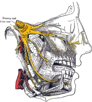
Smell and taste sense work by chemoreception. They are connected to the so- matosensory trigeminal nerve(s) that pass the ethmoid plate. The somatosensory trigeminal nerve is the so called fifth cranial nerve. The fifth nerve is primarily a sensory nerve, but it also has certain motor functions (biting, chewing, and swal- lowing) Wikipedia, The Free Encyclopedia (2009).
1.1 Smell
Diseases of smell are called dysosmies. The can be divided into hyposmies (reduc- tion of smell), anosmie (no smell at all), kakosmie (every smell seems to be bad), and phantosmie (smell hallucination).
The epithelium of smell sensation is very enlarged. The neurons act as chemore- ceptors and alter chemical signals/stimuli in neuronal signals. In contrast to every until now known sensory system based on primary sensory cells the neurons in the nose are are continuously replaced, about all eight weeks, by basal/supporting cells.
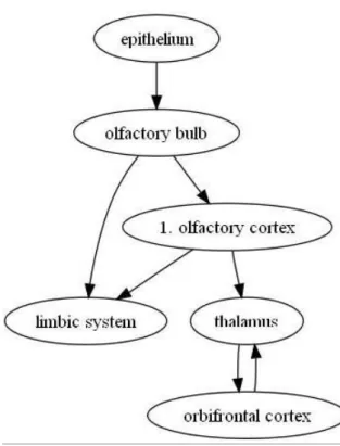
The sensory transduction occurs in the cilia of the olfactory neurons. Here smells are immediately detected by odorant receptors (7 transmembrane receptors) and reported overtwo pathways:
Figure 1.1: The trigeminal nerve (from Wikipedia, The Free Encyclopedia).
Figure 1.2: The two pathways for olfaction (from OpenWetWare, 2006).
The cAMP second messenger response declines again after milliseconds. cAMP activates directly cyclic nucleotide activated cation ion channel (CNG) and leads hereby to a membrane depolarisation. This causes by a not yet understood active transport mechanism chloride efflux. PDE (phosphodiesterase) cleaves cAMP in AMP and thereby inhibits the reaction.
The whole signal perception takes place on (at least) four different pathways.
Good to remember:
G protein-coupled receptors (GPCRs), also known as seven transmembrane domain receptors, 7TM receptors, heptahelical receptors, serpentine receptor, and G protein- linked receptors (GPLR), comprise a large protein family of transmembrane recep- tors that sense molecules outside the cell and activate inside signal transduction pathways and, ultimately, cellular responses. G protein-coupled receptors are found only in eukaryotes, including yeast, plants, choanoflagellates, and animals. The ligands that bind and activate these receptors include light-sensitive compounds, odors, pheromones, hormones, and neurotransmitters, and vary in size from small molecules to peptides to large proteins. G protein-coupled receptors are involved in many diseases, and are also the target of around half of all modern medicinal drugs.
GPCRs can be grouped into 6 classes based on sequence homology and functional similarity.
The human genome encodes roughly 350 G protein-coupled receptors, which detect hormones, growth factors and other endogenous ligands. Approximately 150 of the GPCRs found in the human genome have unknown functions.
GPCRs are integral membrane proteins that possess seven membrane-spanning do- mains or transmembrane helices. The extracellular parts of the receptor can be glycosylated. These extracellular loops also contain two highly-conserved cysteine residues that form disulfide bonds to stabilize the receptor structure. Some seven transmembrane helix proteins (such as channelrhodopsin) that resemble GPCRs may contain different functional groups, such as entire ion channels, within their protein.
G protein-coupled receptor are activated by an external signal in the form of a li- gand or other signal mediator. This creates a conformational change in the receptor, causing activation of a G protein. Further effect depends on the type of G protein.
Olfactory receptors expressed in the cell membranes of olfactory receptor neurons are responsible for the detection of odor molecules. Activated olfactory receptors are the initial player in a signal transduction cascade which ultimately produces a nerve impulse which is transmitted to the brain. These receptors are members of the class A rhodopsin-like family of G protein-coupled receptors (GPCRs).
Wikipedia, The Free Encyclopedia (2009) Odorant receptors are the family of G protein-coupled receptors with the most types (over 1000 members).
There are a wide range of different odor receptors, with as many as 1,000 in the mammalian genome which represents approximately 3 % of the genes in the ge- nome. However not all of these potential odor receptor genes are expressed and are functional. According to an analysis of data derived from the human genome project, humans have approximately 400 functional genes coding for olfactory re- ceptors and the remaining 600 candidates are pseudogenes. The reason for the
large number of different odor receptors is to provide a system for discriminating between as many different odors as possible. Even so, each odor receptor does not detect a single odor. Rather each individual odor receptor is broadly tuned to be activated by a number of similar odorant structures. Analogous to the immune sys- tem, the diversity that exists within the olfactory receptor family allows molecules that have never been encountered before to be characterized. However, unlike the immune system, which generates diversity through in-situ recombination, every single olfactory receptor is translated from a specific gene; hence the large portion of the genome devoted to encoding OR genes. Furthermore most odors activate more than one type of odor receptor. Since the number of combinations and per- mutations of olfactory receptors is almost limitless, the olfactory receptor system is capable of detecting and distinguishing between a practically infinite number of odorant molecules. (From Wikipedia, The Free Encyclopedia, 2009)
Doron Lancet, Israel, localised the olfactory receptor genes in the human genome.
Nearly all chromosomes, especially chromosome 11, bear olfactory receptor genes, but Y and 20.
In 2004 Linda B. Buck and Richard Axel won the Nobel Prize in Physiology or Medicine for their work on olfactory receptors.
The ligand spectrum of odorant receptors is very unspecific:
• only one receptor type per cell
• one odorant can activate different receptors
• one receptor can detect different odorants
• the combination of odorant receptors detects an odorant
• one receptor gives different responses to different stimuli
• each odorant has a characteristic pattern of activation
Cells with the same receptor type project to the same glomerulus. There is a topographic projection field in the olfactory epithelium. (this sentence seems to be wrong ...)
Neuropil1 = network of nerve combinations. Figure 1.3 shows the pathway that olfactory information has to go to reach the brain.
The olfactory system is the only known system to project first to the cortex, then to the thalamus.
1.1.1 Summary
• 1000 odorant receptors (second biggest family behind the immune system)
1Neuropil consists of unmyelinated neuronal processes (axonal and dendritic) within the gray matter of the central nervous system. Wikipedia, The Free Encyclopedia (2009)
Figure 1.3: The network of nerve combinations in the neuropil.
• odorant receptors are encoded in nearly all chromosomes
• odorant receptor - G-protein - cAMP-cascade (& IP3 cascade for insiders)
• axons project to nervus olfactorius
• axons ofprimarysensory cells
• projection through the cibriform plate of the bulb
• axons are not myelinated (10 - 100 axons in bundle with Glia sheet); later (in the brain) they get myelinated
• axons terminate on dendrites of mitral cells within the bulb (neuropil struc- ture), first synaptic transfer in the brain
• 2000 glomeruli (mouse)
• high specific convergence (25.000 cells –> 1 cell)
• one odorant includes activation of several glomeruli & towards lateral inhi- bition withing the glomeruli a characteristic spatial pattern in the cortex
2 Senses II - Secondary sensory cells (Knipper)
2.1 Taste
Taste cells are not primary sensory cells, but secondary sensory cells, this means, they make contact to the brain by synapses and not directly.
Everyone knows the famous picture of the taste areas on the tongue. It’s wrong.
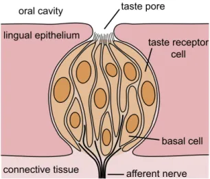
Special tastes like e.g. sour can’t only be tasted in one special area, every taste bud contains 50 - 100 taste cells representing everyone of the five taste sensations (sour, sweet, salty, bitter, umami).
The surface of a tongue is covered with papillae1. There are four different shapes a papilla can have: circumvallate, foliate, fungiform, filiform (what was not men- tioned in the lecture, as far as my notes say). Pictures of these shapes can be seen in figure 2.1.
In the invaginations between the papillae the taste buds are located (nice to be seen in figure 2.1 in the circumvallate and foliate papilla). Sensory hairs on the taste buds detect chemicals dissolved in saliva. Taste bud cells are replaced every 10 days. The number of taste buds differs between species; a chicken has 24, humans around 9000, pigs 1500.
2.1.1 Salty
The receptor that allows us to percept salty tastes is an ion channel through that Na+ ions directly enter the cell. The Na+ influx depolarises the cell and a voltage- dependent Ca2+ channel opens; according to measurements this must be a L-type Ca2+ channel. It is called ENaC, epithelial Na+ channel.
1The term papilla (plural: papillae) generally means a nipple-like structure (from Wikipedia, The Free Encyclopedia)
Figure 2.1: Faithful reproductions of lithograph plates from the 20th U.S. edition of "‘Henry Gray’s Anatomy of the Human Body"’, 1918. In the upper left panel you see the plate titelled "‘Vertical section of papilla foliata of the rabbit, passing across the folia. (Ranvier.)"’, the upper right shows a filiform papilla, the lower left panel was captioned "‘Circumvallate papilla in vertical section, showing arrangement of the taste-buds and nerves."’, and the lower right one was labelled "‘Section of a fungiform papilla. Magnified."’.
Figure 2.2: Schematic drawing of a taste bud. From Wikipedia, The Free Encyclo- pedia
Good to know:
The epithelial sodium channel (short: ENaC, also: sodium channel non-neuronal 1 (SCNN1) or amiloride sensitive sodium channel (ASSC)) is a membrane-bound ion-channel that is permeable for Li+-ions, protons and especially Na+-ions. It is a constitutively active ion-channel. It is arguably the most selective ion channel.
ENaC consists of three different subunits: α, β, γ. The stoichiometry of these su- bunits is still to be verified, but ENaC is very likely to be a heterotrimeric protein like the recently analyzed acid-sensing ion channel 1 (ASIC1), which belongs to the same family. Each of the subunits consists of two transmembrane helices and an extracellular loop. The amino- and carboxy-termini of all polypeptides are located in the cytosol.
ENaC is located in the apical membrane of polarized epithelial cells particularly in the kidney, the lung and the colon. It is involved in the transepithelial Na+ ion transport which it accomplishes together with the Na+/K+-ATPase.
It plays a major role in the Na+ and K+ ion homeostasis of blood, epithelia and extraepithelial fluids by resorption of Na+ ions. The activity of ENaC in colon and kidney is modulated by the mineralcorticoid aldosterone. It can be blocked by either triamterene or amiloride, which are used medically to serve as diuretics.
Wikipedia, The Free Encyclopedia (2009) The hormone aldosterone increases the number of salt receptors on the tongue.
The function of aldosterone is to maintain the normal sodium levels in the body:
increased Na+ sensitivity helps animals suffering from sodium deficiency to find Na+ containing food (often observed in cattle and deer).
Good to know:
Aldosterone is a hormone that causes the tubules of the kidneys to retain sodium and water. This increases the volume of fluid in the body, and drives blood pres- sure up. Many drugs, such as spironolactone, lower blood pressure by blocking the aldosterone receptor. Aldosterone is part of the renin-angiotensin system.
Aldosterone is a steroid hormone (mineralocorticoid family) produced by the outer- section (zona glomerulosa) of the adrenal cortex in the adrenal gland, and acts on the distal tubules and collecting ducts of the kidney to cause the conservation of sodium, secretion of potassium, increased water retention, and increased blood pressure. The overall effect of aldosterone is to increase reabsorption of ions and water in the kidney.
Its activity is reduced in Addison’s disease and increased in Conn syndrome.
It was first isolated by Simpson and Tait in 1953.
Wikipedia, The Free Encyclopedia (2009)
2.1.2 Sour
The sensation of sour is simply proton perception via a amiloride-sensitive Na+ channel and a H+-sensitive cation channel. The H+ ions block the K+ channels and hereby block depolarisation (is this correct? My notes are a bit strange here).
PKD2L1, polycystic kidney disease 2-like 1, is a human gene encoding the TRPP2 protein. This gene encodes a member of the polycystin protein family, TRPP2.
TRPP2 contains multiple transmembrane domains, and cytoplasmic N- and C- termini. TRPP2 may be an integral membrane protein involved in cell-cell/matrix interactions. TRPP2 functions as a calcium-regulated nonselective cation channel.
Alternative splice variants have been described but their full length sequences have not been determined. (From Wikipedia, The Free Encyclopedia, 2009)
This channel is gated by pH (pH = H+ ion concentration). It is found in circum- vallate and foliate taste buds.
2.1.3 Sweet
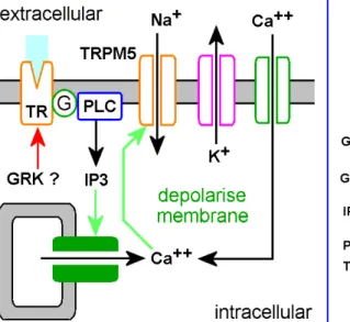
Sweet substances bind to G protein-coupled receptors, the taste receptors (TRs), at the cell surface. These receptors belong to a big group (over 10 different mem- bers) and have a huge N-terminal part. Natural sweeteners such as saccharides activate the GPCR, which releases gustducin. The gustducin then activates the mo- lecule adenylate cyclase, which is already inside the cell. This molecule increases concentration of the molecule cAMP, or adenosine 3’, 5’-cyclic monophosphate.
This protein will either directly or indirectly close potassium ion channels, lea- ding to depolarization and neurotransmitter release. (From Wikipedia, The Free Encyclopedia, 2009)
T1R2 and T1R3 form dimers. Only by those dimers sweet sensation is transduced (Charles Zuker, Univ. of California and San Diego, La Jolla).
2.1.4 Bitter
The sensation of bitter tastes also works over G protein-coupled receptors (gust- ducin), but over different pathways as sweet sensation. T1R2 is the receptor for bitter sensation. There are around 30 different T2R, many within one cell. T2R use the PLC cascade. cAMP acts to release Ca2+ ions from the ER.
Good to know:
There are many different classes of bitter compounds which can be chemically very different. It is interesting that the human body has evolved a very sophisticated sense for bitter substances: we can distinguish between the many radically different compounds which produce a generally "‘bitter"’ response. This may be because the sense of bitter taste is so important to survival, as ingesting a bitter compound may lead to injury or death.
Wikipedia, The Free Encyclopedia (2009) Bitter compounds act through structures in the taste cell walls called G protein- coupled receptors (GPCRs). Recently, a new group of GPCRs was discovered, known as the T2Rs, which it is thought respond to only bitter stimuli. When the bitter compound activates the GPCR, it in turn releases gustducin, the G pro- tein it was coupled to. Gustducin is made of three subunits. When it is activated by the GPCR, its subunits break apart and activate phosphodiesterase, a nearby
Figure 2.3: The fundamental sensing mechanism for sweet, bitter and umami is believed to involve GPCR receptors linked through phospholipase Cβ2 which hydrolyses phosphatidyl inositol to yield IP3 and diacylglycerol.
The IP3 releases internal Ca++stores which open TRPM5 channels, de- polarising the cells and stimulating neurotransmitter and neuropeptide release. Neuroendocrine cells similar to taste buds are present throu- ghout the GI tract, where they regulate the later stages of the digestive process as well as food intake through the mouth. Picture from Rozen- gurt & Sternini (2007) Taste receptor signaling in the mammalian gut.
Current Opinion in Pharmacology 7(6), 557-562.
enzyme, which in turn converts a precursor within the cell into a secondary mes- senger, which closes potassium ion channels. As well, this secondary messenger can stimulate the endoplasmic reticulum to release Ca2+, which contributes to de- polarization. This leads to a build-up of potassium ions in the cell, depolarization, and neurotransmitter release. It is also possible for some bitter tastants to interact directly with the G protein, because of a structural similarity to the relevant GPCR.
(From Wikipedia, The Free Encyclopedia, 2009)
2.1.5 Umami
It is thought that umami receptors act much the same way as bitter and sweet re- ceptors (they involve GPCRs), but not much is known about their specific function.
It is thought that the amino acid L-glutamate bonds to a type of GPCR known as a metabotropic glutamate receptor (mGluR4). This causes the G protein complex to activate a secondary receptor, which ultimately leads to neurotransmitter release.
The intermediate steps are not known (Wikipedia, The Free Encyclopedia, 2009).
The umami receptors response to salts of the glutamic acid (monosodium gluta- mate, MSG) which are often found in asian food, processed meats and cheeses.
T1R1 and T1R3 mediate the responses.
2.1.6 Signal transduction
Taste buds are the sensory endorgans for gustation. In mammals, taste buds com- prise a collection of ca. 50-100 elongate epithelial cells and a small number of proliferative basal cells. Ultrastructural studies reveal three distinct anatomical types of elongate taste cells within each taste bud: Type I, Type II and Type III.
Type I cells, sometimes called "‘dark cells"’, extend lamellate processes around other types of taste cells and express GLAST, a glial glutamate transporter. These features suggest a glial function for Type I cells, e.g. transmitter clearance and functional isolation of other taste cell types (Finger, 2005). Salty, ENaC (what could these two words mean?)
Type II taste cells have a characteristic large, round nucleus and express all of the elements of the taste transduction cascade for sweet, umami and bitter, including T1R or T2R families of taste receptors (for bitter, sweet and umami), the downs- tream transduction components, PLCβ2 and IP3R3, and gustducin. These taste cells thus are the transducing cells for these taste qualities (Finger, 2005). Ex- press taste receptors, TRPM5, PLC (bitter), cAMP (sweet, umami) pathways. Lack synapses with afferent nerve fibers, lack voltage-gated Ca2+ channels.
Type III cells are characterized by morphologically identifiable synaptic contacts with the gustatory nerve fibers and expression of the synaptic membrane protein SNAP25 as well as the neural cell adhesion molecule (NCAM). The presence of a
prominent synaptic contact implicates these cells in transmission of information to the nervous system (Finger, 2005). These cells have neuronal features.
A continuing question in the field is how the different types of elongate taste cells come to be replaced from the proliferative basal cell population. Recent studies entailing chimeric analysis in mice demonstrate that multiple lineages must exist within a taste bud, i.e that the three cell types are not merely different stages of development of a single taste cell type (Finger, 2005).
In humans, the sense of taste is conveyed via three of the twelve cranial nerves.
The facial nerve (VII) carries taste sensations from the anterior two thirds of the tongue, the glossopharyngeal nerve (IX) carries taste sensations from the posterior one third of the tongue while a branch of the vagus nerve (X) carries some taste sensations from the back of the oral cavity (Wikipedia, The Free Encyclopedia, 2009). All nerves terminate in the nucleus of the solitary tract.
2.2 Summary
• a single taste bud contains 50 - 100 taste cells representing all five taste sensations
• each taste cell has receptors on its apical surface
• taste receptors as ENaC are transmembrane proteins which admit Na+influx that gives rise to the sensation of salty; fungiform in type I (?????)
• taste receptors as PKD (?????)
3 BK and its physiologic relevance in the cardiovascular system - an
innovative target for drug design?
(Sausbier)
In mammals there are 75 different K channels. Small conductance K channels (they have a conductance of 10 - 60 pS) are only activated by intracellular Ca2+. The so called large conductance channels have a conductance of 100 - 300 pS.
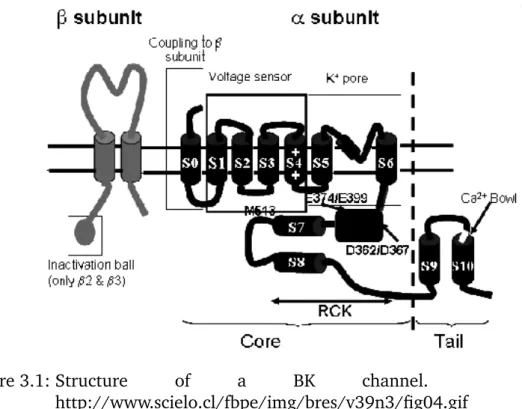
The BK channel, coded by KCNMA1, potassium large conductance calcium- activated channel, subfamily M, alpha member 1, is also called Maxi K channel or Slo(1).
KCa4.1 and KCa 4.2 were found in 2003, but the function is unknown until today.
The BKCa:
• consists of 4 pore formingαsubunits
• consists of 4 auxiliary β subunits (there are 4 different auxiliary subunits, but in the cardiovascular system there’s only β1); but BK doesn’t need the auxiliaryβsubunits for function!
• is the only known channel in the family activated by both, intracellular Ca2+
and membrane depolarisation
• has a large intracellular C-terminus, around 800 kDa; between S9 and S10 (see figure 3.1) the calcium bowl is located, which activates the opening propability, but the interaction mechanism with the pore remains unknown
3.1 The meaning of BK for vasoconstriction
Gq-coupled receptor → PLCβ → IP3 → IP3R1 →release of intracellular Ca2+ (e.
g. ER) → DAG (secondary messenger) → activation of TRP channels, type C
→ depolarisation of the membrane (local) → opens L-type Ca2+ → Ryanodine Receptor (sarcoplasmic reticulum, near membrane of smooth muscle cell)→Ca2+
spark (1 - 2 mM (normal concentration: 100µM)) → BK activation, K+ efflux → closing of L-type Ca2+ channel →negative feedback
Figure 3.1: Structure of a BK channel. From http://www.scielo.cl/fbpe/img/bres/v39n3/fig04.gif
Boldly printed steps increase the Ca2+ concentration in the cell
There exists a knockout mouse for the BK channel in which the pore forming S5 and S6 are deleted (see figure 3.1).
Good to know:
The Royal Swedish Academy of Sciences has decided to award the Nobel Prize in Chemistry for 2003 "‘for discoveries concerning channels in cell membranes"’, with one half of the prize to Peter Agre, Johns Hopkins University School of Medicine, Baltimore, USA, "‘for the discovery of water channels"’ and one half of the prize to Roderick MacKinnon, Howard Hughes Medical Institute, The Rockefeller University, New York, USA, "‘for structural and mechanistic studies of ion channels"’.
From the press release:
The other type of membrane channel which is the subject of this year’s Prize is the ion channel. Roderick MacKinnon surprised the whole research community when in 1998 he was able to determine the spatial structure of a potassium channel. Thanks to this contribution we can now "‘see"’ ions flowing through channels that can be opened and closed by different cellular signals.
The ion channels are important for, among other things, the function of the nervous system and the muscles. What is called the action potential of nerve cells is gene- rated when an ion channel on the surface of a nerve cell is opened by a chemical signal sent from an adjacent nerve cell, whereupon an electrical pulse is propagated along the surface of the nerve cell through the opening and closing of further ion channels in the course of a few milliseconds.
This year’s Prize illustrates how contemporary biochemistry reaches down to the atomic level in its quest to understand the fundamental processes of life.
The determinants of blood pressure are
• CNS (via the sympathicus)
• kidney (RAAS, renin-angiotensin-aldosterone system)
• heart (via power and frequency)
• blood vessels (via contractility)
In vitro blood pressure measurements in awake wildtype and knockout mice du- ring their circadian rhythm correlated to physical activity led to the following re- sults:
• knockout mice are sometimes less active so the measurements were only performed when the activity was comparable
• knockout mice have a higher blood pressure
• the heartbeat frequency of wildtype and knockout mice did not differ
The BK channel could be a good target for drug design. There are therapeutics against L-type Ca2+channels, but not yet against BK. Sildenafil increases the cGMP concentration and hereby acts against coronar problems and erectile dysfunction.
BK knockout mice have erectile dysfunction what means that the BK channel is linked to the NO-cGMP-pathway.
4 The vestibular system (Engel)
Our feeling of balance is not only influenced by the vestibular system, where the vestibular hair cells detect movements, but also by vision and our somatosensory sense. But a defect in the vestibular system causes heavy problems with balance.
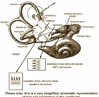
4.1 Structure of the vestibular system
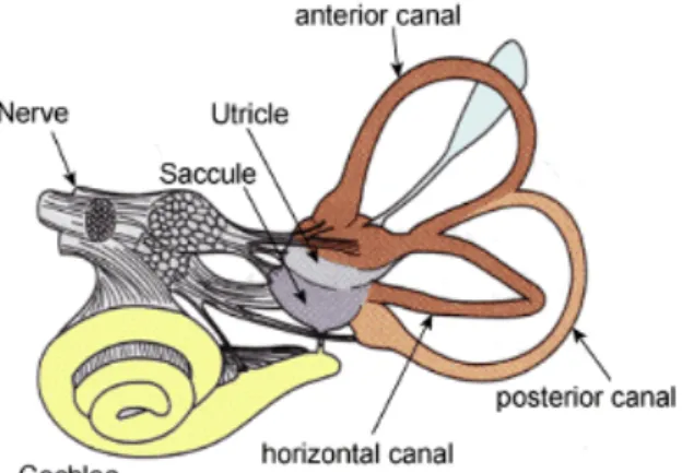
Figure 4.1: The labyrinth of the inner ear, from the left ear. It contains i) the cochlea (yellow), which is the peripheral organ of our auditory sys- tem; ii) the semicircular canals (brown), which transduce rotational movements; and iii) the otolithic organs (in the blue/purple pouches), which transduce linear accelerations. The light blue pouch is the en- dolymphatic sac, and contains only fluid. From Wikipedia, The Free Encyclopedia
The most obvious parts of the vestibular system are the three semicircular canals.
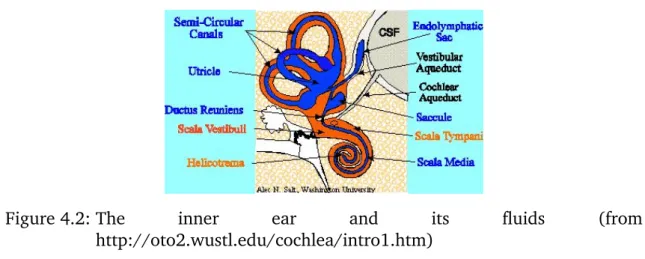
All of them, the interior, the posterior, and the horizontal semicircular canal, begin and end in the vestibulum. The semicircular canals are approximately orthogonal to each other. The two other parts are the utricle, that bears a huge spot of sensory and supporting cells, and the saccule, that also has a sensory epithelium. The vestibular system is filled with endolymphe as can bee seen in figure 4.2. The endocochlear potential in the vestibular system is a bit lower than in the scala media - perhaps that means that the potential is less important for the vestibular system than for hearing?
Figure 4.2: The inner ear and its fluids (from http://oto2.wustl.edu/cochlea/intro1.htm)
Here should be an explanation, how nystagmus is related to the vestibular system and its use in the clinical diagnosis. Anyone?
The vestibular system can be divided in two distinct components:
1. otolithic organs like saccule and utricle for detecting linear acceleration 2. cristae ampullaris in the semicircular canals for detecting angular accelera-
tions
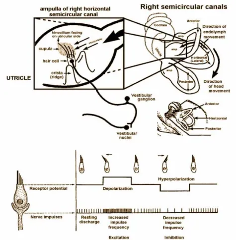
The cristae ampullaris are the places where rotational movements are perceived.
On a crista ampullaris vestibular hair cells are located (see figure 4.3). You can find afferent and efferent nerves connecting to the vestibular hair cells. Vestibular hair cells have, in contrast to cochlear hair cells, a true stereocilium, the so called kinocilium, and stereovilli (like cochlear hair cells). A gelly cupula embeds the sensory cells and is tethered to the roof of the semicircular canal, hereby partially blocking the semicircular canal. Between the cupula and the endolymphe there is no difference in density. The endolymphe is inert, angular deflection leads to deflection of the stereocilia and the kinocilium, effecting in hyperpolarisation or depolarisation (see figure 4.4). Deflections of the stereovilli leads to K+ influx and hereby activates a T-type Ca2+ channel.
Vestibular hair cells spike all the time, also without excitation. If the tip links are compressed because of the stereovilli being deflected in direction to the kinocilium, the hair cell is deactivated, the spiking rate decreases. If the tip links are stretched, the hair cell is activated and the spiking rate is increased.
The otolithic organs that make us sense linear acceleration consist of huge patches of sensory epithelium, one in the saccule, one in the utricle (see figure 4.5). The vestibular hair cells in these epithelia are covered by a gel-like structure, that is not tethered, but bears otoliths (other names: otoconia, statoliths), CaCO3 crystals, that are anyhow fixed to this gel-like structure. The turnover rate of this crystals is unknown, but they are renewed after some time. They make the gel-like structure heavy (ca. 3 g/cm3). The mechanical inertia of the otolith-loaded gel deflects the stereovilli and the kinocilium, the hereby generated vector from the vilii to the stereocilium makes us feel the direction of the linear acceleration. As you can see in figure 4.6, although there are only two spatial dimensions of the epithelium
Figure 4.3: Slide showing a crista ampullaris. From http://faculty.une.edu/com/abell/histo/histolab3b.htm
Figure 4.4: From http://www.neuroanatomy.wisc.edu/virtualbrain/BrainStem/13VNAN.html.
patches we can sense all different acceleration directions because the different hair cells in the patches are arranged in different directions.
Figure 4.5: From http://www.neuroanatomy.wisc.edu/virtualbrain/BrainStem/13VNAN.html In the vestibular system two different types of hair cells can be found, termed
type I and type II (see figure 4.7). Type II hair cells are more elongated and have afferent and efferent nerve endings. Type I hair cells are shaped more like a pear, the afferent synapse engulfes the whole hair cell as a calyx. Calyx-like synapses have advantages for the cell, but of course also disadvantages. (Examples?)
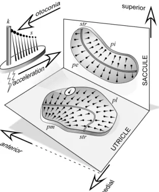
Figure 4.6: Orientation of the maculae of the utricle and saccule. In head coordi- nates, the saccular macula is in a vertical plane aligned anteroposterior.
The utricular macula is mostly horizontal plane but with a backward tilt. The fanlike alignments of the hair cells on the macula surface are depicted with the kinocilia (k) at the thick end and stereocilia (s) at the thin end. Each hair cell responds optimally to acceleration in the direction of its alignment, increasing firing rate when the stereo- cilia are deflected toward the kinocilium. Thus linear acceleration of the head from kinocilium toward stereocilia will cause an increase in discharge. A striola (str) divides each macula into two regions of rever- sed hair-cell polarity, so that when all afferents of a macula are excited or inhibited, the net signal will be determined by the relative weigh- ting of each region. pe, Pars externa; pi, pars interna; pl, pars lateralis;
pm, pars medialis. [Adapted from drawings by Spoendlin (93).]. From http://jap.physiology.org/cgi/content-nw/full/96/6/2301/FIG3.
Figure 4.7: The two different types of vestibular hair cells. From http://www.unmc.edu/physiology/Mann/mann9.html.
5 Senses III - Coding of sensory information (Knipper)
If we understand hearing, vision, taste, and smell, we understand the whole brain - because of the coding, because taste, hearing, and vision cells are nothing more than synapses. In Prof. Knipper’s opinion, transducing "‘outside"’ to "‘inside"’ in- formation was most complicated for evolution in hearing.
5.1 Afferent and efferent nerves
Inner hair cells have afferent (cochlear nucleus) and efferent (ipsilateral) nerves.
Efferents = axodendritic feedback (this feedback doesn’t exist in olfaction, if it’s there in taste is unknown, in vision it exists in horizontal and amakrine cells.
Always one type I afferent neuron (also called "‘radial fibers"’) contacts one IHC, but one IHC is contacted by 8 - 30 of these neurons simultaneously (8 in humans, ca. 36 in gerbils), so one cell has several connections what is a strange system.
These type I afferent neurons make up 85 - 90 % of the auditory nerve and carry information about heard sounds into the brain.
OHCs are contacted by type II afferent neurons (also called "‘outer spiral fibers"’) and contralateral directly at the base by efferent neurons. Each type II neuron contacts many OHCs, together they make up 5 - 15 % of the auditory nerve.
The most important group of efferent neurons consists of the medial olivocochlear (MOC) neurons that contact the OHCs. In a second group there are the lateral olivocochlear (LOC) neurons contactint the type I afferent neurons, not the IHCs themselves.
The spiral ganglion comprises afferent fibers type I (from IHCs) and afferent fi- bers type II (from OHCs). The spiral ganglion cells are bipolar cells (like e.g. in photoreceptors).
5.2 The coding of sound frequency
Sound is encoded according to its frequency via the travelling sound wave. The frequencies are encoded by their places in the cochlea (tonotopy). High frequen- cies have the best amplitude at the cochlear’s base, low frequencies at the apex.
This is due to the composition of the basilar membrane: at the base it’s narrow and tight, at the apex wide and loose. If you divide the human cochlea in three parts, you can estimate the frequencies as follows:
• high≈1 500 - 20 000 Hz
• medium≈600 - 1 500 Hz
• low≈600 - 200 Hz
The frequencies in which speech is taking place are between 250 - 8 000 Hz.
Consonants have high, vowels low frequencies.
IHCs are tuned to their so called characteristic frequency (CF) (also: best fre- quency, BF). This is the frequency the IHC reacts best to a sound at the lowest sound level. The CF is due to basilar membrane velocity which is influenced by the active amplifier of the OCHs. The transduction process of the IHCs is amplified upon depolarisation-induced motile cell responses of OHCs. The motile cell res- ponses enhance the resonance behaviour of the whole basilar membrane structure.
The OHC movement is due to Prestin, a sulphate anion transporter (SLC26A5), independent of the basilar and apical membrane. Intensities of frequencies are coded in distinct spiking patterns (the louder, the more).
For tones at best frequencies, basilar membrane velocity grows linearly at low sound levels, then it’s compressed because the cochlear amplifier gain is reduced.
If a patient suffers from OHC loss, his hearing his therefore impaired (see figure 5.1).
Figure 5.1: The influence of the cochlear amplifier (OHCs) on basilar membrane (BM) velocity in normal hearing and OHC loss.
When looking at figure 5.1 the question arises "‘How is loudness and frequency
> 5 kHz encoded?"’.
Humans have around 3 200 inner hair cells. These do the frequence coding by the following means. One of these means is the so called phase-locking, where the IHC fires one action potential per phase of a pure tone. Hereby the response pattern of the nerve fibres would reflect the frequency of the sound wave what means that there is a frequencey code. But this code only works for frequencies up to 2 000 Hz. Higher frequencies are coded by another mean, the combined response of fibres (see figure 5.2). But the combined response of fibres only
works up to 5 000 Hz. Over 5 .000 Hz there is the firing rate hypothesis, but the principle is not understood yet.
Figure 5.2: Principle of the combined response of fibres. The upper three lines show the signals created by single fibers, the lowest line shows the signal that is perceived in the brain.
5.3 The coding of loudness
Sound intensity, that means loudness, is differently encoded. If a sound stimulus is more intense (louder), more depolarisation takes place and the nerve fibres at the synapses fire at greater rates. This response increase in firing rate is linear over circa 20 dB, but then it’s saturated, the nerves can’t fire faster. So how can loudness be encoded at various intensities?
The sound levels between the threshold and the saturation are called thedynamic range. There exist AN (does that mean afferent neuron or auditory nerve?) fibre discharge rates for two theoretical fibres: a low threshold, high spontaneous rate fibre, and a high threshold, low spontaneous rate fibre (see figure 5.3). According to Liberman (1978) and Heinz and Young, 2004, fibres are sorted in three groups:
• high spike rate = > 18 spikes per second (ca. 60 %)
• medium spike rate = 0,5 - 18 spikes per second (ca. 30 %)
• low spike rate = < 0,5 spikes per second (ca. 10 %)
Humans have 3 200 IHCs and therefore 32 000 AN (afferent neurons?). 17 000 AN (afferent neurons?) (61 %) have a high spiking rate, and 4 500 (16 %) have a low spiking rate. A single fibre response is < 35 dB - a large dynamic range is needed to hear all loudnesses. The combination of all of our different fibre types encodes sound levels and hereby leads to loudness sensation.
Low spiking rate fibres are more sensitive to damage - this is another explanation for different loudness sensation in age-related hearing loss.
Figure 5.3: Auditory nerve (AN)-fiber discharge rates for 2 theoretical fibers, a low-threshold high spontaneous-rate fiber (light solid line) and a high- threshold low spontaneous-rate fiber (heavy solid line). The low thre- shold fiber is saturated at the sound level at which the BM becomes compressive (vertical dotted line), so its discharge rate does not reflect the compression. By contrast, the high-threshold fiber has a knee in its rate-level function at the threshold for BM compression. In the impai- red ear (dashed lines), the low-threshold fiber’s rate-level function is shifted without a change of slope, but the high-threshold fiber’s is stee- pened at suprathreshold levels. From Heinz and Young, 2004, figure 1 b.
6 Mast cell activation and
anaphylaxis is blunted in mice with targeted deletion of the SK4
channel (Ruth)
Good to know:
EntrezGene summary for KCNN4: The protein encoded by this gene is part of a potentially heterotetrameric voltage-independent potassium channel that is activa- ted by intracellular calcium. Activation is followed by membrane hyperpolarization, which promotes calcium influx. The encoded protein may be part of the predomi- nant calcium-activated potassium channel in T-lymphocytes. This gene is similar to other KCNN family potassium channel genes, but it differs enough to possibly be considered as part of a new subfamily.
National Center for Biotechnology Information, National Library of Medicine, National Institutes of Health, Bethesda, MD 20892-6510, USA Good to know:
UniProtKB/SwissProt function description of SK4: Forms a voltage-independent po- tassium channel that is activated by intracellular calcium. Activation is followed by membrane hyperpolarization which promotes calcium influx. The channel is blo- cked by clotrimazole and charybdotoxin but is insensitive to apamin.
UniProt Consortium
6.1 What is a SK4 channel?
The calcium-activated potassium channels (KCachannels) have been classified into 3 groups. Large conductance (BK) channels are gated by the concerted actions of internal calcium ions and membrane potential and have a unit conductance of 100 to 220 picoSiemens (pS). Intermediate conductance (IK) and small conductance (SK) channels are gated solely by internal calcium ions, with a unit conductance of 20 to 85 pS and 2 to 20 pS, respectively, and are more sensitive to calcium than are BK channels. Each type of channel shows a distinct pharmacology. KCNN4 belongs to a family of IK and SK channels (Ishii et al., 1997). The BK channel has an unit conductance of 250 pS, SK4 has 15 pS.
The official name of the SK4 channel is intermediate conductance calcium- activated potassium channel protein 1, it’s gene has the official symbol KCNN4
and is also known as IK1, SK4, KCa4, hSK4, IKCa1, or KCa3.1. SK4 is widely ex- pressed in blood cells, the function in the cochlea is not clear yet. What is known is that SK4 is not expressed in hair cells.
The SK4 channel consists of 6 transmembrane domains with the pore between number 5 and 6, both the N- and the C-terminus are intracellular. At the C- terminal part there is a calmodulin-binding domain (see figure 6.2).
Figure 6.1: Schematic representation of structure of the rat intermediate conduc- tance Ca2+-activated K+ channel (ImK). From Saito et al., 2002, fi- gure 2.
6.2 Mast cells and their role in allergic disorders
At the second contact of the body with the antigen the antigen molecules cross-link the Fc receptors on the surface of the mast cell. This induces cell activation of the mast cell via intracellular pathways causing the TRPC channels (transient receptor potential cation channels) to open and hereby evoking a Ca2+ influx (see figure 6.2). This allows degranulation and histamine release what causes vasodilata- tion, bronchoconstriction, oedema, tissue damage, and can end up in anaphylaxis, asthma bronchiale or worse.
6.3 The SK4 knockout mouse
At the lab of Prof. Ruth they are working on a conditional SK4 knockout mouse.
The constitutive knockout mouse is already available, it was made by a targeted deletion of the SK4 channel pore-coding exon. These knockout mice have nor- mal bone marrow-derived maste cell (BMMC) maturation, but the BMMCs lack SK4 currents on antigen presentation and have under standard conditions less K+ outflow. The normal antigen-mediated hyperpolarisation of BMMCs is absent in sentisized SK4−/− mice, also the BMMCs have a reduced Ca2+ entry in response to as well antigen as endothelin stimulation. The degranulation is also attenuated in SK4−/− mice. So mast cell hypoactivity is a result from SK4 knockout as is visible in figure 6.3.
Figure 6.2: The function of the SK4 channel in mast cells. Depicted in red is the increase in intracellular calcium concentration. The SK4 channel helps by potassium efflux and hereby depolarisation to increase the calcium influx through the TRPC channel.
Figure 6.3: SK4 takes part in mast cell activation by influencing the calcium influx in the cell. In SK4 knockout mice the mast cells lack the "accelerator".
In an experiment the acute anaphylactic response in wildtype (WT) and SK4−/−
(knockout, KO) mice was measured. Therefore the animals were "‘loaded"’ with IgE against DNP, 16 hours later they got contact with DNP. During the following anaphylactic shock the body temperature was measured (the body temperature can be an indicator for the severity of the anaphylactic shock). WT animals had a decrease in body temperature of about 3 to 3,5 °C whereas KO animals had only a decrease of 1,5 °C. This indicates that the KO mice had a less severe anaphylaxia.
Could SK4 blockers be a treatment for anaphylaxia?
7 Senses IV - Vision (Knipper)
The retina is a point-to-point visual system producting a reduced, inverted picture (see figure 7.1 and 7.2) of the parts of our world that we can see. We see light of the wavelength 370 - 740 nm. The quality of the light is altered by the lens.
The diameter of the pupil, that controls how many light reaches the retina, is also controlled by our agitation: The parasympathicus makes the circular muscle of the iris (the sphincter) contract so the pupil turns small, the sympathicus causes the radial muscle of the iris (the dilator) contract whereby the pupil grows bigger.
Figure 7.1: Optical layout of the eye. The image projected onto the retina is inver- ted due to the optics of the eye. From Wikipedia, 2009b.
7.1 Accommodation and its disorders
If we focus a picture, our eye accommodates (the procedure is called accommo- dation). The mechanism is not really clear until now, but this is the theory of Helmholtz: If the object we want to focus is close to us, the parasympathicus makes the ciliary muscle contract itself, the lense rounds up. If the object is more far away, the ciliary muscle relaxes and the lense flattens. We heard it like that in the lecture, but this theory is only one of three as you can read in Wikipedia, 2009a.
The most common disorders of accommodation are myopia (near sightedness), where the eye bulbus is too long, and presbyopia (far sightedness), where the eye bulbus is too short. Myopia requires a divergent (minus) lens, whereas hyperopia requires convergent (plus) lens. In Tübingen, Prof. Frank Schäffel does research on the length of eyes etc.
Figure 7.2: Scheme showing central connections of the optic nerves and optic tracts. From Wikipedia, 2009b.
Figure 7.3: Light from a single point of a distant object and light from a single point of a near object being brought to a focus by changing the curva- ture of the lens. From Wikipedia, 2009a.
Figure 7.4: Structure of the mammalian retina. From Benjamin Neunstöcklin.
7.2 The structure of the retina
The structure of the retina can be described as follows (cf. figure 7.4) from the most outer to the most inner part ("‘wrong"’ direction, most descriptions from Wikipedia):
Pigment epithelium At the back of the retina there is a pigment epithelium the light cannot pass.
Photoreceptors Before this epithelium, the secondary sensory cells, rods and cones, are located. Rods are for brightness detection, their image resolu- tion is not so high as in cones, they are highly sensitive to light (circa 8x more sensitive than cones) and used for night vision. They cannot discrimi- nate colours. Cones are for colour vision, need bright light to function, they build the fovea and have there a very good image resolution.
Horizontal cells Horizontal cells are the laterally interconnecting neurons in the outer plexiform layer of the retina of mammalian eyes. They help integrate and regulate the input from multiple photoreceptor cells. Among their func- tions, horizontal cells are responsible for allowing eyes to adjust to see well under both bright and dim light conditions.
Bipolar cells As a part of the retina, the bipolar cell exists between photoreceptors (rod cells and cone cells) and ganglion cells. They act, directly or indirectly, to transmit signals from the photoreceptors to the ganglion cells.
Amacrine cells Amacrine cells are responsible for 70 % of input to retinal gan- glion cells. Bipolar cells, which are responsible for the other 30 % of input to retinal ganglia, are regulated by amacrine cells.
Ganglion cells Ganglion cells receive visual information from photoreceptors via two intermediate neuron types: bipolar cells and amacrine cells. Retinal gan- glion cells collectively transmit visual information from the retina to several regions in the thalamus, hypothalamus, and mesencephalon, or midbrain.
Retinal ganglion cells vary significantly in terms of their size, connections, and responses to visual stimulation but they all share the defining property of having a long axon that extends into the brain. These axons form the optic nerve, optic chiasm, and optic tract.
Membrane First the light strikes the membrane that contains blood vessels and nerve fibres.
7.3 Photoreceptors and how they work
amount/eye temporal resolution speciality
rods 100 Mio 100 ms dark +++
cones 6 Mio 1 - 10 ms colour +++
In the outer part of the photoreceptors (see figure 7.5) lies the most important seg- ment for vision: the disks, ca. 1000 per cell, that contain the rhodopsin molecules (108per outer segment).
Good to remember:
Rhodopsin consists of the protein moiety opsin and a reversibly covalently bound cofactor, retinal. Opsin, a bundle of seven transmembrane helices, binds retinal, a photoreactive chromophore, in a central pocket. Retinal is produced in the retina from Vitamin A. Isomerization of 11-cis-retinal into all-trans-retinal by light induces a conformational change in opsin that activates the associated G protein and triggers a second messenger cascade.
Rhodopsin of the rods most strongly absorbs green-blue light and therefore appears reddish-purple, which is why it is also called "visual purple". It is responsible for monochromatic vision in the dark. Several closely related opsins exist that differ only in a few amino acids and in the wavelengths of light that they absorb most strongly. Humans have four different other opsins beside rhodopsin. The photopsins are found in the different types of the cone cells of the retina and are the basis of color vision. They have absorption maxima for yellowish-green (photopsin I), green (photopsin II), and bluish-violet (photopsin III) light. The remaining opsin (melanopsin) is found in photosensitive ganglion cells and absorbs blue light most strongly.
From Wikipedia, 2009g Seven transmemberane receptors (7TM receptors) like rhodopsin are metabotro- phic, not ionotrophic receptors (that means they are never ion channels them- selves).
One photon activates up to 3 000 rhodopsins to change their conformation. Each conformational change of 11-cis-retinal to all-trans-retinal activates one associated G protein, transducin. Each activated transductin molecule activates 2 000 PDE6 molecules that transform cGMP to GMP molecules. Alltogether this means a signal amplification factor of 1:6 000 000. The following little table summarises what happens in a photoreceptor cell:
Figure 7.5: Functional parts of the rods and cones. From http://en.wikipedia.org/wiki/Photoreceptor_cell.
dark PDE↓ cGMP↑ Na+ current↑ light PDE↑ cGMP↓ Na+ current↓
There is a phenomenon in photoreceptor cells called "‘noise"’, that means the flow of current independent of a signal. In cones this noise is much higher than in rods:
in cones the amplitude of the noise is 0,12 pA, in rods only 0,03 pA.
7.4 Colour vision disorders
Besides disorders of accommodation disorders of colour seeing are quite well known. In colour deficiencies deuteranopia (no green) and protanopia (no red) are the most important types. Abenefitof bein colour blind is the better protection from light damage over time (has anyone more about that? I don’t understand the reasons.)
7.5 Retinal circuitry
The physiology of vision is strange compared to hearing. If a photoreceptor cell detects a light signal it closes its Na+ channels, but the K+ channels stay open, what leads to a hyperpolarisation of -70 mV and reduced transmitter (glutamate) release. If a receptor doesn’t detect a signal both channels are open and the mem- brane has a potential of -30 mV.
Figure 7.6: The cellular organization of the foveal (right) and nonfoveal (left) re- tinas. Notice that higher density of cones, their one-to-one connection with bipolar cells, and the absence of ganglion and other cells in the foveal area. Rods, located only outside the fovea, converge in large numbers onto bipolar cells. Cones in the fovea are also smaller and more closely packed than in the nonfoveal retina. From Mann, 2008.
Retinal circuitry (see figure 7.6) is different in the fovea from the periphery. In the periphery the signals of many photoreceptors are sent to different bipolar cells that collect data from a lot of photoreceptors. In the fovea, where only cones can be found, each cone is connected to a single bipolar cell.
The retina, unlike a camera, does not simply send a picture to the brain. The retina spatially encodes (compresses) the image to fit the limited capacity of the optic nerve. Compression is necessary because there are 100 times more Photoreceptor cells than ganglion cells as mentioned above. The retina does so by "decorrelating"
the incoming images in a manner to be described below. These operations are carried out by the center surround structures as implemented by the bipolar and ganglion cells.
There are two types of center surround structures in the retina - on-centers and off- centers. On-centers have a positively weighted center and a negatively weighted surround. Off-centers are just the opposite. Positive weighting is more commonly known as excitatory and negative weighting is more commonly known as inhibi- tory (see figure 7.7).
These center surround structures are not physical in the sense that you cannot see them by staining samples of tissue and examining the retina’s anatomy. The center surround structures are logical (i.e., mathematically abstract) in the sense that they depend on the connection strengths between ganglion and bipolar cells.
It is believed that the connection strengths between cells is caused by the number and types of ion channels embedded in the synapses between the ganglion and bipolar cells. [...]
The center surround structures are mathematically equivalent to the edge detec- tion algorithms used by computer programmers to extract or enhance the edges in
Figure 7.7: On-centers and off-centers of the retina. From Wikipedia, 2009f.
a digital photograph. Thus the retina performs operations on the image to enhance the edges of objects within its visual field. For example, in a picture of a dog, a cat and a car, it is the edges of these objects that contain the most information. In order for higher functions in the brain (or in a computer for that matter) to extract and classify objects such as a dog and a cat, the retina is the first step to separating out the various objects within the scene. [...]
Once the image is spatially encoded by the center surround structures, the signal is sent out the optical nerve (via the axons of the ganglion cells) through the optic chiasm to the LGN (lateral geniculate nucleus). The exact function of the LGN is unknown at this time. The output of the LGN is then sent to the back of the brain.
Specifically the output of the LGN "radiates" out to the V1 Primary visual cortex.
(Cited from Wikipedia, 2009f)
To summarise these information: Light information is encoded by spiking patterns.
The dual response of the bipolar cells (on and off cells) allows coding of spatial information of light.
8 Forensic genetics (Blin)
"‘Forensic"’ means dealing with cases of crime. Next to forensic genetics there are also forensic anthropology, toxicology, serology and dactyloscopy (and propably even some more).
8.1 A little history of forensics
1880 Dr Henry Faulds published his first paper on the subject in the scientific journal Nature. Returning to the UK in 1886, he offered the concept to the Metropolitan Police in London but it was dismissed.
1892 Sir Francis Galton published a detailed statistical model of fingerprint ana- lysis and identification and encouraged its use in forensic science in his book Finger Prints.
1892 Juan Vucetich, an Argentine police officer who had been studying Galton pattern types for a year, made the first criminal fingerprint identification. He successfully proved Francisca Rojas guilty of murder after showing that the bloody fingerprint found at the crime scene was hers, and could only be hers.
1897 The world’s first Fingerprint Bureau opened in Calcutta (Kolkata), India af- ter the Council of the Governor General approved a committee report (on 12 June 1897) that fingerprints should be used for classification of criminal records. Working in the Calcutta Anthropometric Bureau (before it became the Fingerprint Bureau) were Azizul Haque and Hem Chandra Bose. Haque and Bose were the Indian fingerprint experts credited with primary deve- lopment of the fingerprint classification system eventually named after their supervisor, Sir Edward Richard Henry.
8.2 Identification of individuals
Individuals can be identified by
• morphology (e.g. the shape of the ear is purely genetically determined)
• classic serum diagnostics (AB0, MNS, Rhesus, Kelly, Duffy, Kidd, Lewi)
• isoenzyme polymorphisms
• HLA (human leucocyte antigen); 4 genes, totalling 61 alleles
• new tools: DNA tests for polymorphisms – RFLP (ca. 10µg DNA needed)
– hypervariable regions (VNTR) (Jeffreys et al., 1985)
– DNA profiling (testsoutside genes→no translucant humans) – DNA fingerprinting / profiling
* single-locus probes don’t produce individual-specific results
* even with five tests 1:1 000 is the same in relatives
To overcome the problems of DNA profiling, 16 different systems are used in fo- rensic genetics, this results in a 1 to4·109likeliness of match.
8.2.1 Technical limitations
DNA profiling doesn’t work if
• one component of a mixed probe is < 10 %
• the mixed probe contains > 3 parts
• DNA degradation results in fragments of < 100 bp
• the trace is < 100 pg DNA (that is ca. 12 cells)
8.2.2 Wrongful diagnosis
The following events can lead to wrong data in DNA profiling:
• misleading DNA patterns
• mix-up of samples
• genetic mosaicism
• blood donation
• bone marrow donation
• monozygotic twins
Due to these eventualities the estimated error rate is at 1:100 - 1:1 000
8.2.3 Quality controls
To ensure good data, each profiling lab has to undergo quality controls. Up to 1992 US courts disputed the DNA evidence because of its unreliability. So quality controls were established. They include
• blind proficiency tests
• allelic frequencies of different populations
• GLP (good laboratory practice)
• accepted statistical methods
8.3 The O.J. Simpson story
O.J. Simpson, a famous football player, was accused to have murdered his ex-wife and a friend of her. All DNA tests showed that the murderer was him, but the lawyers achieved that he was not senteced. They said the DNA tests were not reliable because they were done by white guys whereas O.J. Simpson was a black man. So he was set free (but at the end of 2008 they sent him to prison because of new facts).
8.4 Famous puzzles forensic genetic could help to solve
As forensic genetics can of course also be used for death bodies, there are some old puzzles that could be solved by this means.
Caspar Hauser There was a discussion and rumours going around that Caspar Hauser was a nobel offspring that had been hidden and then been killed because he was found. Forensic genetics could proof he was not the child of the supposed parents.
The Romanov family The last tsar and tsarina of Russia were killed after the Fe- bruary Revolution. The bodies were hidden, but 1991 some bodies were found near the place where the Romanov family had been placed under house arrest. Forensic genetics could show good results to proof that these were the Romanov family members.
Schiller The coffin containing Friedrich Schiller’s skeleton is in the Weimarer Fürs- tengruft. On 3 May 2008 it was announced that the DNA tests have shown that the skull of this skeleton is not Schiller’s.
Unfortunately the background radioation of around 10µGray degrades DNA in circa 1 million years so very old bodies cannot be analysed for resolving historic questions.
8.5 Other purposes for (forensic) genetics
Another very important possibilityfor us scientists is to find out if the cell line we use is really the one we think it would be ... (Azari et al., 2007)
9 Otosclerosis - new
pathophysiologic insights (Pfister)
Good to know:
Clinical description of Otosclerosis
Chronic conductive hearing loss (CHL) is the finding in almost all cases of otoscle- rosis (in fact should a person present with sensorineural hearing loss they would likely never be diagnosed with otosclerosis). This usually will begin in one ear but will eventually affect both ears with a variable course. On audiometry, the hearing loss is characteristically low-frequency, with higher frequencies being affected later.
Sensorineural hearing loss (SNHL) has also been noted in patients with otosclerosis;
this is usually a high-frequency loss, and usually manifests late in the disease.
Approximately 0.5 % of the population will eventually be diagnosed with otosclero- sis. Post mortem studies show that as many as 10 % of people may have otosclerotic lesions of their temporal bone, but apparently never had symptoms warranting a diagnosis. Whites are the most affected race, with the prevalence in the Black and Asian populations being much lower. In clinical practice otosclerosis is encountered about twice as frequently in females as in males, but this does not reflect the true sex ratio. When families are investigated it is found that the condition is only slightly commoner in women. Usually noticeable hearing loss begins at middle-age, but can start much sooner. The hearing loss often grows worse during pregnancy.
The disease can be considered to be heritable, but its penetrance and the degree of expression is so highly variable that it may be difficult to detect an inheritance pattern. Most of the implicated genes are transmitted in an autosomal dominant fashion.
From Wikipedia, 2009e Otosclerosis affects mostly women, in affected women the symptoms always wor- sen after the first pregnancy. It’s primary a metabolic bone disease and is inherited autosomal dominant with incomplete penetrance.
9.1 A very short history of otosclerosis
1704 Valsalva described stapes fixation
1890 Bacon says about otosclerosis that no surgery is to be considered at the mo- ment
9.2 Epidemiology
Otosclerosis is not equally distributed in mankind:
• caucasian: 10 %
• asian: 5 %
• african american: 1 %
• native american: 0 %
The most common age range of otosclerosis patients is 15 - 45 years. Histologi- cally otosclerosis is found to be equally distributed between both sexes, but clini- cally there are twice as many women affected as men. This could be due to the progression of otosclerosis during pregnancy.
9.3 Pathophysiology
Otosclerosis (osseous dyscrasia) is limited to the temporal bone and the ossicles.
The inciting event is unknown, the cause for the malformations are resorption and formation of new bone.
9.4 Pathology
There are two phases in otosclerosis. The first phase is the active (otospongiosis) phase, the second is the sclerotic phase. The most common sites for this disease are the round window, the fissula ante fenestrum, and diverse others (has anyone more?).
9.5 Treatment
There are two possibilities to treat otosclerosis patients. As otosclerosis is a conductive hearing loss, not a sensorineural one, hearing aids can help: They amplify the sound that much that it can be perceived despite the lack of amplifica- tion via the ossicles. This of course doesn’t help if the patients suffer not only from a conductive, but from a combined hearing loss.
The other possibility is a surgery where the incorrectly grown bone is removed. But the ossicles are the smallest bones in our body and the surgery is very complicated - there is the danger of becoming deaf in consequence of this surgery that was intended to restore the sense of hearing.
Another problem of the surgery is that is just corrects the symptoms, not the source of the disease. The bone can grow wrongly again and impaire the movement of the ossicles, there can arise a hyalinisation of the stria or there can even be an erosion into the inner ear.
9.6 Diagnosis
If a patient with hearing impairment has some of the following characteristics he or she could suffer from otosclerosis:
• women between 20 - 30 years of age
• conductive / mixed hearing loss
• often: tinnitus (the ear is damaged or you hear the pulse of the growing bone)
• family history of otosclerosis (this is the case in 2/3 of patients)
• vestibular symptoms (25 % of patients)
To substantiate this suspicion pure tone audiometry and computed tomography (CT) are used.
9.7 Otosclerosis loci
To find the otosclerosis loci different means were and are used. Two of these means are very important: linkage analysis and association analysis. Both methods are model-free analysis, the difference is the fact that you do linkage studies in families whereas association analysis isn’t dependent on relations between the patients.