Charged Dendrimers as Antimicrobial Coatings
for Biomaterials
Dissertation
zur Erlangung des Doktorgrades
der Naturwissenschaften (Dr. rer. nat.) der Fakultät IV
Chemie und Pharmazie der Universität Regensburg
vorgelegt von
Diplom-Chemikerin Verena Katzur aus Geretsried (geboren Evander, Südafrika)
2015
Die Daten zur Anfertigung der Dissertation wurden in der Zeit vom 10/2007 - 7/2010 an der Universität Regensburg akquiriert und im Zeitraum
8/2010 - 7/2015 innerhalb der Elternzeit zusammengefasst und ausgewertet
Kolloquiumstermin: 15. September 2015
Die Arbeit wurde angeleitet von: Priv. Doz. Dr. Rainer Müller
Prüfungsausschuß: Priv. Doz. Dr. Rainer Müller
Prof. Dr. Dipl.-Ing. Jürgen Geis-Gerstorfer Prof. Dr. Werner Kunz
Vorsitzender: Prof. Dr. David Díaz-Díaz
Beauty is the first test:
there is no permanent place in this world
for ugly mathematics
(Godfrey Harold Hardy)
Might this also apply to biochemistry?
Abstract
According to a survey conducted in October 2012 a third of all dental implants is affected by peri-implantitis ten years after insertion. Unfortunately, the impact of surface properties, such as charge, wettability and coating agility on protein adsorption and subsequent bio- logical events, like bacterial adhesion and biofilm formation, is still not fully unravelled. To date there are no bi-functional coatings that suppress the adhesion and growth of pathogenic organisms while simultaneously stimulating the integration of the implant into the surround- ing tissue. A coating combining both properties is believed to reduce the incidence of peri- implantitis at the trans-gingival passage. Therefore, this thesis focuses on the preparation and characterization of covalent surface modifications terminated by functional groups, ca- pable of generating charge in aqueous solution. The following functionalities were chosen for this purpose: the ammonium (-NH3+), the pyridinium (-N+C5H5), the sulfonate (-SO3–), and the carboxylate (-COO–) functionality. They were inserted in two diverse basic coatings.
On the one hand as a terminal group in the well known "self-assembled monolayer" (SAM) system based on linear alkyl silanes on silicon dioxide surfaces and on the other hand at the periphery of immobilised polyamidoamine (PAMAM) dendrimers. Non-ionizable methyl terminated coatings, poly(ethylene glycol) functionalized surfaces and highly hydrophilic oxidized SiO2 surfaces served as references. Dendrimers were chosen since they are an ef- ficient and omnipresent structural element in nature (see e.g. nerves). The functionalities dendritically arranged at a surface are believed to be of higher concentration and possess a higher degree of freedom in comparison to the functionalities anchored within the rigid SAM system. The immobilization of PAMAM onto the substrata was achieved by a thin siloxane coating terminated by carboxylic acid anhydride moieties.
The first part of this thesis focuses on the preparation and physico-chemical characteri- zation of the above mentioned coatings. Diffuse-reflectance infrared Fourier transformed spectroscopy (DRIFT), X-ray photoelectron spectroscopy (XPS) and a chemiluminescence based assay for the detection of terminal amino groups were performed to determine the preparation success of the surface modifications. The impact of the functional groups in their corresponding fixation on the electrokinetic potential was assessed by electrokinetic measurements. Furthermore, the wettability of the surfaces was determined by static water contact angle measurements.
The dendrimer backbone resulted in a higher or comparable wettability of the surface in com- parison to the fixation of the identical functionality via linear alkyl chains. At SAM-coatings a mainly pronounced negative electrokinetic potential (-30 mV to -120 mV) was detected in neutral aqueous solution, with the exception of the amino-group terminated SAM at which a value around zero was determined. In contrast, the novel coatings based upon dendrimers
dence of the pH of the surrounding solution at the SAM-coatings was observed to depend solely on the ionization behaviour of the surface confined functionalities, whereas the "effec- tive" ζ-potential-pH profiles at the PAMAM-coatings could only be explained considering film swelling and structural rearrangements. That the dendrimer maintained its flexibility upon immobilization was also supported by pilot sum frequency spectroscopy (SFG) mea- surements at the coating/air interface. No unambiguous correlation was recognized between wettability and charge.
The second part of this thesis investigates the influence of the surface modifications on protein adsorption from saliva and serum, as well as bacterial adhesion and viability ofStreptococcus gordonii DL1 and Staphylococcus aureus in the presence and absence of a pre-coat from the former mentioned physiological solutions. In addition the impact of the surfaces coatings on human MG-63 osteoblast attachment was inspected.
The PAMAM-coatings showed comparable or lower amounts of protein upon the surfaces after the exposure to human whole saliva in comparison to their linear analogues. Serum exposure resulted in an adverse effect, maintaining or supporting the adsorption of proteins on PAMAM-coatings in comparison to the alkyl chain based films. Both bacteria investi- gated preferred dendrimer based coatings terminated by cationic functionalities and avoided the settlement on surfaces functionalized by dendrimers with anionic or neutral periphery.
PAMAM-COOH is as effective and PAMAM-CH3 nearly as effective as the gold standard SAM-PEG in prohibiting bacterial attachment. This property was even slightly enforced after serum conditioning, whereas it was slightly reduced after saliva contact. In contrast all SAM coatings were colonized by both bacteria irrespective of the terminal functionality.
Regarding the vitality of the adhering bacteria, S. gordonii, was only influenced by pyri- dinium coated samples, whereas the bactericidal effect was stronger on PAMAM-Py than at the SAM-Py coated surfaces. This effect was lost after saliva pre-conditioning. The vitality of S. aureus was also reduced on pyridinium terminated coatings, but the most pronounced effect was achieved with PAMAM-CH3. After serum exposure this bactericidal property was maintained. The antimicrobial property of PAMAM-NH2, reported in literature, could not be preserved after tethering the molecule to the surface. Cell adhesion studies with human MG-63 osteoblasts showed comparable cell numbers on the dendrimer based coatings in comparison to the alkyl chain based coatings terminated by the same functionality, but after serum exposure higher cell numbers could be detected on the dendrimer based coatings in comparison to their linear analogues bearing the same functionality.
This study demonstrates, that a surface fixation of the terminal groups via a dendrimer possesses a higher potential of modulating the oral biological response, than the supplement
of the same functional groups via linear alkyl chains. Since PAMAM-CH3 revealed a gentle antimicrobial property towardsS. aureus and reduced the settlement of bacteria, PAMAM- CH3 might be promising in reducing peri-implantitis at the trans-gingival passage without minimizing the attachment of bone forming cells.
Keywords: Electrokinetic surface properties, ζ-potential, dendrimers, self-assembled mono- layers (SAM), protein adsorption, bacterial adhesion, biomaterials, anti-microbial coatings
Gemäß einer Umfrage vom Oktober 2012 sind ein Drittel aller dentalen Zahnimplantate zehn Jahre nach ihrem Einsatz von Periimplantitis betroffen. Leider ist der Einfluss von Ober- flächeneigenschaften, wie Ladung, Benetzbarkeit und Beweglichkeit auf Proteinadsorption und den darauf folgenden biologischen Ereignissen immer noch nicht vollständig geklärt. Bis heute gibt es keine bifunktionalen Beschichtungen, welche die bakterielle Adhäsion und de- ren Wachstum unterbinden, während sie die Integration des Implantats in das umgebende Gewebe unterstützen. Eine Beschichtung, welche beide Eigenschaften vereint, ist vermut- lich geeignet, das Auftreten einer Periimplantintis an der transgingivalen Durchtrittstelle zu vermindern. Deshalb konzentriert sich diese Arbeit auf die Präparation und Charakteri- sierung von kovalenten Oberflächenmodifikationen, terminiert durch funktionelle Gruppen, welche in der Lage sind in wässriger Lösung Ladungen auszubilden. Hierfür wurden folgen- de Funktionalitäten ausgewählt: die Ammonium- (-NH3+), die Pyridinium- (-N+C5H5), die Sulfonat- (-SO3–) und die Carboxylatgruppe (-COO–). Sie wurden in zwei unterschiedliche Basisbeschichtungen eingebracht. Zum einen als endständige Gruppen in das wohl bekannte
„self-assembled monolayer“ (SAM) System basierend auf linearen Alkylsilanen auf Silizi- umdioxid und zum Anderen an die Peripherie immobilisierter Polyamidoamin Dendrimere (PAMAM). Nicht ionizierbare methylterminierte Beschichtungen, Polyethylenglykol funk- tionalisierte Oberflächen und oxidierte Siliziumoberflächen dienten als Referenzoberflächen.
Dendrimere wurden deshalb gewählt, weil sie ein effizientes, häufig in der Natur verwendetes Strukturelement (siehe z.B. Nerven) sind. Zudem wurde bei einer dendritischen Anordnung sowohl eine höhere Oberflächenkonzentration als auch eine bessere Zugänglichkeit der Funk- tionalitäten erwartet als durch die Verankerung in einem starren SAM-System. Die Immobili- sierung von PAMAM wurde mittels einer dünnen Siloxanschicht mit terminalen Anhydriden erreicht.
Der erste Teil dieser Arbeit ist auf die Präparation und physikalisch-chemische Charakte- risierung der oben genannten Beschichtungen fokussiert. Infrarotspektroskopie (DRIFT) in diffuser Reflexion, Röntgen-Photoelektronen Spektroskopie (XPS) und ein Aminogruppen detektierender, auf Chemilumineszenz basierender Test wurden angewandt um die erfolgrei- che Darstellung der Oberflächenmodifikationen zu ermitteln. Der Einfluss der funktionellen Gruppen in ihrer jeweiligen Fixierung auf die effektive Oberflächenladung wurde mit Hilfe elektrokinetischer Messungen untersucht. Des Weiteren wurde die Benetzbarkeit der Ober- flächen durch statische Kontaktwinkelmessung mit Wasser erfasst.
Hinsichtlich Ihrer Benetzbarkeit zeigt sich eine PAMAM-Beschichtung in etwa gleich hy- drophil oder hydrophiler als die SAM-Beschichtung mit gleicher Funktionalität. Für alle SAM-Beschichtungen wurde in neutraler wässriger Lösung ein vorwiegend stark negativ aus-
geprägtes elektrokinetisches Potential (-30 mV bis -120 mV) detektiert, mit Ausnahme der mit Aminogruppen terminierten SAM-Beschichtung, deren Wert um Null lag. Im Gegen- satz dazu, zeichneten sich die neuen Beschichtungen, basierend auf Dendrimeren, durch eine moderate Oberflächenladung (-20 mV to 30 mV) bei pH 7 aus. Während dasζ-potential in Abhängigkeit vom pH der umgebenden Lösung an den SAM-Beschichtungen ausschließlich von dem Ionisierungsverhalten der gebundenen chemischen Funktionalitäten abhängt, konn- ten die „effektiven“ ζ-potential-pH Verläufe an den Dendrimerbeschichtungen nur unter der Annahme von Quellvorgängen und strukturellen Umlagerungen erklärt werden. Vorversuche mit Hilfe von Summenfrequenzspektroskopie (SFG) an der Beschichtungs/Luft Grenzfläche unterstützten die Annahme, dass das Dendrimer nach der Anbindung an die Oberfläche eine gewisse Flexibilität behält. Es konnte kein eindeutiger Zusammenhang zwischen Benetzbar- keit und Oberflächenladung hergestellt werden.
Der zweite Teil dieser Arbeit untersucht den Einfluss der Oberflächenmodifikationen auf die Proteinadsorption aus Speichel und Serum, als auch die Bakterienadhäsion und Bakteri- envitalität von Streptococcus gordonii DL1 und Staphylococcus aureus in Anwesenheit und Abwesenheit einer Präadsorptionsschicht aus den zuvor genannten physiologischen Lösun- gen. Zusätzlich wurde die Adhäsion von humanen MG-63 Osteoblasten untersucht.
Die PAMAM-Beschichtungen zeigten vergleichbare oder geringere Mengen an Protein nach Kontakt mit humanem Speichel im Vergleich zu deren linearen Analogen. Gegensätzliches wurde nach dem Kontakt mit Serum beobachtet, wo die Dendrimerbeschichtungen ver- gleichbare oder höhere Mengen an Protein adsorbierten als ihre auf linearen Alkylketten basierenden Filme. Beide in der Arbeit untersuchten Bakterientypen banden bevorzugt an kationisch terminierte PAMAM-Beschichtungen und vermieden die Besiedelung von Dendri- merbeschichtungen mit anionischer oder neutraler Peripherie. PAMAM-COOH zeigte sich als effektiv und PAMAM-CH3 ähnlich effektiv wie der Goldstandard SAM-PEG in der Un- terbindung von Bakterienadhäsion. Diese Eigenschaft wurde sogar leicht verstärkt nach Vor- konditionierung mit Serum, während sie nach Speichelkontakt leicht reduziert wurde. Im Gegensatz zu den PAMAM-Beschichtungen wurden alle SAM-Beschichtungen von beiden Bakterien unabhängig von der terminalen Gruppe besiedelt. Die Vitalität des adhärieren- den BakteriumsS. gordonii wurde nur von pyridiniumbeschichteten Oberflächen beeinflusst, während der antibakterielle Effekt auf PAMAM-Py stärker war als auf der SAM-Py Oberflä- che. Dieser Effekt wurde durch die Konditionierung mit Speichel aufgehoben. Die Vitalität von S. aureus wurde auch von den Pyrdinium-Beschichtungen reduziert, aber der stärkste Effekt wurde an der PAMAM-CH3Oberfläche erzielt. Nach Konditionierung mit Serum wur- de diese Eigenschaft beibehalten. Die antibakterielle Eigenschaft von PAMAM-NH2, gemäß Literaturangaben, konnte nach der Anbindung des Moleküls nicht beibehalten werden. Zell- studien mit humanen MG-63 Osteoblasten zeigten vergleichbare Zellzahlen auf den PAMAM
mit Serum konnten auf den PAMAM- Beschichtungen höhere Zellzahlen ermittelt werden als auf den analogen SAMs.
Diese Arbeit zeigt, dass die Fixierung der funktionalen Gruppen durch das Dendrimer ein höheres Potential besitzt, die orale biologische Antwort zu modellieren, als eine Bereitstel- lung der gleichen funktionellen Gruppe mittels einer linearen Alkylkette. Da PAMAM-CH3 einen leichten antibakteriellen Charakter bezüglichS. aureusaufweist und antiadhäsiv wirkt, scheint diese Beschichtung primär in der Lage zu sein, die Periimplantitis zu reduzieren, ohne die Anheftung von knochenbildenden Zellen entschieden zu vermindern.
Schlüsselwörter: Elektrokinetische Oberflächeneigenschaften,ζ-Potential, Dendrimere, Selbst- organisierende Monoschichten (SAM), Proteinadsorption, Bakterielle Adhäsion, Biomateria- lien, Antibakterielle Beschichtung
Preface
This PhD thesis is based on scientific research that was carried out at the department of Physical and Theoretical Chemistry at the University of Regensburg and is part of a com- prehensive study on novel biomaterial coatings based on dendrimers. Cooperating partners contributing to the investigation of this new coating type are the Department of Operative Dentistry and Periodontology at the University Hospital of Regensburg, the Section of Med- ical Materials and Technology at the Centre of Dentistry in Tübingen, and the Department of Oral Biology at the State University of New York Buffalo (NY, USA). It was due to the financial support of the Deutsche Forschungsgemeinschaft (DFG) that this project could be realized.
This work would not have been possible without the help and guidance of the following people:
First of all, I would like to thank Priv. Doz. Dr. Rainer Müller for encouraging and supervis- ing this thesis. Furthermore, for introducing me to the fascinating, comprehensive science of biomaterials. For constantly encouraging me during the progress of this work and benefiting from his expert knowledge in this field. And especially, for his patience!
Likewise, I would like to thank Prof. Dr. Werner Kunz for providing premises and instrumen- tation requisite for accomplishing this work. Short conversations were always path-breaking, entertaining and impressive.
Furthermore, my special thanks goes to the group of Prof. Dr. Motschmann and his staff, Dr. Christiane Stage und Dr. Peter Karageorgiev. They are an outstanding team!
I owe my gratitude to Prof. Dr. Geis-Gerstorfer and Dr. F. Rupp for their pleasant col- laboration and memorable meetings in Regensburg and Tübingen. I thank Prof. Dr. Dr.
Helmut Schweikl and Prof. Dr. Gottfried Schmalz for the cordial openness and introduction into dental science. Furthermore, I would like to thank Dr. Karl-Anton Hiller for the data management of the biological investigations.
In particular, I would like to thank Andreas Eidt and Dipl. Chem. Erika Deigele, for the pleasant teamwork. I owe the accomplishing of this work in particular to Mrs E. Deigele.
Her relentless request about the progress of this work and the unbroken connection to Re- gensburg helped me to never lay this work aside.
I owe the staff of the company Anton Paar (Austria), especially Dr. Alexandra Ewers,
providing me with the necessary knowledge to perform a few measurements on the Electro Kinetic Analyser (EKA - SurPASS) and even lending this instrument to me for a short while.
That was a very valuable contribution in the accomplishment of this thesis.
My sincere thanks goes to all students that supported me in my laboratory work, especially in the time of pregnancy. Alexandra Mattschas, Eva Maria Schöll, Tatjana Leibßle, Alexan- der Spengler, Matthias Knorn, Ulrike Vogl all distinguished by exemplary dedication.
There are no words which can adequately express my thanks to my husband, Michael Moschberger, and our kids, Konstantin und Cosima. If it was not for my husband, I could not have spent all those hours in front of the computer and above literature.
Unimaginable would have been the managing of this thesis, if I would not have been so lucky to have such dedicating parents and parents in law. Both performed outstanding in day caring for our children! Thank you so, so much! Furthermore, it is dear to my heart to express the sincere appreciation and gratitude to my parents, who supported and guided me in all aspects of my life.
Last but not least, I would like to thank my sister, Dipl.-Ing. Eva-Maria Katzur and my dearest friend, Dr. Eva-Maria Wagner, for their thorough proof-reading.
Publications
Surface-immobilized PAMAM-dendrimers modified with cationic or anionic ter- minal functions: physicochemical surface properties and conformational changes after application of liquid interface stress
Verena Katzur, Mirjam Eichler, Erika Deigele, Christiane Stage, Peter Karageorgiev, Jürgen Geis-Gerstorfer, Gottfried Schmalz, Stefan Ruhl, Frank Rupp, Rainer Müller
Journal of Colloid and Interface Science, Volume 366, Issue 1, Pages 179-190, 2012
The impact of dendrimer-grafted modifications to model silicon surfaces on pro- tein adsorption and bacterial adhesion
Mirjam Eichler, Verena Katzur, Lutz Scheideler, Michael Haupt, Jürgen Geis-Gerstorfer, Gottfried Schmalz, Stefan Ruhl, Rainer Müller und Frank Rupp
Biomaterials 32 (2011) 9168-9179
Influences of protein films on antibacterial or bacteria-repellent surface coatings in a model system using silicon wafers
Rainer Müller, Andreas Eidt, Karl-Anton Hiller, Verena Katzur, Michael Subat, Helmut Schweikl, Satoshi Imazato, Stefan Ruhl und Gottfried Schmalz
Biomaterials 30 (28):4921-9 (2009)
Conference Contributions
Surface-immobilized charged dendrimers - a new type of antiadhesive or antimi- crobial surface modification?
Rainer Müller, Verena Katzur, Mirjam Eichler, Helmut Schweikl, Karl-Anton Hiller, Got- tfried Schmalz, Stefan Ruhl, Lutz Scheideler, Jürgen Geis-Gerstorfer, Frank Rupp
Jahrestagung der Deutschen Gesellschaft für Biomaterialien, Gießen, Germany, 10.-12. November 2011
Human salivary protein adsorption onto chemically modified silica beads
Gregory White, Verena Katzur, Molakala Reddy, Rainer Müller, Robert Baier, Stefan Ruhl General Session of the International Association of Dental Research,
San Diego, USA, 16.-19. March 2011
Adhesion and Viability of Oral Bacteria on Charged Self-Assembled Monolayers Helmut Schweikl, Andreas Eidt, Karl-Anton Hiller, Gottfried Schmalz, Verena Katzur, Rainer Müller, Mirjam Eichler, Frank Rupp, Jürgen Geis-Gerstorfer, Stefan Ruhl
Tampere, Finnland, 11.-15. September 2010
Conditioning layer formation on charged dendrimers
Mirjam Eichler, Ingrid Stephan, Verena Katzur, Rainer Müller, Lutz Scheideler, Jürgen Geis-Gerstorfer, Frank Rupp
Jahrestagung der Deutschen Gesellschaft für Biomaterialien,
Tübingen, Germany, 8.-10. October 2009; BIOmaterialien 2009, 10, 105
Surface-functional groups of model substrates as determinant for protein adsorp- tion
Verena Katzur, Rainer Müller, Mirjam Eichler, Jürgen Geis-Gerstorfer, Frank Rupp, Karl- Anton Hiller, Helmut Schweikl, Gottfried Schmalz, Stefan Ruhl
International Association for Dental Research - Continental European Division, Munich, Germany, 9.-12. September 2009
Adhesion of Streptococcus Gordonii to positively and negatively charged self- assembled monolayers
Verena Katzur, Andreas Eidt, Mirjam Eichler, Karl-Anton Hiller, Gottfried Schmalz, Stefan Ruhl und Rainer Müller
3rd International Symposium of Interface Biology of Implants,
Warnemünde, Germany 13.-15. May 2009; BIOmaterialien 2009, 10(S1), 76
A systematic investigation of physico-chemical surface parameters influencing the adsorption of saliva proteins and the adherence of oral bacteria
Verena Katzur, Mirjam Eichler, Frank Rupp, Jürgen Geis-Gerstorfer, Karl-Anton Hiller, Helmut Schweikl, Gottfried Schmalz, Stefan Ruhl und Rainer Müller
Jahrestagung der Deutschen Gesellschaft für Biomaterialien,
Hamburg, Germany 20.-22. November 2008; BIOmaterialien 2008, 9, 203-204
Contents
I Introduction 1
II Fundamentals 6
1 Biomaterial Surfaces 7
1.1 Adsorption and Adhesion Processes at Biomaterials . . . 7
1.1.1 Protein Adsorption . . . 8
1.1.2 Bacterial Adhesion . . . 10
1.2 Current Antimicrobial Concepts . . . 13
1.2.1 Anti-Adhesive Coatings. . . 13
1.2.2 Antimicrobial-Releasing Coatings . . . 14
1.2.3 Contact-Killing Surface Coatings . . . 15
1.2.4 Tissue-Integrating Surface Coatings . . . 15
1.2.5 Auspicious Concept: Multifunctional coatings . . . 15
2 Surface Coatings based on Polyamidoamine (PAMAM) 17 2.1 Dendrimers . . . 17
2.1.1 Structural Features . . . 18
2.1.2 Dendrimer Synthesis . . . 19
2.1.3 PAMAM Properties. . . 21
2.2 Dendrimer based Antimicrobials . . . 25
2.3 PAMAM Immobilization Strategies . . . 27
3 Surface Coatings based on Linear Alkyl Chains (SAM) 29 3.1 SAM Component: Organosilanes . . . 29
3.2 SAM Preparation and Transformation . . . 30
3.3 SAM Formation Mechanism and Siloxane Structure . . . 32
4 Electrokinetic Surface Charge 33 4.1 Electrochemical Double Layer (EDL) Models . . . 34
4.2.1 Particle Surfaces: Electrophoresis . . . 38
4.2.2 Flat Surfaces: Streaming Current/Potential . . . 40
4.3 Soft Interphases and Hard Interfaces . . . 42
III Results and Discussion 43 5 Substrata 44 6 Coatings based on Linear Alkyl Chains (SAM) 48 6.1 Cationic SAMs . . . 48
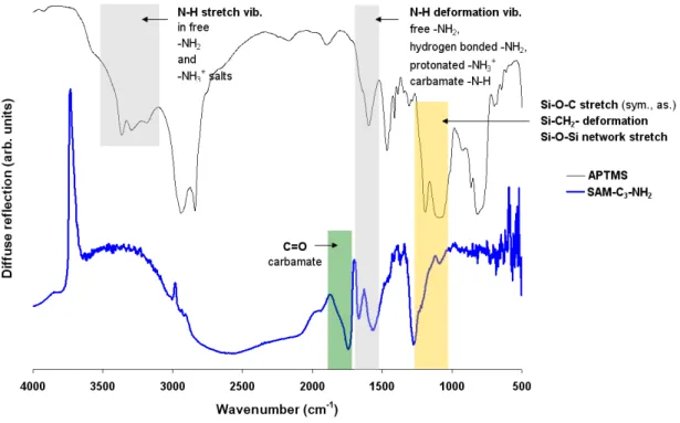
6.1.1 SAM-C3-NH2 . . . 48
6.1.2 SAM-C11-NH2 . . . 54
6.1.3 SAM-C11-N+C5H5 (=b SAM-Py) . . . 58
6.2 Anionic SAMs . . . 62
6.2.1 SAM-C6-COOH . . . 62
6.2.2 SAM-C11-SO3H . . . 67
6.3 Non-ionizable SAMs . . . 71
6.3.1 SAM-PEG . . . 71
6.3.2 SAM-C7-CH3 . . . 71
7 Coatings based on Dendrimers (PAMAM) 72 7.1 Immobilization of PAMAM-NH2 . . . 73
7.1.1 "Diluted" Carboxylic Acid Terminated SAMs . . . 74
7.1.2 Carboxylic Acid Anhydride Terminated Surfaces . . . 76
7.1.3 PAMAM-NH2 Coupling . . . 79
7.2 PAMAM with Cationic Periphery: PAMAM-N+C5H5 (=b PAMAM-Py) via PAMAM-Br . . . 86
7.3 PAMAM with Anionic Periphery . . . 90
7.3.1 PAMAM-COOH . . . 90
7.3.2 PAMAM-SO3H . . . 93
7.4 PAMAM with Non-Ionizable Periphery: PAMAM-CH3 . . . 95
8 Surface Charge Characteristics: Electrokinetic Potentials 97 8.1 Zeta-Potentials of SAM Coated Surfaces . . . 98
8.1.1 SAMs Terminated by Anionic Groups . . . 98
8.1.2 SAMs Terminated by Cationic Groups . . . 100
8.1.3 SAMs terminated by Non-Ionizable Groups. . . 103
8.2 Zeta-Potentials of PAMAM Coated Surfaces . . . 104
8.2.1 Comparison: PAMAM-NH2 and PAMAM-CH3 . . . 104
8.2.2 PAMAMs Terminated by Cationic Groups . . . 107
8.2.3 PAMAMs Terminated by Anionic Groups . . . 108
8.3 Charge Characteristics of S. gordonii DL1 and S. aureus . . . 110
9 Coating Characterization by Sum Frequency Generation Spectroscopy (SFG): A Pilot Experiment 113 10 Biological Response to Modified Surfaces 116 10.1 Protein Adsorption from Saliva and Serum . . . 116
10.2 Adhesion ofS. gordonii DL1 and S. aureus . . . 120
10.3 Vitality of S. gordonii DL1 and S. aureus . . . 123
10.4 Adhesion of Eukaryotic Cells . . . 125
IV Conclusion and Perspective 127 V Experimental Part 134 11 Materials 135 11.1 Substrata . . . 135
11.2 Chemicals and Solvents . . . 136
12 Surface Functionalizations 138 12.1 Substrata Cleaning and Oxidation . . . 138
12.2 Silanisation . . . 141
12.3 Self-Assembled Monolayer (SAM-X) Preparation . . . 147
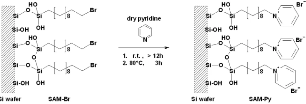
12.3.1 SAM-NH2 including SAM-Br and SAM-N3 . . . 147
12.3.2 SAM-Py . . . 149
12.3.3 SAM-COOH including SAM-CH=CH2 . . . 149
12.3.4 SAM-SO3H including SAM-SCOCH3 . . . 149
12.3.5 SAM-PEG (reference) . . . 150
12.3.6 SAM-CH3 (reference) . . . 150
12.4 Immobilization of PAMAM-NH2 . . . 151
12.4.1 via Carboxylic Acid Groups . . . 152
12.4.2 via Anhydride Groups . . . 153
12.5 Post-Modifications of tethered Polyamidoamine (PAMAM-X). . . 154
12.5.1 PAMAM-Py including PAMAM-Br . . . 154
12.5.3 PAMAM-SO3H . . . 155
12.5.4 PAMAM-CH3 . . . 155
13 Surface Characterization Methods 156 13.1 Electrokinetic measurements . . . 156
13.1.1 Streaming Current - Flat Surfaces . . . 156
13.1.2 Electrophoresis - Particles . . . 158
13.2 X-ray Photoelectron Spectroscopy (XPS) . . . 158
13.3 IR-Spectroscopy . . . 159
13.4 Contact Angle Measurement - Wettability . . . 159
13.5 Atomic Force Microscopy (AFM) . . . 159
13.6 Chemiluminescence Based Assay: Amino Group Detection on Flat Surfaces . 160 13.7 Kaiser Test: Amino Group Detection on Silica . . . 160
13.8 Sum Frequency Generation (SFG) Spectroscopy . . . 161
14 Testing Biological Response 162 14.1 Protein Adsorption - Bicinchoninic Acid (BCA) . . . 162
14.2 Bacterial Response . . . 163
14.2.1 Bacterial Adhesion . . . 163
14.2.2 Bacterial Viability . . . 164
VI Annex 166 Preparation & Characterization: SAM-N+Me3/PAMAM-N+Me3 . . . 167
List of Figures. . . 170
List of Tables . . . 178
Bibliography . . . 193
Declaration . . . 194
Part I
Introduction
Bacterial attachment to surfaces is an ubiquitous phenomenon [1]. In the field of medicine, microbial adhesion of pathogenic germs on implants leads to biomaterial-associated infec- tions (BAI). A BAI is a dreaded incidence after implantation because a biofilm formed by colonising bacteria is difficult to eradicate by antibiotics, antimicrobials or the hosts immune system [1][2]. This is due to the dense structure [2], a small accessible surface area, and a re- duced metabolic activity of the biofilm [3]. Has this inauspicious case occurred, a removal of the biomaterial is often inevitable and a secondary surgery indispensable [1][2][4]. According to a recent survey at least a third of all dental implants are affected by peri-implantitis (see Fig.: 0.1) 10 years after implantation (Dr. R. J. Meissen [updated: 31.10.2012, accessed:
17.03.2013]www.zp-aktuell.de).
Figure 0.1: Peri-implantitisat the gingival sulcus
PD Dr. Sönke Harder, Praxisklinik für Zahnmedizin und Implantologie, Munichc
At present biomaterial research is endeavoured to create surfaces that suppress the adhesion and growth of pathogenic organisms and simultaneously stimulate the integration of implants into the surrounding tissue, especially for those that are intended to remain in the body for long durations, as in the case of dental implants. Up to now, numerous systems have been invented and investigated for the application on implants, such as coatings with non-adhesive character, coatings with the ability to release antibiotics, coatings impregnated with silver, surface modifications bearing bactericidal components to kill bacteria on contact, and sur- faces facilitated by protein sequences to promote host cell adhesion [3]. Unfortunately, none of the mentioned strategies combines antimicrobial and cell integration properties.
Many antimicrobial substances possess cationic structural elements as for example the well known cetylpyridinium chloride (CPC), a cationic quaternary ammonium compound present in plenty products for dental hygiene. Positive charges are known to destabilize and disrupt the in netto negatively charged cell surface of bacteria. Regarding cell adhesion, it was found that charged surfaces promote the adhesion of bone-forming cells in comparison to uncharged
I. Introduction
surfaces [5]. Besides electrostatic interactions, multivalent interactions, a key concept in bio- recognition [6], play a significant role in biological processes. This PhD-thesis describes the preparation, characterization and evaluation of coatings combining the auspicious concepts of surface charge and multivalency as potential biomaterial coatings.
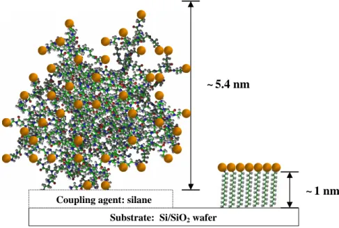
For this purpose smooth silicon wafers were covalently functionalised with dendrimeric struc- tures terminated by common ionic organic groups (X) of different structure and polarity ca- pable of generating charge in an aqueous medium at the physiological pH of 7.4. Additionally, silicon wafers were functionalised by self-assembled monolayers (SAMs) of unbranched alkyl chain siloxanes comprising the same functional groups as the dendrimer based coatings. The following picture (view Fig.:0.2) illustrates the concept this thesis focuses on.
~ 1 nm
~5.4 nm
Substrate: Si/SiO2 wafer Coupling agent: silane
Figure 0.2: Schematic representation of the two types of surface modifications investigated within this thesis. The coating on the right hand side represents the thin, stiff monolayer functionalisation based on self assembled alkyl chains, whereas the coating on the left hand side represents the rather thick, flexible monolayer functionalisation based on dendrimers. The orange spheres symbolise the positions of terminal groups. For a better overview only 46 terminal groups are indicated at the dendrimer, whereas 128 are present.
The coating on the right hand side represents the thin, rigid, highly ordered monolayer functionalisation based on SAMs. From the perspective of an approaching molecule SAMs are a monofunctional flat surface. In contrast, the coating on the left hand side represents the rather thick, flexible monolayer functionalisation based on dendrimers. Dendrimers are globular in shape and thus approaching molecules face a multifunctional, wavy surface. An increase in surface charge was expected by expanding the coating dimensions from a two di- mensional distribution of charge carriers to a three dimensional distribution. A dendrimeric
molecule was chosen to provide the centres of charge because the terminal functionalities within a dendrimer are much closer to each other than in linear polymers. Furthermore, dendrimers possess a well-defined structure [7] ideal to study surface structure-reactivity relationships.
The dendrimer chosen for this project is polyamidoamine (PAMAM-NH2) of the fifth gen- eration (G5). Polyamidoamine contains numerous amide bonds just like a protein and is of comparable size to natural proteins. Moreover polyamidoamine has shown antimicrobial properties in solution, as well as anti-inflammatory activity [8]. The terminal groups sym- bolised by orange balls in Fig. 0.2 and selected for this study are ammonium (-NH3+), and pyridinium (-N+C5H5) moieties to generate positive surface charge. Negatively charged sur- faces were aimed at by introducing carboxylate (-COO–) and sulfonate (-SO3–) moieties.
Plain oxidized unmodified surfaces of silicon dioxide (-Si−O–), methyl terminated coatings (-CH3) and polyethyleneoxide (-PEG) coatings were chosen as references.
It is only possible to correlate observed biological responses to physico-chemical surface prop- erties if the surface characteristics are analysed as precisely and comprehensively as possible.
An IR-spectrometer with a diffuse reflectance accessory recording surface modifications on silica particles, X-ray photoelectron spectroscopy (XPS) and wettability measurements were carried out on functionalised silicon wafers, and a chemiluminescence based assay detecting free amino groups was used to verify the successful preparation of the surface coatings. Intro- ducing IR-Vis sum frequency spectroscopy measurements were performed on some coatings revealing molecular orientations at the solid/air interface. Information on the charge forma- tion processes at the solid/liquid interface was gained by electrokinetic measurements.
In order to evaluate the applicability of these ultra-thin films for biomaterial purposes the impact of the surface modifications on microbial adhesion, microbial vitality and cell attach- ment were examined considering protein adsorption (determined by a colorimetric assay) readily occurring from surrounding body fluids (serum or saliva) after implantation. The conditioning film, resulting from serum or saliva, mediates between the biomaterial surface and the physiological environment. Its constitution depends on the functionalisation of the substratum, and is thus depended on the surface charge, and influences the adhesion of cells and bacteria as well as their further destiny. Therefore, only a surface coating maintaining antimicrobial and cell integration properties after the exposure to physiological solutions may be regarded as a promising biomaterial coating for implants. Streptococcus gordonii DL1 an early colonizer of oral surfaces, and Staphylococcus aureus a common pathogenic bacteria very often responsible for infections at metal implants were chosen for the biological investigations.
I. Introduction
This PhD thesis was performed at the Institute of Physical and Theoretical Chemistry at the University of Regensburg (Germany) and was part of a cooperation project with the Depart- ment of Operative Dentistry and Periodontology at the University Hospital of Regensburg (Germany), the Section of Medical Materials and Technology at the Center of Dentistry in Tübingen (Germany) and the Department of Oral Biology at the State University of New York (Buffalo). It was a further task of this PhD thesis to develop coating strategies for nu- merous silicon-based substrata of different geometry and stability (quartz crystal resonators, glass slides, silica spheres, silica beads, silicon dioxide particles, double-side polished silicon wafers and single-side polished silicon wafers) and supply all departments with sufficient numbers of model surfaces. The substratum diversity was necessary because of the numer- ous analytical methods that were applied in this project by the participating departments:
Static protein adsorption and bacterial adhesion data acquired by A. Eidt (MTA) under the supervision of Prof. Dr. S. Ruhl (Department of Oral Biology at the State University of New York, Buffalo, USA) at the Department of Operative Dentistry and Periodontology at the University Hospital of Regensburg (Germany) are included in this thesis. Furthermore, cell adhesion experiments performed by C. Waha (MTA) under the supervision of Prof. Dr. Dr.
H. Schweikl at the Department of Operative Dentistry and Peridontology at the University Hospital of Regensburg (Germany) are mentioned here to complete the biomaterial testing.
Dynamic protein adsorption and bacteria adhesion studies, as well as surface energy calcu- lations were performed at the Section of Medical Materials and Technology at the Center of Dentistry in Tübingen (Germany) by Msc M. Eichler in the course of her PhD thesis under the supervision of Dr. F. Rupp and Prof. Dr. Geis-Gerstorfer utilizing quartz crystal microbalance and dynamic contact angle analysis. Static and dynamic examinations were carried out because biofilm formation may occur supra-gingivally and sub-gingivally in the oral cavity. The former surfaces are exposed to shear forces, whereas the later are not.
Even though various processes and factors that influence adhesion have been unravelled, no generally valid theory exists to explain the complex process of interactions between bacteria, cells, proteins and surfaces. An improved knowledge of the interactions between biomaterial surface, the mediating protein layer and the subsequent adherence of cells and/or bacte- ria is essential for the development of biomaterial coatings or functionalized surfaces with properties optimized for their field of application.
Part II
Fundamentals
1 Biomaterial Surfaces
1.1 Adsorption and Adhesion Processes at Biomaterials
Within seconds after the insertion of an artificial object into a physiological fluid (e.g. body fluids, such as blood or saliva) protein adsorption will occur. The layer formed by the ad- sorbed proteins is referred to as a conditioning film (within the oral cavity the conditioning film caused by saliva is termed "pellicle"). Only later, micro-organisms and cells reach the biomaterial. They face a surface covered by proteins upon their arrival instead of the orig- inally implanted surface. The conditioning film mediates between the substratum and the surrounding fluid and controls proceeding reactions, thus making it extremely relevant in the design of biomaterials. [9]
The physico-chemical properties of a substratum surface and the composition of the contact- ing physiological fluid provoke conditioning films of different protein composition, packing, density and/or configuration [9]. Whereas the composition and properties of the biofluid and the micro-organisms/biomolecules are pre-given, surface characteristics can be altered using surface modification strategies. Surface modifications of varying chemical structure and composition influence the physico-chemical surface characteristics such as hydrophobic- ity, surface charge, and topography, addressing only a few of them [10]. These factors effect protein adsorption and eventually the pace and composition of the proceeding biofilm or cell-attachment. Therefore, changes in physico-chemical substratum parameters influence biofilm formation and tissue adhesion indirectly.
The complex composition of physiological fluids, containing numerous different proteins and highly adaptive bacteria [4], changing their physico-chemical properties depending on envi- ronmental conditions, nutritional status and their state of growth [11], make both, protein adsorption and subsequent cell/bacteria adhesion, a very complex process. Furthermore, biomechanical conditions determined by the location of the process are also of importance, i.e. if the centre of action is exposed to hydrodynamic shear or not.
Initial protein adsorption and bacterial adhesion was attempted to be described by the the- ories applied in colloid and interface chemistry, neglecting metabolic processes, growth or
conformational rearrangements [12]. Therein, initial adhesion/adsorption is described to be governed by a combination of basic forces. These forces are, besides the always present attractive van der Waals forces, electrostatic interactions, hydrogen bonding, and the Brow- nian motion. Further forces are direct or indirect consequences of the former mentioned basic forces. Two physico-chemical approaches are mainly mentioned in literature: surface thermodynamics [13] and the extended-Derjaguin-Landau-Verwey-Overbeek (x-DLVO) the- ory. [1][14][9]
Thethermodynamic approach describes adsorption as a process of minimizing the Gibbs energy of a system. The surface free energies (SFE) of the substratum and the bacterium are determined by wettability measurements. The energy between the bacterium and the surface is calculated based on the assumption that the interfaces bacterium/water and surface/water are replaced by a surface/bacterium interface. Critical is the use of thermodynamic equilib- rium equations in a mainly irreversible process. Furthermore, no conformational changes, appendages at bacteria, or inhomogeneous bacterial envelops are taken into account. Up to now, no universal relationship between interfacial free energies and adhesion/adsorption has been found. [15][9]
The x-DLVO theory describes the interaction energy as function of the distance between the colloidal particle (here, the approaching micro-organism or proteins) and the surface [1].
The Gibbs energy is a sum of van der Waals forces, electrostatic forces and short range Lewis acid-base (i.e., hydrogen bridges) interaction energies [15].
The following two subsections will give a small impression on the topics protein adsorption and bacterial adhesion. The section thereafter addresses methods already known for a very long time, and new approaches in current research, to prohibit the formation of biofilms on surfaces.
1.1.1 Protein Adsorption
Commonly proteins accumulate at solid/liquid interfaces. A proteophobic behaviour is ob- served in only very few cases and will be addressed in section1.2.
Proteins are biopolymers composed of amino acids with hydrophobic and hydrophilic side chains. Hydrophilic side chains can be of ionisable or non-ionisable chemical moieties. Be- cause ionisable moieties are able to form positive and negative charges, proteins can be referred to as polyampholytes [16]. Proteins are folded in such a way that the hydrophilic side chains interact with water, whereas most of the hydrophobic side chains are hidden within the biomolecule to prevent water contact [17].
IIFundamentals
Brownian motion (i.e., diffusion) and liquid flow (i.e., convection) transport proteins towards a surface. Long range (attractive van der Waals forces > 50 nm, and repulsive non-specific electrostatic interactions about 10 - 20 nm) and short range forces (specific electrostatic inter- actions 2 - 10 nm, displacement of water by hydrophobic groups 0.5 nm, specific interactions
< 1 nm) govern primary adhesion that is, theoretically, reversible [9]. Subsequent confor- mational changes, optimizing interactions with the substratum [18], and the attachment to the surfaces via several segments make protein adsorption an almost irreversible process [16]. Protein removal can only be achieved utilizing detergents or by inducing changes in pH and/or ionic strength. Even diluting the adjacent solution does not lead to protein des- orption. However, a replacement of proteins with higher mobility, by proteins with limited mobility but higher affinity for the surface may still change the protein composition upon a surface ("Vroman effect" [16]) with time.
W. Norde [16] studies protein adsorption onto surfaces already since the early 1970s and has acquired a comprehensive knowledge in this field. He divides proteins into two classes:
structurally labile proteins and structurally stable proteins (meaning their ability to remain in their native state) and refers to them as "soft" (e.g. α-lactalbumin) and "hard" (e.g.
lysozyme) proteins. Upon adsorption hard proteins undergo limited structural changes, whereas soft proteins undergo larger structural changes. He published a table with predic- tions regarding protein adsorption onto charged surfaces of different wettability, that all his experiments were in line with. A transcript of this table is presented below (Table 1.1).
He concludes that proteins of any type adsorb onto hydrophobic surfaces, irrespective of
Protein Surface
Hydrophobic Hydrophilic
+ - + -
Hard
+ 4 4 8 4
- 4 4 4 8
Soft
+ 4 4 4 4
- 4 4 4 4
Table 1.1: Scheme proposed by W. Norde to predict whether a protein adsorbs (4) to a surface or not (8) [16]; "+" and "-" signs refer to the netto charge of the protein and the surface, respectively.
the surface charge. The reason why all proteins adhere to hydrophobic surfaces (apolar), even if electrostatically unfavourable, maybe the dominant influence of the dehydration of the hydrophobic surface (i.e., liberating water molecules and increasing entropy). In some cases additional support is obtained by hydrophobic interactions between protein and surface
upon unfolding ("denaturation"). At polar (hydrophilic) surfaces hard proteins only attach, if they are electrostatically attracted, whereas soft proteins even adsorb if the surface is elec- trostatically hostile (i.e., same charge). The driving force for hard proteins to adsorb are the ionic interactions and the liberation of counter-ions. The driving force for soft proteins to accumulate at polar surfaces may originate from the gain of conformational entropy as the protein reorientates on the surface and looses secondary and tertiary structures (i.e., order).
This seems to outweigh unfavourable dehydration of hydrophilic surfaces and electrostatic repulsion. [16]
The observations and illustrations made by W. Norde et al. help to develop a slight un- derstanding on the nature of proteins and their behaviour upon surface approach. But, even though this classification is useful to explain the result of many investigations, it is not a generally valid principle. This is attributed to the heterogeneous character of proteins (each protein has its own "personality" [16]) and the multiple additional factors influencing adhesion.
1.1.2 Bacterial Adhesion
As already mentioned in the introduction (I) of this thesis, microbial surface contamination is a dreaded incidence in many fields of life, like food packaging, marine biofouling, surgical instruments, biomaterials, and many others. Great efforts have been undertaken, since many years, to design surface coatings, which prohibit bacterial attachment. Unfortunately, just like in the case of proteins mentioned in the previous subsection (1.1.1), little knowledge has been gained on the relationship between material surface characteristics, such as surface energy expressed by hydrophilicity/hydrophobicity, charge, polarity and topography, and bacterial adhesion. The mechanism of initial bacterial colonization is suggested to be de- pendent on both, physico-chemical (non-specific; dependent on the overall characteristics of the adhering species) and specific bio-chemical interactions (localized adhesion sites on cell surfaces) [10], just like in the case of proteins. Bacterial adhesion is strongly related to the type and amount of proteins upon the surface, which is dependent on the surface properties of the biomaterial. [19]
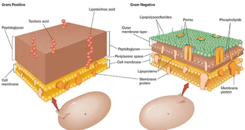
The size of a typical bacterial cell is 0.5 - 5.0 micrometers, whereas an average human cell is about 10 micrometers in size. Most common bacteria can be divided into two classes, namely gram-positive (e.g. Staphylococcus aureus) and gram-negative (e.g. Escherichia coli) bacte- ria. This classification is based on a staining technique, developed in 1884 by Hans Christian Gram, characterizing bacteria by structural characteristics of their cell wall. Gram-positive bacteria appear purple upon staining with crystal violet, whereas gram-negative bacteria
IIFundamentals
Figure 1.1: A scheme illustrating the arrangement of components at the envelopes of the two main classes of bacteria: gram-negative and gram-positive; cThe McGraw-Hill Companies, Inc. Permis- sion required for reproduction or display
do not retain the stain and are counter stained by safranin or fuchsine appearing red or pink under the microscope. Figure 1.1 illustrates the structural differences among the cell envelops of two main types of bacteria.
Gram-positive bacteria are surrounded by many layers of peptidoglycan (linear polysaccha- ride with a short peptide sequence) which are penetrated by lipoteichoic acids (glycerol phos- phate, ribitol phosphate). Peptidoglycan is found in prokaryotes only. The peptidoglycan is composed of N-acetyl glucosamine and N-acetyl muramic acid as alternating sugars. The glycan chains are cross-linked by peptide chains. It is worth mentioning that most enzymes in mammalian hosts are unable to degrade peptidoglycan with one important exception:
lysozyme. The overall charge of gram-positive bacteria is negative due to the phosphoryl groups in the teichoic and teichuronic acid residues. Carboxylic acid moieties along the te- ichuronic acid intensify the overall negative charge [20].
The cell envelope of gram-negative bacteria is thinner but of higher complexity. Gram- negative bacteria also possess a peptidoglycan layer but much thinner than gram-positive bacteria and not positioned at the outer envelope of the cell. The peptidoglycan sheet is surrounded by an additional layer composed of lipopolysaccharides (LPS), phospholipids (only confined on the inner side of the membrane), and porins. Phosphoryl and 2-keto-3- deoxyoctonate carboxylate groups control the charge of gram-negative bacteria [20]. There are no teichoic or lipoteichoic acids present.
The outer cell wall also possess binding sites for Ca2+ and Mg2+ ions stabilizing the structure
of the outer membrane. Due to the structural properties mentioned above, the cell surface of bacteria possess an overall negative electrostatic charge [20][21]. In contrast to bacteria, the cell membrane of human erythrocytes (representatives of normal mammalian cells) is mainly composed of zwitterionic phosphatidylcholine, phosphatidylethanolamine and sphingomyelin penetrated by sterols. Phosphatidyl serine is also present carrying a negative charge. The overall charge of mammalian cells is rather neutral to slightly acidic [22]. The higher nega- tive charge on the envelope of bacteria makes them more susceptible to be deteriorated by cationic antimicrobials than mammalian cells.
The net cell surface charge of bacteria can be determined by estimating their electrophoretic mobility in an electric field [20]. In the past, determined electrophoretic mobilities of bacteria were transferred into zeta-potentials using Smoluchowski formalism (for further information view subsection4.2.1). This is a very questionable method and has been dismissed by many authors, especially by J. Duval. He criticises the implementation of bacterial surface proper- ties into colloidal interaction theory because bacteria are no hard and impermeable particles.
They are heterogeneous, permeable and soft, forming rather an interphase to the surround- ing fluid than a well-defined interface. As such, the location of the slip plane at a bacterial interphase is ambiguous and he recommends the abundance of concepts like zeta-potential, surface potential and surface charge. He proposes a diffuse soft particle electrokinetic (DSPE) formalism for deriving interphasial properties. The DSPE model is based on a full numerical integration of the governing electrohydrodynamic equations of soft particles and allows the exploration of the effects of chemical heterogeneities and the physically diffuse character of the soft microbial interphase on electrophoretic motion. Interphasial characteristics ob- tained by using the DSPE formalism on microbial electrophoretic mobilities may shed light into correlations between electro-hydrodynamic characteristics of the microbial interphase and bacterial attachment. [23][16]
IIFundamentals
1.2 Current Antimicrobial Concepts
There are four main types of antimicrobial coating concepts currently used for biomaterials.
One concept is based on the anti-adhesive character of the surface modification, preventing micro-organisms, cells and bio-macromolecules from settling onto the surface. A further con- cept is based on coatings comprising immobilized antimicrobial substances killing bacteria on contact. Also coatings containing a reservoir of the biocidal molecules, referred to as antibiotic/antimicrobial release systems, are used for biomaterial implants. Surface modi- fications bearing tissue-integrating factors are also part of antimicrobial concepts because surfaces with adhering cells are not prone to microbial colonization. All four types have their advantages, disadvantages and application fields, which will be addressed shortly in this section. At the end of this section a rather new type of coating will be introduced.
1.2.1 Anti-Adhesive Coatings
The gold standard in anti-adhesive coatings is poly(ethylene glycol) (PEG). Also surfaces coated by oligo(ethylene glycol)n (n = 3 - 6) (OEG) groups possess anti-adhesive properties.
Even though studied for many years the mode of action has not been fully unravelled yet.
Organisms and proteins are said to be sterically repelled by the PEG chains, except for Pseudomonas aeruginosa strains [15]. PEG chains are of high agility and this is suppressed by the arrival of large molecules. A decrease in conformational freedom reduces entropy and is energetically unfavourable [24]. Upon compression the polymer concentration increases on the surface but water diffuses back into the PEG layer causing hydration and expansion of the chains. Therefore two main parameters counter compression and induce repulsion: the entropic reset force and the osmotic force. Both, the formation and the concentration of the ethylene glycol units and thus their ability in binding interfacial water are most probably decisive for the proteophobic character of a PEG-surface [25]. Chains containing a higher amount of ethylene oxide units were found to be more proteophobic than shorter chains.
The strong disadvantage of this surface modification is its susceptibility to auto-oxidation [26][27], thus having no long term stability.
Following the structure of PEG coatings, hydrophilic polymer chains end-grafted at high density onto surfaces seem currently the most promising strategy in resisting adherence [16].
This type of modification is referred to as polymer brush coatings because the chains stretch away from the surface into the adjacent medium [2][1][19]. Just like in PEG coatings, compression of the strongly hydrated polymer chains upon macromolecule/micro-organism approach increases the osmotic pressure and decreases the mobility (conformational entropy) of the polymer chains in the brush, causing repulsion of the approaching biomolecule [1].
Hydrophilic polymers like poly(N-vinyl pyrrolidone) (PVP), poly(oxazoline) and poly(N,N- dimethylacrylamide) also show reduced adhesion capacity. This is attributed to the fact that these surface coatings swell strongly in aqueous medium and the hydrogels formed causes unfavourable bacterial attachment conditions as the surface properties are similar to the water phase.
Comprehensive tests to shed light into the mechanism of protein resistant surfaces were con- ducted by Whitesides et al.. They tried to correlate protein resistance to molecular structure patterns and found the following molecular characteristics to be apparently essential to cre- ate protein resistant coatings : polar functional groups with hydrogen bond accepting groups (electronegative atom) but no hydrogen bond donating groups and the presence of no net charge (zwitterionic structures). [26]
Non-adhesive coatings are for example used for contact lenses and catheters [3], but may not be used for areas where tissue integration is desired.
1.2.2 Antimicrobial-Releasing Coatings
Coatings loaded with silver, silver salts and silver composites are already applied in urinary catheters and wound bandages. The bioactive components are solubilised silver ions. In vitro experiments in the oral cavity with silver-based glass-ionomer cement provided no promis- ing results [1]. Silver sulfonamides, particularly silver sulfadiazine, were a classic treatment for wound burns over the years [28], but bacteria were observed to develop Ag sulfadiazine resistance.
Antibiotic-releasing systems (gentamycin) are used in total hip and knee arthroplasties, but the implant can only incorporate a specific weight percent of antibiotics without affecting its physical stability. The shady side of such release systems is the fact that after an initially high release (even at toxic concentration levels) of antibiotics the release continues for a long period of time at concentrations lower than needed to be effective, leading to the develop- ment of bacterial resistance. [1]
The general drawback of this concept is the difficulty to provide sustainable concentrations of the biocides for a prolonged period of time after the burst release, especially in areas like the oral cavity where the adjacent solution is continuously replaced. Furthermore, mobile biocidal molecules may diffuse away from the target area causing undesired side effects in other regions. Therefore, the immobilization of biocides was the focus of research in the recent years. [1][29]
IIFundamentals
1.2.3 Contact-Killing Surface Coatings
Molecules containing one or two cationic moieties (e.g. benzalkonium chloride, cetylpyri- dinium chloride) are well known antimicrobial agents in aqueous solution. Their mode of action is based on non-specific interactions. Cationic antimicrobials adsorb readily onto the net negatively charged bacterial cell surface [6]. The cationic molecules disrupt the cell membrane and cause the cell to disintegrate [6]. Electrolytes like potassium ions and phos- phate leave the cell, followed by nucleic materials [6][21]. Eventually the cell dies. Generally an enhanced adsorption is expected for polycationic molecules, but the increased size may reduce the permeability into the cell [6]. The most common family of cationic biocides are quaternary ammonium compounds (QACs). They have a detergent like structure, that is a hydrophobic hydrocarbon chain with a polar head group, but whereas the head groups in detergents contain a negative charge, these molecules contain a positive charge at their head groups. They are effective, because of ionic and hydrophobic interactions between the antimicrobial and the microbial cell [1]. They are most effective when they posses an alkyl chain with at least eight carbon atoms [30][6][31]. QACs retain their antibacterial properties even after tethering to a surface without being released into the body [1][29][32]. Unfor- tunately the killing on contact leaves a layer of dead bacteria providing a good platform for the adherence of new bacteria [1]. The application of these coatings is only reasonable where few bacteria need to be killed or where the immune system helps to get rid of the dead micro-organisms [1]. The removal may be achieved by brushing teeth and thus these coatings may be promising for the application in the oral cavity [1].
1.2.4 Tissue-Integrating Surface Coatings
In biomaterial research the phrase: "race for the surface" was coined by A. G. Gristina.
He presumes that the fate of the biomaterial is dependent on who wins the competition for the surface, tissue-integrating cells or bacteria. If the tissue cells win the race, they will occupy the surface and make bacterial colonization unlikely [4]. Therefore, tissue in- tegration promoted by films bearing cell adhesion mediators (e.g. fibronectin) or peptide sequences like RGD (RGD: arginine-glycine-aspartic acid), a component of many extra cel- lular matrix molecules (e.g., fibronectin, fibrinogen, and vitronectin), are a further concept for antimicrobial surface coatings [1].
1.2.5 Auspicious Concept: Multifunctional coatings
Coatings killing bacteria upon contact are more promising than antimicrobial release coat- ings, especially, for dental applications [1]. Most coatings for biomaterials are mono-functional, i.e. either aiming on inhibiting biofilm formation or in stimulating tissue integration [1]. Mul- tifunctional coatings providing both, anti-adhesive or on contact-killing properties in com-
bination with tissue integrating factors are an auspicious concept for biomaterial coatings [3]. Bifunctional coatings may be especially promising for the prevention of peri-implantitis, since the fate of dental implants also depends on a race for the surface between biofilm for- mation and tissue integration [1].
2 Surface Coatings based on Polyamidoamine (PAMAM)
Polyamidoamine is a dendrimer. Dendrimers are a remarkable sub-class of hyperbranched polymers. They possess a beautiful radial symmetry, are highly branched, mono-disperse, almost spherical in shape, and emanate from a central core [30]. The following section will briefly introduce dendrimers and in particular the structural features and properties of polyamidoamine. Thereafter, the advantages of applying dendrimers in antimicrobial concepts will be addressed. The terminatory section summarizes immobilization strategies for polyamidoamine already mentioned in literature. Joining both, effective immobilization techniques and antimicrobial enhancement by dendrimers seems to be a very promising approach for bactericidal coatings.
2.1 Dendrimers
Dendritic structures are ubiquitary motifs in nature and have been successfully applied throughout evolution. They enable a particular function to be available at multiple, densely packed positions at the same time. This can sometimes lead to an amplification of this function, exceeding the effectiveness that would be obtainable when summarizing all single functional units involved. This is referred to as a synergistic effect achieved by the dendritic arrangement of a particular function. The high local concentration of functional groups may also be denoted as "multivalency", especially if charged moieties are involved. [17]
Figure 2.1
"Four trees"
by Egon Schiele (1890-1918) Trees, for example, utilize dendritic motifs to en-
hance photosynthesis by exposing numerous leaves to sunlight (view Figure 2.1). Below the surface a dendritically shaped root system accomplishes peak performance in collecting water from the soil. In our body the network of bronchioles and alveoli within the lungs, the arterial network and the cen- tral nervous system are designed in dendritic struc- tures in order to maximize the exchange of mate-
rial/information with the surroundings. [17]
In 1978 Vögtle and co-workers prepared the first dendritic molecules by organic synthesis.
Taking up on this idea, Tomalias group at Dow Chemicals developed larger, amide bond containing, star-like macromolecules and termed this new class "dendrimers". The word
"dendrimer" is of Greek origin, where "dendros" means "tree" and "meros" means "part".
Tomalia and co-workers invented polyamidoamine (PAMAM) in the 1980s.
2.1.1 Structural Features
Dendrimers are composed of three constitutive parts as depicted by a second generation polyamidoamine in Figure2.2: a core (here: ethylene diamine), concentric homo-structural layers around the core called "generations" (indicated by dotted circles in2.2), and terminal groups.
N H
NH2
N N
O O
O O NH
NH
NH NH
N
N N
N O O
O O O
O
NH HN
NH
NH N
H
NH
N N
N
N N
N
O NH
N O
N H
N O
O O
O
O O
O
O
O O
O
O NH HN
NH2 N
H2
O
O NH N H2
N H N H2
NH N H2
NH N H2
NH
N H2
NH NH2
NH NH2 NH
NH2
NH
NH NH2 O
O NH2 NH
N H2
NH NH2 NH
N H2
NH2 Terminal group
Focal point
core
16
Figure 2.2:Schematic representation of a second generation (G2) polyamidoamine (PAMAM) terminated by amino groups. Circles depict the layers of re-occurring units. Branching points, referred to as focal points, are highlighted in blue. Terminal groups are marked in green and the central core is presented in black. The four ovals in grey accentuate the large voids occurring within dendrimers. Within one dendron all amide bonds are highlighted in red. At the bottom right corner an convenient abbreviation for the voluminous molecule is shown.
Each generation is terminated by focal/branching points (marked in blue) if not situated at the dendrimer periphery. Generations at the rim of the dendrimer are terminated by end
IIFundamentals
groups. The number of focal points counted along one dendrimer branch from the centre to the periphery represents the generation number of the presented dendrimer. There are discrepancies in literature regarding the counting of generations. Some researchers refer to the inner core as "G0" [17] as originally proposed by Tomalia [7], but the main manufacturer of PAMAM, Dendritech Inc., refers to the first actual dendrimer as "G0" - Tomalia uses this terminology as well, in later publications [33]. The word "Generation" is abbreviated by a capital "G" followed by the respective number of re-occurring layers around the core. The outermost functional groups are termed "end groups" or "terminal groups". Furthermore, dendrimers possess large inner cavities between the dendrons as shown by the grey ovals in Figure 2.2. These are well shielded from the exterior. Because of the large molecular dimensions of dendrimers they may be abbreviated by a "black ball" (presented in Figure 2.2, bottom right corner) only representing the terminal groups by chemical structures. This representation is especially favoured if reactions at the periphery are of interest only [17].
All three structural elements of a dendrimer - the core, the region between core and periphery (dendrons), and the outermost multivalent surface - can be tailored by organic synthesis in such a way that the dendrimer obtains different molecular properties, functions, sizes, molecular weights, and solvent interactions [30]. Voids within the dendrimer serve as endo- receptors and the terminal functional groups as exo-receptors [34]. A skilful design of both, endo- and exo-receptors, has been applied to design dendrimers for gene delivery [8], anti- sense oligonucleotide transfection agent [35], chemical sensing, metal complexing agents [36], nanoreactors for particle synthesis [36], nanoscale catalysts [37], micelle mimics [37], high- performance polymers [37], adhesives [37], and drug transport agents [30][38]. As an example, most drugs are hydrophobic and not very soluble in water [30]. Therefore, dendrimers with an apolar interior and water soluble terminal groups have been designed to transport the hydrophobic drugs in the bloodstream [17][30][8].
2.1.2 Dendrimer Synthesis
Dendrimers are either synthesized by divergent or by convergent approach [30]. Dendrimer preparation by divergent approach starts from a multi-functional core. Successive reaction and purification steps cause radial growth of the dendrimer [30]. Dendrimers synthesized by this method are, e.g. PAMAM from Dendritech Inc. (Midland, MI, USA) and PPI (poly(propylene)imine) from DSM (Geleen, Netherlands). The molecular weight (MW) and the number of terminal groups increase rapidly with each generation, as demonstrated for PAMAM-NH2 in Table2.1.
![Figure 2.3: PAMAM-NH 2 synthesis by divergent strategy as invented by Tomalia and co-workers [7].](https://thumb-eu.123doks.com/thumbv2/1library_info/4645878.1608030/39.892.317.717.129.537/figure-pamam-synthesis-divergent-strategy-invented-tomalia-workers.webp)