Dissertation
zur Erlangung des Doktorgrades der Naturwissenschaften (Dr.rer.nat.) der Fakultät für Biologie und Vorklinische Medizin der Universität Regensburg
vorgelegt von
Alexandra Kristin Perrasaus
Münchenim Jahr
2017Das Promotionsgesuch wurde eingereicht am: 13.01.2017
Diese Arbeit wurde angeleitet von: Prof. Dr. Christine Moissl-Eichinger Prüfungsausschuss:
Vorsitzender Prof. Dr. Peter Poschlod
1.Gutachter: Prof. Dr. Christine Moissl-Eichinger
2.Gutachter: Prof. Dr. Reinhard Wirth
3. Prüfer: Prof. Dr. Rainer Merkl
by
Alexandra Kristin Perras from Munich
2017
Key words:
Mars-analogues, microbial ecology, Archaea, Candidatus Altiarchaeum hamiconexum, hami, extreme environments, the ISS microbiome, MASE
The dissertation was performed under the supervision of Prof. Dr. Christine Moissl-Eichinger (PhD supervisor) Prof. Dr. Reinhard Wirth (1st mentor)
Prof. Dr. Charles Cockell (2nd mentor)
At the Naturwissenschaftliche Fakultät 3, Biology and Preclinical Medicine and the Medical University of Graz under fulfilment of all requirements of the Regensburger Graduate School
4. Scope and publication guide ... 32
III. Manuscripts and publications ... 34
III.1. Grappling archaea: ultrastructural analyses of an uncultivated, cold-loving archaeon, and its biofilm ... 35
Abstract ... 36
Introduction ... 36
Material and methods ... 39
Results ... 39
Discussion ... 48
III.2. S-layers at second glance? Altiarchaeal grappling hooks (hami) resemble archaeal S-layer proteins in structure and sequence... 52
Abstract ... 53
Introduction ... 53
Material and methods ... 55
Results ... 58
Discussion ... 66
III.3. Resilient microorganisms in dust samples of the International Space Station – Survival of the adaptation specialists ... 71
Abstract ... 72
Introduction ... 72
Materials and methods... 74
Results ... 79
Discussion ... 93
III.4. Mars exploration begins on Earth: Systematic comparison of the anaerobic, intact and cultivable microbiome of extreme, anoxic, Mars-analogue environments ... 100
Abstract ... 101
Introduction ... 101
Results ... 105
Discussion ... 122
IV. General Discussion ... 125
1. Small size matters! Recent findings on the grappling SM1 Euryarchaeon ... 125
2. Survival specialists deriving from the ISS ... 133
3. Terrestrial extreme, Mars-like habitats: Implications for extraterrestrial life? ... 136
V. Zusammenfassung ... 144
VI. Bibliography... 146
VII. Content of supporting CD ... 167
VIII. Eidesstattliche Erklärung ... 168
Auch bei meinem dritten Mentor und Leiter des MASE-Projektes, Prof. Charles Cockell, möchte ich herzlichst für die Betreuung während der letzten drei Jahre bedanken! Die Arbeit im MASE-Projekt hat sehr viel Spaß gemacht und mir die wertvolle Erfahrung gegeben, mit sehr vielen, internationalen Wissenschaftlern gemeinsam an einem Projekt zu arbeiten. An dieser Stelle ein großes Dankeschön an alle MASE-Teammitglieder für die ausgezeichnete Zusammenarbeit, die spannenden sampling trips und meetings.
Ein großes Dankeschön geht auch an Prof. Rainer Merkl für das Übernehmen des Drittprüfers!
Die Anfänge dieser Arbeit durfte ich am Lehrstuhl der Mikrobiologie in Regensburg anfertigen. Ein großes Dankeschön geht hier an alle Mitglieder, ganz besonders an Harald Huber und alle „Kellerkinder“ für die schöne Zeit! Ganz besonders möchte ich mich bei Anna Auerbach bedanken, für ihre Hilfe und Freundschaft, viele Spaziergänge mit unseren „Kleinen“ und für die schönen Fotografien! Anna, wir waren ein super Team!
Der Großteil dieser Arbeit fand am Zentrum für medizinische Forschung der Medizinischen Universität Graz statt. Auch hier möchte ich allen Mitgliedern für die freundliche Aufnahme und die Hilfe beim Umzug und Aufbau des Labors bedanken! Ebenso bedanke ich mich bei allen Beteiligten der Karl-Franzens-Universität Graz und der Technischen Universität Graz, insbesondere bei Dagmar Kolb-Lenz, Gerhard Leitinger, Martin Grube, Gabriele Berg und Alexander Mahnert.
Ich möchte mich bei allen Co-Autoren bedanken, deren harte Arbeit zu zahlreichen Publikationen geführt haben! Außerdem möchte ich mich auch bei allen Mitgliedern diverser anderer, ausgesprochen spannender Projekte bedanken (u. a. besonders AMADEE-15, das Antarktis-Projekt, das Throat-Projekt und das Dallol- Projekt)! Vielen Dank für die Möglichkeit der Mitarbeit!
Meinen Kollegen Maximilian Mora, Kaisa Koskinen, Manuela Pausan, Lisa Wink und Stefanie Duller möchte ich für eine sehr schöne Zeit im Labor danken! Ein herzliches Dankeschön geht auch an alle Praktikanten, Bacheloranden und Masteranden!
Ein ganz, ganz großes Dankeschön geht an alle Korrekturleser für das Ausmerzen großer und kleiner Fehler!
Am meisten danke ich meinen Eltern, meinem Zwilling Patrizia, meinem Bruder Elias und meinen Freunden Rainer, Kati, Steffi, Kamil und den Michi(s) für die enorme Unterstützung, ohne die das Anfertigen der Arbeit nicht möglich gewesen wäre!
Vielen Dank an meinen Freund Mina für seine Unterstützung, unsere wunderschöne Zeit zusammen und dass er es war, der Graz doch noch zu meinem „Zuhause“ gemacht hat!
List of publications
Publications listed here have appeared or will appear in peer-reviewed journals. All publications appeared in the frame of the PhD program. Copies of all accepted and submitted publications are available on the supporting CD (./All publications/Published manuscripts and ./All publications/Submitted manuscripts)
Published manuscripts (sorted by year)
01 Probst AJ, Birarda G, Holman HY, DeSantis TZ, Wanner G, Andersen GL, Perras AK, Meck S, Völkel J, Bechtel HA, Wirth R, Moissl-Eichinger C (2014): Coupling genetic and chemical microbiome profiling reveals heterogeneity of archaeome and bacteriome in subsurface biofilms that are dominated by the same archaeal species. PLoS One 9(6): e99801. Doi:10.1371/journal.pone. 0099801
02 Probst, AJ; Weinmaier, T; Raymann, K; Perras, A; Emerson, JB; Rattei, T; Wanner, G; Klingl, A; Berg, IA;
Yoshinaga, M; Viehweger, B; Hinrichs, KU; Thomas, BC; Meck, S; Auerbach, AK; Heise, M; Schintlmeister, A;
Schmid, M; Wagner, M; Gribaldo, S; Banfield, JF; Moissl-Eichinger, C (2014): Biology of a widespread uncultivated archaeon that contributes to carbon fixation in the subsurface. Nature Communications 5: 5497- 5497
03 Perras, AK*, Wanner, G*, Klingl, A; Mora, M; Auerbach, AK; Heinz, V; Probst, AJ; Huber, H; Rachel, R; Meck, S; Moissl-Eichinger, C (2014): Grappling archaea: ultrastructural analyses of an uncultivated, cold-loving archaeon and its biofilm. Frontiers in Microbiology. Doi: 10.3389/fmicb.2014.00397. * authors contributed equally
04 Perras A; Daum, B; Ziegler, C; Takahashim, LK; Ahmed, M; Wanner, G; Klingl, A; Leitinger, G; Kolb-Lenz, D;
Gribaldo, S; Auerbach, A; Mora, M; Probst, AJ; Bellack, A; Moissl-Eichinger, C (2015): S-layers at a second glance? Altiarchaeal grappling hooks (hami) resemble archaeal S-layer proteins in structure and sequence.
Frontiers in Microbiology. Doi: 10.3389/fmicb.2015.00543
05 Mora, M*; Mahnert, A*; Koskinen, K; Pausan, MR; Oberauner-Wrappis, L; Krause, R; Perras, A; Gorkiewicz, G; Berg, G; Moissl-Eichinger, C (2016): Microorganisms in Confined Habitats: Microbial Monitoring and Control of Intensive Care Units, Operating Rooms, Cleanrooms and the International Space Station. Frontiers in Microbiology. Doi: 10.3389/fmicb.2016.01573. * authors contributed equally
06 Mora, M*; Perras, A*; Alekhova, TA; Wink, L; Krause, R; Aleksandrova, A; Novozhilova, T; Moissl-Eichinger, C (2016): Resilient microorganisms in dust samples of the International Space Station – Survival of the adaptation specialists. Microbiome. Doi: 10.1186/s40168-016-0217-7. * authors contributed equally
Submitted manuscripts
07 Perras, A; Grube, M; Berg, G; Steinmetz, I; Krause, R; Moissl-Eichinger C. (2016): Ready for change: the role of the environment on emerging diseases through opportunistic pathogenic microorganisms. Submitted to Frontiers in Microbiology (2016)
08 Perras, A; Wink, L; Duller, S; Monaghan, E; Schwendner, P; Cockell, C; Rettberg, P; Beblo-Vranesevic, K;
Bohmeier, M; Gaboyer, F; Westall, F; Walter, N; Cabezas P; Garcia-Descalzo, L; Gomez, F; Malki, M; Amils, R;
Ehrenfreund, P; Vannier, P; Marteinsson, V; Erlacher, A; Mahnert, A; Bashir, M; Moissl-Eichinger, C (2016): Mars exploration begins on Earth: Systematic comparison of the anaerobic, intact and cultivable microbiome of extreme, anoxic, Mars-analogue environments. Submitted to Nature Communications (2016)
12 Cockell, C; Schwendner, S; Perras, A; Rettberg, P; Beblo-Vranesevic, K; Bohmeier, M; Rabbow, E; Moissl- Eichinger, C; Wink, L; Marteinsson, V; Vannier, P; Gomez, F; Garcia-Descalzo, L; Ehrenfreund, P; Monaghan, E;
Westall, F; Gaboyer, F; Amils, R; Malki, M; Pukall, R; Cabezas, P; Walter, N (2016) Anaerobic Microorganisms in Astrobiological Analog Environments: From Field Site to Culture Collection. Submitted to Astrobiology (2016)
Manuscripts in preparation
13 Wolf, A; Moissl-Eichinger, C; Perras, A; Koskinen, K; Tomazic, PT; and Thurnher D (2016) The salivary microbiome mirrors tumor stage and preconditions of patients with oropharyngeal squamous cell carcinoma.
14 Monaghan, E; Cockell, C; Schwendner, P; Perras, A; Rettberg, P; Beblo-Vranesevic, K; Bohmeier, M; Rabbow, E; Moissl-Eichinger, C; Wink, L; Marteinsson, V; Vannier, P; Gomez, F; Garcia-Descalzo, L; Ehrenfreund, P;
Westall F; Gaboyer, F; Amils, R; Malki, M; Pukall, R; Cabezas, C; and Walter, N (2016): Mars on Earth? Even the best analogs bear the imprint of 2.5 billion years of oxygenated, carbon-rich world.
Other contributions:
Patent
Christine Moissl-Eichinger, Alexandra K. Perras, Robert Huber and Alexander J. Probst (2015): Microbial Nano- Tool. EP15166985.0, publication number: WO2016180762 A1
Conference Proceedings
Contributions at international conferences included >20 posters and talks presented as first author (date:
January 2016). A full list of all contributions can be requested from the author (akperras@gmail.com).
Proposals (as principal investigator)
From nature to humans – or vice versa? The niche differentiation of microbial pathogens (2015): Submitted to:
Starter proposal project of the Medical University of Graz. Rejected.
Etablierung der Lebend-Tot Analyse in Stuhlproben (2016): Approved and funded by: Stadt Graz, Österreich
I. Abstract
Extreme environments are characterised by the presence of various physical and chemical stressors and offer the opportunity to understand the terrestrial and extraterrestrial borders of life (e.g. onboard the International Space Station and on Mars). Some terrestrial extreme environments resemble Mars-like conditions and enable us to monitor potential Martian habitability. A key factor here is the limitation of oxygen on Mars. Anoxic environments are the most defiant samples in sample collection and cultivation and thus vastly unexplored.
However, the assessment of anaerobic microorganisms enables researcher to draw conclusions on possible extinct and extant extraterrestrial life. Also, the logistics of travel to Mars has to be considered. Long lasting, manned space missions to Mars require more knowledge on the diversity and behaviour of microorganisms in space-environments to maintain a secure working place. This thesis encompasses the assessment of the microbial diversity in space-relevant, extreme environments and analysis of members thereof by combining cultivation-based and cultivation-independent methods.
One extreme environment is represented by the confined indoor environment of the International Space Station (ISS). While the presence of numerous human-associated bacteria onboard the ISS was already reported, the occurrence of Archaea remained unclear. Archaea are omnipresent on Earth and were frequently detected in cleanrooms, where the spacecraft hardware is assembled. The molecular investigation of long-term stored dust samples of the ISS confirmed the presence of human-associated microbiota and uncovered also archaeal signatures, which were detected for the first time in an extraterrestrial environment. Moreover, obtained bacterial cultures and subsequent stress-tests uncovered an exceptional resistance against desiccation and antibiotics. These findings strongly enlighten insights into the extraterrestrial microbiome and the archaeome and have to be considered for future space missions.
Terrestrial Mars analogues are in the scope of interest, because they offer the opportunity to study microbial model organisms and model communities, which might be able to withstand Mars-analogue conditions. Several Mars-analogue settings have been investigated before; however, a substantial lack of knowledge is given on oxygen-depleted settings. Several extreme, anoxic settings, which resemble abiotic Early and Present Martian conditions, were investigated in the frame of the MASE project (Mars Analogues for Space Exploration).
Extensive cultivation efforts delivered numerous valuable, novel anaerobic model organisms, which were unconsidered for astrobiology so far. Findings of cultivation-independent methods uncovered an overall environment-specific diversity including highly adapted microorganisms. Deeper analysis revealed a cosmopolitan group of bacterial signatures present in all Early Mars-analogue settings. A small fraction of these signatures were also present in the Present Mars-analogue environment. These results emphasise a high microbial diversity in anoxic, extreme environments including versatile core genera with significant implications for astrobiology.
Subsurface environments, such as the MASE sulphidic springs, were already known to harbour an exceptional archaeon. In the nutrient-poor aquifers, uncultivated SM1 euryarchaeal biofilm (now: “Candidatus Altiarchaeum hamiconnexum”) flocks are washed up and have been investigated thoroughly upon their phylogeny and life-style throughout the last decade. The work carried out along in this thesis helped to decipher novel findings on the ultrastructure. Candidatus Altiarchaeum hamiconnexum possesses an Archaea- atypical double membrane and cell surface appendages (“hami”), which are anchored and assembled (most likely) similarly to bacterial type 4 pili. The hami are formed by one major protein species, namely hamin protein, which showed high N-terminal similarity to S-layers. These findings propose the divergent evolution of a highly-ordered proteinaceous sheath into exceptionally organized filaments. The full-length hamin-encoding gene was identified and served as foundation for heterologous expression attempts. The hami filaments pose exceptional tools in nanobiotechnology.
In summary, the findings presented in this thesis remarkably contribute to understand life in extreme extraterrestrial and terrestrial settings with high impacts in astrobiology and industry.
1.3 billion years after the formation of the Earth (~4.5 billion years ago), after the Hadean Eon, the planet cooled down, solid crusts and oceans formed and established the major prerequisite for the origin of life. The oldest biosignatures of life found so far are
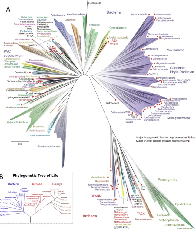
“stromatolites”, dome-shaped clumps formed by mats of microorganisms, and are dated back to around 3.4 billion years ago in the Archean Eon (Allwood et al., 2007; Schopf et al., 2007). The world back then was very different from the Earth today. It was characterised by extreme conditions including high temperatures, and an oxygen-depleted atmosphere comprised of large amounts of ammonia, methane, water vapour, and carbon dioxide. One hypothesis speculates that first lifeforms were extremophiles (meaning “loving the extreme”; Brock, 1978; Fredrickson and Onstott, 1996), which had adapted to anaerobic and hyperthermophilic conditions. Many of these microorganisms are evolutionary relics and belong to the domain Archaea. The existence of Archaea was first recognized in 1977, when Carl Woese and George Fox rearranged the tree of life based on universal molecular markers (small ribosomal subunits; Woese and Fox, 1977; Woese et al., 1990; Figure II.1-1). Archaea are morphological similar to Bacteria at first sight and are comparable in size and shape.
Nonetheless, closer investigations on genetic and ultrastructural levels disclosed high dissimilarities of Archaea to Bacteria as well as to Eukaryota. Hence, Archaea were grouped in a separate branch in the tree of life. For a long time, archaeal existence was thought to be restricted to extreme environments on Earth: high/low temperatures, high acidic or alkaline conditions, high amounts of salts, very low water activity and low nutrient availability (Rothschild and Mancinelli, 2001). This may be due to the fact that the majority of Archaea were isolated from specialized and extreme niches. However, archaeal as well as bacterial signatures are detected in almost every environment on Earth. Their omnipresence demonstrates that microorganisms are equipped with physiological versatility and are consequently assigned as “survival specialists”.
The detailed assessment of the microbial diversity has challenged microbiologists for a long time. Thus far, ~11.000 microbial species have been cultivated and validly described and
~600 are added each year (Rosselló‐Móra, 2012). Traditional cultivation techniques in the laboratory involve plating and inoculation in broth medium including or excluding the presence of oxygen. Anaerobic cultivation demands much more effort due to the strict avoidance of oxygen. The preparation of anoxic media requires the expulsion of all bound oxygen molecules and has been standardized over years (Hungate, 1969; Miller and Wolin, 1974). The air in the atmosphere is replaced by gas mixtures of H
2, CO
2, N
2depending on the metabolic demand. This technique is carried out using suitable equipment such as gas stations and anaerobic glove boxes (Aranki and Freter, 1972). Growth of microorganisms takes place in gas-tight vessels, which do not allow atmospheric oxygen to enter the medium.
When looking at the estimated microbial richness of 10
7-10
12species, the amount of cultivated microorganisms count a very small proportion (Dykhuizen, 1998; Pedrós-Alió, 2006). One explanation is that the majority of microorganisms are considered to be uncultivable (Amann et al., 1995; Joseph et al., 2003). There are many reasons for this: (i) cells are known to enter a non-culturable state when exposed to unsuitable growth conditions (Roszak et al., 1984, Xu et al., 1982), (ii) lack of knowledge on growth conditions for unknown species (Dewi Puspita et al., 2012), (iii) time-demanding slow growth (especially cold-loving psychrophiles; Franzmann et al., 1997) and (iv) dependence on symbioses (one or more), which does not allow pure cultures of single species (Jahn et al., 2007; Morris et al., 2013).
A second explanation is that obtaining pure cultures is in many cases time-consuming as well as labour intensive. In particular, cultivation becomes increasingly challenging while working with microorganisms that require complex metabolic demands.
Despite the obstacles, cultivated microorganisms are valuable and indispensable for
microbiologists. A valid description of a microbial species requires a cultivated
representative, which are subjected to various techniques to guarantee a clear classification
(e.g. DNA-DNA hybridization and spectrometric techniques, such as MALDI-TOF
by promoting the microorganisms` growth directly within their natural biotope (Gavrish et al., 2008; Nichols et al., 2010). In the best case, one microorganism is trapped within a cell- device and required growth factors are delivered throughout a semipermeable membrane, which keeps other microorganisms out of the trapping system and prevents overgrowth.
This system has improved over the years and nowadays even high-throughput chip-systems are available (isolation chip; iChip), allowing the simultaneous in situ isolation and cultivation of thousands of microorganisms in the same biotope (Ling et al., 2015).
As promising as the
in situ cultivation method is described, it still has drawbacks. The firstand most obvious limitation is restricted access to some extraordinary biotopes such as the hydrothermal vents in the Deep Sea. Hence, sampling and monitoring is limited by a long preparation time and very cost-intensive. A second limitation is represented by very slow growers which do not show up in a researcher`s lifetime. Lastly, the purification of interdependent communities is still not feasible with this method.
Yet, it is possible to analyse certain traits of uncultivated microorganisms by culture- independent methods such as stable isotope probing (SIP; Radajewski et al., 2000;
Radajewski et al., 2003), microscopy techniques (including scanning, transmission and fluorescence-based microscopy such as fluorescence
in situ hybridisation; Wagner et al.,1994) and molecular methods (e.g. sequencing techniques). The latter attracted increasing and justified attention, since the methods are rapidly improving. Sequencing methods are enabling the assessment of entire microbial communities, which are not sufficiently covered by cultivation.
Sanger and colleagues established the first sequencing method which enabled us to decipher
complete gene sequences and entire genomes (Sanger et al., 1977). The Sanger technique
lead to the ground-breaking novel tree of life suggested by Woese and Fox (Woese and Fox,
genome (Consortium, 2004). The diversity of microbial communities can be estimated by community fingerprinting, whereas PhyloChip analyses enables the quantification and identification thereof (Muyzer and Smalla, 1998; DeSantis et al., 2007). However, the aforementioned methods are more and more replaced by high-throughput sequencing methods.
The trend nowadays is to focus on Next Generation Sequencing (NGS) methods, which cover the small subunit ribosomal RNA (SSU rRNA) genes of a microbial population. Different sequencing platforms are available, ranging from Roche 454 pyrosequencing (Quince et al., 2009), Pacific Biosciences (PacBio RS II; Hoefler et al., 2013) to Illumina sequencing (MiSeq and HiSeq; Caporaso et al., 2012). The sequencing technique has evolved so rapidly that nowadays sequencing is even possible on the International Space Station. In an experiment referred to as the Biomolecule Sequencer, the MinION device demonstrated successfully sequencing of DNA in microgravity (Castro-Wallace et al., 2016). All technologies provide high speed, massively parallel sequencing, but inherently introduce inherent error rates (Quail et al., 2012). The least biased method is represented by Illumina MiSeq sequencing, which is currently the method of choice.
NGS allows a deep characterisation of the composition of the biological world. However, there are also limitations, which falsify the “real picture”. For instance, organisms from novel lineages remain hidden due to an inappropriate primer matching (Bergmann et al., 2011;
Brown et al., 2015). Other biases are introduced by (i) DNA extraction (Feinstein et al., 2009), (ii) PCR amplification (Pinto and Raskin, 2012), (iii) sequencing artefacts (Lee et al., 2012), (iv) DNA copy number (Kembel et al., 2012), and (v) sampling depth (Lagier et al., 2012).
Some errors can be tackled by performing replicates, suitable DNA extraction methods
(Feinstein et al., 2009), multiple combinations of primer sets, reducing the number of PCR
cycles to avoid chimera formation (Ahn et al., 2012) and negative controls (meaning no
template controls), which are carried along each step. Although researchers are aware of
these problems and try to eliminate them, microbiome projects are still lacking a standard
operation procedure (SOP), which guides each step of sample preparation and data
processing (Brooks et al., 2015).
reconstructed tree contains a vast number of uncultivated representatives, in particular, of
organisms deriving from extreme, yet unexplored environments (Hug et al., 2016; Figure II.1-
1). When compared to the tree of Woese and collegues, which was calculates almost 30
years ago, there has been tremendous progress in capturing the uncultivated microbial
diversity by NGS as is illustrated in Figure II-1.1.
Figure II.1-1 The new tree of life (Hug et al., 2016) (A) is facing the old tree (B, Woese et al., 1990). The old tree was calculated on single sequences (16S/18S rRNA gene of Prokaryotes and Eukaryotes, respectively), while the new tree is based on entire (draft) genomes. While comparing their dimensions, it becomes clear that the knowledge on the diversity is rapidly growing. The major part of organisms remains uncultivated (for instance in the Candidate Phyla Radiation, coloured in purple). In (A) red dots and non-italicized names highlight lineages, which lack an isolated representative.
However, as described above, culture-dependent and culture-independent methods are
facing the same problem: On a standalone basis, each method does not create a true
environments, namely the International Space Station and Mars-analogue sites. An extensive cultivation approach allows drawing conclusions on stress resistances of isolated microbes.
Cultivation is not always possible in the laboratory and the following chapter shows how in
situ cultivation combined with cultivation-independent methods sheds insights into thelifestyle of an archaeon, which is hosted in one of the investigated extreme, environments.
These sulphidic springs are also in the scope of astrobiological research as Mars-analogue aquifers. In the subsequent chapter, extreme environments on Earth and in space are described with respect to their relevance for the search of extraterrestrial life.
2. A grappling archaeon: Traits of the SM1 Euryarchaeon
“I have never been disappointed upon asking microorganisms for whatever I wanted”
22.1. Profiling the SM1 Euryarchaeon
More than 15 years ago, the discovery of an archaeon was reported, which exhibited a very unusual lifestyle (Rudolph et al., 2001). In the Sippenauer Moor, a sulphide-rich anoxic spring in close vicinity to Regensburg, Bavaria (Germany), whitish pearl-like assemblages were found to be attached to solid surfaces and float on the subsurface. Their eye-catching and macroscopically visible appearance drew interest on their composition. Different microscopy techniques such as scanning electron or phase contrast microscopy and fluorescence in situ hybridization (FISH) revealed that the pearls and the connected threads were formed by two main morphologically as well as phylogenetically different microorganisms (Rudolph et al., 2001; Moissl et al., 2002; Moissl et al., 2003; Rudolph et al., 2004b): Filamentous, sulfur- oxidizing bacteria (Thiothrix sp. SipK4, Gammaproteobacteria; Rudolph et al., 2001; Moissl et al., 2002) connect whitish pearls, which mainly consist of anaerobic coccoid archaea (referred to as “SM1 Euryarchaeon”, based on the location Sippenauer Moor).
It was already hypothesised at this point that this unusual partnership relies on an atypical form of cohabitation based on a syntrophic or even symbiotic relationship (Moissl et al., 2002). The partnership was proposed to rely on a sulfur cycling process, where the anaerobic SM1 Euryarchaeon potentially reduces sulphate to H
2S, which is metabolized by their surrounding sulfur-oxidizing bacterial partners and
vice versa. Cultivation experiments onthe archaeal enrichment failed and the authors speculated on a partner-dependent lifestyle (Rudolph et al., 2001; Moissl et al., 2002).
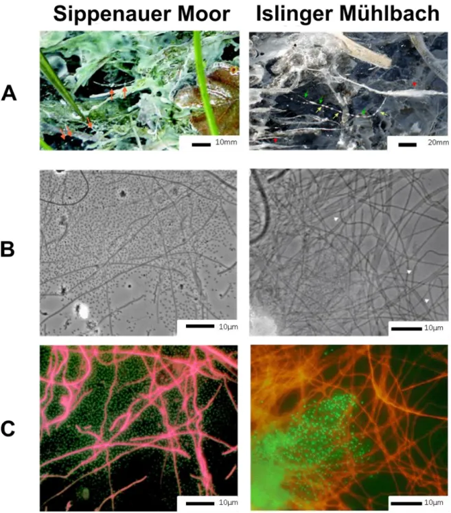
The unusual lifestyle in form of the “strings-of-pearls community” (SOPC) was not restricted to the Sippenauer Moor. Another sulphidic spring in Bavaria, the Islinger Mühlbach, shows the presence of similar structures. The spring was artificially formed by a drill hole and is characterised by a similar water chemistry and temperature compared to the Sippenauer Moor (~10°C throughout the year; Probst et al., 2013b). Near the outflow, coccoid archaeal cells accumulate to macroscopically visible pearls, surrounded and connected by filamentous bacteria, which, however, belonged to a different bacterial class (IMB1;
Epsilonproteobacterium; Rudolph et al., 2004b; later classified as Sulfuricurvum sp.; Kodama
and Watanabe, 2004). The phylogenetic different cohabitation was explained by the lower
oxygen concentration in the Islinger Mühlbach compared to the Sippenauer Moor, which is
enhancing the growth of IMB1 and outcompetes the growth of
Thiothrixsp. (Probst et al.,
2013b). The macroscopic and microscopic view on the SOPC is shown in Figure II.2-1.
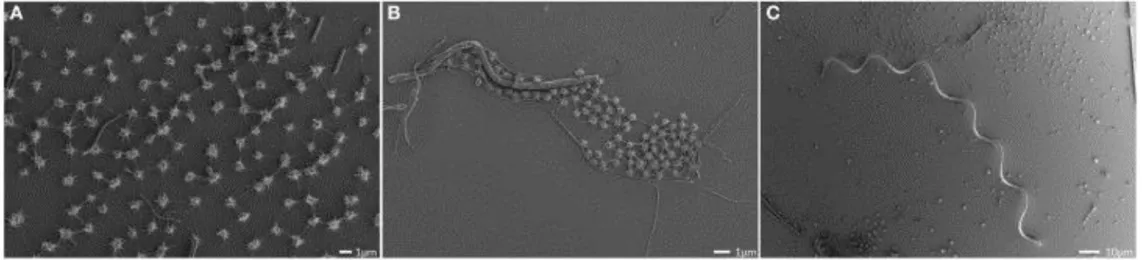
Figure II.2-1 String-of-pearl community differently imaged. The structure can already be observed by naked eye (A). It is formed by coccoid and filamentous microorganisms (B, phase contrast microscopy), which were identified as Archaea and Bacteria, respectively (Fluorescence in situ hybridisation imaging (C)). Images were taken from Rudolph et al. 2001 and 2004.
Further studies focused on the microcolony-forming archaea encapsulated within the pearls. The cocci with a diameter of 0.5 µm were analysed by clone libraries and taxonomic analyses classified the archaeal strains from both springs. They belong to a single phylotype and branch separately
within the Euryarchaeota (Rudolph et al., 2001; Moissl et al., 2002; Moissl et al., 2003; Rudolph et al., 2004b). Signatures of the SM1 Euryarchaeon were also detected in environmentally remote locations (e.g. in Turkey) and proved them to be widely distributed (Rudolph et al., 2004b, Bird et al., 2016;
Figure II.2-2).
revealed the flocks to be dominated by the same archaeal species, with only a minor proportion (5%) of their respective bacterial partners and thus forming an almost pure archaeal biofilm (Henneberger et al., 2006; Probst et al., 2013b; Figure II.2-3). The composition of the core microbiome of the biofilm was assessed using 16S rRNA gene microarray (PhyloChip G3) analysis, and was further confirmed by dsrB gene targeting FISH.
Quantitative PCR assays revealed representatives of the Deltaproteobacteria (sulphate- reducing group) as significantly enriched (Probst et al., 2013b). It was speculated that these bacteria provided a valuable function within the biofilm, as they were consistently abundant over a long period of time (Henneberger et al., 2006; Probst et al., 2013b). In addition, the sensitive PhyloChip technique detected additional archaeal species embedded in the biofilm, however, in a small proportion (Probst et al., 2013b). This finding was confirmed by FISH and clone library experiments.
The SM1 euryarchaeal cells, as previously seen in SOPCs, were surrounded by a thick matrix of extracellular polymeric substance (EPS; Costerton et al., 1995). The EPS layer consisted of polysaccharides and proteins and was most likely also produced by the SM1 euryarchaeal cells (Henneberger et al., 2006). The EPS of archaeal biofilms is, similar to bacterial EPS matrices, the key mediator for structure and function of biofilms (Flemming and Wingender, 2010). Only a few archaea-containing biofilms are known (Edwards et al., 2000; Battin et al., 2001; Couradeau et al., 2011) and most information is based on static incubation systems conducted under laboratory conditions (LaPaglia and Hartzell, 1997; Schopf et al., 2008;
Baker-Austin et al., 2010; Fröls et al., 2012; Bang et al., 2014; Di Meglio et al., 2014).
Naturally formed archaeal mono-species biofilms, as formed by the SM1 Euryarchaeon, are
rare. The best knowledge available is on the ARMAN consortium which dominates one
environment (Orphan et al., 2001). Hence, archaeal mono-species biofilms pose a special
feature within natural archaeal lifestyles.
The biofilm formation of the SM1 Euryarchaeon was observed in deeper, anoxic subsurfaces of two biotopes (Islinger Mühlbach and Sippenauer Moor). Interestingly, they exhibit macroscopically different appearances, presumably caused by different strains (Probst et al., 2013b).
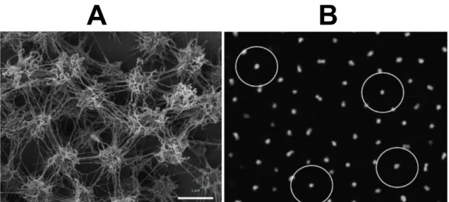
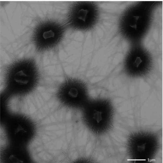
Strikingly, as observed in all SM1 euryarchaeal lifestyles (SOPC and biofilms), the cells were arranged in a regular, three-dimensional pattern and each cell kept a certain distance from the other (Figure II.2-3).
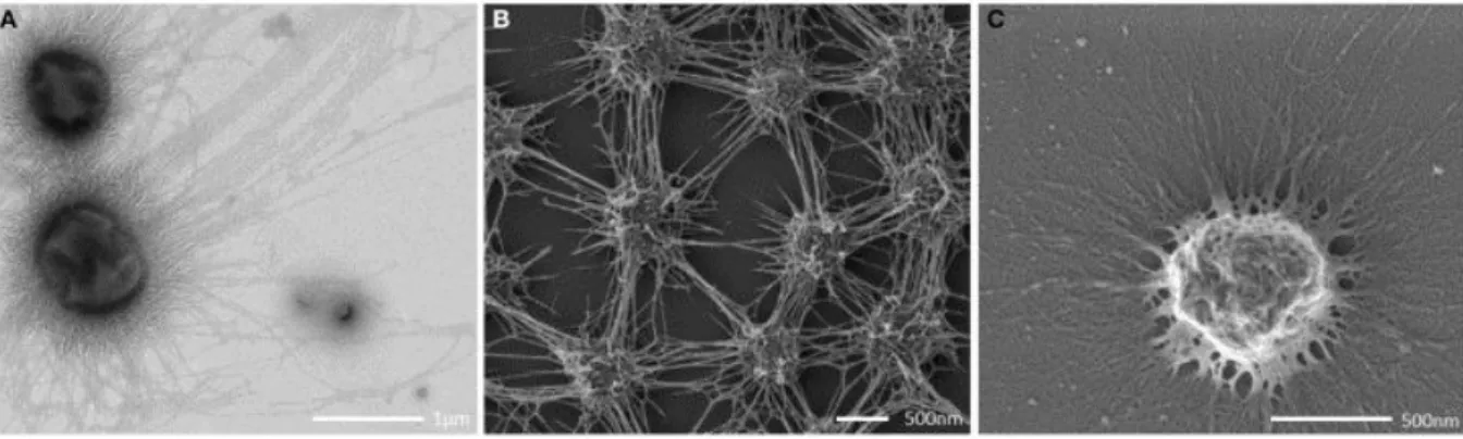
Figure II.2-3 Scanning electron micrograph depicting the dense, well-ordered biofilm (A). Each cell is kept in a certain distance by spacers (Bar: 1 µm; Probst and Moissl-Eichinger, 2015). This phenomenon is also observed in confocal scanning micrographs (B, each circle indicates for a diameter of 8 µm; Henneberger et al., 2006).
The spacers were observed as proteinaceous cell surface appendages exhibiting an extraordinary appearance and architecture never seen before.
2.2. Archaeal ninjas
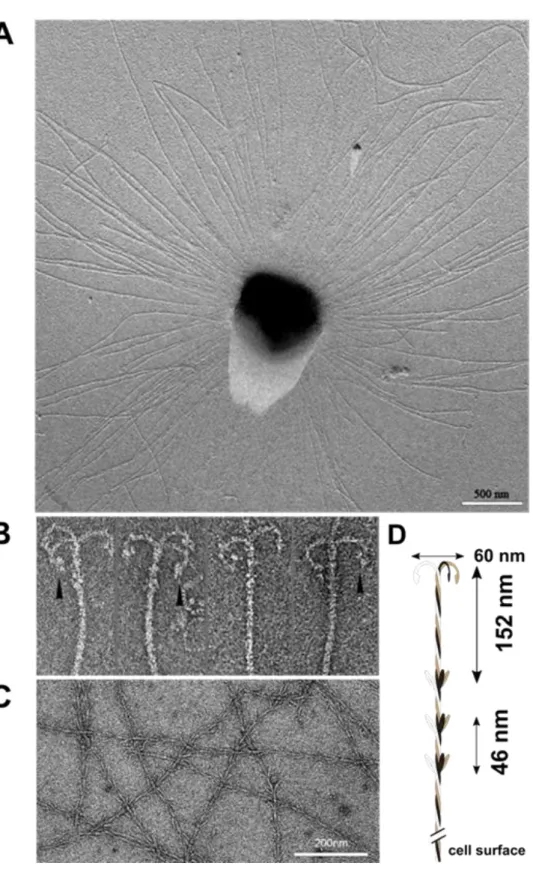
Each SM1 euryarchaeal cell analysed from the Sippenauer Moor and Islinger Mühlbach was observed to be covered by more than a hundred surface appendages, which exhibit an extraordinary architecture (Moissl et al., 2005). The barbed-wire filaments had a length of
~1-3 µm and carried so-called prickles (length: 30 nm) sticking out in regular intervals (46
nm) with a tripartite, grappling hook (diameter: 60 nm) at its distal end. The hook region was
responsible for the name assignment of the unique filaments: hami (sing. hamus; Latin for
anchor, hook). Tomographic reconstructions suggested a helical, twisted structure, which
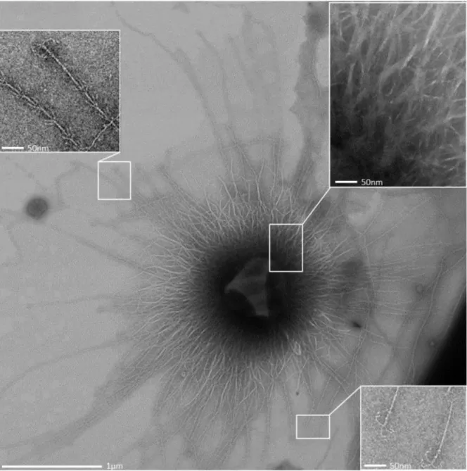
Figure II.2-4 Each SM1 Euryarchaeal cell is surrounded by numerous cell surface appendages, referred to as
“hami” (Moissl et al., 2005; A). The hami carry a tripartite, grappling hook at the distal end (Moissl et al., 2005;
B), the filaments are reminiscent of barbed-wires with prickles sticking out in regular intervals (Moissl et al., 2005; Probst and Moissl-Eichinger, 2015; C). Based on microscopic investigations, a hamus model was created
The hamus structure was described to be unique in nature; however, marginal structural similarities were drawn with actin filaments (Milligan et al., 1990; Holmes et al., 2003; Moissl et al., 2005). The hami filaments exhibit a well-defined top-to-bottom organisation, which speculates a complex assembly involving a vast variety of proteins similar to eukaryotic actin filaments (Pollard, 2007).
Interestingly, denaturing SDS-PAGE and immunological experiments revealed that the proto- filaments were composed of one major protein subunit with a molecular weight of 120 kDa without sequence homologies to any deposited proteins sequences in databases (Moissl et al., 2005).
These experiments revealed a rather simple assembly design, which applies to findings on archaeal flagella filaments. Archaeal flagella systems are composed of less than 13 different proteins (Thomas et al., 2001). The archaeal flagella filament is composed of one to several proteins with no homologues to eukaryotic or bacterial flagellar proteins, suggesting a convergent evolution (Cohen-Krausz and Trachtenberg, 2002). In fact, the archaeal flagella synthesis is similar to synthesis of bacterial type IV pili (Albers et al., 2006; Trachtenberg and Cohen-Krausz, 2006; Jarrell and McBride, 2008; Ng et al., 2008).
Up to this day, nothing was known about the hami assembly process and anchoring system, which was speculated to be complex (Moissl et al., 2005), but requires further investigation.
The stability of hami filaments was remarkable in both a wide pH and temperature range:
The filaments kept their three dimensional characteristic structure up to 70°C and in a pH range of 0.5 to 11.5 (Moissl et al., 2005). This finding was surprising, since the host thrives in a pH neutral and cold environment.
The stable filaments most likely function as a spacer and mediate the highly ordered cell- pattern within the biofilm. The hooks serve as attachments with strong adhesive forces between the cells and might even trap various organic and inorganic materials in their natural biotope (Moissl et al., 2005; Moissl-Eichinger et al., 2012a).
In summary, the extraordinary hami filaments show unusual traits. They are (i) complex in
even exhibit more extraordinary traits, which remained hidden so far and awaits further examination.
3. The era of astrobiology
“
Everything is everywhere, but the environment selects”
3Astrobiology is the study of the origin, evolution, and distribution of life in the entire Universe. It addresses the question whether life exists/ existed beyond Earth and how it can be detected. In recent years, tremendous technical progress enabled mankind to explore and study extra-terrestrial settings such as the moon (in manned explorations) and Mars (in unmanned explorations using numerous orbiters and rovers). Mars is supposed to be the most promising planet for extant and extinct life in our Solar System, and thus attracts the interest for future missions (next to icy moons). Mars rovers like Mars Science Laboratory (better known as “Curiosity rover”) and orbiters detected frequent outbursts of methane gas on the Martian surface (Formisano et al., 2004; Geminale et al., 2008; Fonti and Marzo, 2010; Geminale et al., 2011; Webster et al., 2015). These outbreaks arise spontaneously and are speculated to emerge from subsurface aquifers, where methane has previously been locked, or where basalt is serpentinised. However, the methane might also have a biogenic source. On Earth, the atmospheric methane is primarily produced by methanogenic microorganisms and for this reason there is hope to detect life on Mars (Atreya et al., 2007).
Several rovers and orbiters deliver valuable information on the Mars, but future, long-term missions to Mars aspire to search for life by human beings. The travel to Mars itself will take place in confined space shuttles and lasts for several months. The mission requires extensive preparation and a microbiological-safe work place has to be provided. This scenario is already trained using the International Space station (ISS).
In 1998, the first component of the ISS launched into the low Earth orbit and set another milestone for human space flight. At the present time, it consists of several modules organized by five space agencies, namely National Aeronautics and Space Administration (NASA; USA), European Space Agency (ESA; Europe), Russian Federal Space Agency (Roscosmos; Russia), Canadian Space Agency (CSA; Canada), and Japanese Aerospace Exploration Agency (JAXA; Japan). The spacecraft provides the platform for several scientific experiments on astrobiology, astronomy, physical science, material science, space weather meteorology and human research including space life and medicine.
The ISS represents a completely sealed, confined (meaning restricted to a defined number of parameters) man-made environment. It is characterised by extreme parameters such as higher radiation levels compared with Earth settings, low nutrient levels due to reduced exchange with the environment and microgravity. In addition, the indoor-environment is characterised by a stable, constant temperature (~22°C) and humidity (~60%; Coil et al., 2016). Since 2000, the ISS has been inhabited by humans – being accompanied by millions of microorganisms (the human body carries up to 4x10
13microbial cells; Sender et al., 2016).
The monitoring and control of the ISS microbiome is of uttermost interest as some species may impose a life-threatening hazard to the inhabiting crew. It has been shown that the increased microgravity and (presumably) starvation conditions about 400 km above ground as experienced onboard the ISS lead to an enhanced pathogenicity of
Salmonella typhimurium (Wilson et al., 2007; Zea et al., 2016). Not only may the crew be affected, butthe spacecraft is also endangered. Technophilic microorganisms (in particular fungi) were already responsible for major problems in the Russian space station (Mir) corroding alloys and polymers (Novikova et al., 2001; Novikova, 2004; Alekhova et al., 2005). Consequently, the cleaning procedure onboard the ISS is very strict including periodical disinfection procedures and air/subsurface controls, which check for microbial and fungal contaminations. The vast investigation on the bacterial community onboard the ISS revealed that they were human-associated (Venkateswaran et al., 2014).
Respective ground controls on Earth, cleanrooms, where the spacecraft hardware is
assembled, are reflecting the same image. Cleanrooms are highly controlled, and defined
most likely derived from dead cells in subsequent PCR reactions (Nocker et al., 2007), while DNA originating from viable cells is protected by the intact cell wall. The main proportion of detected microorganisms was uncovered to be human-associated (Moissl et al., 2007; Bashir et al., 2016), which is in accordance with the microbial population detected on the ISS (Checinska et al., 2015). However, these microorganisms are considered to be very hardy and adapt very well to the harsh conditions prevalent in cleanrooms and the ISS.
Possible adaptation of microorganisms towards space stresses are addressed within an flight project originally named “ARBEX” (Archaeal and Bacterial Extremophiles on board the ISS;
now “EXTREMOPHILES”; Moissl-Eichinger et al., 2016a) and focuses on the investigation of adaptation processes of moderate and extremotolerant microorganisms onboard the ISS.
Studies on the ISS provide important knowledge to future crewed space missions to Mars. So far, no human being has reached Mars, and it still requires rovers and orbiters equipped with technologies to search for Martian life signatures. The most recent project was the ExoMars mission a program of ESA and Roscosmos (Montroni et al., 2016). The Trace Gas Orbiter (TGO) already reached the Martian orbit and collects atmospheric data. The landing demonstrator Schiaparelli reached the Martian surface, however, unfortunately could not demonstrate a successful landing and crashed on the Martian surface at 300 km/h (Clery, 2016). In 2020, the next ExoMars mission will be equipped with hardware enabling the search for signatures of life on the Martian surface and in its subsurface by drilling experiments. In the course of planning this project, two main questions arise:
Do we expect Earth-derived life (contamination), which is brought along with the hardware?
Forward contamination is a main issue in planetary protection. Forward contamination means the transfer of microbes to other planets (e.g. the Mars). The microbial hitchhikers would interfere with life-detection procedures and cause false positive signals in life-detection measurements. For this reason, possible contamination, in particular on life-detection systems must be kept to a minimum.
How does possible Martian life look like?
No one knows what lifeforms we are facing on Mars. Consequently, we have to sustain and draw our knowledge on lifeforms we are also facing in Mars-like settings on Earth. Based on this, it is possible to develop life-detection systems, which can search purposefully for Martian life.
The first question can be addressed by assessing the microbial communities, which are in contact with any hardware designated to be sent to Mars. In addition, an uttermost care of decontamination is carried out to minimize this undesired scenario.
The second question is more challenging. All speculations are based on the assumption that Mars provides or rather has provided prerequisites for the origin and persisting of life.
The appearance of Mars underwent a dramatic change throughout the last billions of years and it has to be emphasised that the conditions of Early Mars and Present Mars are highly dissimilar and have to be regarded individually.
Today, Mars is a cold, desert world. The globally averaged atmospheric pressure at the surface was measured to be about 7 mBar and is consequently extreme thin. Hence, the temperature is only slightly above the freezing point of water near the equator during the warmest part of the day and does not allow water to stay permanently in its liquid form (Liu et al., 2003). In fact, a high amount of water is supposed to be present on Mars, however, due to the inappropriate conditions, water remains in ice-rich permafrost and is not accessible (Séjourné et al., 2012).
Yet, the climate on Mars may not always have been as extreme as it is today and might once
have supported the origin of life. In the Noachian period (~4.1–3.7 Ga ago; Bibring et al.,
2006; Carr and Head, 2010) the atmosphere of Mars was expected to be significantly denser
and consisted primarily of carbon dioxide, admixed with methane, sulphur gases, and only
traces of oxygen. The dense atmosphere might have resulted in increased Greenhouse
warming leading to stable, warm temperatures above the freezing point (Jakosky and
proximity (Westall et al., 2011). Carbon on Mars is likely to originate from meteorites and comets (Duprat et al., 2010), which impacted the planet in a regular manner and in hydrothermal systems such as Martian volcanic environments (Martin and Russell, 2007;
Hofmann and Bolhar, 2007). Volcanic rocks simultaneously provide the source for essential elements (H,N,O,P,S) and trace minerals (Nisbet et al., 2007). This scenario has already been observed for subaqueous volcanic sediments of similar shaped Early Earth (Westall et al., 2011). The sediments contained essential elements and energy sources and represented appropriate biotopes for heterotrophic microorganisms (Furnes et al., 2004; Nisbet et al., 2007; Westall, 2009; Southam et al., 2015). Thus, the Early Mars fulfilled all criteria necessary for a habitable planet. The geochemical settings on Early Mars are described to range from (i) acidic conditions (for instance indicated by the presence of jarosite in Burns formation at Meridiani Planum; Squyres and Knoll, 2005; Bibring et al., 2006; Knoll and Grotzinger, 2006), to (ii) pH neutral aquifers with low salinity and variable redox stages of iron, phosphor and sulphur (for instance the Fluvio-Lacustrine Environment at Yellowknife Bay, Gale Crater; Bristow and Milliken, 2011; McLennan et al., 2014; Grotzinger et al., 2014).
These data show that Early Mars appears to have hosted localized environments that would have been compatible with the requirements of primitive terrestrial life (Grotzinger et al., 2014). However, life may not have been persisted throughout the change into a hygroscopic planet. In this scenario, we expect to find preserved fossilised microorganisms on present Mars (Friedmann, 1986; Hofmann et al., 1997). Another scenario represents life, which has adapted to a very low water activity and three different possibilities are supposed:
a) It is speculated that the evolution of life might have been formed in hygroscopic
surfaces by obtaining water solely from the atmosphere (Davila and Schulze-Makuch,
2016). The speculations are based on a similar setting on Present Earth: The Atacama
Desert, which is considered to be the driest region on Earth. Within the desert an
amazing diversity of microorganisms adapted to desiccation, high radiation and high salinity reside on the surface (e.g. the Terrabacteria, which include Cyanobacteria, Chloroflexi and Deinoccus-Thermus members). Considering the capacity of terrestrial microorganisms to adapt to arid conditions, Early Martian life could have adapted and evolved throughout the transformation of Mars.
b) Recently delivered data describe occasionally occurring last remnants of narrow, up to ~5 meter in diameter, liquid water streamlets (recurring slope lineae, RSL; Chevrier and Rivera‐Valentin, 2012; Ojha et al., 2014; Ojha et al., 2015). The RSLs are restricted to small scale flow features and appear mostly during the Martian spring and summer, which suggests a melting result of frozen liquid solution. The large amounts of sedimentary salts lead to high salinity conditions within the RSL and thus, they are reflected as brines (Wang et al., 2006; Chevrier and Melchiorri, 2016).
Similar brines on Earth host halophilic microorganisms, which demonstrates their habitability (Leuko et al., 2010).
c) Permafrost settings on Mars might enclose life in a metabolically very slow state as in similar settings, which have been investigated on Earth (Soina and Vorobyova, 1996).
Life might still exist on Mars in a cryptobiotic state and bloom whenever water becomes available. Eventually, settings on Early and Present Mars are considered as potentially habitable and thus, there is a possibility of extant/extinct life. The search for Martian life is supported by model organisms, which thrive in terrestrial Mars-analogue environments.
Terrestrial extreme, harsh environments host model organisms, which are widely adapted to
withstand the challenging circumstances. A range of Mars-like environments were studied
from the aforementioned Atacama Desert to permafrost settings and caves (Fernández-
Remolar et al., 2008). However, in the body of literature, one main Martian feature is widely
ignored: The atmospheric oxygen abundance is ~10
-5times less than the present
atmospheric levels on Earth (Franchi et al., 1999; Mahaffy et al., 2013). Earth is not exposed
to comparable harsh physical and chemical conditions (i.e. high radiation dose, high amount
of perchlorates) as experienced on Mars. Still, terrestrial model conditions must be at least
very close to Martian conditions. Anaerobic analogue settings must include at least a few of
model organisms for astrobiology. This will advance the knowledge of possible life in Mars- analogue conditions.
The MASE (Mars Analogues for Space Exploration; http://mase-eu.org/
4) project is a collaborative research project with more than 20 members from 7 different European countries. It was kicked-off in the beginning of 2014 with the major aim to fill in knowledge gaps upon anaerobic, Mars similar microorganisms. The work plan aimed to cover the following main points:
a) Sampling of anoxic, Mars-like settings and development of a standard operation protocol for anoxic sampling
b) Anaerobic cultivation of microorganisms using a designated standard medium and providing the resulting anaerobic culture collection to the scientific community c) Assessing and describing the overall microbial community using molecular methods d) Study the response of cultivated isolates to combined environmental stressors also
experienced on Mars
e) Artificial fossilisation of selected microorganisms (stressed and unstressed counterparts) and assess the potential of their preservation
f) Development of biosignature detection techniques based on fossilised and unfossilised microorganisms
The tasks of the MASE project are divided and every member is allocated certain responsibilities. This dissertation covers the microbiological part (points a-c, see above), however, everyone in the team contributed to all work packages. The outcome of this
dissertation will help to provide knowledge on extreme, Mars-like life – an essential prerequisite for detecting extraterrestrial life.
4. Scope and publication guide
The scope of this thesis was to investigate extreme environment, namely the International Space Station (in the frame of the ESA flight EXTREMOPHILES project) and Mars-like environments within the frame of the MASE project upon their microbial community using cultivation-independent and cultivation-based techniques. One extreme setting, also involved in the MASE project, namely the sulphidic springs, hosts an extraordinary archaeon with exceptional traits (e.g. unusual lifestyles, and ultrastructure) – the SM1 Euryarchaeon.
The uncultivated archaeal cells were thoroughly investigated upon ultrastructure. The following paragraphs provide the guidance for the respective publications, where major findings are presented and discussed in detail.
Elucidating the grappling ultrastructure of the SM1 Euryarchaeon
The SM1 Euryarchaeon has attracted the interest of researchers for more than a decade.
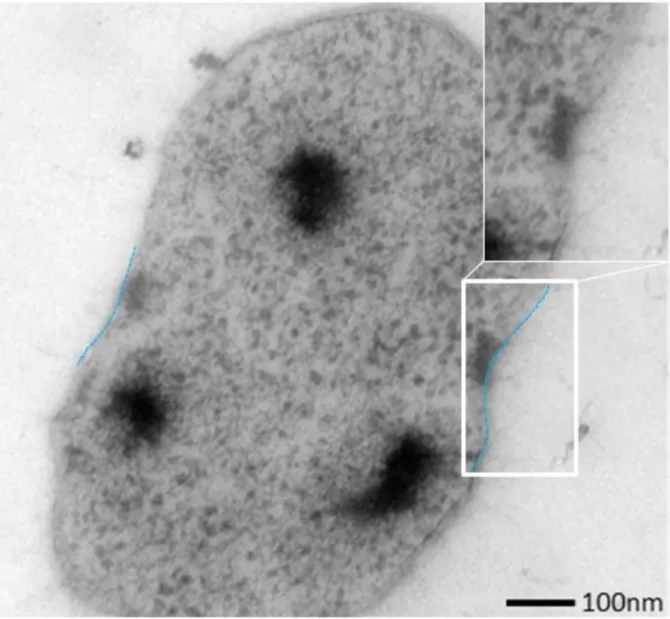
Although thoroughly studied, not all knowledge gaps on the ultrastructure are filled. The publication “Grappling Archaea: ultrastructural analyses of an uncultivated, cold-loving archaeon and its biofilm” reports on the microbial interaction within the biofilm and ultrastructure using electronic microscopy. The results confirmed previous findings (Probst et al., 2014a) which evidenced a divergence in the biofilms of the two biotopes. Moreover, it was shown that the SM1 Euryarchaeon exhibits a double membrane, a rarity in the archaeal domain. This observation was confirmed in another study carried out in the frame of this thesis (Probst et al., 2014b).
Notwithstanding this archaeon being uncultivable in the laboratory, a thorough study based
on the ultrastructure and genetic features of the “hami” structures was feasible and the
results are described in detail in “S-layers at a second glance? Altiarchaeal grappling hooks
(hami) resemble archaeal S-layer proteins in structure and sequence”. The full length of the
hami-encoding gene was obtained and the N-terminus region revealed a high similarity to S-
layer proteins. The results expand the knowledge on archaeal cell surface appendages and
microbial community of the US-module manifest a different composition, however, both harbour a substantial part of human-associated signatures. The results are presented in the publication “Resilient microorganisms in dust samples of the International Space Station – Survival of the adaptation specialists.”
The MASE project: Expanding the knowledge on extraterrestrial life
The body of literature contains solid knowledge on terrestrial Mars-like life, however lacks substantial information on the anaerobic proportion of microorganisms thereof. In the frame of the MASE project, we assessed the microbial diversity of (Present and/or Early) Mars-similar environments. Again, cultivation and cultivation-independent methods were combined and delivered impacts for extraterrestrial life. The findings are presented in “Mars exploration begins on Earth: Systematic comparison of the anaerobic, intact and cultivable microbiome of extreme, anoxic Mars-analogue, environments”. Studies, which were carried out along the MASE project contained stress tests upon selected MASE organisms, fossilisation thereof, physiochemical description of sampling sites, and a detailed description of the MASE sampling procedure (Beblo-Vranesevic et al., 2016; Cockell et al., 2016b;
Gaboyer et al., 2016). Taken together, they merge to a detailed picture on potential Mars-
similar life.
III. Manuscripts and publications
Overview
This cumulative dissertation contains four articles. Three manuscripts were already published, and one has been submitted to Nature communications. In any case, the PhD candidate, Alexandra K. Perras, has authored every manuscript printed in the following as a first author.
Concerning the data structure on the supporting CD the reader is referred to Chapter VII.
The PhD student’s contributions to the manuscripts are as follows:
III-1. Perras AK, Wanner G et al., (2014): Alexandra performed sample processing, contributed to transmission electron microscope imaging and wrote the paper. The manuscript and supporting information can be found on the supporting CD (./Selected publications/*03*).
III-2. Perras AK et al., (2015): Alexandra performed experiments, analysed data and wrote the paper. The manuscript and supporting information can be found on the supporting CD (./Selected publications/*04*).
III-3. Mora M, Perras AK et al., (2016): Alexandra performed bioinformatics, biostatistics and wrote the paper. The manuscript and supporting information can be found on the supporting CD (./Selected publications/*06*).
III-4. Perras et al., submitted (2016): Alexandra planned the study, participated during sampling trips, performed experiments, bioinformatics and wrote the manuscript. The manuscript and supporting information can be found on the supporting CD (./Selected publications/*08*).
A full list of publications, authored by the PhD candidate, is listed on pages 8 and 9. The
2Department of Biology I, Biozentrum Ludwig Maximilian University of Munich, Planegg-Martinsried, Germany;
3Zellbiologie, Philipps-Universität Marburg, Marburg, Germany; 4LOEWE Research Centre for Synthetic Microbiology (Synmikro), Marbug, Germany
Edited by: Luis Raul Comolli, Lawrence Berkeley National Laboratory, USA
Reviewed by: Luis Raul Comolli, Lawrence Berkeley National Laboratory, USA; Ariane Briegel, Caltech, USA
*Correspondence: Christine Moissl-Eichinger, Department of Internal Medicine, Medical University Graz, Auenbruggerplatz 15, 8036 Graz, Austria ; Email: christine.moissl-eichinger@medunigraz.at
†These authors have contributed equally to this work.
This article was submitted to Terrestrial Microbiology, a section of the journal Frontiers in Microbiology.
Publication information:
Frontiers in microbiology (2014), 5: 397 doi: 10.3389/fmicb.2014.00397
Received: 2 Jun 2014 Accepted: 14 Jul 2014 Published online: 5 Aug 2014
Abstract
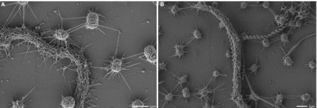
Similarly to Bacteria, Archaea are microorganisms that interact with their surrounding environment in a versatile manner. To date, interactions based on cellular structure and surface appendages have mainly been documented using model systems of cultivable archaea under laboratory conditions. Here, we report on the microbial interactions and ultrastructural features of the uncultivated SM1 Euryarchaeon, which is highly dominant in its biotope. Therefore, biofilm samples taken from the Sippenauer Moor, Germany, were investigated via transmission electron microscopy (TEM; negative staining, thin-sectioning) and scanning electron microscopy (SEM) in order to elucidate the fine structures of the microbial cells and the biofilm itself. The biofilm consisted of small archaeal cocci (0.6 μm diameter), arranged in a regular pattern (1.0–2.0 μm distance from cell to cell), whereas each archaeon was connected to 6 other archaea on average. Extracellular polymeric substances (EPS) were limited to the close vicinity of the archaeal cells, and specific cell surface appendages (hami; Moissl et al., 2005) protruded beyond the EPS matrix enabling microbial interaction by cell-cell contacts among the archaea and between archaea and bacteria. All analysed hami revealed their previously described architecture of nano- grappling hooks and barb-wire basal structures. Considering the archaeal cell walls, the SM1 Euryarchaea exhibited a double-membrane, which has rarely been reported for members of this phylogenetic domain. Based on these findings, the current generalized picture on archaeal cell walls needs to be revisited, as archaeal cell structures are more complex and sophisticated than previously assumed, particularly when looking into the uncultivated majority.
Keywords: Archaea, Biofilm, Ultrastructure, Hami, EPS, SEM, TEM, Microbial interaction
Introduction
Understanding the microbial “dark matter” has become one of the driving desires of the scientific community (Rinke et al., 2013). In particular, deep-branching, uncultivated archaea have attracted the interest, being largely unexplored but widespread and likely major drivers of the nutrient cycles in various ecosystems (Cavicchioli et al., 2007). Systems that allow unbiased and direct analyses of uncultivated microorganisms on microscopic and macroscopic levels due to one organism's predominance are extremely rare. However, such systems are of utmost importance to understand the functioning of microorganisms in the environment, their natural cellular composition, their actual metabolic activity and their interactions with the abiotic and biotic environment (Morris et al., 2013).
The majority of microorganisms remain uncultivable using standard methods (Amann et al.,
1995; Joseph et al., 2003). The unsatisfying success in this regard might be rooted in the
interwoven interactivity of microorganisms in their natural biotope, such as natural
ecosystems, or macrobes, such as plants or the human body. The human body itself is
colonized by 10–100 times more microbial cells than own cells (Schleifer, 2004). Analyzing
the (human) microbiome has become a major scientific focus, benefitting from state-of-the-
partner and not harmful for the other. Parasites, however, strongly affect the fitness of one partner (Moissl-Eichinger and Huber, 2011). A well-documented model system of a bacterial symbiotic interaction is “Chlorochromatium aggregatum”, a clearly structured consortium of immobile green sulfur bacteria epibionts and a motile beta-proteobacterium (Müller and Overmann, 2011). This association provides mobility to the epibionts and, in exchange, amino acids and 2-oxoglutarate to the inner partner. Detailed ultrastructural analyses revealed that hair-like filaments protrude from the epibionts and directly interconnect with the central bacterium. The latter connects with the epibionts via periplasmic tubes, which attach to the epibiont's outer membrane (Wanner et al., 2008).
In general, structural analyses of syntrophic and interactive consortia and communities that include an archaeal partner have rarely been reported, and information on the structure of natural archaeal populations in the literature is scarce. A likely syntrophic interaction between two hyperthermophilic archaea was artificially established under laboratory conditions: during co-culture conditions,
Pyrococcus furiosus attaches to Methanopyrus kandleri forming an unusual bi-species biofilm on provided surfaces (“fried-egg colonies”;Schopf et al., 2008). The contact between the two types of archaeal cells is mediated by flagella and possibly by extracellular polymeric substances (EPS). One example for a natural and uncultivated archaeal-archaeal interactive community is the ARMAN (archaeal Richmond Mine acidophilic nanoorganisms) system, where the ARMAN cells interact closely with Thermoplasmatales cells leading to a potential nutrient or molecule exchange (Comolli et al., 2009; Baker et al., 2010; see also article in this issue).
A model system for archaeal interspecies relationships is represented by the “intimate association” of Ignicoccus hospitalis and its partner
Nanoarchaeum equitans(Huber et al., 2002; Jahn et al., 2008). The relationship is based on the attachment of
N. equitans to theouter cellular membrane (OCM) of I. hospitalis (Jahn et al., 2004). It has been shown that this obligate dependence on
I. hospitalis is a consequence of the transfer of membrane lipids,amino acids and probably even ATP from
I. hospitalis to N. equitans(Huber et al., 2012).
Other investigations gave evidence for the lateral transfer of genetic material in both directions, during the co-evolution of these two archaeal cells (Podar et al., 2008). While I.
hospitalis is able to grow in pure culture, N. equitans still resists cultivation without its host.