Deposition of metal nanoparticles on magnetic nanobeads
and evaluation of their catalytic activity
Doctoral Thesis presented by
Francesca Besostri
For the degree of
Doctor of Sciences by Universität Regensburg and
Doctor of Chemical Science and Technology by Universitat Rovira i Virgili
Regensburg 2016
This work was supervised by: Prof. Dr. Oliver Reiser
Prof. Dr. Miquel A. Pericas Brondo
Graduation request filled on: 17. May 2016
Oral examination done on: 27. June 2016
This thesis was performed inside the EU-ITN Network Mag(net)icFun in accordance to the cotutelle agreement between the University of Regensburg and the Universitat Rovira i Virgili for a joint PhD degree programme.
The experimental work from February 2013 to March 2015 was done under the supervision of Prof.
Dr. Oliver Reiser, at the Institut für Organische Chemie der Universität Regensburg. From April 2015 to January 2016 the experimental work was done under the supervision of Prof. Dr. Miquel A. Pericas at the Institut Català d’Investigació Química (ICIQ, Tarragona).
Prof. Dr. OLIVER REISER and Prof. Dr. MIQUEL A. PERICÀS BRONDO,
STATE, that the present Doctoral Thesis entitled: "Deposition of metal nanoparticles on magnetic nanobeads and evaluation of their catalytic activity", presented by Francesca Besostri to recieve the degree of Doctor, has been carried out under their supervision between the University of Regensburg (UR) and the Institute of Chemical Research of Catalonia (ICIQ).
Regensburg, 4th May 2016 Tarragona, 5th May 2016
Prof. Dr. Oliver Reiser Prof. Dr. Miquel Àngel Pericàs Brondo
Table of contents
Thesis abstract 1
Zusammenfassung 3
Resumen 5
Introduction: Metal nanoparticles on solid supports: synthesis and applications 7
1.1 Supports 8
1.2 Deposition methods 14
1.3 Applications 21
1.4 Conclusions 27
1.5 References 28
Main Part
1. Magnetic supports: iron oxide and carbon-coated cobalt nanobeads 31
1.1. Introduction 31
1.2. Outline 32
1.3. References 33
2. Pd@Co/C and Pd@SiO2@Co/C nanobeads: evaluation of the catalytic
activity for the Suzuki-Miyaura coupling reaction and recyclability test 34
2.1 Introduction 35
2.2 Results and discussion 37
2.3 Conclusions 52
2.4 Experimental section 53
2.5 References 60
3. Deposition of platinum and gold nanoparticles on the graphene-like coating
of cobalt nanobeads and evaluation of their catalytic activity 62
3.1 Introduction 63
3.2 Results and discussion 64
3.2.1 Deposition of Au nanoparticles on graphene-coated cobalt nanobeads
and evaluation of their catalytic activity 65
3.2.2 Deposition of Pt nanoparticles on graphene-coated cobalt nanobeads
and evaluation of their catalytic activity 75
3.3 Conclusions 79
3.4 Experimental section 80
3.5 References 87
4. Deposition of nickel nanoparticles on magnetic nanobeads and
evaluation of their catalytic activity 89
4.1 Introduction 90
4.2 Results and discussion 91
4.2.1 Nickel nanoparticles on Co/C nanobeads (Ni@Co/C) 91 4.2.2 Nickel nanoparticles on Fe3O4 nanobeads (Ni@Fe3O4) 96
4.3 Conclusions 102
4.4 Experimental section 103
4.5 References 110
NMR Spectra 112
List of abbreviations 121
Curriculum Vitae 123
Acknowledgments 126
Thesis abstract
The present dissertation presents the scientific work developed in the last three years at the University of Regensburg (UR, Regensburg, Germany) and at the Institut Català d’Investigació Química (ICIQ, Tarragona, Spain). The work was focused on the development of nanocatalysts composed of metal nanoparticles deposited on the surface of magnetic nanobeads and the evaluation of their catalytic activity.
The first chapter describes the two types of magnetic supports employed in this work. On one hand, cobalt magnetic nanobeads with an average diameter of 50 nm were used. These nanobeads are composed of a metallic core surrounded by a graphene-like shell, which protects the core from oxidation and ensures thermal and chemical stability. On the other hand, iron oxide nanobeads with an average diameter of 6 nm, synthesised by thermal decomposition of Fe(acac)3 in the presence of oleic acid and oleylamine as surfactant agents, were employed as magnetic support.
The second chapter deals with the evaluation of the activity for the Suzuki-Miyaura cross-coupling reaction of a catalyst composed by palladium nanoparticles deposited on the surface of the carbon- coated Co nanobeads. High catalytic activity was found for the coupling of phenylboronic acid with aryl iodides and bromides. The use of microwave irradiation was proven to be particularly advantageous for this transformation, allowing significantly shorter reaction times. In fact, full conversions of the aryl iodides and bromides were obtained after only 5 and 15 minutes respectively, when the reaction mixture was heated to 100 and 120 °C by microwave irradiation. Moreover, low metal loading of 0.1 and 0.5 mol% were required and no further chromatographic purification was needed to isolate the products. The recyclability and metal leaching in the final products were also evaluated. The nanocatalyst could be easily recovered from the reaction mixture by applying an external magnetic field and directly reused for subsequent reactions, being active for at least six runs.
However, the detected metals leaching (Co and Pd) was above the allowed limits. Thus, a functionalisation consisting on a silica coating over the carbon shell and incorporation of a palladium complex was studied in order to minimize the leaching of metals. The resulting catalyst showed high catalytic activity for the Suzuki-Miyaura cross-coupling of phenylboronic acid with aryl iodides and bromides, proving its recyclability for four consecutive runs. The silica coating was found to minimize the metals leaching, being the palladium amount found in the final products below the required limits whereas the cobalt still exceeding them.
The third chapter describes the deposition of noble metals (Au and Pt), on the carbon coating of the cobalt nanobeads and the assessment of their catalytic activity. Aiming to obtain small (~ 5 nm) catalytically active metals nanoparticles, different synthetic pathways were studied. The synthesis of an active gold catalyst was achieved proving its potential for the hydrogenation of para-nitrophenol.
Contrarily, synthesising an active supported platinum catalyst was more difficult. High metal loading were achieved but the Pt was forming big clusters, without a catalytic activity, on the surface of the magnetic support.
The fourth chapter deals with the deposition of nickel nanoparticles on the surface of two different magnetic nanobeads and the evaluation of their catalytic activity for a broad range of reactions. On one hand, the carbon coated cobalt nanobeads were employed as support, taking in consideration the tendency of carbonaceous materials to incorporate metal nanoparticles. On the other hand, the ability of nickel to link to C-C double bonds present on the surface of iron oxide nanobeads was investigated.
Unfortunately, in no case, an active catalysts was obtained due to the formation of nickel oxide nanoparticles and agglomeration of the metal on the surface of the magnetic supports.
Zusammenfassung
Die vorliegende Dissertation zeigt die wissenschaftliche Arbeit, die in den letzten drei Jahren an der Universität Regensburg (UR, Regensburg, Deutschland) und am Institut Català d’Investigació Química (ICIQ, Tarragona, Spanien) entwickelt wurde. Die Arbeit konzentrierte sich auf die Entwicklung von Nanokatalysatoren bestehenden aus Metallnanopartikeln auf der Oberfläche von magnetischen Nanobeads und auf die weitere Auswertung ihrer katalytischen Aktivität.
Das erste Kapitel beschreibt die zwei Arten von magnetischen Trägern, die in dieser Arbeit verwendet wurden. Einerseits wurden magnetische Nanobeads mit einem durchschnittlichen Durchmesser von 50 nm bestehend aus einem Cobaltkern umgeben mit einer graphenähnlichen Schicht verwendet, die den Kern vor Oxidation schützt und thermische und chemische Stabilität sichert. Andererseits wurden Eisenoxidnanobeads als mögliche magnetische Träger mit einem durchschnittlichen Durchmesser von 6 nm verwendet, die durch thermische Zersetzung von Fe(acac)3 in Anwesenheit von Ölsäure und Oleylamin als oberflächenaktive Substanz synthetisiert wurden.
Das zweite Kapitel beschäftigt sich mit der Auswertung der Aktivität für die Suzuki-Miyaura Kreuzkupplungsreaktion eines Katalysators bestehend aus Palladiumnanopartikel abgeschieden auf der Oberfläche kohlenstoffbeschichteter Cobaltnanobeads. Es konnte eine hohe katalytische Aktivität für die Kupplung von Phenylborsäure mit Aryliodid und Arylbromid gezeigt werden. Die Verwendung von Mikrowellenbestrahlung erwies sich durch Minimierung der Reaktionszeit in der Praxis als vorteilhaft für die Transformation. In der Tat wurde kompletter Umsatz der Ausgangsmaterialien nach nur jeweils 5 und 15 Minuten für Aryliodid und Arylbromid beim Erhitzen der Reaktionsmischung auf 100 und 120 °C mit Mikrowellenbestrahlung erhalten. Darüber hinaus wurde eine niedrige Metallbeladung von 0.1 und 0.5 mol% benötigt und keine weitere chromatographische Aufreinigung war nötig. Ebenfalls wurde die Recycelbarkeit und das Leaching der Produkte untersucht. Der Nanokatalysator konnte von der Reaktionsmischung einfach mit Hilfe eines externen magnetischen Feldes wiedergewonnen werden und direkt für weitere Reaktion wiederverwendet werden mit Aktivität für mindestens sechs weitere Läufe. Jedoch war das detektierte Metallleaching (Co und Pd) über den erlaubten Grenzwerten. Daher wurde eine Funktionalisierung bestehend aus einer Silicaschicht über der Kohlenstoffhülle und Aufnahmen eines Palladiumkomplexes untersucht um das Metallleaching zu minimieren. Der entstandene Katalysator zeigte hohe katalytische Aktivität für Suzuki-Miyaura Kreuzkupplung von Phenylborsäure mit Aryliodid und Arylbromid mit Recycelbarkeit für vier aufeinanderfolgende Läufe. Die Silicaschicht reduzierte das Metallleaching des Palladiums im Endprodukt unter den benötigten Grenzwert, wobei der Cobaltwert immer noch darüber liegt.
Das dritte Kapitel beschreibt die Ablagerung von Edelmetallen (Au und Pt) auf die Kohlenstoffhülle der Cobaltnanobeads und die Bewertung der katalytischen Aktivität.
Hierbei war das Ziel kleine (~ 5 nm) katalytisch aktive Metallnanopartikel zu erhalten, es wurden verschiedene synthetische Weg erforscht. Die Synthese eines aktiven Goldkatalysators wurde erreicht und dessen Potential für dir Hydrierung von para-Nitrophenol gezeigt. Im Gegensatz dazu war die Synthese eines aktiven Platinkatalysators schwieriger. Hohe Metallbeladungen konnten erreicht werden, aber Pt bildete große Cluster ohne jegliche katalytische Aktivität auf der Oberfläche der magnetischen Träger.
Das vierte Kapitel beschäftigt sich mit der Abscheidung von Nickelnanopartikeln auf den Oberflächen von zwei verschiedenen magnetischen Nanobeads und der Bewertung ihrer katalytischen Aktivität für eine breite Auswahl an Reaktionen. Einerseits wurde die kohlenstoffumhüllten Cobaltnanobeads als Träger verwendet, mit der Überlegung dass kohlenstoffhaltige Materialien eine Tendenz zur Aufnahme von Metallnanopartikeln aufweisen. Andererseits wurde die Fähigkeit von Nickel zum Binden an C-C Doppelbindungen, die auf der Oberfläche der Eisenoxidnanobeads vorhanden sind, untersucht. Unglücklicherweise wurde in keinem Fällen ein aktiver Katalysator erhalten, wegen der Bildung von Nickeloxidnanopartikeln und Agglomeration des Metalls auf den magnetischen Trägern.
Resumen
La presente tesis doctoral expone el trabajo doctoral realizado durante los últimos tres años en la Universidad de Regensburg (UR, Regensburg, Alemania) y el Institut Català d’Investigació Química (ICIQ, Tarragona, España). El trabajo se enfocó en el desarrollo de nanocatalizadores compuestos de nanopartículas metálicas depositadas en la superficie de nanoperlas magnéticas y su consecuente evaluación catalítica.
El primer capítulo describe los dos tipos de soportes magnéticos utilizados en este trabajo. Por un lado, se emplearon nanoperlas magnéticas de cobalto con un diámetro promedio de 50 nm. Dichas nanoperlas están compuestas por un núcleo de Co0 rodeado por una capa de tipo grafeno la cual brinda protección al núcleo metálico, evitando su oxidación y garantizando su estabilidad química y térmica.
Por otro lado, nanoperlas de óxido de hierro con un diámetro promedio de 6 nm fueron usadas como posible soporte magnético. Estas perlas fueron sintetizadas por medio de la descomposición térmica de Fe(acac)3 en presencia de ácido oleico y oleilamina como surfactantes.
El segundo capítulo trata la evaluación catalítica en la reacción de acoplamiento Suzuki-Miyaura de un catalizador compuesto por nanopartículas de paladio depositadas en la superficie de nanoperlas de cobalto envueltas en carbono. Una alta actividad catalítica fue encontrada para el acoplamiento de ácido fenilborónico con yoduros y bromuros de arilo. El uso de irradiación con microondas fue de particular utilidad para esta transformación ya que permitió reducir apreciablemente los tiempos de reacción. Conversiones completas de yoduros y bromuros de arilo fueron obtenidas en 5 y 15 minutos respectivamente, cuando la mezcla de reacción fue calentada a 100 y 120 º C usando irradiación de microondas. Adicionalmente, bajas cargas de catalizador (0.1 y 0.5 mol %) pudieron ser usadas y no se requirió purificación por cromatografía para el aislamiento de los productos. La reciclabilidad del catalizador y la contaminación de los productos por lixiviación metálica fueron evaluadas. El nanocatalizador pudo ser fácilmente separado de la mezcla de reacción mediante la aplicación de un campo magnético externo mostrando actividad catalítica en al menos 6 ensayos catalíticos, sin embargo, el contenido metálico (Co y Pd) en los productos se encontró por encima de los límites permisibles. Con el objetivo de contrarrestar este efecto se exploró el uso de un recubrimiento de sílica sobre la capa de carbono en la cual se incorporó un complejo de paladio. El catalizador resultante mostró alta actividad catalítica para el acoplamiento Suzuki-Miyaura de ácido fenilborónico con yoduros y bromuros de arilo pudiendo ser también reutilizado en al menos 4 reacciones consecutivas.
El recubrimiento de sílica redujo efectivamente la pérdida de metales del catalizador, encontrándose los niveles de paladio por debajo de los límites requeridos aunque la contaminación por cobalto aún los supera.
El tercer capítulo describe la deposición de los metales nobles (Au y Pt) en la cubierta de carbono de las nanoperlas de cobalto y su evaluación catalítica.
Con el objetivo de obtener nanopartículas pequeñas (~ 5 nm) que fuesen catalíticamente activas se estudiaron diversas metodologías de síntesis. Nanopartículas de oro catalíticamente activas pudieron ser sintetizadas y su potencial en la hidrogenación de para-nitrofenol fue estudiado. Por otro lado, la síntesis de un catalizador activo de platino soportado presentó dificultades. Altas incorporaciones de Pt fueron observadas, sin embargo, la formación de clústeres de gran tamaño, sin actividad catalítica, fue predominante en la superficie del soporte magnético.
El cuarto capítulo trata la deposición de nanopartículas de níquel en la superficie de dos diferentes nanoperlas y la evaluación de su actividad catalítica en una gran variedad de reacciones. Por un lado se utilizaron nanoperlas de cobalto recubiertas con carbono, teniendo en cuenta la tendencia de los materiales carbonaceos a la incorporación de nanopartículas metálicas. Del otro lado, nanoperlas de óxido de hierro fueron empleadas. La alta afinidad del níquel por los dobles enlaces C-C presentes en la superficie de las nanoperlas fue usada para unir el catalizador al soporte. Desafortunadamente en ningún caso se obtuvieron catalizadores activos, principalmente por causa de la formación de nanopartículas de óxido de níquel y aglomeración del metal en la superficie de ambos soportes magnéticos.
Introduction
Metal nanoparticles on solid supports: synthesis and applications
Nanoscience, the emerging science of objects with dimensions in the range of few to 100 nm,[1]
(Figure 1) has received in the last decade great attention from different disciplines (i.e. physics, medicine, chemistry, biology and engineering) and still plays a main role in the technological progress for the development of efficient approaches for the synthesis of chemicals, materials and energy generation.[2,3] It mostly focuses on a) the development of methods and tools for the synthesis of nanostructures, b) the evaluation of the physical and chemical properties of the obtained nanomaterials and c) their final application in different fields.[4]
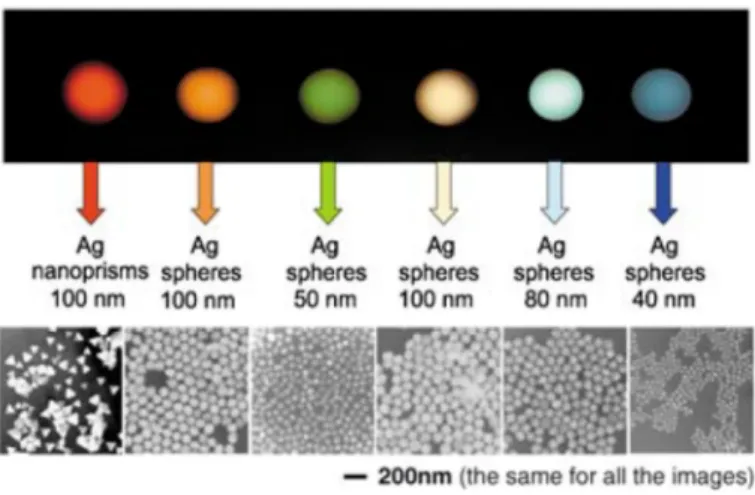
Figure 1 Depiction of the size regime of nanoparticles related to common “nano” scale objects. (A) prokaryotic cell, (B) ultraviolet wave, (C) virus and (D) enzyme. Blue sphere represents a 50 nm metal nanoparticle. Reproduced from Reference [9], with permission from The Royal Society of Chemistry.
Nanomaterials (i.e. carbon-based and inorganic materials) have been widely investigated due to their application as building blocks for the assembly of nanoscale machines and heterogeneous catalysis, in particular, significant interest has been placed on metallic nanoparticles (MNPs). These, display different properties compared to the related bulk materials, such as large surface-to-volume ratio, degenerated density of energy states, higher catalytic activity and optical, mechanical and chemical properties that can be tuned by modifying their size and composition (Figure 2).[4]
Figure 2 Size, shape and composition matters. This statement is illustrated here by considering the Rayleigh light-scattering properties of various nanoparticles (quoted nanoparticles sizes are all approximated). Reprinted from Reference [4]. Copyright © 2005 Wiley-VCH Verlag GmbH & Co. KGaA, D-69451 Weinheim.
Due to their size, metal nanoparticles are thermodynamically unstable because of the high surface energies.[5] Thus, their synthesis and growth must be performed under controlled conditions. Two different methods can be applied to obtain stable metal nanoparticles. On the one hand, organic ligands, capping materials or polymers can be employed to form protected core-shell nanocomposites.[6] An alternative and widely investigated procedure, is their immobilization on solid supports.[7] The latter method minimizes some of the disadvantages encountered employing bare metal nanoparticles, such as aggregation, poisoning and deactivation.[8] The properties of supported metal nanoparticles are strongly related to their morphology (size and shape), dispersion and interaction with the support material, rendering them suitable for a broad spectrum of applications.[9]
In the following sections an overview of the most widely employed supports, common deposition methods and applications of the corresponding nanocomposites is presented.
1.1 Supports
A variety of materials is employed as support for metal nanoparticles, each of them possessing specific advantages and influencing the final characteristics of the nanohybrids.[9,10] Among these, carbon based materials, metal oxides, polymers and magnetic nanobeads are the most widely applied families in the synthesis of supported metal nanoparticles.
Carbon-based materials
Carbon nanotubes (CNTs) are an important class of materials frequently investigated in the last years because of their intrinsic and specific characteristics such as high mechanical strength, high surface area and high electrical and thermal conductivity, which make them attractive for different applications.[11] Carbon nanotubes are one-dimensional tube-like materials with the carbon layer rolled up into a long, thin cylinder with diameters in the nanoscale range. They exist in two forms: single- walled carbon nanotubes (SWCNTs) have a diameter between 1 and 10 nm and are usually capped at the end while the multi-walled ones (MWCNTs), consisting in concentric cylinders held together by Van der Waals forces, are consequently larger with diameters between 5 to a few hundred nm (Figure 3).[12,13]
Figure 3 Schematic representation of single-walled (left) and multi-walled carbon nanotubes (right).
Moreover, the surface of CNTs can be tailored by covalent and non-covalent functionalisation strategies. These modifications involve the incorporation of new elements (e.g. oxygen and nitrogen), the anchoring of organic functionalities (e.g. biological macromolecules, polymers or surfactants) and also the deposition of metal nanoparticles.[11,13] In 2010, Bi et al. successfully deposited RuO2
nanoparticles on the surface of MWCNTs by a solution-based method. The structure of the MWCNTs with a diameter of 60-100 nm was retained after the treatment and scanning electron microscopy (SEM) showed a rougher surface indicating the presence of RuO2 nanoparticles. Transmission electron microscopy (TEM) and high resolution transmission electron microscopy (HRTEM) revealed carbon nanotubes coated with very small (< 2 nm) and well-dispersed RuO2 nanoparticles (Figure 4).[14]
Similarly, Choi et al. selectively deposited small Pt and Au nanoparticles on the sidewall of MWCNTs.[15]
Figure 4 Upper panel: SEM picture of the multi-walled CNTs used (left) and the as prepared RuO2/CNT nanocomposites (RuO2/CNT = 6:7 in wt %) (right). Bottom panel:
TEM (left) and HRTEM (right) images of the as prepared RuO2/CNT nanocomposites (RuO2/CNT = 6:7 in wt %). Images adapted with permission from Reference [14].
Copyright © 2010 American Chemical Society.
Graphene is another carbon-based material commonly used as a support for metal nanoparticles.[16,17]
It is a two-dimensional single-layer sp2 carbon atom network that is densely packed in a honeycomb structure.[16,18] As in the case of CNTs, graphene possesses several interesting properties as large surface area, high adsorption capacity, excellent electrical and thermal conductivity and high mechanical strength that render it attractive for a number of applications.[19] Functionalisation of the surface of graphene sheets allows to obtain versatile hybrid materials with use in different fields (i.e.
solar cells, fuel cells, water treatment and catalysis).[16] A common modification is the introduction of defects on their surface to improve the electronic transport properties.
In fact, intrinsic graphene is a zero bandgap material with small on/off ratios that lead to a limitation for the electronic applications.[20] Disadvantages that can be encountered working with graphene are connected to the possible agglomeration caused by the high π-π stacking between the nanosheets, leading to a decrease in activity as the catalytic sites are blocked.[21]
Nevertheless, several examples of well-dispersed ultrafine metal nanoparticles on graphene are reported in literature. A uniform coating of FePt nanoparticles with an average diameter of 7 nm was obtained by Guo et al. applying a solution-phase-assembly strategy.[18]
Similarly, Shang et al. deposited Pt nanoparticles on reduced graphene oxide sheet,[21] obtained by oxidation of graphite and composed of exfoliated sheets.[19] A uniform distribution of ultrafine Pt nanoparticles with an average diameter of 1.6 nm was achieved and the nanohybrid was successfully applied in catalysis.[21]
Figure 5 Left: TEM image of the G/Fe18Pt82 NCs. Adapted with permission from Reference [18]. Copyright © 2012 American Chemical Society. Right: TEM image of Pt- rGO (etched). Reprinted from Reference [21]. Copyright © 2014 Wiley-VCH Verlag GmbH & Co. KGaA, Weinheim.
(Bio)polymers
Biopolymers are a class of organic materials characterized by their robustness, chemical stability and easy chemical modification. They have been widely investigated as supports for metal and metal oxide nanoparticles leading to the synthesis of hybrid materials with unique properties and applications. The dispersion of the metal nanoparticles inside the polymer network takes place through a combination of different mechanisms (i.e. complexation and electrostatic interactions) and strongly depends on the morphology, porosity and surface area of the support.[22]
Chitosan is a polysaccharide biopolymer derived by deacetylation of natural chitin that displays unique characteristics as film-forming and chelating properties due to the presence of amino and hydroxyl functional groups. Moreover, it can be easily separated from the reaction mixture and is nontoxic, biocompatible and biodegradable, making its use feasible for pharmaceutical and biomedical applications.[23] In 2010, Wei et al. achieved the synthesis of Au and Ag-chitosan bio-conjugates, where the chitosan was able to directly reduce the metal source to metal nanoparticles (Figure 6).
Similarly, homogeneously distributed Pd nanoparticles with an average diameter of 5 nm were described by Sarkar et al. showing a different nanoparticles size depending on the metal loading, which indicates the presence of agglomerates when the loading was too high (Figure 6).[22]
Figure 6 Left: Image of metal NPs-chitosan bio-conjugates by exposure of 30 mg chitosan flakes to 6.0 mM AgNO3 at 95 °C. Reprinted from Reference [23]. Copyright © 2009 Elsevier Ltd. Right: TEM analysis of chitosan-Pd catalyst. Adapted with permission of Springer from Reference [22]. Copyright © 2012 Springer Science+Business Media B.V.
Nanocelluloses are another class of biopolymer obtained from vegetal cellulose. They are characterized by a rod-like or ribbon-like shape with length between 50-1000 nm and width in the range of 3-50 nm. The final structure depends on the preparation conditions and the starting cellulose that can be derived from several different sources (i.e. wood, cotton, bacteria, bamboo). Nevertheless, nanocelluloses have high surface area, high aspect ratio, high crystalline order and chirality, good mechanical strength and feasible surface modification due to the presence of hydroxy and sulphate ester groups. Because of these unique characteristics, together with the ability to form stable suspensions in water, nanocelluloses have been widely investigated as support for metal nanoparticles and organometallic species for catalytic applications.
Metal oxides
Another family of widely used supports for metal nanoparticles are metal oxides, which in turn can be classified as inert (SiO2) and active (TiO2, CeO2, ZrO2, Al2O3). In general, they possess high thermal and chemical stability together with a well-developed porous structure and high surface area.[2] Their use enhances the stability of the metal nanoparticles and allows their application in different fields as optics, analytic chemistry and catalysis.[24]
Moreover, active metal oxide supports participate in catalytic reactions through interactions with the metal nanoparticles. An example is the application of Pt@CeO2 catalyst in water/gas shift reactions.[25]
In this case, on one hand, the ceria drives the activated oxygen to the Pt and, on the other hand, the Pt nanoparticles enhance light absorption and charge-carrier separation in the metal oxide semiconductor.
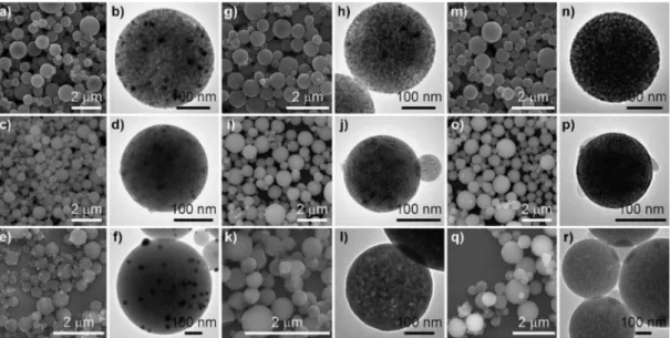
Jin et al. achieved the synthesis of nanohybrids where metal nanoparticles were uniformly distributed on the surface of different metal oxides (Figure 7).[25] An additional example comes from the application of precious metals supported on TiO2 nanospheres for photocatalytic water-splitting
reactions. It has been demonstrated that metal nanoparticles induce a shift in the Fermi level to more negative potentials, leading to the formation of nanohybrids with improved energetics and efficiency in the interfacial charge-transfer process.[26]
Figure 7 SEM (first, third and fifth column) and TEM (second, fourth and sixth column) images of the mesoporous metal oxides microspheres loaded with metal nanoparticles. a, b) Au@TiO2. c, d) Au@ZrO2. e, f) Au@Al2O3. g, h) Pd@TiO2. i, j) Pd@ZrO2. k, l) Pd@Al2O3. m, n) Pt@TiO2. o, p) Pt@ZrO2. q, r) Pt@Al2O3. Reprinted from Reference [25]. Copyright © 2012 Wiley-VCH Verlag GmbH & Co. KGaA, Weinheim.
Magnetic nanobeads
The use of magnetic nanobeads as support has also been widely investigated in the recent years because of their properties as high surface area to volume ratio, chemical inertness and thermal stability. Moreover, they can be easily recovered from the reaction mixture by applying an external magnetic field, which greatly facilitates the reaction work-up.[27] In fact, time-consuming and tedious protocols as filtration and centrifugation can be avoided and the catalyst can be reused.[28]
Furthermore, the surface of the magnetic nanobeads can be functionalised in different ways such as silica/carbon coating or by the addition of surfactants and polymers.
This leads to a stabilization of the nanoparticles themselves and also introduce the possibility to anchor different molecules or deposit metal nanoparticles on the surface.[27]
1.2 Deposition Methods
Different preparation routes, physical or chemical, can be applied to achieve the deposition of metal nanoparticles onto solid supports.[9] Today’s focus on sustainable chemistry drives the development of synthetic pathways that employ less toxic precursors, environmentally friendly solvents, ambient reaction temperatures and few synthetic steps. Moreover, minimum quantities of by-products and waste should be generated.[29]
Atomic layer deposition
Atomic layer deposition (ALD) is a technique employed for the deposition of thin films[30] on different supports such as nanoparticles, nanowires, nanotubes, soft and biological materials.[31,32] This technique is extensively used as a consequence of the reproducibility of the obtained films together with the large range of applications of the synthesised hybrid structures (i.e. catalysis, microelectronics, energy storage and sensing). ALD is based on the reaction of precursors that are separated into successive surface reactions. In this way, the reactants are kept separated and react with the surface in a self-limiting process. Every surface reaction is separated by a purge step, where unreacted precursor and by-products are removed. In each cycle, a film with a defined thickness is deposited and the cycles are continued until the final desired film is obtained (Figure 9).[32]
Nevertheless, sometimes nanoparticles can be deposited instead of films, being particularly convenient for catalysis applications due to the higher surface area. In 2009, Liu et al. achieved the deposition of uniform and well-dispersed Pt nanoparticles on carbon nanotubes as it is possible to observe in Figure 8 where the Pt nanoparticles appear as white dots.[31]
Figure 8 Left: SEM cross-section of the carbon nanotubes on Si wafer after ALD of Pt for 100 cycles. The carbon nanotubes were acid treated for 6 h. Right: SEM images of carbon cloth, with acid treatment for 6 h, after ALD of Pt. Reprinted from Reference [31].
Copyright © 2009 Wiley-VCH Verlag GmbH & Co. KGaA, Weinheim.
Figure 9 Schematic illustration of one ALD reaction cycle. Reprinted from Reference [30], with the permission of AIP Publishing. Step 1: pulse of the reactant A leading to its absorption on the surface. Step 2: purge of the unreacted precursor A and of the by- products. Step 3: pulse of the reactant B, which reacts with the surface species created by precursor A. Step 4: purge of the unreacted precursor B and of by-products.
Microwave irradiation
Microwave (MW) heating found in the recent years large application in different fields such as synthesis of nanomaterials, solid-state chemistry, nanotechnology and organic synthesis.[33] MW application in the synthesis of organic, inorganic and hybrid materials allows to obtain nanoparticles with uniform size/shape because of the fast and uniform heating of the reaction mixture. Moreover, it often allowed a fine dispersion of the metals on the supports, a generally difficult goal with methods as co-precipitation and impregnation.[34] In 2008, Campelo et al. developed an easy and fast procedure to deposit metal nanoparticles on polysaccharide-derived mesoporous materials by microwave irradiation and avoiding the use of reducing agents. The synthesis required microwave heating for 2 min at 100- 140 °C (300–450 W) of an ethanol/acetone or ethanol/water solution of the desired metal salt. They demonstrated how microwave irradiation was critical for the synthesis of nanoparticles with uniform size, shape and distribution. Moreover, the controlled temperature allowed the deposition of only reduced metal nanoparticles. A comparison with nanohybrids obtained by conventional heating was also made. In this case, low metal loading was achieved together with an irregular nanoparticle shape, size and distribution.[35] Similarly, Guo et al. employed microwave irradiation for the deposition of platinum nanoparticles on graphene sheets with a one-step protocol. High loading of Pt nanoparticles with a uniform distribution and small size (2.6 nm) was achieved. The nanohybrids were then successfully applied for electroanalytical applications.[36]
Sonochemistry
Sonochemistry is an additional deposition methodology that can be applied to the synthesis of supported metal nanoparticles. The advantages related to this procedure such as controllable size distribution, environmentally friendly protocol and ambient temperature reaction conditions, are due to the acoustic cavitation phenomena involving the formation, growth and collapse of bubbles in a liquid.[37] During the sonochemical process, three different regions are formed in the liquid medium: 1) the gas phase of the collapsing bubbles, where high temperatures (> 5000 °C) and pressures (> 20 MPa) are reached, causing water to pyrolyse into H and OH radicals; 2) interfacial region with temperatures high enough (few hundred °C) to induce reactions and 3) the bulk solution at ambient temperature.[38] Pol et al. demonstrated how sonochemical processes lead to the deposition of small (~
5 nm) Ag and Au nanoparticles on silica nanospheres with a uniform distribution. Furthermore, no reducing agents were added to the reaction mixture, being the high temperature and pressure reached during the sonochemical process, responsible for the reduction of the metal ions to the amorphous form.[37,39]
Chemical vapour deposition
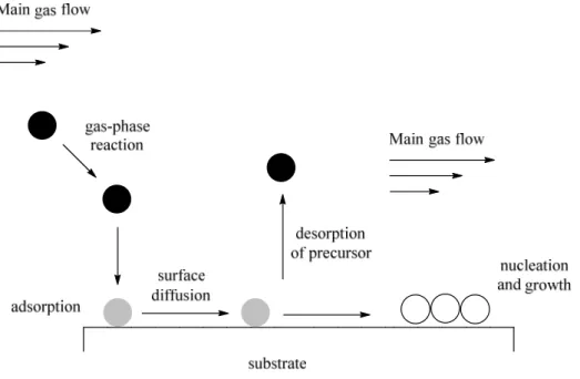
Hybrid materials such as microelectronic devices, optics and membrane reactors synthesised by chemical vapour deposition (CDV) of thin films on solid supports can be used in a variety of applications. Moreover, CDV allows a conformal and selective deposition of metals. The process involves the thermally induced reaction of a metal precursor on a heated surface. In particular, a volatile metal precursor (inorganic, metal-organic or organometallic) is transported in the reactor and to the support where it adsorbs and reacts to liberate the ligand that is subsequently desorbed and transported out of the reactor. The metal atoms then diffuse to form stable nucleus and the film coating (Figure 10).[40] This methodology was employed by Okumara et al. to deposit small (2-3 nm) and well- dispersed gold nanoparticles on different metal oxide supports, Al2O3, SiO2 and TiO2. The same good results could not be achieved applying liquid phase preparation methods.[41]
Figure 10 Schematic representation of the key steps involved in the chemical vapour deposition process.
Pulsed laser deposition
Pulsed laser deposition (PLD) is a clean and low-cost physical technique employed for the deposition of thin metal films on solid supports.[42] In PLD, a high power pulsed laser is focused on the target surface. Applying a sufficiently high laser fluence (~1 J/cm2), each laser pulse is able to vaporise or ablate a small amount of material which expands rapidly from the surface in vacuum and provides the deposition flux of thin film growth.[43] The dimension and morphology of the nanoparticles are usually controlled by the number of laser pulses, leading to spherical small nanoparticles (1 nm) with a narrow size dispersion for low metal loading.[44] In 2012, Dai et al. deposited CdSe quantum dots on ternary metal oxide nanoparticles Zn SnO for photovoltaic application by pulsed laser deposition.
This methodology allowed to use non-toxic chemicals and to obtain a uniform monolayer of small quantum dots varying laser parameters.[42]
Deposition-precipitation method
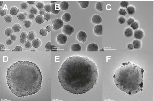
Currently, this methodology is particularly employed for the deposition of metal nanoparticles on functionalised/coated magnetic nanobeads. It consists in the sonication of the support in the desired metal salt solution followed by addition of a reducing agent. Zhu et al. applied this method to incorporate Pd nanoparticles on the surface of iron oxide (Fe3O4) nanobeads coated with a carbon layer derived from glucose carbonization.[45] Similarly, Liu et al. deposited Ag, Pd and Au on the polymer coating of magnetic nanospheres (Fe3O4@P(MBAAm-co-MAA). The loading of the metal nanoparticles was achieved by exchange of sodium cations present in the ionized microsphere, leading to the formation of uniform and well-dispersed Ag and Pd nanoparticles on the shell layer of the microspheres. In the case of Au, nanoparticles with irregular shape and distribution were formed, probably because of the weak interaction between the Au and the polymer coating (Figure 11).[46]
Figure 11 TEM images of (A, D) Fe3O4@P(MBAAm-co-MAA)/Ag microspheres, (B, E) Fe3O4@P(MBAAm-co-MAA)/Ag microspheres and (C, F) Fe3O4@P(MBAAm-co- MAA)/Au microspheres. Adapted with permission from Reference [46]. Copyright © 2011 American Chemical Society.
Microemulsion
Water-in-oil microemulsion is a well-known procedure to synthesise supported metal nanoparticles. In fact, the interaction between the microemulsion itself and the support is believed to enhance the hydrophobicity of the latter rendering it more chemically-compatible.[9]
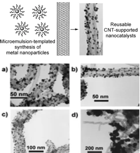
Yoon et al. employed this method to synthesise a catalyst composed of Pd and Rh nanoparticles uniformly distributed on carbon nanotubes, of average size between 2 and 10 nm. Hexane was added to an aqueous solution of the desired metal containing a surfactant agent, obtaining a two-phase system. The solution was purged with nitrogen and hydrogen, leading to the formation of the nanoparticles within the microemulsion. The nanoparticles were anchored on the support by stirring the carbon nanotubes into the microemulsion. It has been moreover demonstrated that, with a direct deposition of metal nanoparticles on the carbon nanotubes, not employing the microemulsion method, large Pd and Rh clusters (1 order of magnitude larger) were obtained (Figure 12).[47]
Figure 12 Upper panel: Schematic representation of the microemulsion-based method for the deposition of metal nanoparticles on carbon nanotubes. Bottom panel: TEM images of the microemulsion-templated synthesis of CNTs-supported metal nanoparticles: a) Pd/CNTs, b) Rh/CNTs, c) Pd/Rh bimetallic nanoparticles from the microemulsion methods and d) Pd/CNTs from an aqueous Na2PdCl4 solution. Adapted with permission from Reference [47]. Copyright © 2005 American Chemical Society.
Impregnation
This approach, also known as wetness impregnation, consists in the “wetting” of a solid support with a solution containing the desired metal. Usually, nanoparticles with a broad distribution of sizes is observed.[9] In 2011, Choi et al. developed graphene-supported metal nanoparticles employing impregnation method obtaining materials composed of small nanoparticles (Pt = 1.9 nm; Pd = 2.0 nm;
Rh = 3.9 nm; Ru = 3.0 nm) with a fine distribution on the graphene sheets. The synthesis was achieved by mixing the graphene with a solution of the metal in acetone and stirring the mixture by ultrasonication. After collecting and drying, the hybrid materials were treated with 4% H2 in N2 at 250
°C for 3 h, to reduce the metal nanoparticles.[48]
Centrifuge
In 2014, Markelonis et al. showed the possibility to employ a common laboratory centrifuge to deposit uniform films of Au and PbS nanoparticles on different types of materials, from flexible to rigid sheets as well as flat and rough surfaces. The synthesis involves the placement of the chosen support on the bottom of a vial followed by addition of a well-dispersed solution of the colloidal nanoparticles. The mixture was at this point centrifuged, leading to the formation of uniformly supported metal nanoparticles, the thickness of the film correlated to the concentration of the colloidal nanoparticles solution employed (Figure 13).[49]
Figure 13 Upper panel: PbS NPs deposited by centrifugation method on different substrates. Left: blank substrates and right: PbS NPs deposited on a) aluminium foil, b) paper, c) plastic sheet and d) glass. Bottom panel: Schematic diagram to illustrate the centrifuge deposition process. Adapted from Reference [49], with permission of Springer
1.3 Applications
Supported metal nanoparticles have been successfully applied in a great variety of different fields. A small overview of the most relevant uses of these nanohybrids is presented in the following section.
Sensors
Metal-graphene nanocomposites have found various applications as electrochemical energy storage and sensing due to their large electrochemically active surface areas, which are able to efficiently increase the electron transfer between the electrode and the detected molecules, leading to more rapid and sensitive response than traditional sensors.[36,50,51] Determination of hydrogen peroxide (H2O2) in biological systems is of high importance in biology, biomedicine and environmental protection, being a by-product of several biological and chemical processes (i.e. pharmaceutical, paper and chemical industries). In biological systems, hydrogen peroxide can generate reactive oxygen species (ROS) that, in excess, can cause different diseases (i.e. Parkinson, Alzheimer, heart attack and cancer).[50]
Similarly, detection of glucose in blood serum is particularly important in the control of diabetes mellitus, a current worldwide health problem. In 2012, Zhang et al. developed a new nanohybrid material composed of Ag nanoparticles deposited on graphene, employing tannic acid as reducing agent. The nanocomposite was then immobilized on the surface of a glassy carbon electrode (GCE) by addition of chitosan to form a modified electrode (chitosan/Ag NPs-G/GCE), evaluated for the sensing of H2O2 and glucose. The Ag NPs-G nanocomposites exhibit high electrocatalytic activity for the reduction of H2O2, being the current response of the modified chitosan/Ag NPs-G/GCE electrode significantly higher than the current obtained with the bare electrode. For the detection of glucose, the enzyme glucose oxidase (GOD) was immobilized on the already described electrode.
The detection mechanism involves the oxidation of glucose in the presence of oxygen by GOD leading to the formation of H2O2, that is electrochemically detected. A good linear relationship was observed between the current response and the glucose concentration, with a detection limit of 100 µmol/L, which enabled the modified electrode to be successfully used for the detection of glucose in blood samples.[51] In 2014, Maji et al. developed a hybrid material composed of well-dispersed small (~ 3 nm) Au NPs deposited over reduced graphene oxide (rGO) sheets covered with periodic mesoporous silica (PMS) (rGO-PMS@AuNPs). The nanocomposite was successfully employed as an electrode material to fabricate a sensor for the detection of H2O2 and glucose in biological samples (human urine). Additionally, as a result of the very low achievable detection limit (60 nM), it was used also for the detection of H2O2 released by cancer cells (Figure 14).[50]
Figure 14 Schematic diagram for in vitro detection of H2O2. Adapted with permission from Reference [50]. Copyright © 2014 American Chemical Society.
Different cell lines were employed, human embryonic kidney cells (HEK 293), human cervical cancer cells (HeLa) and human hepatoma cancer cells (HepG2) together with a stimulating agent, the phobol 12-myristate-13-acetate (PMA), which induces the generation of hydrogen peroxide from tumour cells in a short time period. The RGO-PMS@AuNPs was demonstrated to be sensitive and reliable in the detection of H2O2 generated by tumour cells, allowing the distinction between normal and cancerous cell lines.[50]
The surface-enhanced Raman scattering (SERS) has attracted great attention for its microanalytical abilities, allowing the detection of analyte with high sensitivity and low detection limits. In 2011, Han et al. prepared silver-coated magnetic nanospheres (Fe3O4/Ag) that serve as a SERS substrate and can also be easily handled by applying an external magnetic field. Malachite green (MG) is a fungicide and antiseptic used in aquaculture industry but suspected to be genotoxic and carcinogenic, making its monitoring in water at trace level particularly important.[52] This scenario has been selected to evaluate the SERS-property of the silver-coated magnetic nanospheres.[53] The SERS signal obtained for this hybrid materials were much stronger than the one detected for the bare microspheres. A quantitative analysis was also made. Stock solutions of the MG were firstly mixed with the Fe3O4/Ag. A sample solution was then dropped on a glass slide, dried and analysed by Raman spectroscopy, using the relative Raman intensity to quantitatively evaluate the MG content.
A good linear relationship was observed in the concentration range of 10-350 ppb, demonstrating that the microspheres can be used to detect MG in water, in concentration as low as 10 ppb. The Fe3O4/Ag microspheres were also used to develop a SERS-based optofluidic system (Figure 15), where the microspheres and the MG analytes were mixed in a microfluidic channel and the SERS analysis was performed under continuous flow conditions. A mini-solenoid was introduced in the system, in order to generate a magnetic field that can trap the microsphere in the channel. In this way, they could adsorb the analyte molecules and be effective. A valve system was also introduced to regulate the flows. First, the microspheres were introduced and trapped on the channel. Afterwards, the analyte was circulated through the channel and absorbed on the microspheres. Here, “hot spots” generated by Fe3O4/Ag were detected by SERS.[52]
Figure 15 Layout of a SERS-based optofluidic sensor integrated with a solenoid.
Adapted with permission from Reference [52]. Copyright © 2011 American Chemical Society.
Fuel cells
The current increase in energy consumption combined with the depletion of fossil fuel reserves and the environmental concerns associated with green-house gas emissions have motivated the development of alternative energy sources. Fuel cells, such as proton exchange membrane fuel cells (PEMFCs) and direct methanol fuel cells (DMFCs) are considered one of the most promising class of energy conversion devices allowing high densities and efficiencies.[54] Fuel cells might play a key role as possible future energy sources for zero-emission vehicles, distributed home power generators and power source for small electronics.[55] Fuel cells consist in an electrochemical device that is able to convert chemical energy of fuels (i.e. hydrogen, methanol, formic acid and ethanol) to electricity by separating the electron flow from the mass transport of two main chemical reactions involved, fuel oxidation on the anode and oxygen reduction on the cathode (Figure 16).[54]
Figure 16 Schematic representation of a fuel cell. Adapted with permission from Reference [54]. Copyright © 2014 American Chemical Society.
In 2011, Qiu et al. described a new hybrid nanomaterial composed of small (~ 4.6 nm) and well- dispersed Pt nanoparticles on functionalized graphene sheets, successfully employed in a direct methanol fuel cell (DMFC) where methanol is oxidized with oxygen to carbon dioxide and water, generating electricity. Platinum is the most widely used catalyst in fuel cells and graphene has been used as support for these applications due to the high conductivity and surface area. The synthesised nanocomposite exhibits high electrochemical activity for methanol oxidation and oxygen reduction.
Moreover, good tolerance towards CO was observed.[56]
Photocatalysis
Metal nanoparticles supported on semiconductor materials have been extensively studied for a variety of photocatalytic reactions. In 2011, Tsukamoto et al. studied the oxidation mechanism under visible- light irradiation of small Au nanoparticles (< 5 nm) deposited on the interface of anatase/rutile TiO2
particles. Au nanoparticles are characterized by a strong light absorption in the visible region that generate oscillation of free electrons, known as surface plasmon resonance (SPR). In a general mechanism of visible-light-driven oxidation (Figure 17), surface plasmon resonance transfers electrons from Au nanoparticles to the semiconductor support conduction band and the positively charged Au nanoparticles receive electrons from a substrate that is in this way oxidized.
Simultaneously, the electrons in the conduction band of the support are consumed by oxygen reduction.[57]
Figure 17 Proposed mechanism for visible-light-driven aerobic oxidation by Au particles supported on semiconductor particles. Adapted with permission from Reference [57].
Copyright © 2012 American Chemical Society.
It was demonstrated that small Au nanoparticles are required to observe a photocatalytic activity and also their position on the support influences the entire process. In fact, the Au nanoparticles need to be located at the interface of the anatase/rutile TiO2 particles in order to obtain an active joint site that facilitates the electron transfer, leading to an efficient transformation. Applying the described nanocomposite, the oxidation of alcohols was performed at room temperature using sunlight. This opens the way to the design of more efficient photocatalysts that can be applied in a range of organic transformations.[57] Supported metal nanoparticles can also be applied to the photodegradation of toxic pollutants. In 2010, Hu et al. evaluated the photocatalytic activity of silver-silver iodide nanoparticles supported on alumina (Ag-AgI/Al2O3) for the degradation of phenolic compounds that are highly toxic and difficult to degrade (i.e. 2-chlorophenol (2-CP), 2,4-dichlorophenol (2,4-DCP) and trichlorophenol (TCP)). Different irradiation wavelengths (λ) were tested and comparison with AgI/Al2O3 was done to understand the reaction mechanism. Firstly, pollutants were absorbed on the surface of the nanocomposite under dark conditions. Subsequently, degradation (dechlorination) and mineralization (degradation of by-products) was carried out in an aqueous dispersion. The Ag-AgI/Al2O3
demonstrated to be an effective and stable plasmon induced catalyst that could be recycled for six consecutive runs. A two electron process was involved in the reaction. A first electron transfer occurred from photoexcited Ag nanoparticles to the AgI conduction band, leading to the formation of O2
∙− species and plasmon induced positive holes h+, responsible of the pollutants degradation. A second electron transfer occurs from the pollutants to the Ag nanoparticles that resulted in this way significantly stable.[58]
Catalysis
Heterogeneous catalysis has been extensively studied in the past decades due to several related advantages such as easy separation from the reaction mixture, minimized metal contamination, recyclability, eco-friendliness and more economic transformations.[59] Several examples of supported metal nanoparticles employed in catalytic reactions are present in the most recent literature. In 2014, Deng et al. developed efficient hydrogen evolution reaction (HER) electrocatalysts by encapsulating 3d transition metals Fe, Co and FeCo nanoparticles on N-doped carbon nanotubes (NCNTs). The production of hydrogen is important for the development of technologies related to energy supply (i.e.
hydrogen fuel cells). A general mechanism for the production of hydrogen by HER in acid media involves three steps: 1) absorption of an H atom by combination of a proton and an electron, 2) formation and desorption of an H2 molecule by reaction of an H atom with a proton from the electrolyte and an electron from the CNT or 3) formation and desorption of an H2 molecule by combination of two H atoms. Extraordinary catalytic activity was observed for this nanocomposite, showing, moreover, long-term durability.[60]
Similarly, Chen et al. immobilised small and well-dispersed Pd nanoparticles on graphene oxide sheets, employing the nanocomposite for the electro-oxidation of formic acid and ethanol. The nanohybrid showed superior catalytic activity compared to standard Pd/C electrode in both transformations. Moreover, high stability was exhibited, being still active after 100 potential cycles.[61]
Important catalytic reactions are also the hydrogenation of multiple carbon-carbon bonds and their formation. Liu et al. embedded Pd nanoparticles on the inner surface of carbon nanotubes and employed the nanocatalyst for the Suzuki-Miyaura cross-coupling reaction. The Pd nanoparticles were found uniformly distributed on the carbon surface and exhibited an enhanced sinter-resistance compared to the commercially available CNTs-supported Pd catalyst. In fact, no decrease in the catalytic activity was observed after 4 runs.[62]
α-β-Alkynyl ketones are an important class of compounds present in biologically active molecules and in intermediates of natural products. They can be obtained by the metal-catalysed Sonogashira coupling of terminal alkynes and aryl iodides in the presence of CO. Liu et al. applied immobilized Pd nanoparticles on iron oxide nanobeads to catalyse this transformation. A broad scope was investigated, including aryl iodides bearing both electron-withdrawing and electro-donating group in ortho, meta and para position. In all the cases, the desired product was afforded in good yields. Moreover, the nanocatalyst exhibited high stability and recyclability, being still active after seven consecutive runs.[63] Palladium is often used as catalysis in an exceedingly array of reactions, among those, hydrogenation and coupling reactions play an important role in pharmaceutical and chemical industries. In 2014, Linhardt et al. immobilised Pd nanoparticles on magnetic nanobeads previously modified with an ionic liquid, capable of stabilizing the nanoparticles through electrostatic interactions or coordination. The obtained nanocatalyst was firstly successfully applied in the hydrogenation of trans-stilbene with molecular hydrogen.
High yields were obtained in short reaction times and working at room temperature. Furthermore, the catalyst could be recycled for at least 11 runs without loss of activity and it was also possible to vary the substrates, from different olefins to nitro derivates.[64]
1.4 Conclusions
In combination with the development of nanoscience and the possibility to work and manipulate materials on a nano-scale, the rising environmental concerns and growing demand for energy and fast consumption of organic fuels, have created the need to promote “green” and sustainable processes.
The immobilisation of metal nanoparticles on a variety of solid supports, leading to the formation of nanomaterials that can be recycled, allows a reduction of the generated waste. Furthermore, the synthesis of these nanocomposites can be achieved employing naturally occurring materials as supports and also methodologies that use alternative energy input (i.e. ultrasound and microwave). The development of more efficient catalytic system is one of the most intensely investigated topics, of particular importance is the simplification and shortening of reaction protocols that would enable increased productivities. The use of nanocatalysts, particularly those employing magnetic supports, show great promise at fulfilling these objectives. As a consequence of their easy separation from reaction media, the development of nanosystems that can be recycled for several consecutive runs without loss of activity is now a hot topic. At the same time, supported metal nanoparticles provide significant contributions in biomedical applications, allowing to detect molecules connected to human diseases in biological samples.
In conclusion, substantial advances have been made in the last years in nanoscience regarding the synthesis and applications of supported metal nanoparticles and the on-going research will surely lead to the creation of improved nanosystems with an even broader application range.
Objectives
Goal of the present doctoral thesis developed at the University of Regensburg and at the Institut Català d’Investigació Química was the synthesis of magnetic nanocatalysts that can be easily recovered from the reaction mixture just applying an external magnetic field and recycled for consecutive runs. The deposition of metal nanoparticles (Pd, Pt, Au and Ni) on the surface of two different magnetic supports, iron oxide and carbon-coated cobalt nanobeads, was investigated. Different deposition methods were considered aiming to obtain small and catalytically active supported metal nanoparticles. The activity of the synthesised nanocomposites was afterwards evaluated for a wide range of reactions, from Suzuki-Miyaura cross-coupling to hydrogenation of C-C double bonds and nitro-derivatives. Once the catalytic activity was proved, the recyclability of the nanocomposites and the metals leaching in the final product was evaluated.
1.5 References
[1] G. M. Whitesides, Small 2005, 1, 172–179.
[2] J. M. Campelo, D. Luna, R. Luque, J. M. Marinas, A. A. Romero, ChemSusChem 2009, 2, 18–45.
[3] R. Luque, R. S. Varma in Sustainable Preparation of Metal Nanoparticles: Methods and Applications, The Royal Society of Chemistry, 2013.
[4] C. A. Mirkin, Small 2005, 1, 14–16.
[5] M. Chen, Y. Cai, Z. Yan, D. W. Goodman, Journal of the American Chemical Society 2006, 128, 6341–6346.
[6] A. M. Doyle, S. K. Shaikhutdinov, S. D. Jackson, H. Freund, Angewandte Chemie International Edition 2003, 42, 5240–5243.
[7] D. Barkhuizen, I. Mabaso, E. Viljoen, C. Welker, M.Claeys, E. Van Steen, J. C. Q. Fletcher, Pure and Applied Chemistry, 2009, 78, 1759-1769
[8] T. N. Rostovshchikova, V. V. Smirnov, V. M. Kozhevin, D. A. Yavsin, M. A. Zabelin, I. N.
Yassievich, S. A. Gurevich, Applied Catalysis A: General 2005, 296, 70–79.
[9] R. J. White, R. Luque, V. L. Budarin, J. H. Clark, D. J. Macquarrie, Chemical Society Reviews 2009, 38, 481–494.
[10] Y. Kobayashi, Y. Tadaki, D. Nagao, M. Konno, Journal of Colloid and Interface Science 2005, 283, 601–604.
[11] S. R. N. Jha, Nanoscale 2010, 2, 806–810.
[12] K. A. Wepasnick, B. A. Smith, J. L. Bitter, F. D. Howard, Analytical and Bioanalytical Chemistry 2010, 396, 1003–1014.
[13] G. G. Wildgoose, C. E. Banks, R. G. Compton, Small 2006, 2, 182–193.
[14] R. Bi, X. Wu, F. Cao, L. Jiang, Y. Guo, L. Wan, The Journal of Physical Chemistry C 2010, 114, 2448–2451.
[15] H. C. Choi, M. Shim, S. Bangsaruntip, H. Dai, Journal of the American Chemical Society 2002, 124, 9058–9059.
[16] Y. Cheng, Y. Fan, Y. Pei, M. Qiao, Catalysis Science & Technology 2015, 5, 3903–3916.
[17] W. Hong, H. Bai, Y. Xu, Z. Yao, Z. Gu, G. Shi, The Journal of Physical Chemistry C 2010, 114, 1822–1826.
[18] S. S. S. Guo, Journal of the American Chemical Society 2012, 134, 2492–2495.
[19] P. V. Kamat, The Journal of Physical Chemistry Letters 2010, 1, 520–527.
[20] H. Vedala, D. C. Sorescu, G. P. Kotchey, A. Star, Nano Letters 2011, 11, 2342–2347.
[21] L. Shang,T. Bian, B. Zhang, D. Zhang, L. Wu, C. Tung, Y. Yin, T. Zhang, Angewandte Chemie International Edition 2014, 53, 250–254.
[22] S. Sarkar, E. Guibal, F. Quignard, A. K. SenGupta, Journal of Nanoparticle Research 2012, 14, 1–24.
[23] W. Dongwei, Y. Yongzhong, J. Xueping, Y. Chao, Q. Weiping, Carbohydrate Research 2010, 345, 74–81.
[24] G. V. Krylova, Y. I. Gnatyuk, N. P. Smirnova, A. M. Eremenko, V. M. Gun’ko, Journal of Sol- Gel Science and Technology 2009, 50, 216–228.
[25] Z. Jin, M. Xiao, Z. Bao, P. Wang, J. Wang, Angewandte Chemie International Edition 2012, 51, 6406–6410.
[26] V. Subramanian, E. E. Wolf, P. V. Kamat, Journal of the American Chemical Society 2004, 126, 4943–4950.
[27] R. K. Sharma, S. Dutta, S. Sharma, R. Zboril, R. S. Varma, M. B. Gawande, Green Chemistry 2016, accepted manuscript.
[28] M. B. Gawande, R. Luque, R. Zboril, ChemCatChem 2014, 6, 3312–3313.
[29] J. A. Dahl, B. L. S. Maddux, J. E. Hutchison, James E., Chemical Reviews 2007, 107, 2228–2269.
[30] R. L. Puurunen, Journal of Applied Physics 2005, 97, 121301.
[31] C. Liu, C. Wang, C. Kei, Y. Hsueh, T. Perng, Small 2009, 5, 1535–1538.
[32] C. Marichy, M. Bechelany, N. Pinna, Advanced Materials 2012, 24, 1017–1032.
[33] M. B. Gawande, S. N. Shelke, R. Zboril, R. S. Varma, Accounts of Chemical Research 2014, 47, 1338–1348.
[34] E. A. Anumol, P. Kundu, P. A. Deshpande, G. Madras, N. Ravishankar, ACS Nano 2011, 5, 8049–8061.
[35] J. M. Campelo, T. D. Conesa, M. J. Gracia, M. J. Jurado, R. Luque, J. M. Marinas, A. A. Romero, Green Chemistry 2008, 10, 853–858.
[36] S. Guo, D. Wen, Y. Zhai, S. Dong, E. Wang, ACS Nano 2010, 4, 3959–3968.
[37] V. G. Pol, A. Gedanken, J. Calderon-Moreno, Chemistry of Materials 2003, 15, 1111–1118.
[38] K. S. Suslick, IEEE Transactions on Ultrasonics, Ferroelectrics, and Frequency Control 1986, 33, 143–147.
[39] V. G. Pol, D. N. Srivastava, O. Palchik, V. Palchik, M. A. Slifkin, A. M. Weiss, A. Gedanken, Langmuir 2002, 18, 3352–3357.
[40] M. J. Hampden-Smith, T. T. Kodas, Chemical Vapor Deposition 1995, 1, 8–23.
[41] M. Okumura, S. Nakamura, S. Tsubota, T. Nakamura, M. Azuma, M. Haruta, Catalysis Letters, 51, 53–58.
[42] Q. Dai, J. Chen, L. Lu, J. Tang, W. Wang, Nano Letters 2012, 12, 4187–4193.
[43] C. A. Smyth, I. Mirza, J. G. Lunney, E.M. McCabe, Applied Surface Science 2013, 264, 31–35.
[44] J. Gonzalo, A. Perea, D. Babonneau, C. N. Afonso,N. Beer, J. P. Barnes, A. K. Petford-Long, D.
E. Hole, P. D. Townsend, Physical Review B 2005, 71, 125420.
[45] G. D. M. Zhu, The Journal of Physical Chemistry C 2011, 115, 24743–24749.
![Figure 9 Schematic illustration of one ALD reaction cycle. Reprinted from Reference [30], with the permission of AIP Publishing](https://thumb-eu.123doks.com/thumbv2/1library_info/3945377.1534178/23.892.178.720.119.939/figure-schematic-illustration-reaction-reprinted-reference-permission-publishing.webp)

![Figure 16 Schematic representation of a fuel cell. Adapted with permission from Reference [54]](https://thumb-eu.123doks.com/thumbv2/1library_info/3945377.1534178/32.892.269.621.109.376/figure-schematic-representation-fuel-cell-adapted-permission-reference.webp)
