Original article:
VARIABILITY OF ANTIOXIDANT AND BIOLOGICAL ACTIVITIES OF RHUS TRIPARTITUM RELATED TO PHENOLIC COMPOUNDS Hanène Ben Miled
a-b*, Mariem Saada
a, Ines Jallali
a, Zaineb Ben Barka
b, Mounira Tlili
b, Hichem Alimi
b, Mohsen Sakly
b, Khémais Ben Rhouma
b, Manef Abderrabba
c,
Hafedh Abdelmelek
b, Olfa Tebourbi
b, Riadh Ksouri
aa
Laboratoire des Plantes Aromatiques et Médicinales (LPAM), Centre de Biotechnologie, Technopôle de Borj-Cédria (CBBC), BP 901, 2050, Hammam-Lif, Tunisie
b
Laboratoire de Physiologie Intégrée, Faculté des Sciences de Bizerte, Université de Carthage, 7021, Jarzouna, Tunisie
c
Laboratoire Matériaux Molécules et Applications, IPEST, Université de Carthage BP51, 2070 La Marsa, Tunisie
* Corresponding author: Tel: (+216) 97 671 660, Fax: (+216) 72 590 566 E-mail: benmiled.fsb@gmail.com
http://dx.doi.org/10.17179/excli2016-735
This is an Open Access article distributed under the terms of the Creative Commons Attribution License (http://creativecommons.org/licenses/by/4.0/).
ABSTRACT
Rhus species are known in traditional medicine for their therapeutic virtue and their extracts showed numerous important properties including antimalarial, antimicrobial, antiviral, and hypoglycemic and anticonvulsant activi- ties. Rhus tripartitum (Ucria) is a medicinal plant widely used in Tunisia folk medicine against chronic diarrhea and gastric ulcer. This study was designed to examine in vitro and ex vivo antioxidant, anti-inflammatory and anticancer activities of four extracts of Rhus tripartitum root cortex with increasing solvent polarity (hexane, di- chloromethane, methanol and water). HPLC was used to identify and quantify phenolic compounds in Rhus ex- tract. Water extract showed the highest antioxidant activity using oxygen radical absorbance capacity (ORAC method) with 8.95 ± 0.47 µmol Trolox/mg and a cell based-assay with 0.28 ± 0.12 µmol Trolox/mg as compared to the other fractions. Moreover, methanol extract displayed the strongest anti-cancer activity against human lung carcinoma (A-549) and colon adenocarcinoma cell lines (DLD-1) with an IC
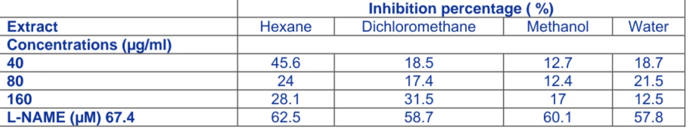
50value of 60.69 ± 2.58 and 39.83 ± 4.56 µg/ml (resazurin test) and 44.52 ± 5.96 and 55.65 ± 6.00 µg/ml (hoechst test), respectively. Besides, the highest anti-inflammatory activity, inhibiting nitric oxide (NO) release, was exhibited by dichloromethane extract with 31.5 % at 160 µg/ml in lipopolysaccharide (LPS)-stimulated RAW 264.7 macrophages. The HPLC analysis showed that catechol and kaempferol were the major phenolics. These data suggest the richness of all fractions of Ucria root on interesting bioactive molecules with different polarity and confirm the known traditional therapeutics virtues of this species for the treatment of dysentery, diarrhea and gastric ulcer.
Keywords: Rhus tripartitum, phenolics, anticancer ability, antioxidant capacity, anti-inflammatory activity
INTRODUCTION
The human body is equipped with many antioxidant defense systems to preserve healthy cell membranes against free radicals and active oxygen species (Kaur and Kapoor,
2001). A large diversity of free radical scav-
enging molecules found in plants, such as
phenolic compounds, carotenoids, vitamins,
and some other endogenous metabolites, are
found to exhibit powerful antioxidant activity
(Cai et al., 2003). Therefore, antioxidant com- pounds provided by the diet may not only re- tard oxidative degradation of lipids and thereby promote food quality and nutritional value (Ksouri et al., 2009), but also enrich the antioxidative status of living cells and there- fore decrease the damage, mainly in the el- derly (Shukla et al., 1997). Phenolic com- pounds as powerful antioxidants in plants play a crucial role to quench reactive oxygen species responsible in oxidative stress, these bioactive antioxidants are increasing and may reach 8000 (Havsteen, 2002; Wollgast and Anklam, 2000). Previous studies have shown that many of these antioxidant compounds possess biological activities such as anti-in- flammatory, anti-atherosclerotic, antitumor, antimutagenic activities (Sala et al., 2002).
Besides, it has been shown that there might be a relation between the intake of natural anti- oxidants and the lowering of risks of several diseases related to ageing like cancer, cardio- vascular disease, and diabetes (Ksouri et al., 2012; Yang et al., 2001). Accordingly, some phenolic antioxidants are assumed as preven- tive and curative mediators against UV-radia- tions (Weiss and Landauer, 2003) which are responsible for skin diseases (Eberhardt et al., 2000; Ganesan et al., 2011). In Tunisia, Rhus tripartita (Ucria) Grande is a known medici- nal plant used mainly for the treatment of di- arrhea and dysentery (Abbassi and Hani, 2012). Rhus tripartitum root cortex extract was reported to contain interesting phenolics (proanthocyanidic oligomers and polymers) with strong antioxidant capacity and prevent thymocytes apoptosis in rat (Tebourbi et al., 2006). Other phenolic compounds have been identified in this plant which exhibit anti-in- flammatory and antimicrobial properties (Mahjoub et al., 2006; Abbassi and Hani, 2012). So far, no other study of biological ac- tivities of Rhus tripartitum root was per- formed, that's why we propose to investigate in vitro and ex vivo antioxidant, anti-inflam- matory and anticancer abilities and to identify and quantify their phenolic content using HPLC system after extraction by soxhlet us- ing different solvents.
MATERIALS AND METHODS Preparation of crude plant extracts
Roots of Rhus tripartita (Ucria) Grande [=R. tripartitum (Ucria) D.C. = R. oxya-can- thoides Dum. Cours. = R. oxyacantha Shousb.
Ex. Cav.] (Pottier-Alapetite, 1979; Le Floc’h and Boulous, 2008) were collected in April 2012 from Labayadh–Hajeb layoun city. This medicinal plant was identified at the Biotech- nology Centre of Borj-Cédria, and a voucher specimen (RT-CBBC-53) was placed in our Laboratory. To assess biological activities, 30 g of powdered roots were extracted in a soxhlet system with 4 increasing polarity sol- vents (hexane, dichloromethane, methanol and water). Then, root extracts were filtered, and the solvent was evaporated using rotary vacuum evaporator. At last, root samples were lyophilized and the residue was dis- solved in dimethyl sulphoxide prior to analy- sis.
Assessment of antioxidant activities ORAC
FLassay
The procedure was modified from the method described by Ou et al. (2001). The ORAC assay was accomplished in black round bottom 96-well microplates (Costar) on a Fluoroskan Ascent FL™ plate reader (Lab- systems) using an automated injector. The test was directed at 37.5 °C and in pH 7.4 phos- phate buffer, with a blank sample in parallel.
Different concentrations of Trolox as the con- trol standard were used in quadruplicate, and a gradient of 16 concentrations of the extracts was set without replication. The fluorimeter was planned to register the fluorescence (λ ex.: 485 nm/em.: 530 nm) of fluorescein each minute once adding of 375 mM of 2,2-azobis (2-amidinopropane) dihydrochloride (AA- PH), for 35 min. The final results were calcu- lated using the net area under the curves of the extract concentrations for which reduction of at least 95 % of fluorescence was detected at 35 min and which also showed a linear dose–
response pattern. ORAC values were stated in
micromoles of Trolox equivalents (TE) per
gram (µmol TE/g).
Antioxidant cell assay using 2', 7'-dichloro- fluorescin-diacetate (DCFH-DA)
Antioxidant activity was assessed via the DCFH-DA test as reported by Legault et al.
(2003), with changes. Human skin fibroblast cells were plated in 96 microwell plates at 10,000 cells per well and incubated for 48 hrs at 37 °C and 5 % CO
2. Next, the cells were washed with 150 µl Hank’s balanced salt so- lution (HBSS) at pH 7.4 and incubated for 30 min with 100 µl HBSS (pH 7.4) holding 5 µM DCFH-DA (Sigma–Aldrich). Then, the cells were washed with 150 µl HBSS. To as- sess antioxidant activity, the cells were incu- bated either with a growing concentration of samples from Rhus tripartitum, quercetin or Trolox, in the presence or absence of 200 µM tert-butylhydroperoxide (tBH). Fluorescence was quantified after 1 and 4 hrs on the auto- mated 96-well plate reader (Fluoroskan As- cent FL™, Labsystems) via an excitation wavelength of 485 nm and an emission wave- length of 530 nm.
Cell culture
The human lung carcinoma A-549 (ATCC #CCL-185) and colon adenocarci- noma DLD-1 (ATCC #CCL-221) cell lines were purchased from the American Type Cul- ture Collection (ATCC, Manassas, USA). The A-549 and DLD-1 cell lines were developed in Minimum Essential Medium with Earle’s salts. The media was complemented with 10 % foetal calf serum (Hyclone, Logan, USA) for (A-549 and DLD-1), solution of vit- amins (1×), sodium pyruvate (1×), nonessen- tial amino acids (1×), penicillin (100 IU) and streptomycin (100 µg/ml) (Mediatech Cellgro). Cells were cultivated in a humidi- fied atmosphere at 37 °C in 5 % CO
2.
Cytotoxicity assay
Exponentially growing cells were plated at a density of 5×10
3cells per well in 96-well microplates (Costar, Corning Inc.) in 100 µl of culture medium and were permitted to paste for 16 hrs before treatment. Then, 100 µl of growing concentrations of sample dis- solved in the suitable solvent (DMSO) were
added. The final concentration of solvent in the culture medium was preserved at 0.5 % (v/v) to evade solvent toxicity. The cells were incubated for 48 hrs in the presence or in the absence of sample. Cytotoxicity was meas- ured by means of the resazurin reduction test as reported by O’Brien et al. (2000). Fluores- cence was quantified on an automated 96-well Fluoroskan Ascent Fl™ plate reader (Labsys- tems) with an excitation wavelength of 530 nm and an emission wavelength of 590 nm.
Cytotoxicity was stated as the concentration of extract inhibiting cell growth by 50 % (IC
50).
Measurement of anti-inflammatory activity by nitrite quantification
Exponentially growing cells were plated in 24-well microplates (BD Falcon) at a den- sity of 2 ×10
5cells per well in 400 µl of cul- ture medium and were allowed to adhere overnight. Cells were then processed with or without N(G)-nitro-L-arginine methyl ester (L-NAME) as positive control, or growing concentrations of samples dissolved in the proper solvent, and incubated at 37 °C, 5 % CO
2for 24 hrs. The ultimate concentration of solvent in the culture medium was sustained at 0.5 % (v/v) to evade solvent toxicity. Then, cells were stimulated with 100 µg/ml lipopol- ysaccharide (LPS). After 24 hrs, cell-free su- pernatants were gathered and deposited at 80 °C until NO measurement using the Griess reaction (Green et al., 1990) with slight changes. In the beginning, 100 µl aliquots of cell supernatants were incubated with 50 µl of 1 % sulphanilamide and 50 µl of 0.1 % N-1- naphtylethylenediamine dihydrochloride in 2.5 % H
3PO
4at room temperature for 20 min.
Then, absorbance at 540 nm was evaluated by
an automated 96-well Varioskan Ascent plate
reader (Thermo Electron) and the existence of
nitrite was measured by comparison with an
NaNO
2standard curve.
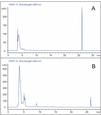
High performance liquid chromatography analysis
The identification of phenolic compounds in methanol and water root fractions was done using an HPLC system equipped with a Hypersil ods-C18 analytical column of 4.6 x 100 mm and 0.5 μm particle size. Column temperature was sustained at 25 °C. The flow- rate of mobile phase was 0.7 mL/min and the injected extract volume was 2 μl. Mobile phase C was milli-Q, water was composed of 0.2 % formic acid and mobile phase B was ac- etonitrile. The gradient program was as fol- lows: 35B/65C (0-6 min); 60B/40C (6- 9 min); 80B/20C (9-14 min); 100B/0C (14- 25 min) and 35B/65C (25-30 min). The dif- ferent detected compounds were identified by comparing their retention time with those of injected authentic standards.
Statistical analysis
Means were compared statistically via the PRISMA PAD program (version 5), with Stu- dent’s t-test at the P < 0.05 significance level.
A one-way analysis of variance (ANOVA) and Newman–Keuls multiple range test were carried out to test any significant difference between species at P < 0.05.
RESULTS AND DISCUSSION In vitro and ex vivo antioxidant activity in several extracts of Rhus tripartitum roots
ORAC and DCFH are among the most re- liable in vitro and ex vivo antioxidant test re- spectively. In fact, the oxygen radical absorb- ance capacity (ORAC) method has been found to be the most relevant one for biologic samples (Huang et al., 2005). Besides, the rapid cell-based assay using dichlorofluo- rescin (DCFH) oxidation indirectly measure the effect of intracellular antioxidant activi- ties in scavenging the reactive oxygen species (ROS) and assess the pro- and antioxidant po- tential of various pure compounds, fruits and vegetable juices (Girard-Lalancette et al.,
2009). Results (Table 1) showed that root wa- ter extract exhibited a potent antioxidant ac- tivity that could inhibit the tBH-induced oxi- dation of DCFH with an IC
50value of 0.28 µg/ml. Contrariwise, inhibition of DCFH-oxidation by methanol, hexane and di- chloromethane extract was lower (respec- tively, 14.39, 35.25 and 20.52 µg/ml). In ad- dition, this value of water extract is higher than those found in other plants (Suaeda fru- ticosa, Zygophyllum album and Solanum elaeagnifolium) from Tunisia (Oueslati et al., 2012; Ksouri et al., 2013; Mejri et al., 2014 respectively) suggesting the strongly pro-oxi- dation effect of Rhus tripartitum roots. The antioxidant activity of root extracts (hexane, dichloromethane, methanol and water) was assessed in vitro using the ORAC assay. Re- sults indicate that water and methanol extracts have statistically similar and strong antioxi- dant activity with respectively an ORAC value of 8.95 and 8.55. Quercetin, used as an antioxidant standard exhibited the highest ORAC value (21.45 µmol Trolox/ml). Hex- ane and dichloromethane extract exhibited a lower activity with ORAC values of 0.11 and
0 .
06 µmol Trolox. These results clearly demonstrate the superiority of polar extract (water and methanol) to non-polar ones (hex- ane and dichloromethane) and that this anti- oxidant activity may be due to the richness of water extract mainly in phenolic compounds.
Previous works on edible plants showed that polar extract has a better ORAC activity than non-polar one (Oueslati et al., 2012; Ksouri et al., 2013). Moreover, a comparison of antiox- idant activities of guava fruit extracts showed a superiority of the methanol extract against dichloromethane extract (Thaipong et al., 2006). Accordingly, Kratchanova et al.
(2010) reported the important impact of sol-
vent extracting on the assessment of antioxi-
dant activity and phenolic content, suggesting
the use of several extraction methods to im-
prove the investigation of the antioxidant ac-
tivity of the natural compound.
Table 1: Oxygen radical absorbance capacity (ORAC) values and antioxidant cell assay expressed as IC
50of four extracts from Rhus tripartitum roots and standards. Each value represents the mean ± SD of three determinations.
EXTRACTS ORAC (µmolTrolox/mg) IC
50(µg/ml) Hexane 0.11 ± 0.02 35.25 ± 5.81 Dichloromethane 0.89 ± 0.06 20.52 ± 4.91 Methanol 8.55 ± 1.4 14.39 ± 1.80 Water 8.95 ± 0.47 0.28 ± 0.12 Quercetin 21.45 ± 1.17 0.23 ± 0.05
Trolox - 0.02 ± 0.01
Evaluation of Rhus tripartitum roots cortex extract cytotoxicity against tumor cell lines Over the past years, there was a lot of fo- cus on natural compound as potential drugs to prevent or treat pathologies such as cancer which was always associated with inflamma- tory responses (Kim et al., 2012) and suggest- ing that inflammation played a crucial role in cancer progress. Extracts from Rhus root cor- tex were evaluated on the anticancer effects with the purpose of better understanding their relationship with their anti-inflammatory ef- fect. In this context, the methanol, hexane, di- chloromethane and water extracts of Rhus tri- partitum root cortex were subjected to in vitro screening, to evaluate their potential cytotoxic activity against human cancer cell lines. The results presented in Table 2 showed that, in particular, the methanol extract inhibited the proliferation of the tumor cell lines unlike other three extracts which were poorly active
against carcinoma cell lines. Also, respec- tively, the IC
50values of methanol extract ac- tive against the two carcinoma A-549 and DLD-1(60.69 and 39.83µg/ml), were compa- rable to those of etoposide used as standard (35.52 µg/ml). Contrariwise, other recent re- search reported that hexane (Tundis et al., 2011) and dichloromethane (Oueslati et al., 2012; Ksouri et al., 2013) extracts were mostly active against the two carcinoma and showed no significant cytotoxicity towards healthy human skin fibroblast cell lines WS1.
In addition, methanolic twig extract from Le- dum groenlandicum was also active against DLD-1 colon carcinoma and A-549 lung car- cinoma cells (Dufour et al., 2007). Alto- gether, these results advocate the appreciable anti-tumour activity of Rhus tripartitum which prompt it to be considered as a poten- tial source of anticancer compounds.
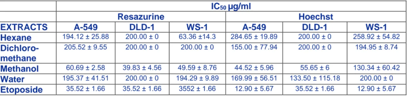
Table 2: Cytotoxic activity of several extracts from Rhus tripartitum against two tumour (A-549, DLD-1) and one healthy (WS1) cell lines.
IC
50µg/ml
Resazurine Hoechst
EXTRACTS A-549 DLD-1 WS-1 A-549 DLD-1 WS-1 Hexane
194.12 ± 25.88 200.00 ± 0 63.36 ±14.3 284.65 ± 19.89 200.00 ± 0 258.92 ± 54.82Dichloro- methane
205.52 ± 9.55 200.00 ± 0 200.00 ± 0 155.00 ± 77.94 200.00 ± 0 194.95 ± 8.74