The Corticotropin-Releasing Factor System in Lactating Rats: Implications in the Regulation of Maternal Behavior with Special Focus on the BNST
DISSERTATION ZUR ERLANGUNG DES DOKTORGRADES DER NATURWISSENSCHAFTEN (DR. RER. NAT.)
DER FAKULTÄT FÜR BIOLOGIE UND VORKLINISCHE MEDIZIN DER UNIVERSITÄT REGENSBURG
vorgelegt von Stefanie Klampfl
aus
Vilshofen an der Donau
im Jahr 2014
Das Promotionsgesuch wurde eingereicht am:
26.09.2014
Die Arbeit wurde angeleitet von:
PD Dr. rer. nat. Oliver J. Bosch
Unterschrift:
“Around here, however,
we don’t look backwards for very long.
We keep moving forward, opening new doors and doing new things, because we’re curious…
And curiosity keeps leading us down new paths.”
Walt Disney
Declaration of Included Manuscripts
Chapter 2: Reduced brain CRF receptor activation is required for adequate maternal care and maternal aggression in lactating rats.
Authors‘ contribution:
Stefanie Klampfl: experimental design, performance of experiments, data analysis, first draft of manuscript
Inga Neumann: revision of manuscript
Oliver Bosch: experimental design, performance of experiments, revision of manuscript
Taken and partly adapted from: Klampfl SM, Neumann ID, Bosch OJ (2013) Reduced brain CRF receptor activation is required for adequate maternal care and maternal aggression in lactating rats. Eur J Neurosci 38:2742-50.
Chapter 3: Hypo-activation of CRF receptors, predominantly type 2, in the medial-posterior BNST is vital for adequate maternal behavior in lactating rats.
Authors‘ contribution:
Stefanie Klampfl: experimental design, performance of experiments, data analysis, first draft of manuscript
Paula Brunton: performance of in situ hybridization, revision of manuscript Doris Bayerl: performance of experiments, revision of manuscript
Oliver Bosch: experimental design, performance of experiments, revision of manuscript
Taken and partly adapted from: Klampfl SM, Brunton PJ, Bayerl DS, Bosch OJ (2014) Hypo-activation of CRF receptors, predominantly type 2, in the medial- posterior BNST is vital for adequate maternal behavior in lactating rats. J Neurosci, 34:9665-76.
Chapter 4: Opposing effects of subtype-specific CRF receptor activation in the adBNST on maternal care and the stress axis in lactating rats
Authors‘ contribution:
Stefanie Klampfl: experimental design, performance of experiments, data analysis, first draft of manuscript
Doris Bayerl: performance of experiments
Oliver Bosch: experimental design, performance of experiments, revision of manuscript
Chapter 5: The CRF binding protein: an underestimated brain neuropeptide indispensable for the expression of maternal behavior in lactating rats
Authors‘ contribution:
Stefanie Klampfl: experimental design, performance of experiments, data analysis, first draft of manuscript
Doris Bayerl: performance of experiments
Oliver Bosch: experimental design, performance of experiments, revision of manuscript
Table of Contents
1.1 Maternal behavior ... 4
1.1.1 Hormonal basis of maternal behavior ... 4
1.1.2 Neuropeptidergic basis of maternal behavior ... 6
1.1.2.1 Maternal care ... 6
1.1.2.2 Maternal motivation ... 9
1.1.2.3 Maternal aggression ... 10
1.1.2.4 Maternal anxiety ... 13
1.1.3 Rat animal model for extremes in anxiety and maternal behavior ... 14
1.2 The CRF system ... 17
1.2.1 CRF family members ... 17
1.2.1.1 CRF ... 17
1.2.1.2 Ucn 1 ... 23
1.2.1.3 Ucn 2 and Ucn 3 ... 25
1.2.1.4 CRF receptors ... 27
1.2.1.5 CRF-BP ... 33
1.2.2 CRF as regulator of the HPA axis ... 35
1.2.3 The CRF system and mood disorders-focus on the peripartum period . 38 1.3 The CRF system in the peripartum period ... 41
1.4 Aim of the thesis ... 43
2.1 Abstract ... 46
2.2 Introduction ... 47
2.3 Materials & Methods ... 48
2.3.1 Animals ... 48
2.3.2 In situ hybridisation for CRF mRNA expression ... 49
2.3.3 Implantation of icv guide cannula in lactating rats ... 50
2.3.4 Acute icv infusion of CRF or D-Phe in lactating rats ... 50
2.3.5 Experimental design ... 51
2.3.6 Behavioral tests ... 51
Abstract ... 1
Chapter 1 - General Introduction ... 3
Chapter 2 - Reduced brain CRF receptor activation is required for adequate maternal care and maternal aggression in lactating rats... 45
2.3.7 Histology ... 53
2.3.8 Statistical analysis ... 53
2.4 Results... 54
2.4.1 Experiment 1: CRF mRNA expression in the pPVN of virgin versus lactating rats ... 54
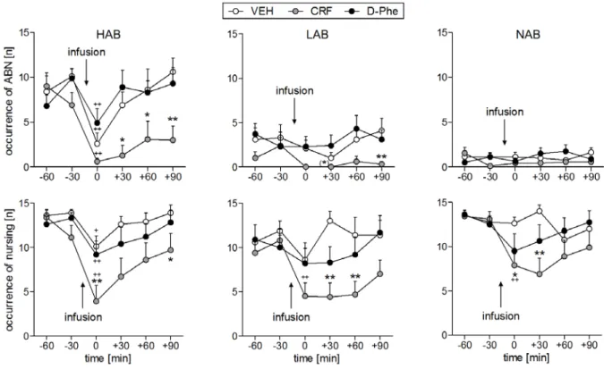
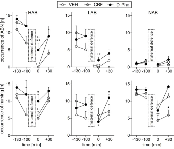
2.4.2 Experiment 2: Behavioral effects of non-specific icv CRF-R1/2 manipulation on maternal behavior in lactating rats ... 55
2.5 Discussion ... 64
3.1 Abstract ... 73
3.2 Introduction ... 74
3.3 Materials & Methods ... 75
3.3.1 Animals ... 75
3.3.2 Behavioral tests ... 76
3.3.3 Experimental design ... 79
3.3.4 Histology ... 82
3.3.5 In situ hybridization for CRF-R mRNA expression ... 82
3.3.6 Statistical analysis ... 83
3.4 Results... 83
3.4.1 Experiment 1: Behavioral effects of non-specific icv manipulation of CRF- R1/2 in lactating rats ... 83
3.4.2 Experiment 2: Expression of CRF-R1 and CRF-R2 mRNA in the BNST of virgin versus lactating rats ... 87
3.4.3 Experiment 3: Behavioral effects of intra-mpBNST CRF-R1 or -R2 manipulation in lactating rats ... 88
3.4.4 Experiment 4 and 5: Behavioral effects of intra-mpBNST CRF-R1 or -R2 blockade on anxiety in virgin and male rats ... 95
3.5 Discussion ... 96
4.1 Abstract ... 103
4.2 Introduction ... 104
4.3 Materials & Methods ... 106
4.3.1 Animals ... 106
Chapter 3 - Hypoactivation of CRF receptors, predominantly type 2, in the medial-posterior BNST is vital for adequate maternal behavior in lactating rats ... 72
Chapter 4 - Opposing effects of subtype-specific CRF receptor activation in the adBNST on maternal care and the stress axis in lactating rats ... 102
4.3.2 Behavioral tests ... 106
4.3.3 Experimental design ... 109
4.3.4 Histology ... 111
4.3.5 Statistical analysis ... 112
4.4 Results... 112
4.4.1 Experiment 1: Intra-adBNST manipulation of CRF-R1 or -R2 in lactating rats ... 112
4.4.2 Experiment 2: Effects of intra-adBNST manipulation of CRF-R1 or -R2 on the HPA axis under basal and stressful conditions ... 118
4.5 Discussion ... 121
5.1 Abstract ... 129
5.2 Introduction ... 130
5.3 Materials & Methods ... 132
5.3.1 Animals ... 132
5.3.2 Behavioral tests ... 133
5.3.3 Experimental design ... 135
5.3.4 Histology ... 137
5.3.5 Statistical analysis ... 137
5.4 Results... 137
5.4.1 Experiment 1: Behavioral effects of icv inhibition of the CRF-BP ... 137
5.4.2 Experiment 2: Behavioral effects of local inhibition of the CRF-BP in the mpBNST ... 141
5.4.3 Experiment 3: Behavioral effects of local inhibition of the CRF-BP in the adBNST ... 145
5.5 Discussion ... 148
6.1. Summary of results ... 155
6.2. The CRF system and its impact on maternal care ... 157
6.3. The CRF system and its impact on maternal motivation ... 163
6.4. The CRF system and its impact on maternal aggression ... 164
6.5. The CRF system and its impact on maternal anxiety ... 166
6.6. The CRF system in the BNST and potential interactions with other neuropeptide systems ... 170
6.7. Translational aspects ... 174
Chapter 5 - The CRF binding protein: an underestimated brain neuropeptide indispensable for the expression of maternal behavior in lactating rats ... 128
Chapter 6 - General Discussion ... 154
6.8. Conclusion and Perspective ... 176
Abbreviations ... 180
References ... 182
Supplementary ... 215
Curriculum vitae ... 223
Publications ... 224
Danksagung ... 226
Abstract
The survival of the offspring strongly depends on the adequate expression of maternal behavior. Any dysregulations during the peripartum period can easily result in behavioral or physiological maladaptations that are detrimental to the offspring as well as to the mother herself. One factor, potentially contributing to dysregulations peripartum, is the corticotropin-releasing factor (CRF) system, which triggers nearly every behavioral and physiological response to stress. Given that the behavioral role of the CRF system during lactation is still under-investigated I aimed to characterize its implications in the regulation of maternal behavior in lactating rats. Therefore, I used a variety of methods including in situ hybridization, ELISA, repeated intravenous blood sampling, and central or local acute pharmacological manipulation of CRF receptors (CRF-R) or the CRF binding protein (CRF-BP) followed by various behavioral tests. For local manipulation, I focused on the bed nucleus of the stria terminalis (BNST) given its role in maternal behavior and its abundant expression of most members of the CRF family.
In the present thesis, I showed that the CRF system needs to be down-regulated postpartum given that central and local activation of CRF-R in the BNST impaired maternal care and maternal aggression, increased anxiety-related behavior but had no effect on maternal motivation. Importantly, the effects of CRF-R activation differed specifically in the various subdivisions of the BNST depending on the CRF-R subtype. In the mpBNST, predominantly CRF-R2 activation reduced maternal care and maternal aggression while both CRF-R1 and CRF-R2 mediated anxiety-related behavior. In the adBNST, CRF-R manipulation solely affected maternal care; CRF- R1 activation reduced maternal care while CRF-R2 stimulation reduced nursing but increased arched back nursing in a time-dependent manner. These behavioral
changes following specific CRF-R manipulation in the adBNST might be indirectly mediated via activation of the hypothalamo-pituitary-adrenal axis. Interestingly, I could demonstrate that the CRF-BP, which appears to sequester extracellular CRF / urocortin 1 and inhibit their binding to the CRF-R, has emerged as potent regulator of the required down-regulation of CRF-R activity as well as of maternal aggression, especially in the mpBNST.
In conclusion, both central and local activation of CRF-R in the BNST are detrimental to the appearance of maternal behavior in lactating rats. Therefore, CRF-R activation needs to be down-regulated postpartum, which is partly achieved and supported by altered expression patterns of CRF-R and its ligands as well as by the CRF-BP. This well balanced interplay of the CRF family members is essential to enable the mother showing adequate maternal behavior and, thus, to increase her fitness.
- Chapter 1 -
General Introduction
Across all species reproduction and, hence, successful rearing of the young represents the major aim in life. In mammals, this aim is achieved by the recruitment of a wide array of behavioral and physiological adaptations of the parents. Parental behavior is defined as “any behavior of a member of a species toward a reproductively immature conspecific that increases the probability that the recipient will survive to maturity” (Numan and Insel, 2003). Approximately 90 % of all mammalian species have established a promiscuous, uniparental care system with the mother as primary caregiver due to her ability to lactate (Numan and Insel, 2003).
Thus, maternal behavior has emerged as the most important pro-social female behavior and guarantees the survival and development of the offspring.
1.1 Maternal behavior
1.1.1 Hormonal basis of maternal behavior
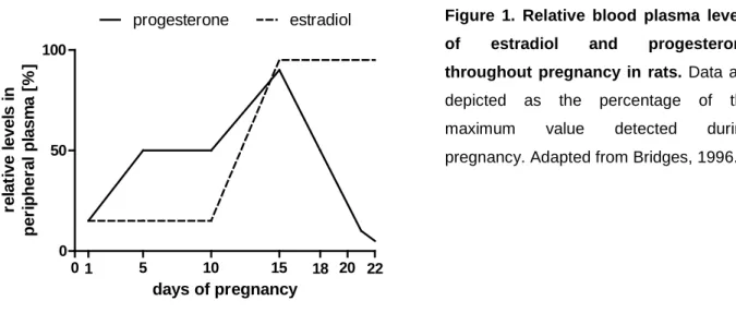
A variety of mammals show a basic level of responsiveness to the young that is activated by stimulation with the infant. However, a fundamental change in the (neuro-)endocrine system is indispensable in many species to prepare for motherhood (Numan and Insel, 2003). In rats, the onset of maternal behavior is induced by steroid hormones that are secreted in gestation-specific patterns throughout pregnancy (Bridges, 1996). Plasma estradiol concentrations are low during the first part of pregnancy and rise around day 15, after which peak levels are maintained until parturition. In contrast, progesterone levels are high throughout the first part of pregnancy, peaking at day 15 followed by a decline that becomes abrupt after day 20. Hence, shortly before parturition, a reversal of the estrogen-to- progesterone ratio can be observed (Figure 1). In primates and humans, pregnant females show similar changes in their hormonal system; however, the drastic drop in
progesterone is not required for the onset of labor and maternal behavior (Bahr et al., 2001).
0 5 10 15 20
0 50 100
progesterone estradiol
22 18
1
days of pregnancy relative levels in peripheral plasma [%]
Figure 1. Relative blood plasma levels of estradiol and progesterone throughout pregnancy in rats. Data are depicted as the percentage of the maximum value detected during pregnancy. Adapted from Bridges, 1996.
These hormonal changes are accompanied by increased production and secretion of pituitary prolactin (PRL) and placental lactogens throughout most of pregnancy (Grattan, 2001). During the first half of pregnancy, PRL is released from the pituitary in two daily surges, while during most of the second half of pregnancy PRL secretion is low except for an increase on the final day of pregnancy. The low PRL release in the second half of pregnancy is accompanied by rising plasma levels of lactogens.
Thus, pregnant rats are most of the time exposed to chronic lactogenic and progestin stimulation. The high levels of estradiol, PRL and lactogens act on neurons in the medial preoptic area (MPOA) and prime them for a successful onset of maternal behavior (Numan et al., 1977; Bridges et al., 1990; Bridges et al., 1997). This event is finally initiated by central neurotransmitter systems; in addition to PRL, the neuropeptides oxytocin (OXT) and arginine-vasopressin (AVP) are released to promote an immediate onset of maternal behavior (Pedersen and Prange, 1979;
Pedersen et al., 1982; Pedersen et al., 1994).
1.1.2 Neuropeptidergic basis of maternal behavior
The nonapeptides OXT (van Leengoed et al., 1987; Numan and Insel, 2003; Bosch et al., 2005; Bosch and Neumann, 2012) and AVP (Bosch and Neumann, 2008, 2010; Bosch et al., 2010; Bosch, 2011; Bosch and Neumann, 2012) are not only fundamentally involved in the onset of maternal behavior but play also a crucial role in maintaining appropriate levels of maternal behavior (for further details see below).
Before going into detail with the neuropeptidergic regulation, I will introduce the behavioral variety of maternal behavior, which can be divided into maternal care, maternal motivation, and maternal aggression and is accompanied by adaptations in maternal anxiety.
1.1.2.1 Maternal care
During motherhood, a rat dam shows a variety of pup-directed behaviors, which serves to ensure the well-being and development of the offspring. The dam licks and grooms the pups, which helps them to urinate and defecate, having a high impact on their social and emotional development (Caldji et al., 1998; Champagne, 2008).
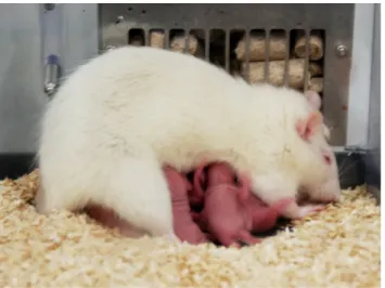
Additionally, the dam employs various nursing positions including blanket posture, hovering over the pups, and arched back nursing (ABN; Figure 2) in order to provide the young with sufficient nourishment.
Figure 2. Lactating rat fully engaged in ABN. ABN is characterized by a quiescent kyphotic posture, which enables the pups a perfect access to the teats in a protective environment (Stern and Johnson, 1990).
ABN is the most characteristic nursing behavior of rats and is classified as the only active nursing position with the dam being fully engaged in a quiescent kyphotic posture (Stern and Johnson, 1990). The quiescence of the dam is important not only to enable the pups to become and remain attached to the teats, but also for milk ejection to occur in response to the suckling stimuli. Sufficient offspring suckling is required to induce a behavioral quiescence, which is accompanied by slow-wave sleep and immobility (Voloschin and Tramezzani, 1979; Lincoln et al., 1980). The kyphotic posture is critical to prevent pups from being smothered and makes the 12 teats more readily available (Stern and Johnson, 1990). In order to induce kyphosis, which can be viewed as a reflex, ventral somatic sensory stimulation is required (Numan and Insel, 2003). Moreover, suckling as ventral tactile stimulus is able to subsequently maintain the ventroflexion for relatively long durations. Additional stimulation comes from the muzzle pushing against the teats surrounding region and from the paws treading on a wide area of the ventrum, including adjacent nipples (Stern and Johnson, 1990). This ventral stimulation is forwarded via the spinal cord to the caudal part of the periaqueductal gray (cPAG) in the midbrain (Yeziersky, 1991; Bandler and Shipley, 1994). Descending PAG projections regulate further the occurrence of motoric quiescence and immobility (Bandler and Shipley, 1994;
Cameron et al., 1995; Numan and Insel, 2003). Intriguingly, the PAG is interconnected with the bed nucleus of the stria terminalis (BNST) and the MPOA, two brain regions that are heavily involved in the promotion of maternal behavior. It was recently hypothesized that the BNST/MPOA primarily regulate active maternal behaviors like licking / grooming and pup retrieval, which are inhibited by descending projections from the PAG during kyphotic nursing (Numan and Insel, 2003). In order to terminate ABN, the BNST/MPOA send inhibiting projections to the PAG, thereby promoting the occurrence of active maternal behaviors.
The BNST and MPOA are part of the limbic system and are highly implicated in the regulation of maternal behavior (Numan and Insel, 2003; Bosch, 2011). Especially the ventral BNST (vBNST) and the dorsal MPOA (dMPOA) act in concert to form a maternal “super-region” (Numan and Insel, 2003). This neural complex receives input from the medial amygdala, which is the crucial relay site to forward olfactory stimulation from the pups either to a pup fear/avoidance circuit in virgins or to a pup attraction/approach circuit in lactating rats (Numan and Insel, 2003; Numan and Woodside, 2010). Forwarding the signal via the latter circuit includes further projections from the vBNST/dMPOA to hypothalamic nuclei such as the paraventricular nucleus (PVN) (Simerly and Swanson, 1988) thereby most likely stimulating OXT release (Numan and Woodside, 2010) or to extrahypothalamic sites such as the ventral tegmental area thereby mediating maternal motivation (Numan and Stolzenberg, 2009) (see 1.1.2.2). Interestingly, the state of the art indicates that maternal care is primarily mediated by the cPAG and the vBNST/dMPOA while other important limbic brain regions like the dorsal or posterior BNST (pBNST) have been largely neglected so far. To my knowledge, only one study investigated the implications of the pBNST in the occurrence of maternal care and assessed OXT and AVP release in the pBNST and their behavioral implications in lactating rats (Bosch
et al., 2010). They showed that both neuropeptide systems in the pBNST are apparently not involved in mediating maternal care but are rather vital for the regulation of maternal aggression (see 1.1.2.3).
1.1.2.2 Maternal motivation
The dam’s brain needs to undergo further adaptations in order to be responsive to their young and to seek and maintain contact with them. In rats, retrieval behavior occurs when a mother carries pups in her mouth one at a time to transport displaced pups back to the nest or to move them to a new nest site. This process is referred to as maternal motivation and is described as an appetitive response that is voluntary, proactive and goal-directed (Numan and Stolzenberg, 2009; Numan and Woodside, 2010; Pereira and Morrell, 2011). Appetitive maternal motivation is regulated by the MPOA and its interactions with the telencephalon via the mesolimbic dopamine system appear to be the route through which MPOA neurons mediate such goal- directed behavior (Numan and Woodside, 2010). It is currently believed that OXT in the MPOA potentiates presumably glutamatergic input to the ventral tegmental area (Numan and Smith, 1984; Pedersen et al., 1994), thus stimulating the release of dopamine into the nucleus accumbens, which in turn projects to the ventral pallidum (Numan and Stolzenberg, 2009; Numan and Woodside, 2010). However, it is important to note that, even though this model has been established upon logical conclusions, it is still based on speculations and has not been causally proven yet.
Intriguingly and similar to maternal care, the BNST was not shown so far to play a role in the regulation of maternal motivation, which is supported by studies showing that manipulating OXT receptors in the anterior dorsal BNST (adBNST) (Consiglio et
al., 2005) or V1a receptors in the pBNST (Bosch et al., 2010) does not alter retrieval behavior in lactating rats.
1.1.2.3 Maternal aggression
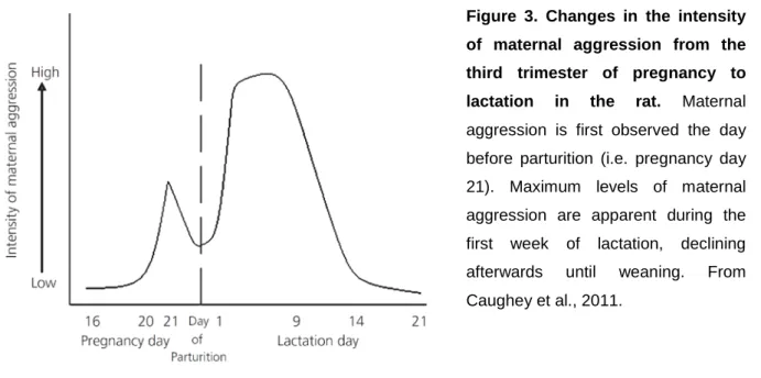
Along with the establishment of maternal care and maternal motivation, lactating females exhibit a dramatic increase in aggressive behavior, which is specifically termed maternal aggression. The term was introduced 1968 (Moyer, 1968) to describe agonistic behavior displayed by females defending their young, and was distinguished from irritable, territorial, sex-related, fear-induced, predatory and intermale aggression. Thus, maternal aggression is not directed against the young but is aimed to protect the offspring from potential external threats, e.g. infanticidal conspecifics. Lactating females have an innate drive to fight against an intruder, which is unique to the dam, as in rodents a virgin female barely ever attacks an intruder (Bosch, 2013). This intruder-targeted maternal aggression is an adaptive and highly conserved behavior that likely increases the fitness of the offspring (Wolff, 1985). Interestingly, a pregnant rat shows first signs of maternal aggression already at the end of pregnancy, which transiently decreases at parturition, and peaks during the first week postpartum (Figure 3; Caughey et al., 2011).
Figure 3. Changes in the intensity of maternal aggression from the third trimester of pregnancy to lactation in the rat. Maternal aggression is first observed the day before parturition (i.e. pregnancy day 21). Maximum levels of maternal aggression are apparent during the first week of lactation, declining afterwards until weaning. From Caughey et al., 2011.
Maternal aggression is assessed in the maternal defense test (Neumann et al., 2001) during which the dam, i.e. the resident, shows a variety of offensive and defensive behaviors toward the conspecific intruder (Erskine et al., 1978b; Lonstein and Gammie, 2002; Bosch et al., 2005). The dam’s attacks are usually directed toward the neck or back of the intruder and can be paired with bites. Additionally, the mother is frequently engaged in threat behaviors such as lateral threat, where the dam approaches the intruder sideways to force the intruder aside, as well as pinning/keeping down and aggressive grooming of the intruder. Furthermore, defensive ‘boxing’ by the dam in an upright posture in front of the intruder can occur and is termed ‘offensive upright’.
In rodents, maternal aggression is modulated by extrinsic and intrinsic factors (Bosch, 2013). On one hand, the presence of the pups, which depend on the dam’s motivation to protect them, immediately after birth is crucial for the onset of maternal aggression (Gandelman and Simon, 1980). Interestingly, the suckling stimulus per se is not sufficient (Moltz et al., 1967; Bridges, 1975) but rather tactile and especially olfactory cues emanating from the pups are essential for stimulating and maintaining
maternal aggressive behavior (Lonstein and Gammie, 2002). On the other hand, the intruder itself which embodies a potential threat to the young’s survival is highly responsible for the occurrence and intensity of maternal aggression (Bosch, 2013).
For instance, the age and/or size (Erskine et al., 1978a; Flannelly and Flannelly, 1985), which is ideally 10 % lower than the resident’s (Bosch, 2013), and the behavior of the intruder, which is usually defensive, directly impacts on the aggressive behavior of lactating rats. Furthermore, the sex of the intruder seems to influence maternal aggression in rats as male intruders receive less aggression from the lactating resident when compared with female intruders (Haney et al., 1989) independent of the intruders’ reproductive status (Neumann et al., 2001).
Besides extrinsic factors, a variety of intrinsic factors modulates the onset and maintenance of maternal aggression. Increasing levels of estrogen, but not dropping progesterone, at the end of pregnancy prime for and stimulate a first onset of maternal aggression (Lonstein and Gammie, 2002). After birth, adaptations of neuropeptidergic systems and the mother’s decreased innate anxiety (see 1.1.2.4) are crucially involved in the appearance of maternal aggression. OXT release in the BNST, PVN, central amygdala (CeA) and lateral septum (LS), and OXT receptor binding as well as general neuronal activity in the BNST and LS of lactating rats is increased during the maternal defense test (Bosch, 2013). Similarly to the adaptations in the OXT system, AVP release in the BNST, CeA and LS, and V1a receptor binding in the BNST and LS is elevated during a maternal aggressive encounter in lactating rats (Bosch, 2013). Intriguingly, the BNST in particular has emerged as a key region in the limbic system modulating maternal aggression.
1.1.2.4 Maternal anxiety
Given the importance of infant contact on almost all aspects of postpartum physiology and behavior, it seems intuitive that infants would be intimately involved in regulating the reduced anxiety seen in lactating mothers (Lonstein, 2007). In humans, breast-feeding is anxiolytic and increases positive mood in recently parturient women compared to bottle-feeding mothers (Fleming et al., 1990; Altshuler et al., 2000; Groer, 2005; Breitkopf et al., 2006). Similarly in rats, late pregnancy and lactation are accompanied by decreased anxiety compared to virgin females (Neumann et al., 2000; Lonstein, 2007). Here, physical contact with the pups, but not suckling, is necessary to reduce anxiety postpartum (Lonstein, 2005). Importantly, this effect appears to be transient as separation from the pups for as little as two hours before testing increases dam’s anxiety (Neumann, 2003). However, not only extrinsic factors but also many neurochemical systems are involved in adapting anxiety-related behavior postpartum. For example, elevated PRL (Torner et al., 2001) and OXT levels peripartum act highly anxiolytic (Neumann et al., 2000; Bosch, 2011).
Interestingly, the brain AVP system is also up-regulated in lactation but, in contrast to OXT, AVP has anxiogenic properties (Bosch and Neumann, 2008). Thus, OXT and AVP affect anxiety in an opposing manner but promote maternal behavior in the same way. Still, it needs to be considered that low anxiety levels do not necessarily entail a good performance in maternal behavior (also see 1.1.3) (Bosch, 2011). The behavioral output is generated by the integration of various signals from numerous brain sites, which most likely do not forward the anxiogenic or anxiolytic properties of neurotransmitters equally. Surprisingly, little evidence exists about specific brain regions mediating anxiety-related behavior in lactating rats. The ventrocaudal part of the midbrain PAG seems to be important as bilateral lesions further decrease anxiety-related behavior in lactating rats (Lonstein et al., 1998) while antagonizing
the OXT receptor in the ventrocaudal PAG increases anxiety to levels typically found in virgins (Lonstein, 2007). Interestingly, the AVP system in the MPOA (Bosch and Neumann, 2008) and the OXT system in the pBNST (Klampfl, Wöster, Bosch, unpublished data) are apparently not involved in the regulation of anxiety postpartum.
However, an interesting model was proposed by Lonstein (2007) involving the vBNST; physical interaction with pups may decrease noradrenaline (NA) release in the vBNST when dams are exposed to an anxiogenic situation (Figure 4). This decrease disinhibits γ-aminobutyric acid-(GABA)ergic projections from the vBNST to various brain regions including the PVN, amygdala, and the ventrocaudal PAG.
Potentially excitatory projections from the PVN and amygdala to the ventrocaudal PAG are also inhibited. Intriguingly, OXT and PRL release within and from the PVN may also be induced by vBNST disinhibition to exert anxiolytic actions postpartum.
Figure 4. Proposed neural network of the anxiolytic effects of pup contact. Physical contact with pups reduces NA release from the locus coeruleus A1 and A2 region into the ventral BNST (BSTv) and promotes a disinhibition of GABAergic projections to the paraventricular nucleus (PVN), amygdala and ventrocaudal PAG (cPAGv). Excitatory projections from PVN and amygdala to cPAGv are inhibited. Decreased cPAGv activation promotes low anxiety behaviors. From Lonstein, 2007.
1.1.3 Rat animal model for extremes in anxiety and maternal behavior
The understanding of anxiety-related behavior has advanced in the last years and has been greatly promoted by the use of specific animal models. Starting in 1993, Wistar rats were selectively bred for extremes in anxiety-related behavior as
measured on the elevated plus-maze (EPM) (Liebsch et al., 1998). Two breeding lines have emerged termed HAB (high anxiety-related behavior) and LAB (low anxiety-related behavior). Rats are selected for experimental purposes and further breeding, when HAB and LAB rats spend less than 10 % and more than 35 % of time on the open arm of the EPM, respectively. The hyper-anxious phenotype of HAB rats, found in both males (Murgatroyd et al., 2004) and females (Bosch et al., 2006;
Bosch and Neumann, 2008), derives from a single nucleotide polymorphism in the AVP promoter region, which leads to a disinhibition of the AVP gene transcription, and to increased AVP synthesis and release (Keck et al., 2002; Wigger et al., 2004).
Furthermore, HAB rats have increased corticotropin-releasing factor (CRF) mRNA levels within the PVN compared to LABs (Bosch et al., 2006), which likely contribute to their hyper-anxious phenotype (see 1.2.3). In addition to the extremes in anxiety- related behavior, HAB and LAB rats are characterized by robust differences in various physiological and behavioral parameters. HAB rats show hyperresponsiveness of the hypothalamo-pituitary-adrenal (HPA) axis (see 1.2.2) (Landgraf et al., 1999; Keck et al., 2003). Additionally, in a behavioral test for depression HAB rats spend more time floating in the forced swim test (FST) indicating increased passive stress coping and, thus, increased depressive-like behavior compared to LABs (Liebsch et al., 1998; Neumann et al., 1998a; Keck et al., 2005; Slattery and Neumann, 2010). Profound differences are also found in the display of social behaviors; male LAB rats show a lack of social preference compared to HAB or non-selected (NAB) Wistar rats (Lukas et al., 2008). Furthermore, LAB rats are prone to show abnormal aggressive behaviors toward the intruder (Beiderbeck et al., 2012) while HAB rats show low intermale aggression in the resident-intruder test (Beiderbeck et al., 2007; Veenema et al., 2007; Neumann et al., 2010). Importantly, profound differences in social and aggressive behaviors are also observed in female
HAB and LAB rats, especially during lactation (Bosch et al., 2005; Neumann et al., 2005a; Bosch et al., 2006; Bosch and Neumann, 2008, 2010). The hyper-anxious phenotype of HAB dams is linked to a protective mothering style combined with high maternal care and motivation (Bosch, 2011). They retrieve faster and even more pups in the pup retrieval test (PRT). Moreover, HAB dams leave their nest less often and spend more time on ABN as well as on nursing or on pup-directed behavior in general (Neumann et al., 2005a; Bosch et al., 2006; Bosch and Neumann, 2008). In addition to the increased display of maternal care, HAB dams are more aggressive against a virgin female intruder in the maternal defense test (Bosch et al., 2005;
Bosch, 2013). This is reflected by an increased number of attacks, reduced attack latency as well as by overall aggression compared to LAB dams. Interestingly, the behavioral performances in maternal care and maternal aggression positively correlate with their innate levels of anxiety (Bosch, 2011). A similar correlation was found for the number of attacks and the overall aggressive behavior during the maternal defense test. These results reveal that the intensity of maternal behavior depends on the dam’s innate anxiety in the HAB/LAB animal model.
Investigating maternal behavior using lactating HAB and LAB rats has advanced our understanding of the neurochemical basis of this vital female social behavior. These studies support the role of AVP and OXT in the regulation of maternal behavior during lactation and demonstrate that high levels of both neuropeptide systems underlie high levels of maternal care and maternal aggression (Bosch, 2011).
Furthermore, a vital implication of other neurotransmitter systems, e.g. the CRF system (see 1.2), is feasible.
1.2 The CRF system
The CRF system plays a key role in a diversity of behaviors accompanying stress, anxiety, and depression and in their underlying physiology. Additionally, substantial research has focused on relationships between social behaviors and the CRF system in a variety of taxa including fish, birds, rodents, and primates (Hostetler and Ryabinin, 2013).
1.2.1 CRF family members
In the last three decades, Wylie Vale’s group has identified four peptides of the CRF family in mammals: CRF (Vale et al., 1981), Urocortin (Ucn) 1 (Vaughan et al., 1995;
Donaldson et al., 1996), Ucn 2 (Reyes et al., 2001) and Ucn 3 (Lewis et al., 2001).
These four peptides bind with different affinities to the two known receptors named CRF receptor 1 (CRF-R1) and CRF-R2 (De Souza et al., 1984; De Souza et al., 1985) as well as to the CRF binding protein (CRF-BP) (Behan et al., 1989; Potter et al., 1991; Behan et al., 1995b). In addition, two further CRF family members were discovered in non-mammalian species, i.e. frog sauvagine (Montecucchi et al., 1980;
Montecucchi and Henschen, 1981) and fish urotensin 1 (Ichikawa et al., 1982).
1.2.1.1 CRF
CRF is a 41 amino acid neuropeptide and was first sequenced from ovine hypothalamus in 1980 (Vale et al., 1981). CRF was finally characterized as the ACTH releasing hormone and has been established as one of the major regulators of behavioral, neuroendocrine and autonomic responses to fear or stress (Behan et al., 1996b). The expression of CRF is encoded by only one gene, which is located on the
long arm of chromosome 8 (8q13). This gene basically codes for a 196 amino acid inactive pro-hormone termed ‘prepro-CRF’, which is transcribed and translated into the functionally active CRF peptide. The CRF gene consists of one functional promoter sequence, one intron of 800 base pairs (bps) and two exons of 582 bps.
Interestingly, 97 % DNA sequence homology is found in the first 270 bps of the promoter in humans, sheep, mice and rats demonstrating a high conservation of the gene among mammals (King and Nicholson, 2007). This suggests that not only the signals, leading to CRF gene expression, are similar across animal species but also that the mechanisms, regulating the response to stress, are conserved. Importantly, this high conservation validates the use of animal models in the investigation of CRF’s functionality and increases construct and face validity.
Given that the CRF gene has only one functional promoter, differences in the regulation of gene expression are partly achieved by the variation in transcription factors that act on response elements (RE) within the promoter (King and Nicholson, 2007). Indeed, a substantial number of RE was identified for the transcription regulatory factors MTF1 (MTF1RE), nuclear hormone receptors (HRE), ecdysone (EcRE), glucocorticoid receptor (nGRE), YY1 (YY1RE), CREB (CRE), and CDXA (CDXARE). The elements MTF1RE, HRE and EcRE activate gene transcription by inhibiting the repressive elements nGRE and YY1RE. This allows cyclic adenosinmonophosphate (cAMP) to stimulate the binding of CREB to CRE and, thus, CRF gene expression. In addition to CREB, CRF gene transcription is under the control of the CREB co-activator CRTC2. The activation, i.e. dephosphorylation, of CRTC2 and its subsequent translocation to the nucleus is essential for a full transcriptional response of the CRF gene (Liu et al., 2008; Liu et al., 2010; Liu et al., 2011). Along with cAMP a variety of molecules can stimulate CRF gene expression.
For instance, biogenic amines such as NA, acetylcholine, serotonin, cytokines and
gaseous neurotransmitters like NO and CO were shown to activate the CRF promoter (King and Nicholson, 2007). In contrast, glucocorticoids (GC) and GABA, among others, are able to inhibit CRF gene expression. The best studied inhibitory mechanism of CRF transcription is the negative feedback regulation by GC.
Activation of the HPA axis induces the production and release of GC, which exert a central negative feedback in the brain hippocampus and PVN (see 1.2.2); the GC-GC receptor complex translocates to the nucleus of CRF neurons and binds the nGRE in the CRF promoter. This inhibits the binding of the transcription machinery to the CRE region, hence reducing CRF gene transcription (Jeanneteau et al., 2012).
CRF is expressed both peripherally and centrally. In the periphery, CRF can be found in blood vessels, skin, lung, testes, ovaries, and placenta (Boorse and Denver, 2006). Main functions for peripheral CRF can be assigned within the immune and the reproductive systems. ‘Immune CRF’ is secreted from spleen, thymus, and inflamed tissues and plays a direct immunomodulatory role as an autocrine or paracrine mediator of inflammation (Karalis et al., 1991; Baigent, 2001). Stimulated by inflammation, CRF is produced in peripheral inflammatory sites acutely from peripheral nerves, i.e. postganglionic sympathetic neurons and primary somatosensory fibers, and chronically from immune cells (Webster et al., 1998).
Even though the exact mechanisms are still virtually unknown, some studies present evidence that CRF leads to the degranulation of mast cells (Theoharides et al., 1995), stimulates the secretion of interleukin 1, 2, and 6 (Singh and Leu, 1990;
Angioni et al., 1993), promotes lymphocyte proliferation (Singh, 1989), and enhances the production of oxygen radicals by macrophages (Koshida and Kotake, 1994).
‘Reproductive CRF’ participates in various reproductive functions (Kalantaridou et al., 2007). Ovarial and endometrial CRF may participate in the regulation of steroidogenesis and the inflammatory processes of the ovary (ovulation and
luteolysis) and endometrium (decidualization and blastocyst implantation). Placental CRF is increasingly secreted mainly during the latter half of pregnancy and is responsible for the onset of labor (McLean et al., 1995; Magiakou et al., 1996;
Chrousos et al., 1998). It has been proposed that there is a ‘CRF placental clock’, which determines the length of gestation and the timing of parturition (McLean et al., 1995).
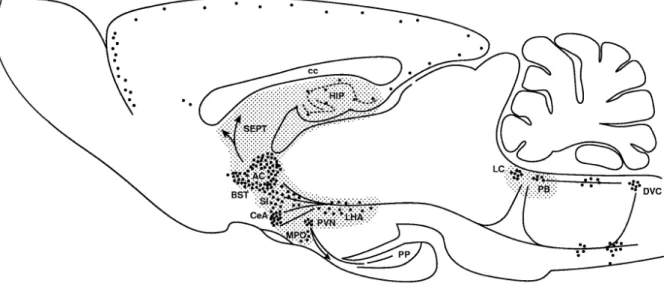
Besides peripheral actions as a hormone, CRF has strong central effects acting as neurotransmitter or neuromodulator. It is expressed in the hypothalamus, BNST, amygdala, hippocampus, the septum, cerebrocortical areas, and some brain stem regions (Figure 5) (Swanson et al., 1983; Sawchenko and Swanson, 1985).
Figure 5. Sagittal section through a rat brain depicting the location of CRF cell bodies and pathways. Shaded areas represent sites identified as mediating the effects of CRF in behavioral responses to stressors. AC, anterior commissure; BST, bed nucleus of the stria terminalis; cc, corpus callosum; CeA, central amygdala; DVC, dorsal vagal complex; HIP, hippocampus, LC, locus coeruleus; LHA, lateral hypothalamus; MPO, medial preoptic area; PB, parabrachial nucleus; PP, pituitary; PVN, paraventricular nucleus; SEPT, septum; SI, substantia innominata. From Koob and Heinrichs, 1999.
Central CRF is involved in triggering nearly every physiological and behavioral response to stress. Expressed centrally in the PVN but secreted into the periphery, CRF is primarily known as hypophysiotropic factor stimulating the HPA axis (see 1.2.2). Within the central nervous system, application of CRF either intracerebroventricularly (icv) or locally at specific brain sites produces a wide variety of behavioral effects. The behavioral pharmacological profile resulting from exogenous administration of this neuropeptide depends on the baseline state of arousal and stress of the animal (Koob and Heinrichs, 1999). In non-stressed animals under low arousal conditions, icv CRF produces a dose-dependent behavioral activation that includes increased locomotor activity, rearing, and grooming when rats are tested in a familiar environment (Sutton et al., 1982; Koob et al., 1984; Sherman and Kalin, 1987; Dunn and Berridge, 1990).
Electrophysiologically, CRF has excitatory properties as icv CRF infusion produces electroencephalographic activation characteristic of arousal at low doses (Ehlers et al., 1983) and seizure-like activity at higher doses (Ehlers et al., 1983; Marrosu et al., 1987). Furthermore, CRF can facilitate learning and memory, enhancing retention at low doses and impairing performance at higher doses (Koob and Bloom, 1985). In a more stressful environment, the profile of the behavioral effects of exogenously administered CRF changes to reflect an enhanced behavioral response to stress.
The same icv doses that produce marked behavioral activation in a familiar environment induce behavioral suppression in a novel, stressful environment (Koob and Heinrichs, 1999). Rodents pretreated with CRF show decreases in locomotion in an open field (Takahashi et al., 1989), decreased exploration in a multi-compartment chamber (Berridge and Dunn, 1986), and decreased exploration in an EPM (Baldwin et al., 1991). Furthermore, CRF increases defensive burying (Diamant et al., 1992) and reduces sexual behavior (Sirinathsinghji et al., 1983), food intake (Arase et al.,
1988) and alcohol intake (Bell et al., 1998), though drug-induced craving is reinstated by CRF (Zorrilla et al., 2014).
Interestingly, the most studied aspect of CRF’s impact on behavioral parameters is its anxiogenic property (Reul and Holsboer, 2002b). CRF increases emotionality in the open field test (Koob and Thatcher-Britton, 1985), enhances the acoustic startle response (Swerdlow et al., 1986), increases conditioned fear in a conditioned suppression test (Cole and Koob, 1988), and enhances stress-induced freezing behavior (Sherman and Kalin, 1988). The anxiogenic effects of CRF are mediated by various brain regions, including the amygdala (Bruchas et al., 2009), LS (Radulovic et al., 1999), and PAG (Miguel and Nunes-de-Souza, 2011). Another brain region which is of particular interest for CRF’s effect on anxiety-related behavior is the BNST (Davis et al., 2010). This part of the extended amygdala contains numerous CRF synthesizing cell bodies (Cummings et al., 1983; Moga et al., 1989; Morin et al., 1999) and CRF-immunopositive fibers and terminals originating from the CeA (Figure 5) (Olschowka et al., 1982; Cummings et al., 1983; Swanson et al., 1983; Morin et al., 1999). Furthermore, various studies revealed stress- or anxiety-like behavioral and autonomic effects of CRF infusions into the BNST of rodents (Lee and Davis, 1997; Liang et al., 2001; Ciccocioppo et al., 2003), whereas infusions of non- selective CRF-R antagonists into the BNST conversely produce anxiolytic-like and anti-stress effects (Greenwell et al., 2004; Jasnow et al., 2004). Intriguingly, microinfusion of CRF into the BNST increases anxiety-related behaviors dose- dependently on the EPM in male rats (Sahuque et al., 2006).
1.2.1.2 Ucn 1
Shortly after the discovery of CRF, it emerged that other CRF-related peptides must exist given that, in addition to the CRF ortholog, fish and amphibians also express a paralog, i.e. urotensin 1 and sauvagine, respectively (Bale and Chen, 2012). Based on the similarities between urotensin 1 and sauvagine, Vale’s group identified a new member of the mammalian CRF family, which was termed Ucn by that time and is now known as Ucn 1 (Vaughan et al., 1995).
The structure of the Ucn 1 gene is very similar to CRF; it contains one intron and two exons with the second exon encoding for the mature protein (Zhao et al., 1998). The promoter region contains transcription factor binding sites such as a TATA-like sequence, several GATA-binding sites, a C/EBP- and a Brn-2-binding site, a CRE sequence, and a NRSE/RE1. The latter two represent activating and inhibiting regulatory sequences, respectively.
Similar to CRF, Ucn 1 is expressed peripherally and centrally. In the periphery, Ucn 1 mRNA is found in lymphocytes, gastrointestinal tract, testes, thymus, spleen, kidney, and cardiomyocytes (Perrin and Vale, 1999; Boorse and Denver, 2006). Although the role of endogenous Ucn 1 in immune function is not completely clear, it is able to mimic some of the actions of CRF. For instance, Ucn 1 suppresses inflammation and cytokine release even more effectively than CRF, independently of endogenous GC (Agnello et al., 1998). Furthermore, the peptide protects myocytes from hypoxia- induced cell death (Okosi et al., 1998), reduces mean arterial blood pressure (Vaughan et al., 1995), and causes inhibition of gastric emptying (Nozu et al., 1999).
In the adult rat brain, Ucn 1 mRNA accumulates in the Edinger-Westphal nucleus and the lateral superior olive (Figure 6). Weaker signals are detected in the supraoptic nucleus (SON) and caudal lateral hypothalamic area as well as in neurons
of the facial, hypoglossal and ambiguual motor nuclei (Vaughan et al., 1995). Some, but not all, localization studies report further Ucn 1 mRNA in the hippocampus, amygdala, PVN, ventromedial hypothalamus, neocortex, olfactory system and cerebellum (Wong et al., 1996; Bittencourt et al., 1999). The highest density of Ucn 1-immunoreactive fibers is found in the LS, with more moderate Ucn 1 innervation in the SON, PVN, PAG, Edinger-Westphal nucleus, several brainstem nuclei, and the spinal cord (Vaughan et al., 1995; Kozicz et al., 1998; Bittencourt et al., 1999).
Many of the behavioral and physiological effects of central Ucn 1 administration are qualitatively similar to those of CRF. Many data indicate a prominent role for Ucn 1 in regulating appetite and feeding behavior. Icv Ucn 1 decreases food intake with an equal or greater potency than CRF (Spina et al., 1996). Interestingly, there is conflicting evidence with respect to a role for Ucn in anxiogenic behavioral responses. Icv Ucn 1 increases anxiety in the EPM in rats as well as in the light-dark box (LDB) and open field in mice (Moreau et al., 1997; Jones et al., 1998). However, in another study, Ucn 1 was reported to be far less potent in producing anxiety in the EPM than CRF, with no significant anxiogenesis at doses up to 1000 times that are required to suppress food intake (Spina et al., 1996). Furthermore, Ucn 1 has no effect on the amplitude of the acoustic startle response while CRF doubles the response (Jones et al., 1998). In one of the few studies of site-specific Ucn 1 administration, Ucn 1 injected into the basolateral amygdala is equipotent with CRF in producing anxiety in the social interaction test (Sajdyk et al., 1999). Similar to CRF, Ucn 1 produces a general increase in activity, i.e. grooming, alertness, locomotion (Spina et al., 1996; Jones et al., 1998), and can induce limbic seizures (Baram et al., 1997).
1.2.1.3 Ucn 2 and Ucn 3
Six years after the discovery of Ucn 1, two further family members were discovered, namely Ucn 2 (also called Stresscopin-related peptide) and Ucn 3 (also called Stresscopin) (Lewis et al., 2001; Reyes et al., 2001). The Ucn 2 gene encodes a 112 amino acid precursor protein where the C-terminus includes the coding region for the putative 38 amino acid mature peptide (Reyes et al., 2001). The Ucn 3 gene encodes a prepro-protein of 161 amino acids and a putative mature protein of 40 amino acids (Hsu and Hsueh, 2001). In contrast to CRF and Ucn 1, the genetic regulation of Ucn 2 and 3 is poorly understood and investigated to date. It was only shown that mouse Ucn 2 gene expression is up-regulated by GC acting on GRE in the promoter sequence (Chen et al., 2003).
Like CRF and Ucn 1, Ucn 2 and Ucn 3 are expressed both in the periphery and the brain. Peripherally, Ucn 2 mRNA is found in the heart, adrenal gland, human pregnant myometrial cells, and peripheral blood cells (Hsu and Hsueh, 2001; Reyes et al., 2001; Karteris et al., 2004). Ucn 2 produces vasodilatation via the adenylate cyclase - protein kinase A (PKA) pathway, inhibits cell death due to hypoxic stress in cardiomyocytes (Suda et al., 2004), reduces skeletal muscle mass and function loss during atrophy, and increases non-atrophying skeletal muscle mass and function (Hinkle et al., 2003). Ucn 3 mRNA is expressed in pancreas, the gastrointestinal tract, muscle, adrenal gland, and skin, though less abundantly than Ucn 2 mRNA (Hsu and Hsueh, 2001; Lewis et al., 2001). Similar to Ucn 1 and Ucn 2, Ucn 3 is able to inhibit cell death due to hypoxic stress in cardiomyocytes (Suda et al., 2004).
Furthermore, Ucn 3 is involved in the local regulation of glucagon and insulin secretions (Li et al., 2003).
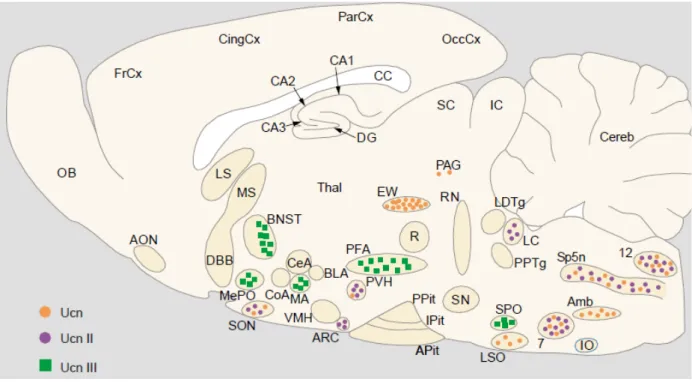
Within the central nervous system, Ucn 2 mRNA is expressed within the magnocellular division of the PVN, SON, arcuate nucleus of the hypothalamus, locus coeruleus (LC), and motor nuclei of the brain stem and spinal cord (Figure 6) (Hsu and Hsueh, 2001; Lewis et al., 2001). Ucn 3 mRNA is expressed in discrete areas including the pBNST, the medial amygdala, the hypothalamus, and brainstem (Figure 6) (Hsu and Hsueh, 2001; Reyes et al., 2001). In the hypothalamus, Ucn 3 mRNA is detected in the median preoptic nucleus, in the rostral perifornical region and in a region lateral to the PVN.
Figure 6. Distribution of Ucn 1, Ucn 2, and Ucn 3 mRNA in a sagittal section of the rat brain. 7:
facial nucleus; 12, hypoglossal nucleus; Amb, amgibuus nucleus; AON, anterior olfactory nucleus;
APit, anterior pituitary; ARC, arcuate nucleus; BLA, basolateral amygdala; BNST, bed nucleus of the stria terminalis; CA1-3, fields CA1-3 of Ammon horn; CC, corpus callosum; CeA, central amygdala;
Cereb, cerebellum; CingCx, cingulate cortex; CoA, cortical amygdala; DG, dentate gyrus; DBB, diagonal band of Broca; EW, Edinger-Westphal nucleus; FrCx, frontal cortex; IC, inferior colliculi; IO, inferior olive; IPit, intermediate lobe of pituitary; LC, locus coeruleus; LDTg, laterodorsal tegmental nucleus; LS, lateral septum; LSO, lateral superior olive; MA, medial amygdala; MePO, median preoptic area; MS, medial septum; OB, olfactory bulb; OccCx, occipital cortex; PAG, periaqueductal gray;
ParCx, parietal cortex; PFA, perifornical area; PPit, posterior pituitary; PPTg, pedunculopontine tegmental nucleus; PVH, paraventricular hypothalamus; R, red nucleus; RN, raphe nuclei; SC, superior colliculi; SN, substantia nigra; SON, supraoptic nucleus; SP5N: spinal trigeminal nucleus;
SPO, superior paraolivary nucleus; Thal, thalamus; VMH, ventromedial hypothalamic nucleus. From Reul and Holsboer, 2002b.
Central administration of Ucn 2 reveals mild motor suppressive effects (Valdez et al., 2002; Skorzewska et al., 2011), enhances a conditioned freezing fear response (Skorzewska et al., 2011), and is anorexic (Pelleymounter et al., 2004). Similarly to Ucn 2, central infusion of Ucn 3 shows acute locomotor suppressive effects, decreases stress-like behaviors (Valdez et al., 2003), and reduces food intake (Pelleymounter et al., 2004). However, the roles of Ucn 2 and Ucn 3 in the regulation of anxiety-related behavior are not clear yet. Valdez and coworkers report anxiolytic behavior 4 h after Ucn 2 infusion and 10 min after Ucn 3 infusion on the EPM (Valdez et al., 2002; Valdez et al., 2003) while Pelleymounter and colleagues show an anxiogenic effect of Ucn 2 and none of Ucn 3 on anxiety-related behavior (Pelleymounter et al., 2004). In another study, Ucn 3 did not affect anxiety in the shock probe test but was anxiolytic in the defensive withdrawal test (Zhao et al., 2007).
1.2.1.4 CRF receptors
CRF and its related peptides exert their physiological and behavioral effects via two receptors, termed CRF-R1 and CRF-R2 (Hauger et al., 2003). In general, CRF-R are G-protein coupled receptors (GPCR) belonging to the class B GPCR family of neuropeptide receptors and exhibit 70 % sequence homology to each other (Perrin and Vale, 1999). These receptors are encoded by distinct genes and have several splice variants expressed in various central and peripheral tissues. CRF-R1 has α and β isoforms in addition to subtypes designated c - h, which have been detected in human and rodent tissues. Most of these isoforms have been shown to be
nonfunctional (for detailed review see Grammatopoulos and Chrousos, 2002). CRF- R2 is expressed in three functional subtypes, i.e. α, β, and γ (Dautzenberg and Hauger, 2002). These isoforms differ in their N-terminus as well as in their distribution in both tissues and species. CRF-R2α and CRF-R2β have been detected in humans and rodents (Lovenberg et al., 1995; Liaw et al., 1996), while CRF-R2γ has only been reported in humans so far (Kostich et al., 1998). Peripherally, CRF-R1 is found in skin, ovaries, testes, and adrenals (Potter et al., 1994; Chalmers et al., 1995) while CRF-R2 is detected in heart, lungs, ovaries, testes, and adrenals (Potter et al., 1994;
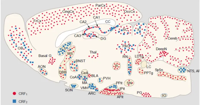
Chalmers et al., 1995; Lovenberg et al., 1995; Sanchez et al., 1999). Within the central nervous system, CRF-R1 and CRF-R2 mRNA show a distinct, but overlapping, distribution (Figure 7) (Potter et al., 1994; Chalmers et al., 1995;
Lovenberg et al., 1995; Van Pett et al., 2000). CRF-R1 is widely distributed in regions involved in sensory information processing and motor control, in the BNST, hippocampus, amygdala, thalamic nuclei, and anterior pituitary. In contrast, CRF-R2 is virtually restricted to subcortical structures such as the BNST, LS, the ventromedial hypothalamic nucleus, SON and certain amygdaloid nuclei.
Figure 7. Distribution of CRF-R1 and CRF-R2 mRNA in a sagittal section of the rat brain. AON, anterior olfactory nucleus; AP, area postrema; APit, anterior pituitary; ARC, arcuate nucleus; Basal G, basal ganglia; BLA, basolateral amygdala; BNST, bed nucleus of the stria terminalis; CA1-3, fields CA1-3 of Ammon horn; CC, corpus callosum; CeA, central amygdala; Cereb, cerebellum; CingCx, cingulate cortex; CoA, cortical amygdala; Deep N, deep nuclei; DG, dentate gyrus; DBB, diagonal band of Broca; FrCx, frontal cortex; IC, inferior colliculi; IO, inferior olive; IPit, intermediate lobe of pituitary; LC, locus coeruleus; LDTg, laterodorsal tegmental nucleus; LS, lateral septum; MA, medial amygdala; MS, medial septum; NTS, nucleus tractus solitarius; OB, olfactory bulb; OccCx, occipital cortex; PAG, periaqueductal gray; ParCx, parietal cortex; PG, pontine gray, PPit, posterior pituitary;
PPTg, pedunculopontine tegmental nucleus; PVH, paraventricular hypothalamus; R, red nucleus; RN, raphe nuclei; SC, superior colliculi; SN, substantia nigra; SON, supraoptic nucleus; SP5N, spinal trigeminal nucleus; Thal, thalamus; VMH, ventromedial hypothalamic nucleus. From Reul and Holsboer, 2002b.
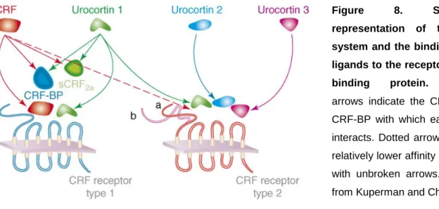
CRF-R bind CRF and its related peptides with different affinities (Figure 8). CRF binds with 10-fold higher affinity to CRF-R1 than to CRF-R2 whereas Ucn 1 shows similar high affinity for both subtypes (Perrin et al., 1999; Hauger et al., 2003). Ucn 2 and Ucn 3 bind exclusively to CRF-R2 (Hsu and Hsueh, 2001; Lewis et al., 2001;
Reyes et al., 2001).
Figure 8. Schematic representation of the CRF system and the binding of the ligands to the receptors or the binding protein. Colored arrows indicate the CRF-R and CRF-BP with which each ligand interacts. Dotted arrow indicates relatively lower affinity compared with unbroken arrows. Adapted from Kuperman and Chen, 2008.
By means of selective agonists / antagonists, antisense oligonucleotides, and transgenic mice, physiological and behavioral effects of the ligands CRF and Ucn 1 – 3 could have been attributed to either CRF-R1 or CRF-R2 (Bale and Vale, 2004).
Most findings of CRF and Ucn 1 can be assigned to signaling via CRF-R1 even though some aspects of Ucn 1 action are mediated via CRF-R2. The effects observed following Ucn 2 or Ucn 3 manipulation are exclusively mediated by CRF- R2. For instance, CRF’s (and Ucn 1’s) prominent role in anxiety-related and depressive-like behavior is mediated by CRF-R1 (Reul and Holsboer, 2002b) while the strong anorexic effects of Ucn 2 and Ucn 3 are mediated via CRF-R2 (Kuperman and Chen, 2008). Due to their specific binding profiles, the potential behavioral effects of Ucn 2 and Ucn 3 on anxiety (Valdez et al., 2002; Valdez et al., 2003;
Pelleymounter et al., 2004; Zhao et al., 2007) might also be mediated via CRF-R2.
However, CRF-R2 play a rather complicated role in anxiety and depressive pathophysiology, which entails the partly contradictory results of the different studies (see 1.2.1.3). One line of research supports the concept that CRF-R2 reestablish homeostasis by counteracting stress response-initiating effects and anxiety-like defensive behavior triggered by CRF-R1 signaling (Coste et al., 2001; Bale and Vale,
2004; Heinrichs and Koob, 2004; Muller and Holsboer, 2006). An alternative hypothesis proposes that CRF-R1 and CRF-R2 contribute to opposite defensive modes, with CRF-R1 mediating active defensive responses triggered by escapable stressors, and CRF-R2 mediating passive anxiety- and depression-like responses induced by inescapable, uncontrollable stressors (Maier and Watkins, 2004;
Valentino and Commons, 2005).
CRF and its related peptides bind to the extracellular domains of CRF-R1 and CRF- R2, which transforms the membrane conformation of the receptors into an active state. CRF-R can signal via three distinct pathways, i.e. the adenylyl cyclase - PKA pathway, the phospholipase C (PLC) - protein kinase C (PKC) pathway, and the extracellular-signal related kinase (ERK) - mitogen-activated protein kinase (MAPK) pathway. In many endogenous and recombinant cell lines, CRF-R1 and CRF-R2 preferentially signal by Gsα coupling to the third intracellular loop, leading to the activation of adenylyl cyclase and generation of the second messenger cAMP.
Increasing cAMP formation, in turn, stimulates PKA to phosphorylate downstream targets in the cytosol and CREB in the nucleus inducing transcription of certain genes (Dautzenberg et al., 2000; Dautzenberg and Hauger, 2002; Hillhouse and Grammatopoulos, 2006; Hauger et al., 2009). Interestingly, abnormalities in adenylyl cyclase – PKA signaling have been implicated in stress maladaptation, anxiety, and depression (Hauger et al., 2009). Signaling via the PLC - PKC pathway is presumably initiated by coupling to Gqα, which in turn stimulates the formation of inositol(1,4,5)-triphosphate and contributes to intracellular calcium mobilization (Gutknecht et al., 2009; Hauger et al., 2009). Dysregulations of PKC signal transduction have been associated with severe anxiety and suicide (Hauger et al., 2009). It is still unknown what factors are responsible for coupling of Gsα or Gqα and thus, the activation of the different signaling pathways. However, it was recently
shown that Ucn 2 and Ucn 3, but not CRF, binding to CRF-R2 induce ERK – MAPK signaling via a PLC-dependent mechanism (Arzt and Holsboer, 2006) whereas activation of CRF-R1 by CRF or Ucn 1 on corticotropes induces a MAPK signal transduction pathway that is downstream of PKA (Kovalovsky et al., 2002). These studies indicate that the activation of the distinct signaling pathways is dependent on ligand specificity and the induction conditions of the intracellular pathways in each cell type (Arzt and Holsboer, 2006).
The magnitude, duration and specificity of cellular signaling by GPCRs depend upon rapid and stringent regulation to prevent deleterious effects of unrestrained, excessive receptor signal transduction. While agonist binding induces conformation changes to activate the receptor and initiate signaling, specific G-protein receptor kinases (GRK) are rapidly recruited to commence homologous desensitization by phosphorylating a specific pattern of serines and threonines in the third intracellular loop and the C-terminus of the receptor (Kohout and Lefkowitz, 2003; Krasel et al., 2005; Moore et al., 2007). This GRK-mediated phosphorylation of the receptor immediately increases its affinity for β-arrestins by approximately 30-fold, triggering their translocation from the cytosol to the cell surface (Kohout and Lefkowitz, 2003;
Krasel et al., 2005; DeWire et al., 2007; Moore et al., 2007). Rapid, high-affinity binding of a single arrestin sterically uncouples the receptor from its cognate Gα protein, thereby terminating signal transduction. Following binding, class B GPCRs form stable complexes with arrestins that internalize as a unit into endocytic vesicles (Oakley et al., 1999; Oakley et al., 2000). Internalized receptors are dephosphorylated in endosomes and recycled as resensitized GPCRs back to the plasma membrane. During very prolonged exposure to high agonist concentrations, internalized GPCRs do not recycle to the membrane but move into lysosomes, where they undergo proteolytic degradation resulting in down-regulation to decrease the
total number of cellular receptors (Kohout and Lefkowitz, 2003; Moore et al., 2007).
Indeed, cell stimulation with CRF for 30 min leads to a prominent internalization of CRF-R1 (Oakley et al., 2007), which resensitize within 1 – 2 h (Hoffman et al., 1985;
Teli et al., 2005; Holmes et al., 2006). However, stimulation with CRF for 24 h decreases CRF-R1 by 70 – 80 %, presumably due to proteolytic degradation of receptors in lysosomes (Hauger et al., 1997; Perry et al., 2005). Intriguingly, following binding and activation by Ucn 2, CRF-R2 desensitize more rapidly and to a greater extent than the rate and magnitude of homologous desensitization of CRF-R1 by CRF (Hauger et al., 1997; Teli et al., 2005; Gutknecht et al., 2008; Markovic et al., 2008).
1.2.1.5 CRF-BP
In addition to the two CRF-R, the activity of CRF and CRF-related peptides is modulated by the CRF-BP, a 37 kDa secreted glycoprotein (Orth and Mount, 1987;
Behan et al., 1989). The CRF-BP gene encodes a 322 amino acid protein, of which the mature protein contains one N-linked glycosylation site (Potter et al., 1991;
Cortright et al., 1995; Jahn et al., 2001). Additionally, the CRF-BP lacks transmembrane domains or a phosphatidyl inositol anchor signal motif, suggesting that the CRF-BP does not have a direct association with the membrane (Potter et al., 1991; Eckart et al., 2001). The gene organization as well as nucleotide and amino acid sequence of the CRF-BP is highly conserved across evolution, suggesting the maintenance of a structural conformation necessary for biological activity. CRF-BP gene expression is positively regulated by stress and GC, providing an important negative feedback mechanism of the HPA axis (McClennen et al., 1998; Lombardo et al., 2001). Interestingly, the stress-induced increase in CRF-BP mRNA can persist up
to 21 h post-stress and has important implications for the effects of future stressors (Herringa et al., 2004). Furthermore, it was shown that CRF, cAMP, and interleukin-6 positively regulate CRF-BP promoter activity (Cortright et al., 1997; Mulchahey et al., 1999; Seasholtz et al., 2001). However, the CRF-BP is not only regulated by stress- related factors but is strongly influenced by reproductive hormones (Westphal and Seasholtz, 2006). For instance, the expression of CRF-BP mRNA varies over the estrous cycle, with nearly 3-fold higher levels at proestrous, when estrogen levels peak (Speert et al., 2002). This is potentially mediated by three estrogen RE half- sites in the CRF-BP promoter (van de Stolpe et al., 2004).
The CRF-BP is expressed in a highly tissue specific pattern (for review see Kemp et al., 1998). Human CRF-BP is found in plasma, amniotic fluid, synovial fluid, placenta, pituitary, and brain. In contrast, CRF-BP is not detected in rodent or ovine plasma and rat, mouse, and ovine CRF-BP mRNA and protein have been found only in brain and pituitary. In the rat brain, the CRF-BP is expressed in many specific regions including the BNST, amygdala, PVN, hippocampus, cerebral cortex, olfactory bulb, and sensory relays associated with the auditory, olfactory, vestibular, and trigeminal systems (Potter et al., 1992). The CRF-BP is expressed in both neurons and astrocytes (Behan et al., 1995a) and overlaps with CRF and CRF-R expression in several areas such as the BNST, amygdala, and pituitary (Kemp et al., 1998).
The CRF-BP is distinct from the CRF-R and binds CRF and Ucn 1, but not Ucn 2 and Ucn 3, with an equal or greater affinity than the CRF-R (Figure 8) (Sutton et al., 1995;
Vaughan et al., 1995). The CRF-BP has important modulatory roles with regards to CRF activity and may have different functions depending on the specific cell-type or context in which it is expressed (Westphal and Seasholtz, 2006). Three major hypotheses exist with regard to the central and pituitary function of CRF-BP. The
glycoprotein could act in an inhibitory fashion to sequester CRF and/or target it for degradation, which would reduce CRF-R activity. Most studies support a role for CRF-BP in limiting the availability of endogenous CRF (Potter et al., 1991; Woods et al., 1994; Cortright et al., 1995; Peto et al., 1999). In contrast to its inhibitory role, the CRF-BP could bind CRF and potentiate CRF signaling by modulating its interaction with the receptor, delivering CRF to the receptor, or extending the half-life of CRF.
Finally, CRF-BP could exert ligand and/or receptor-independent activity given that the CRF-BP is expressed at numerous sites where CRF and Ucn 1 are not detected (Potter et al., 1992).
Intriguingly, very little is known about the functional roles of the CRF-BP. Genetic mouse models have been created to elucidate physiological, neuroendocrine, and behavioral effects of CRF-BP overexpression (Burrows et al., 1998; Lovejoy et al., 1998) or deficiency (Karolyi et al., 1999) (for review see Seasholtz et al., 2001). For example, deletion of the CRF-BP gene results in increased anxiety-related behavior in males (Karolyi et al., 1999) without affecting the HPA axis (Seasholtz et al., 2001).
In order to manipulate the CRF-BP in rats, the peptide can be inhibited by a truncated version of CRF, i.e. CRF(6-33), which binds with high affinity exclusively to the CRF-BP, displaces bound CRF/Ucn 1 and, thus, increases ‘free’ extracellular CRF levels (Behan et al., 1995c). Via this approach, it was shown that inhibition of the CRF-BP leads to cognition-enhancing properties in models of learning and memory. However, barely any other behavior has been investigated using this elegant technique.
1.2.2 CRF as regulator of the HPA axis
One of CRF’s major functions is the activation of the HPA axis. Following stressor exposure, CRF and the co-secretagogue AVP are released from the parvocellular