in lactating rats -
impact of V1b receptors in hypothalamic and limbic brain regions
DISSERTATION ZUR ERLANGUNG DES DOKTORGRADES DER NATURWISSENSCHAFTEN (DR. RER. NAT.)
DER FAKULTÄT FÜR BIOLOGIE UND VORKLINISCHE MEDIZIN DER UNIVERSITÄT REGENSBURG
vorgelegt von Doris Bayerl
aus
Pfaffenhofen an der Ilm im Jahr
2016
Das Promotionsgesuch wurde eingereicht am:
Die Arbeit wurde angeleitet von:
PD Dr. rer. nat. habil. Oliver J. Bosch
Unterschrift:
“It doesn’t matter how beautiful your theory is, it doesn’t matter how smart you are.
If it doesn’t agree with experiment, it’s wrong.”
– Richard Feynman, physicist, Nobel laureate (1918–1988)
Chapter 2: Central V1b receptor antagonism in lactating rats impairs maternal care but not maternal aggression.
Authors’ contribution:
Doris Bayerl: experimental design, performance of experiments, data analysis, first draft of manuscript
Stefanie Klampfl: performance of experiments, revision of manuscript
Oliver Bosch: experimental design, performance of experiments, revision of manuscript
Taken and partly adapted from: Bayerl DS, Klampfl SM, Bosch OJ (2014) Central V1b receptor antagonism in lactating rats: impairment of maternal care but not of maternal aggression. J Neuroendocrinol 26(12):918-26.
Chapter 3: Maternal behaviour in two breeding lines for extremes in anxiety – past and present observations in HAB/LAB dams with special focus on V1b receptors
Authors’ contribution:
Doris Bayerl: experimental design, performance of experiments, data analysis, first draft of manuscript
Stefanie Klampfl: performance of experiments
Oliver Bosch: experimental design, performance of experiments, revision of
manuscript
lactating rats.
Authors’ contribution:
Doris Bayerl: experimental design, performance of experiments, data analysis, first draft of manuscript
Veronika Kaczmarek: performance of experiments
Benjamin Jurek: performance of experiments, revision of manuscript Erwin van den Burg: primer-design for qPCR
Inga Neumann: revision of manuscript Barbara Gaßner: performance of experiments
Stefanie Klampfl: performance of experiments, revision of manuscript
Oliver Bosch: experimental design, performance of experiments, revision of manuscript
Taken and partly adapted from: Bayerl DS, Kaczmarek V, Jurek B, van den Burg EH, Neumann
ID, Gaßner BM, Klampfl SM, Bosch OJ (2016) Antagonism of V1b receptors promotes
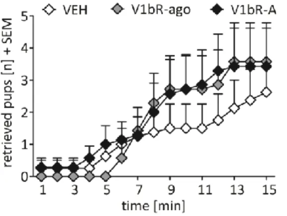
maternal motivation to retrieve pups in the MPOA and impairs pup-directed behaviour
during maternal defence in the mpBNST of lactating rats. Hormones & Behavior 79:18-27
(doi:10.1016/j.yhbeh.2015.12.003).
Authors’ contribution:
Doris Bayerl: experimental design, performance of experiments, data analysis, first draft of manuscript
Jennifer Hönig: performance of experiments
Oliver Bosch: experimental design, performance of experiments, revision of manuscript
Prepared to be submitted to Behavioural Brain Research.
Chapter 6: More than reproduction - central blockade of GnRH and kisspeptin decrease maternal behaviour in lactating rats
Authors’ contribution:
Doris Bayerl: experimental design, performance of behavioural experiments, data analysis, first draft of manuscript
Stefanie Klampfl: performance of behavioural experiments
Yoichi Ueta: experimental design, performance of experiments with eGFP rats/brains
Valery Grinevich: experimental design, performance of IHC staining
Ferdinand Althammer: performance of IHC staining, data analysis of IHC staining
Oliver Bosch: experimental design, performance of behavioural experiments, revision of manuscript
Prepared to be submitted to the Journal of Neuroendocrinology.
ABSTRACT ... 1
ZUSAMMENFASSUNG ... 4
CHAPTER 1 GENERAL INTRODUCTION ... 7
1.1 Maternal Behaviour ... 7
1.1.2 Non-hormonal and hormonal basis of maternal behaviour in rats ... 7
1.1.3 Maternal care and “brain circuitry” ... 9
1.1.4 Pup retrieval and “brain circuitry” ... 11
1.1.5 Maternal aggression and “brain circuitry”... 12
1.1.6 Maternal anxiety and “brain circuitry” ... 14
1.2 The brain AVP system ... 16
1.2.1 AVP ... 16
1.2.2 AVP receptors ... 18
1.2.3 The central AVP system in social behaviours ... 20
1.2.4 The central AVP system in maternal and anxiety-related behaviour ... 21
1.3 Animal model for extremes in anxiety-related behaviour ... 23
1.4 The gonadotropin-releasing hormone (GnRH) and kisspeptin system in maternal behaviour ... 25
1.5 Aim of the thesis ... 28
CHAPTER 2 ... 29
2.1 Abstract ... 30
2.2 Introduction ... 31
2.3 Material and methods ... 33
2.3.1 Animals ... 33
2.3.2 Implantation of an icv guide cannula ... 33
2.3.3 Infusion of receptor-specific V1bR agonist or V1bR antagonist ... 34
2.3.4 Experimental schedule ... 34
2.3.5 Behavioural tests ... 35
2.3.5.1 Maternal care observation in the home cage ... 35
2.3.5.2 Maternal motivation in the PRT ... 35
2.3.5.3 Maternal aggression in the maternal defence test ... 36
2.3.5.4 Anxiety-related behaviour on the EPM ... 36
2.3.6 Verification of guide cannula placement ... 37
2.3.7 Statistical analysis ... 37
2.4 Results ... 38
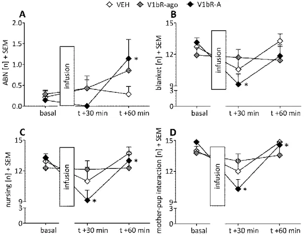
2.4.1 Maternal care was reduced by V1bR antagonism under non-stress conditions ... 38
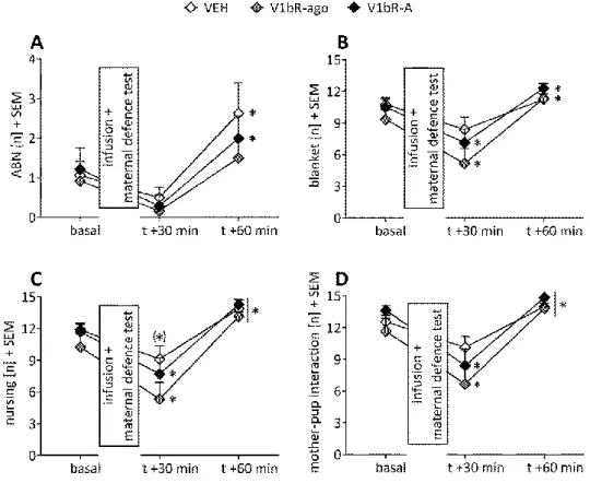
2.4.2 Maternal care was altered by V1bR antagonism and agonism under stress
conditions ... 39
2.4.5 Anxiety-related behaviour was not altered by V1bR manipulation ... 42
2.5 Discussion ... 44
CHAPTER 3 ... 49
3.1 Abstract ... 50
3.2 Introduction ... 51
3.3 Material & Methods ... 53
3.3.1 Animals ... 53
3.3.2 Implantation of an icv guide cannula ... 53
3.3.3 Infusion of receptor-specific V1bR agonist or V1bR antagonist ... 54
3.3.4 Experimental schedule ... 54
3.3.5 Behavioural tests ... 54
3.3.5.1 Maternal care observation in the home cage ... 54
3.3.5.2 Maternal motivation in the PRT ... 55
3.3.5.3 Maternal aggression in the maternal defence test ... 55
3.3.5.4 Anxiety-related behaviour on the EPM ... 56
3.3.6 Verification of guide cannula placement ... 56
3.3.7 Statistical analysis ... 57
3.4 Results ... 58
3.4.1 V1bR manipulation in lactating HAB/LAB dams of generation #47 ... 58
3.4.1.1 Effects of V1bR agonism and antagonism on maternal care under non-stress conditions ... 58
3.4.1.2 Effects of V1bR agonism and antagonism on maternal care under stress conditions ... 59
3.4.1.3 Effects of V1bR agonism and antagonism on maternal motivation ... 61
3.4.1.4 Effects of V1bR agonism and antagonism on maternal aggression ... 62
3.4.1.5 Effects of V1bR agonism and antagonism on maternal anxiety ... 63
3.4.2 Maternal behaviour in non-manipulated HAB/LAB dams of generation #50 ... 64
3.4.3 Maternal behaviour in non-manipulated HAB/LAB dams of generation #51 ... 67
3.5 Discussion ... 69
CHAPTER 4 ... 73
4.1 Abstract ... 74
4.2 Introduction ... 75
4.3 Material and Methods ... 77
4.3.1 Animals ... 77
4.3.2 Experiment 1: qPCR for V1bR mRNA in the MPOA and BNST ... 77
4.3.3 Experiment 2: Western Blot analysis for V1bR protein in the MPOA and BNST ... 78
4.3.4.1 Implantation of local guide cannulae. ... 79
4.3.4.2 Infusion of receptor subtype-specific V1bR antagonist... 80
4.3.5 Behavioural tests ... 81
4.3.5.1 Maternal care observation. ... 81
4.3.5.2 Maternal motivation in the PRT. ... 82
4.3.5.3 Anxiety-related behaviour on the EPM. ... 83
4.3.5.4 Maternal aggression in the maternal defence test. ... 84
4.3.6 Verification of cannulae placement ... 84
4.3.7 Statistical analysis ... 85
4.4 Results ... 86
4.4.1 Experiments 1 & 2: Expression of V1bR in the MPOA and BNST of virgin versus lactating rats ... 86
4.4.2 Experiment 3: Local blockade of V1bR within the MPOA... 86
4.4.2.1 Maternal care under non-stress conditions. ... 86
4.4.2.2 Maternal care under stress conditions. ... 87
4.4.2.3 Maternal motivation in the PRT. ... 88
4.4.2.4 Maternal aggression in the maternal defence test. ... 90
4.4.2.5 Anxiety-related behaviour on the EPM. ... 90
4.4.3 Experiment 4: Local blockade of V1bR within the mpBNST ... 90
4.4.3.1 Maternal care under non-stress conditions. ... 90
4.4.3.2 Maternal care under stress conditions. ... 92
4.4.3.3 Maternal motivation in the PRT. ... 92
4.4.3.4 Maternal aggression in the maternal defence test. ... 92
4.4.3.5 Anxiety-related behaviour on the EPM. ... 93
4.5 Discussion ... 94
CHAPTER 5 ... 98
5.1 Abstract ... 99
5.2 Introduction ... 100
5.3 Material and Methods ... 101
5.3.1 Animals ... 101
5.3.2 V1bR mRNA and protein in the PVN ... 101
5.3.3 Local blockade of V1aR and V1bR within the PVN ... 101
5.3.3.1 Implantation of local guide cannulae. ... 101
5.3.3.2 Infusion of receptor subtype-specific V1aR or V1bR antagonist. ... 102
5.3.4 Behavioural tests ... 102
5.3.4.1 Maternal care observation. ... 102
5.3.4.2 Maternal motivation to retrieve pups in the PRT. ... 102
5.3.4.3 Anxiety-related behaviour on the EPM. ... 103
5.4 Results ... 105
5.4.1 Expression of V1bR in the PVN of virgin versus lactating rats ... 105
5.4.2 Local blockade of V1aR or V1bR within the PVN ... 105
5.4.2.1 Maternal care under non-stress conditions ... 105
5.4.2.2 Maternal motivation in the PRT ... 105
5.4.2.3 Anxiety-related behaviour on the EPM. ... 106
5.5 Discussion ... 107
CHAPTER 6 ... 109
6.1 Abstract ... 110
6.2 Introduction ... 111
6.3 Material & Methods ... 113
6.3.1 Animals ... 113
6.3.2 Experimental schedule ... 113
6.3.2.1 Experiment 1: Icv infusion of GnRH antagonist ... 114
6.3.2.2 Experiment 2: Icv infusion of different doses of GPR54 antagonist ... 114
6.3.3 Behavioural tests ... 114
6.3.3.1 Maternal care observation. ... 114
6.3.3.2 Maternal motivation in the PRT ... 115
6.3.3.3 Maternal aggression in the maternal defence test. ... 116
6.3.4 Verification of guide cannula placement ... 116
6.3.5 Statistical analysis ... 116
6.4 Results ... 118
6.4.1 Experiment 1: Icv infusion of GnRH antagonist ... 118
6.4.1.1 Maternal care under non-stress conditions ... 118
6.4.1.2 Maternal care under stress conditions ... 118
6.4.1.3 Maternal motivation in the PRT. ... 118
6.4.1.4 Maternal aggression in the maternal defence test. ... 118
6.4.2 Experiment 2: Icv infusion of different doses of GPR54 antagonist ... 119
6.4.2.1 Maternal care under non-stress conditions ... 119
6.4.2.2 Maternal care under stress condition ... 120
6.4.2.3 Maternal aggression in the maternal defence test ... 121
6.5 Discussion ... 122
CHAPTER 7 GENERAL DISCUSSION ... 125
7.1 Summary of results ... 125
7.2 Involvement of the brain AVP system, especially of V1bR, in maternal care ... 128
7.3 Involvement of the brain AVP system, especially of V1bR, in maternal motivation ... 132
7.6 Correlation of the AVP, GnRH and kisspeptin system in the view of maternal behaviour
... 140
7.7 Conclusions, translational aspects and outlook ... 142
ABBREVIATIONS ... 146
BIBLIOGRAPHY ... 148
SUPPLEMENTARY ... 182
CURRICULUM VITAE ... 189
PUBLICATIONS ... 191
DANKSAGUNG ... 193
Abstract
Adequate maternal behaviour offers the best chance for the offspring to survive to maturity.
This pro-social behaviour is known to be regulated by the nonapeptide arginine-vasopressin (AVP) amongst other peptidergic and non-peptidergic systems in rodents as well as in humans. Most research regarding the AVP system in maternal behaviour has focussed on the peptide itself and its V1a receptor (V1aR), and revealed a pivotal involvement in the regulation of different aspects of maternal behaviour. In addition, the involvement of AVP and its V1aR was investigated in the animal model of rats selected for extremes in anxiety- related behaviour. Highly anxious dams (HAB) have a single nucleotide polymorphism in the AVP promoter-region, resulting in increased expression and release of AVP. Regarding the phenotype, HAB dams show a hyper-protective mothering style in contrast to dams selected for low anxiety-related behaviour (LAB), which only provide a low amount of maternal behaviour. Since the maternal phenotype of HAB and LAB dams can be partly reversed by blockade or infusion of V1aR or synthetic AVP, respectively, these rats provide a good model to observe the behavioural consequences after pharmacological manipulation of the AVP system on maternal behaviour.
The influence of V1b receptors (V1bR) on mediating maternal behaviour has only been investigated in a single study in V1bR knockout mice to date. This study showed that V1bR knockout dams show decreased maternal aggression compared to wildtype dams.
Therefore, I aimed to investigate in more detail the role of central V1bR on the maternal repertoire via acute central application of V1bR agonist and antagonist during lactation in non-selected rats (Chapter 2) as well as in the animal model of rats selected for extremes in anxiety-related behaviour (Chapter 3). Although the V1bR is widely distributed within the brain, I aimed to investigate the involvement of the receptor subtype in brain-regions known to be involved in maternal behaviour, including the medial preoptic area (MPOA), the bed nucleus of the stria terminalis (BNST) and the paraventricular nucleus (PVN) via acute local infusion of V1bR antagonist. In addition, I determined whether endogenous V1bR protein and/or mRNA expression differ in lactation within these brain regions (Chapter 4 -5).
Further, in a pilot study co-expression of AVP and gonadotropin-releasing hormone (GnRH)
immune-reactivity in the organum vasculosum of the lamina terminalis (OVLT) in lactating
rats was observed. Since mice with a 30 % decrease of GnRH neurons show impaired pup
retrieval, and the co-localization suggests an interaction with the AVP system known to be
involved in maternal behaviour, I was interested in the influence of the GnRH and the interconnected kisspeptin system in the regulation of maternal behaviour in rodents (Chapter 6).
To investigate my aims I used a variety of cellular, molecular and behavioural approaches like quantitative real-time PCR, western blotting, central or local acute pharmacological manipulation of V1bR, GnRH receptors or kisspeptin receptors followed by observations of maternal care, maternal motivation to retrieve pups, maternal aggression and anxiety- related behaviour.
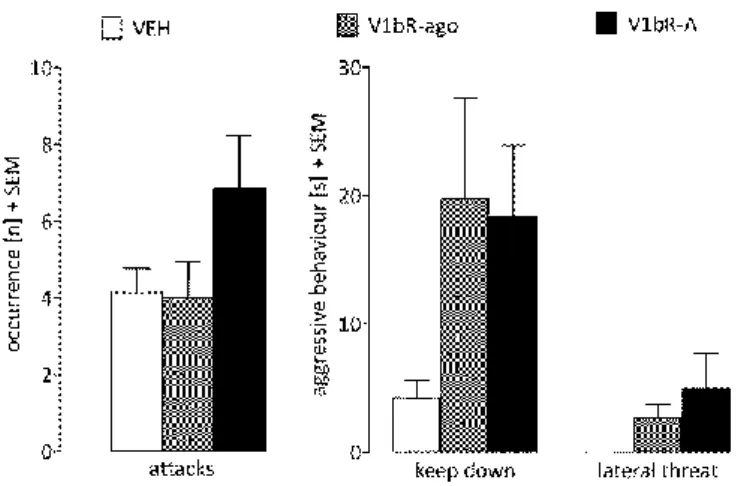
In this thesis I showed that central blockade of V1bR in non-selected rats impaired maternal care but did not affect maternal motivation, maternal aggression or anxiety-related behaviour. V1bR manipulation in HAB and LAB dams had no effect on any maternal behaviour. When investigating the role of V1bR in specific brain regions, I could show that blockade of V1bR within the MPOA increased maternal motivation whereas within the BNST maternal aggression, more precisely pup-directed behaviour, was decreased. Additionally, V1bR blockade in both brain regions decreased nursing, but only under stress conditions.
Interestingly, in the PVN only V1aR, but not V1bR blockade decreased nursing under non- stress conditions in addition to maternal anxiety.
Thus, both V1R subtypes seem to be involved in the regulation of maternal behaviour in a brain region-dependent and context-specific manner, thereby acting exclusively, as complement or as counterpart of the other receptor subtype. Interestingly, this was not associated with alterations in mRNA or protein expression in any brain-region, suggesting that either transport of the receptor-subtype to the membrane, signal transduction or agonist sensitivity may be altered in lactation, which remains to be investigated.
Regarding the involvement of GnRH and kisspeptin in the regulation of maternal behaviour, blocking GnRH decreased maternal aggression, whereas kisspeptin receptor (GPR54) blockade resulted in impaired maternal care, in a dose-dependent manner. These results provide evidence, that the GnRH and kisspeptin system are not only involved in reproduction, but also in various aspects of maternal behaviour. Further investigation of their role, especially in maternal brain circuits, is essentially required.
In conclusion, the presence of V1bR in brain regions involved in maternal behaviour are
mandatory in lactating rodents to show adequate maternal behaviour, especially in stressful
situations. Further, maternal behaviour is regulated by a variety of hormones acting centrally
within the brain like oxytocin and AVP, whereby hormones like GnRH and kisspeptin, thought to be mainly responsible in reproduction also influence maternal behaviour.
Taken all these results into account I could provide further knowledge of the impact of
central hormonal systems on the complexity of regulatory mechanism in maternal
behaviour.
Zusammenfassung
Adäquates mütterliches Verhalten ermöglicht den Nachkommen die bestmögliche Chance bis zum Erwachsenenalter zu überleben. Dieses prosoziale Verhalten wird sowohl im Nager als auch im Menschen, unter anderem durch das Nonapeptid Arginin-Vasopressin (AVP), reguliert.
Im Zusammenhang mit mütterlichem Verhalten lag der Forschungsschwerpunkt bezüglich des AVP-Systems bis jetzt auf dem Peptid selbst, sowie seinem im Gehirn exprimierten V1a Rezeptor (V1aR). Beide sind ein essentieller Bestandteil in der Regulierung von verschiedenen mütterlichen Verhaltensweisen. Des Weiteren wurde der Einfluss von AVP und seinem V1aR im Tiermodell für Ratten, die auf die Extreme im Angstverhalten selektiert sind, untersucht. Rattenmütter mit einem hohen Maß an Angstverhalten (HAB) haben einen Einzelnukleotid-Polymorphismus (single nucleotide polymorphism, engl.) in der AVP Promoter-Region, der zu einer erhöhten AVP Expression und Ausschüttung führt. Bezüglich des Phänotypen, kennzeichnen sich HAB-Mütter durch ein Übermaß an Fürsorge, im Gegensatz zu Rattenmüttern die auf niedriges Angstverhalten selektiert sind (LAB). Diese zeigen nur ein geringes Maß an mütterlichen Verhaltensweisen. Da der mütterliche Phänotyp der HAB und LAB Ratten durch eine Blockade von V1aR bzw. eine Injektion von synthetischem AVP verändert werden kann, eignet sich dieses Tiermodell besonders gut, um die Auswirkungen pharmakologischer Manipulationen des AVP Systems auf mütterliches Verhalten zu beobachten.
Der Einfluss von V1b Rezeptoren (V1bR) auf mütterliches Verhalten wurde bis jetzt nur in einer einzigen Studie untersucht. Diese zeigt, dass V1bR Knockout-Mäuse im Vergleich zu Wildtyp-Mäusen eine verringerte mütterliche Aggression zeigen.
Aufgrund dieser unzureichenden Studienlage war es ein Ziel meiner Arbeit, den Einfluss der
zentralen V1bR auf die unterschiedlichen mütterlichen Verhaltensweisen während der
Laktation in nicht-selektierten Ratten (Kapitel 2), sowie im Tiermodell für Ratten, die auf die
Extreme im Angstverhalten selektiert sind (Kapitel 3), mit Hilfe akuter zentraler
Verabreichung eines V1bR Agonisten sowie Antagonisten genauer zu untersuchen. Obwohl
der V1bR weitflächig im Gehirn exprimiert wird, war es ein weiteres Ziel meiner Arbeit den
Einfluss des Rezeptors auf mütterliches Verhalten, mit Hilfe von akuten lokalen Injektionen
eines V1bR Antagonisten in das mediale präoptische Areal (MPOA), in den bed nucleus der
Stria terminalis (BNST) und in den paraventrikulären Nukleus (PVN), zu untersuchen. Diese
Gehirnregionen sind nachweislich an der Regulation von mütterlichem Verhalten beteiligt.
Des Weiteren, habe ich erforscht, ob sich die Menge an endogenem V1bR Protein und/oder die mRNA Expression in diesen Gehirnregionen während der Laktation unterscheiden (Kapitel 4 und 5).
Auf Basis einer Pilotstudie wurde außerdem im organum vasculosum der lamina terminalis (OVLT) laktierender Ratten eine Koexpression von AVP und des Gonadotropin-releasing Hormons (GnRH) beobachtet. Da Mäuse mit 30 %iger Reduzierung von GnRH Neuronen ein vermindertes Einsammeln ihres Nachwuchses zeigen, und die Kolokalisation einen Zusammenhang mit dem AVP System, das nachweislich mütterliches Verhalten beeinflusst, vermuten lässt, war ich ausserdem am Einfluss des GnRH- und des eng verbundenen Kisspeptin-Systems auf mütterliches Verhalten im Nager interessiert (Kapitel 6).
Zur Untersuchung meiner Ziele habe ich eine Reihe an zellulären, molekularen sowie verhaltensbasierten Methoden angewendet, wie z.B. quantitative real-time PCR, Western Blot, zentrale oder lokale akute pharmakologische Manipulation von V1bR, GnRH- oder Kisspeptin-Rezeptoren, gefolgt von Verhaltensbeobachtungen (mütterliche Fürsorge, mütterliche Motivation zum Einsammeln der Jungen, mütterliche Aggression und Angstverhalten).
In meiner Arbeit konnte ich zeigen, dass die zentrale Blockade von V1bR in nicht-selektierten Ratten zu einer verringerten mütterlichen Fürsorge führt, ohne dabei die mütterliche Motivation, mütterliche Aggression oder das Angstverhalten zu beeinflussen. Die Manipulation von V1bR in laktierenden HAB und LAB Ratten hatte keine Auswirkungen auf etwaige mütterliche Verhaltensweisen. Eine gehirnregionspezifische Blockade von V1bR in der MPOA führte zu einer gesteigerten mütterlichen Motivation zum Einsammeln der Jungen, während die Blockade im BNST die mütterliche Aggression, genaugenommen die Aufmerksamkeit, die dem Nachwuchs während des Aggressionstests entgegengebracht wurde, verringert war. Zusätzlich führte die V1bR Blockade in beiden Gehirnregionen zu einem verminderten Säugeverhalten, jedoch ausschließlich unter Stressbedingungen.
Interessanterweise veranlasste im PVN nur die Blockade von V1aR, jedoch nicht von V1bR, ein niedrigeres Säugeverhalten unter stressfreien Bedingungen, sowie ein geringeres Angstverhalten.
Daraus lässt sich folgern, dass beide V1R-Subtypen in einer gehirnregions- sowie
kontextspezifischen Art und Weise an der Regulation von mütterlichem Verhalten beteiligt
sind. Das resultierende Verhalten erfolgt daher aus der Wirkungsweise eines einzelnen Rezeptorsubtypen, dem Zusammen- oder aber auch dem Gegenspiel beider Rezeptorsubtypen. Allerdings standen die Verhaltensänderungen nicht im Zusammenhang mit der mRNA- oder Protein-Expression. Dies lässt eine Anpassung im Transport des Rezeptors zur Zellmembran, in der Signalweiterleitung oder in der Sensibilität des Agonisten vermuten, was untersucht werden muss.
Hinsichtlich des Einflusses von GnRH und Kisspeptin auf mütterliches Verhalten führte die Blockade von GnRH zu verminderter mütterlicher Aggression, während die Blockade von Kisspeptin Rezeptoren (GPR54) dosisabhängig zu einer verringerten mütterlichen Fürsorge führte. Diese Ergebnisse zeigen deutlich, dass sowohl das GnRH- als auch das Kisspeptin- System nicht nur in der Fortpflanzung, sondern auch in verschiedenen Aspekten von mütterlichem Verhalten eine Rolle spielen. Die weiterführende Erforschung beider Systeme, insbesondere im Zusammenhang mit mütterlichen Verhaltensweisen, ist von großem Interesse.
Es ergibt sich die Schlussfolgerung, dass die V1bR in Gehirnregionen, die mütterliches Verhalten modulieren, zwingend notwendig sind, um angemessenes mütterliches Verhalten vor allem unter Stressbedingungen zu garantieren. Des Weiteren wird mütterliches Verhalten von zahlreichen Hormonen im Gehirn, wie Oxytocin und AVP, reguliert, wobei aber auch Hormone wie GnRH und Kisspeptin, die bis jetzt hauptsächlich dem Fortpflanzungsverhalten zugesprochen wurden, mütterliches Verhalten beeinflussen.
Somit konnte ich mit meiner Arbeit das Wissen über den Einfluss zentraler Hormonsysteme
auf die Regulation komplexer Mechanismen, die mütterliches Verhalten regulieren,
erweitern.
Chapter 1
General Introduction
One of the most important objectives in the life cycle of humans and other mammalian species is reproduction and the successful rearing of the young. The latter is promoted by parental behaviour, which was defined by Numan and Insel as “any behaviour of a member of a species towards a reproductively immature conspecific that increases the probability that the recipient will survive to maturity” (for review see Numan and Insel, 2003). About 90
% of species show uniparental care, with the mother taking care for the offspring which is due to the fact that in mammals only the females provide milk. This is essential for the young as they would die without the nutrition. In preparation for motherhood the female undergoes numerous physiological, cellular and molecular adaptations to be prepared for the upcoming birth, lactation and defence of the young against potential threats. These changes occur as a direct consequence of both hormonal and non-hormonal alterations that occur throughout the peripartum period. All the more remarkable as in most mammalian species females are not maternal per se I want to give insight in these changes that lead to maternal behaviour in more detail. Due to the fact that these mechanisms are most studied in laboratory rodents, the following descriptions refer to rats unless otherwise stated.
1.1 Maternal Behaviour
1.1.2 Non-hormonal and hormonal basis of maternal behaviour in rats
Nulliparous rodent females naturally avoid pups (Fleming and Rosenblatt, 1974; Rosenblatt, 1967) or even show infanticide (Mennella and Moltz, 1989). Interestingly, these virgins show less infanticide after a distinct co-habituation period with the actual mother of the test pups;
after a co-habituation period of 5 to 10 consecutive days 55 % of nulliparous female exhibit
pup killing, whereas after 20 days of co-habituation only 9 % show infanticide (Mennella and
Moltz, 1989). Also the co-habituation of nulliparous rats with foster pups over a period of 4
to 7 days, leads to tolerance of the pups, which is followed by spontaneous maternal
interest, expressed as increased licking of the pups, before retrieving them to a nest and
crouching over them (Fleming and Rosenblatt, 1974; Rosenblatt, 1967; Stern, 1983). This process is known as “sensitization” and occurring as a response to pup stimuli.
In contrast to virgins, primiparous female rats show spontaneous maternal behaviour starting around 3.5 hours before parturition by licking (foster) pups, retrieving them to a nest site and crouching over them to provide milk (Mayer and Rosenblatt, 1984). Such preterm maternal interest in pregnant rats was linked to the hormonal changes, concomitant to non-hormonal adaptations, occurring during the peripartum period and inducing maternal behaviour.
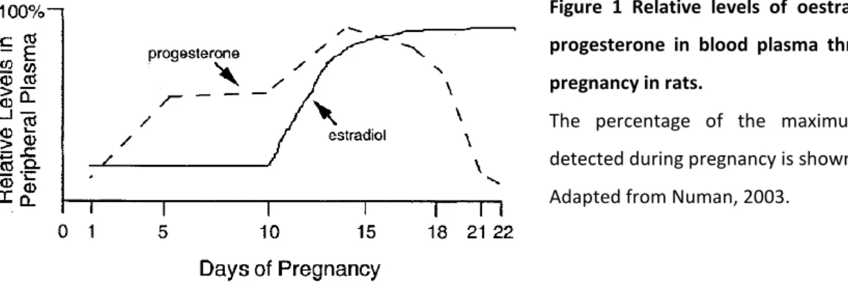
The steroid hormones oestradiol and progesterone - together with lactogenic hormones like prolactin (PRL) and placental lactogens (see below) - are the major factors implicated in the regulation of maternal behaviour in rats (Bridges et al., 1996; Grattan et al., 2001; Southard and Talamantes, 1991). Thereby, oestradiol levels are low during the first trimester, before they start rising on pregnancy day (PD) 10 and peak at a level, 3- to 4-fold higher than at the start of pregnancy, from day 15 until the day of parturition in rats (see Figure 1). In contrast, progesterone is already high during the first two weeks of pregnancy and declines with a robust drop (to 1/6 of magnitude) during the last days of gestation. Finally, a reversal of the oestradiol-to-progesterone ratio occurs leading from long progesterone dominance to a short oestradiol dominance (for reviews see Bridges, 2014; Numan and Insel, 2003).
Figure 1 Relative levels of oestradiol and progesterone in blood plasma throughout pregnancy in rats.
The percentage of the maximum value detected during pregnancy is shown.
Adapted from Numan, 2003.
Lactogenic hormones are secreted throughout pregnancy and are either derived from the
anterior pituitary (PRL) or from the placenta (placental lactogens e.g. placental lactogen I
and II) (Grattan, 2001). PRL is released in two daily surges within the first half of pregnancy
and on the last day of pregnancy, whereas during the second half of pregnancy plasma levels
of placental lactogens increase (Cohick et al., 1996; Southard and Talamantes, 1991). The
dominance of oestradiol, together with the drop of progesterone and increase of PRL at the end of pregnancy are important hormonal adaptations which prime the neural circuits within the medial preoptic area (MPOA) and the bed nucleus of the stria terminalis (BNST) among others. These brain regions are also termed “maternal circuits” and facilitate the appropriate onset of maternal behaviour (Bridges and Ronsheim, 1990; Bridges et al., 1997, 1996).
Besides PRL, the neuropeptides oxytocin (OXT) and arginine-vasopressin (AVP) are released within this maternal circuits around parturition, facilitating the adequate onset of maternal behaviour (Numan et al., 1977; Pedersen and Prange, 1979; Pedersen et al., 1994, 1982).
The OXT system also adapts during pregnancy and lactation in order to decrease stress responsiveness of the HPA axis (Slattery and Neumann, 2008), i.e. with elevated OXT release within the hypothalamus in response to a stressor (Bosch et al., 2004). In addition, changes in sizing and branching patterns of dendritic trees within the supraoptic (SON) and paraventricular nuclei (PVN), alterations of electrophysiological properties and changes in the synthetic and secretory activity of oxitocinergic neurons take place (for review see Neumann, 2003). All these adaptations provide a dynamic OXT system, which is highly responsive to stimuli in the context of reproduction, i.e. labour and suckling of the pups, but attenuated in response to pharmacological, emotional or physical stressors (for reviews see Neumann, 2003; Slattery and Neumann, 2008).
Similar adaptations during the peripartum period occur within the AVP system, described in more detail below (Chapter 1.8). Before going into detail with the AVP system, I will introduce the distinct maternal parameters, which can be observed in lactating laboratory rodents, and will especially refer to brain regions, which are involved in the modulation of maternal behaviour.
1.1.3 Maternal care and “brain circuitry”
Rodent mothers show a variety of distinct behaviours towards their pups to provide them
with milk, warmth and protection against potential threats (for review see Bosch and
Neumann, 2012). This care provided by the mother, i.e. maternal care, is one of the most
important pro-social behaviours displayed by mammalian mothers and helps to raise the
litter to maturity (for review see Bosch, 2011). Therefore, nest-building is an active,
voluntary aspect of maternal behaviour initiated by the dam and mediated via oestradiol, PRL, OXT and dopamine within the MPOA in rodents (for review see Numan and Insel, 2003).
By licking and grooming their pups, rat mothers enable them to urinate and defecate.
Further, it positively influences the social and emotional development of the offspring (Caldji et al., 1998; Champagne, 2008). This behaviour occurs very frequently during the first weeks of life, declining with the growth of the pups and their move to independence (for review see Numan and Insel, 2003). Another important parameter is nursing, which can be subdivided into various distinct nursing postures. The dams show blanket posture to keep the pups warm and safe while lying in a flat position above them. Furthermore, dams will also lay on their side or back during nursing, especially when the pups grow older.
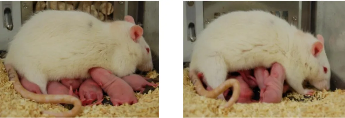
Sometimes dams also hover over their litter by resting on their hind limbs while being engaged in licking the pups or mouthing them. The most intense nursing position is termed arched back nursing (ABN; Figure 2), the only active nursing posture with the dam crouching over the pups in a quiescent kyphosis (Stern and Johnson, 1990). Depending on the angle of her back, one can distinguish between low crouch with a flat or slightly arched back, or high crouch with an intensely arched back and splayed legs (Stern, 1996), thereby ensuring the best possibility for all young (12 on average in Wistar rats (Rosen et al., 1987)) to have access to the 12 nipples (for review see Numan and Insel, 2003).
Figure 2 Lactating rat engaged in arched back nursing (ABN).
Dam showing ABN in either low crouch (left) or high crouch (right) position during quiescent kyphosis.
Kyphosis can be seen as a reflex, which is induced by ventral stimulation caused by the nuzzling, rooting pups searching for the nipples (for review see Numan and Insel, 2003).
Maintenance of this behaviour, including its relatively long upright crouch durations,
depends of the suckling stimulus of the pups, which itself is not essential for the initiation of
kyphosis (Stern and Johnson, 1990; Stern, 1991).
The tactile stimulus from the pups provides positive afferent input to the MPOA/BNST region, which facilitates maternal behaviour. Neurons within the dorsolateral MPOA and the ventral BNST (vBNST) form a functional system, called “maternal super-region” (for reviews see Bosch and Neumann, 2012; Numan and Insel, 2003), and are important for the regulation of maternal behaviour in rats (Numan and Numan, 1996). These neurons express oestrogen receptors, which bind oestradiol and lactogenic hormones, thereby facilitating the onset of maternal behaviour in rats (see above)(Bakowska and Morrell, 1997; Numan and Insel, 2003; Pfaff and Keiner, 1973). Further, the maternal super-region receives input from the medial amygdala, which is the relay site of olfactory information from the pups, and thus leading to a fear/avoidance response of pups in virgins or an attraction/approach circuit in dams (for reviews see Numan and Insel, 2003; Numan and Woodside, 2010). Another brain region projecting to the “maternal super-region” is the PVN (Ingram and Moos, 1992). The PVN is known to be important for the onset, but not the maintenance of established maternal care (Consiglio, 1996; Insel and Harbaugh, 1989; Numan and Corodimas, 1985). In addition, it is the main source of oxitocinergic, but also vasopressinergic projections to other brain regions (de Vries and Buijs, 1983). As both the OXT and the AVP system are increased in the peripartum period (see Chapter 1.8), they implicate a role in the regulation of maternal behaviour (Bosch and Neumann, 2008; Bosch et al., 2010; Neumann et al., 1993, 2000; for review see Bosch and Neumann, 2012).
1.1.4 Pup retrieval and “brain circuitry”
During the period when pups depend on their mother, dams show retrieval behaviour if a
pup is lying outside the nest. This describes mouthing the pups and carrying them back to
the nest if they got scattered or if they are transported to a new nesting site. Pup retrieval is
thought to reflect the dam’s motivation to retrieve the offspring to a single nesting-site and
is described as a voluntary, proactive and goal-directed appetitive response of the mother
(for review see Numan and Insel, 2003). As motivation is referred to an internal process that
regulates changes in responsiveness to a constant stimulus, pup cues evoke this response in
mothers. A study focussing on the brain regions involved in mediating maternal motivation
found that lesions of the MPOA disrupts pup retrieval (Kalinichev et al., 2000). Further, the
processing of information occurs via dopaminergic neurons projecting from the
MPOA/vBNST to the nucleus accumbens (NAc) (Numan and Numan, 1996) both directly and indirectly via synapses in the ventral tegmental area (for review see Numan and Insel, 2003).
More specifically, lesion of the dorsolateral connections of the MPOA/vBNST by knife-cuts extinguishes pup retrieval (Numan et al., 1990). These findings provide insight in brain regions important in maternal motivation to retrieve pups, which are regulated via the neuropeptides OXT and AVP within the MPOA (Bosch and Neumann, 2008; Bosch et al., 2010; Meddle et al., 2007)(see below for more details).
1.1.5 Maternal aggression and “brain circuitry”
Another adaptation occurring in the postpartum female is increased maternal aggression.
This behaviour is not directed towards the pups (Peters et al., 1991), but against any potential threat, which could harm the offspring, like conspecifics.
Maternal aggression can be distinguished from other forms of aggression, like predator aggression, territorial aggression, intermale aggression, defensive aggression or competitive aggression as the trigger to attack the opponent is a different one (Blanchard et al., 2003;
Moyer, 1968). This can also be seen by the fact that lactating females show higher aggression towards a male intruder than non-lactating females (Erskine et al., 1980, 1978a).
Maternal aggression first occurs at the end of pregnancy, vanishes at parturition and peaks during the first week of lactation before declining afterwards (Caughey et al., 2011). During this time the dam shows a variety of offensive and defensive aggressive behaviours when confronted with an opponent (Bosch et al., 2005; Erskine et al., 1978a; for review see Lonstein and Gammie, 2002). In the laboratory, this can be tested in the maternal defence test (Neumann et al., 2001). The lactating mother, as resident, is confronted with an unfamiliar virgin female or male intruder, ideally about 10 % smaller than the resident (to act as an stressor but to prohibit a dominance over the resident; for review see Bosch, 2013).
This encounter leads to a defensive reaction of the intruder followed by aggressive behaviours of the residents (Blanchard et al., 2001; Haney et al., 1989). Aggressive behaviours shown by the resident include offensive behaviours like attacks, which are normally directed towards the neck or the back of an intruder, sometimes paired with bites.
Further, the dam displays threat behaviours, e.g. pushing the intruder from the side termed
“lateral threat”, or standing with all four paws on top of the intruder, termed “keep down”
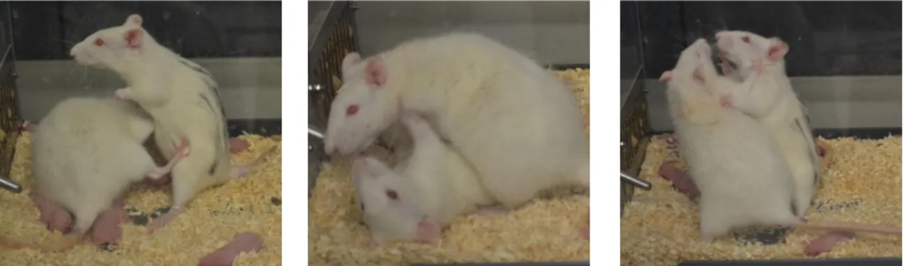
(Figure 3). Also boxing the intruder in an “offensive upright” position and “aggressive grooming” of the intruder are considered threat behaviours. Species-specific cues lead to termination of aggression, i.e. defensive behaviour in rats. Thus, as soon as the intruder shows submissive behaviour, the residents’ attacks and threats decrease/stop.
Figure 3 Lactating dam engaged in threat behaviours during the maternal defence test.
Resident dam showing lateral threat (left), keep down (middle) and offensive upright (right) against a virgin female intruder (marked with black stripes).
A variety of extrinsic and intrinsic factors contribute to the occurrence of maternal aggression (for review see Bosch, 2013). The main extrinsic factor is the offspring; the presence of the pups is essential for the initiation of maternal aggression (Gandelman and Simon, 1980). Short-term removal (4 to 5 hours) of the young decreases, but does not suppress, maternal aggression, whereas 24 hours or more do so (Erskine et al., 1978a;
Ferreira and Hansen, 1986; for review see Lonstein and Gammie, 2002). Importantly, it is not the suckling stimulus per se, but the ventral somato-sensory stimulation (Stern and Kolunie, 1993) and olfactory cues (Ferreira and Hansen, 1986; for review see Lonstein and Gammie, 2002) from the pups, which are important for the onset and the maintenance of maternal aggression.
Another main extrinsic factor is the intruder, which acts as a potential threat to the offspring
and, therefore, is a critical threat which evokes maternal aggression in lactating rats to
protect the offspring from infanticide (for review see Bosch, 2013). Depending on the age
and size of the intruders, the mothers attack smaller intruders up to 80 %, whereas larger
males receive only 30 % attacks (Erskine et al., 1978b; Flannelly and Flannelly, 1985). Also
the sex of the intruder plays a role, with females being attacked more often than males
(Haney et al., 1989); independent of their reproductive status (Neumann et al., 2001).
In addition to these factors, intrinsic factors also influence aggressive behaviour in lactating mothers. On the last days before parturition, aggression already increases, which depends on rising oestrogen, but not dropping progesterone, levels (Mayer and Rosenblatt, 1987; for review see Lonstein and Gammie, 2002). Adaptations in neuropeptidergic AVP (see below Chapter 1.8) and OXT systems during the peripartum period play a crucial role in the modulation of maternal aggression. In response to the maternal defence test, OXT release is increased within the PVN, central amygdala (CeA), BNST and lateral septum (LS), whereas increased neuronal activity and OXT receptor binding is found within the BNST and LS compared to non-aggressive controls immediately after the end of the test, lasting up to 45 min (Caughey et al., 2011; Gammie and Nelson, 2001; for review see Bosch, 2013).
Noteworthy, adaptations in the neuropeptide systems, i.e. increased OXT release in the LS in response to the maternal defence test strongly depends on the rat line used with Sprague Dawleys, but not Wistar, dams showing the effect (Bosch et al., 2004; for review see Bosch, 2013).
1.1.6 Maternal anxiety and “brain circuitry”
The decreased anxiety in postpartum females is important to allow the dam to protect the offspring at the nest site (for review see Numan and Insel, 2003). A reduction in anxiety occurs directly after parturition and continues until mid-lactation, while being more anxious in late pregnancy, compared to virgin rats (Neumann et al., 2000; for review see Lonstein, 2007). In order to achieve this hypoanxious state the dam must be able to assess the stressful stimulus or scenario and adequately cope with it (for review see Neumann et al., 2010). Similar to maternal aggression, it is pup contact and not the suckling stimulus per se that facilitates this adaptation (Lonstein, 2005). Thus, dams separated 4 hours or more from their pups show no reduction in anxiety (Lonstein, 2005). Further, changes in the PRL, OXT and AVP systems are involved in the regulation of anxiety during lactation. For example, increased levels of central PRL (Torner et al., 2002), as well as of central OXT (Neumann et al., 2000; for review see Bosch, 2011), act anxiolytically, whereas central AVP exerts an anxiogenic action (Bosch and Neumann, 2008) during the postpartum period.
Regarding the fact that dams are less anxious but highly aggressive during the first week
postpartum, the increased expression of maternal aggression and protection of the offspring
may require a concomitant reduction in anxiety when facing the intruder (Hansen et al., 1985; Lonstein, 2005). The brain regions and circuitries involved in the regulation of anxiety in lactating rats are still not completely understood but studies suggest an involvement of the periaqueductal gray (Lonstein et al., 1998) as well as the extended amygdala (Davis and Shi, 1999). Indeed, OXT receptor (Figueira et al., 2008) and γ-amino butyric acid (GABA)a receptor (Miller et al., 2010) antagonism in the ventrocaudal PAG increases anxiety-related behaviour in lactating rats. But also pharmacological blockade of V1aR within the BNST tended to decrease anxiety on the elevated plus maze (EPM) in lactating dams, suggesting an involvement of this part of the extended amygdala in the regulation of anxiety-related behaviour in rat dams (Bosch et al., 2010). Further, the corticotropin-releasing factor (CRF) system is known to be involved in the regulation of anxiety in the peripartum period, with central CRF receptor blockade eliciting an anxiolytic effect (Klampfl et al., 2013). In more detail, antagonism of the CRF receptors within the medial-posterior part of the BNST (mpBNST) leads to anxiolysis (Klampfl et al., 2014). Also the noradrenergic system is involved in the regulation of anxiety by releasing noradrenalin into the BNST in response to aversive stimuli. In lactating female rats this response is blunted by disinhibiting GABAergic output cells as response to pup contact, resulting in attenuated anxiety (Smith et al., 2013; for review see Lonstein, 2007).
Although other neurotransmitter systems are implicated in the regulation of maternal
anxiety and the other maternal parameters, the main interest in my thesis is the impact of
the AVP system on maternal behaviour; therefore, I will focus on AVP in the remainder.
1.2 The brain AVP system
In the last decades AVP has not only been linked to the regulation of the hypothalamo- pituitary-adrenal (HPA) axis and anxiety, but also to the facilitation of a wide range of social behaviours including pair bonding, parental care and aggression in rodents.
1.2.1 AVP
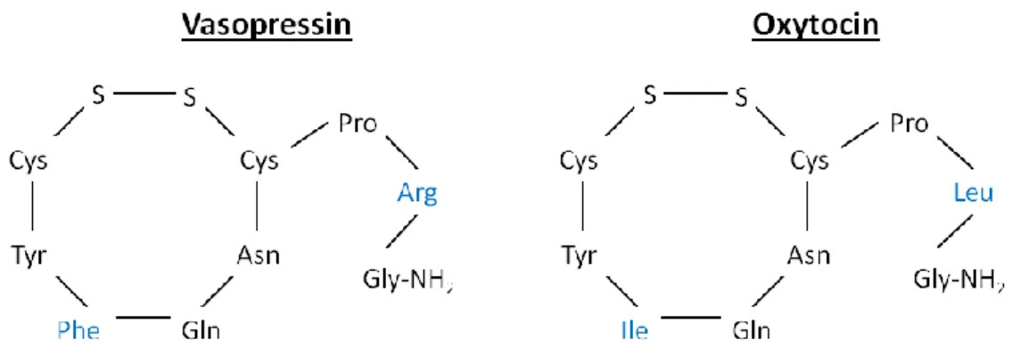
AVP was firstly isolated and synthesized in 1953, together with OXT (for review see Ottenhausen et al., 2015). Both are nonapeptides forming a ring structure, linked by a disulphide bridge connecting two cysteines. AVP differs only in two amino acids at position 3 and 8 from OXT; AVP contains phenylalanine instead of isoleucine in the ring and arginine instead of leucine in the side branch (Figure 4).
Figure 4 Molecular structure of the nonapeptides vasopressin and oxytocin
The peptides only differ in two out of nine amino acids (highlighted in blue) formed to ring structure by a disulphide bridge (S-S).
Arg = arginine, Asn = asparagine, Cys = cysteine, Gln = glutamine, Gly = glycine, Ile = isoleucine, Leu = leucine, NH2 = amidogen, Phe = phenylalanine, Pro = proline, Tyr = tyrosine.
Both hormones are products of several enzymatic cleavage steps. In case of AVP, the
primary protein precursor preprovasopressin is cleaved into a signal peptide and
provasopressin. After translation and several modification steps, provasopressin is further
cleaved into the active nonapeptide itself, neurophysin II protein and the copeptin
glycopeptide (Gainer, 1983; Russell et al., 1980; Sachs et al., 1969). Neurophysin II ensures
the proper targeting, packing and storage of AVP before release into the bloodstream
(Ginsburg and Ireland, 1966). The exact function of the copeptin remains unknown although
it is a stable biomarker for pathologies linked to AVP like stress, diabetes, heart failure and myocardial infarction in human diagnostics (Bolignano et al., 2014; Pozsonyi et al., 2015;
Reinstadler et al., 2015; Urwyler et al., 2015; Wannamethee et al., 2015; for review see Yilman et al., 2015).
The cleavage process takes place within the Golgi complex of neurone cell bodies in the hypothalamic SON and PVN, where neurosecretory granules containing the prohormone are formed. The cleavage products pass down nerve axons to be stored in the posterior pituitary, where they are released from nerve terminals after stimulation into the blood (Brownstein et al., 1980) but also centrally from cell bodies and dendrites of magnocellular neurons of the SON (Ludwig, 1998). In addition, AVP is also synthesized centrally within the suprachiasmatic nucleus (SCN), the olfactory bulb and the LS, among others. In the brain, AVP mediates a wide range of behaviours linked to social behaviours (see Chapter 1.7), emotionality, learning and memory but also circadian rhythms (Laycock et al., 2010).
Further, its importance in HPA axis regulation in response to stress is generally accepted (Aguilera et al., 2008). Thereby, AVP exerts its effect on all three different levels of the HPA axis, the hypothalamus, the pituitary as well as the adrenals (for review see Zelena et al., 2015). Whereas former studies in AVP-deficient Brattleboro rats supported the theory of AVP being the predominant regulator of the HPA axis response during chronic stress (Aguilera et al., 2008, 1994), recent studies also hint to a role of AVP after acute stressor exposure (Makara et al., 2012; for review see Zelena et al., 2015). On the level of the hypothalamus an increased AVP mRNA concentration was observed within the PVN during chronic stress (Aguilera et al., 1994). In addition, AVP was found to compensate for CRF effects in HPA axis regulation (for review see Scott and Dinan, 2002). Interestingly, the response of the HPA axis to stressors is attenuated in human, but also in rodent, mothers towards the end of pregnancy and in lactation (Heinrichs et al., 2001; Neumann et al., 2000, 1998a). Instead, these females show a basal hyper-corticism (Brunton and Russell, 2003;
Neumann et al., 1998a), enhanced sensitivity of the pituitary to AVP (Toufexis et al., 1999) and a down-regulation in the brain CRF system (Klampfl et al., 2013; Neumann et al., 1998b;
Walker et al., 2001) among other factors involved in the change in stress sensitivity (Slattery and Neumann, 2008).
Besides central and neurohypophysial functions, AVP is synthesized in the periphery, i.e.
within the sympathetic ganglia, the adrenals, ovaries and testis among others (Laycock et al.,
2010). Indeed AVP was originally known as antidiuretic hormone, leading to increased blood pressure and water permeability in renal nephrons. In addition, AVP was found to play a role in thermoregulation (Richmond, 2003) and in cardiac function (Wasilewski et al., 2015).
1.2.2 AVP receptors
AVP mediates its action via three receptors, the V1a, V1b (formerly V3) and the V2 subtypes.
Whereas the V1 receptor subtypes are mostly found both centrally and peripherally, the V2 receptor is distributed exclusively in the periphery; mainly within the kidney. There, V2 receptors in the collecting ducts and the loop of Henle are responsible for free water reabsorption (for reviews see Laycock and Hanoune, 1998; Verbalis, 2009). The main focus in this thesis is on the V1 receptors, which I want to further describe in the following.
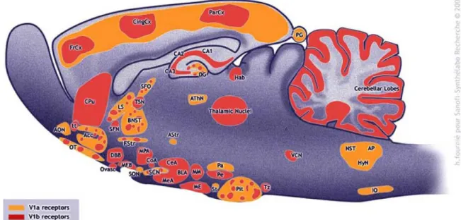
V1a receptors (V1aR) are mainly distributed in the central nervous system, where they have been identified within the hippocampus, BNST and PVN among others (Figure 5).
V1b receptors (V1bR) are mainly found within the adenohypophysis on corticotroph cells, where they mediate adrenocorticotropic hormone (ACTH) secretion, especially under chronic stress conditions (Volpi et al., 2004a, 2004b). Within the brain the receptor-subtype is distributed throughout brain regions like the hippocampus, thalamus, MPOA and amygdala (Hernando et al., 2001; Lolait et al., 1995) among others (Figure 5).
Both V1 receptors are G-protein coupled receptors (GPCRs) of the 1b family. Thus, they have a similar basic structure, which consists of seven transmembrane helices, connected by three intracellular and three extracellular loops, with two cysteine residues forming a disulfide bridge (for review see Bockaert and Pin, 1999).
The primary signalling pathway linked to the receptors has been well-characterised: Gq/G11 for both V1 receptors (Laycock et al., 2010; Thibonnier et al., 1997; Wange et al., 1991).
In detail, the Gαq protein activates phospholipase C, which results in the hydrolysis of
phosphatidylinsositol-4,5-bisphosphate into two 2
ndmessengers (Kirk et al., 1981; Michell et
al., 1979); namely inositol triphosphate (IP3) and diacylglycerol (Laycock et al., 2010). IP3 can
diffuse into the cytosol and bind to calcium channels in the plasma membrane of the
endoplasmatic reticulum, followed by the release of calcium from its intracellular storage or
via calcium dependent channels (Dayanithi et al., 2000; Levy et al., 1990). In return, in the
presence of calcium, diacylglycerol can activate protein kinase C which than phosphorylates
further protein substrates. Additionally, the released calcium can bind to calmodulin, which activates calcium/calmodulin-dependent kinases (Laycock et al., 2010). These kinases have the ability to phosphorylate cyclic adenosine monophosphate response element binding protein (CREB), which stimulates the expression of genes by recruiting a coactivator, thus resulting in new protein synthesis.
Regarding the V1bR, the signalling pathway is similar to that of V1aR. In addition to coupling to a Gq/G11 protein, V1bR have also been found to recruit Gs and Gi proteins, but only in transfected cells, rendering the physiological importance of such mechanisms unclear (Birnbaumer, 2000; Laycock et al., 2010; Thibonnier et al., 2001).
Figure 5 Distribution of V1 receptors in the rat brain Adapted from (Griebel et al., 2005)
Acc = nucleus accumbens, AON = anterior olfactory nucleus, AP = area postrema, Astr = amygdalostriatal area, AthN = anteroventral thalamic nucleus, BlA = basolateral amygdala, BNST = bed nucleus of the stria terminalis, CA1 = hippocampal field CA1, CA2 = hippocampal field CA2, CA3 = hippocampal field CA3, CeA = central amygdala, CingCx = cingulate cortex, CoA = cortical amygdaloid nucleus, Cpu = caudate/putamen, DBB = diagonal band of Broca, DG = dentate Gyrus, FrCx = frontal cortex, FStr = fundus striati, Hab = medial habenula, HyN = hypoglossal nucleus, IO = inferior olive, LS = lateral septum, ME = median eminence, MeA = median amygdala, MFB = medial forebrain bundle, MM = medial mammillary nucleus, MPA = medial preoptic area, NST
= nucleus of the solitary tract, OT = olfactory tubercle, Ovasc = organum vasculosum laminae terminalis, Pa = paraventricular hypothalamic nucleus, ParCx = parietal cortex, Pe = periventricular hypothalamic nucleus, PG = pineal gland, Pit = pituitary gland, SCN = suprachiasmatic nucleus, SFN = septofimbrial nucleus, SFO = subfornical organ, SON = supraoptic nucleus, St = stigmoid nucleus, TSN = triangular septal nucleus, tt = taenia tecta, Tz = trapezoid body, VCN = ventral cochlear nucleus