114
Kariniemi et al., Analysis of heart rate variabilityJ. Perinat. Med.
10(1982)114
Antepartal analysis of fetal heart rate variability by abdominal electrocardiography
V. Kariniemi, A. Siimes, P. Ämmälä
Departments of Obstetrics and Gynecology, Helsinki Üniversity Central Hospital, Haartmanink. 2,00290 Helsinki 29, Finland l Introduction
Antepartal analysis of fetal heart rate variability (FHRV) has been shown to be of value in predict- ing fetal distress in labor [8]. Abdominal fetal electrocardiograin (aFECG) is used äs a trigger signal in the statistical analysis of intervals and the differences between them [7]. The indices of variability thus obtained have a good correlation with indices calculated simultaneously from direct FECG [6, 12]. Interval index (II) describes the long-term variability and differential index (DI) the short-term variability in this analysis. The main disadvantage of this analysis System is a fairly high failure rate of aFECG during the third trimester of pregnancy. Gestational age, the electrode position and the maternal position have been shown to affect the recordability of aFECG [l, 2, 3, 4, 9, 11]. On the other hand, maternal obesity, placen- tal location and the state of the membranes have been shown not to have any effect on the quality of aFECG [l, 3, 9]. The aim of this study was to investigate how often FHRV analysis made by aFECG succeeds and what is the role of certain methodological factors in the recording procedure.
The fottowing methodological factors and patients were üivestigated:
2.1 The success rate of FHRV analysis. The effect of gestational age on the success rate of FHRV analysis was studied by attempting a five-minute FHRV analysis in 80 normal and 554 high risk pregnancies at different stages of gestation. Dis- tribution of the antepartal diagnoses is shown in Tab. I.
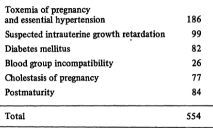
Tab. L Distribution of the antepartal diagnoses in the data of antepartal analyses of fetal heart rate variability in high risk pregnancies.
Toxemia of pregnancy
and essential hypertension 186 Suspected intrauterine growth retaidation 99 Diabetes mellitus 82 Blood group incompatibüity 26 Cholestasis of pregnancy 77 Postmaturity 84
Total 554
2 Patients and methods
FHRV analyses were performed by a previously described method using an on-line microprocessor system [7]. The analysis was regarded äs a failure when less than 30% of the intervals detected were accepted in the analysis. Student's paired t-test (one-sided) was used to measure the significance of the results.
2.2 Maternal position in FHRV analysis. The ma- ternal supine vs. 90-degree lateral position was studied relating to the success rate and to the variability indices in 30 pregnancies. A five-minute analysis was fiist attempted in the supine position immediately followed by a 90-degree lateral posi- tion. The maternal supine and 15-degree lateral positions were similarly compared in 30 pregnan- cies.
0300-5577/82/0010-0114S02.00
Kariniemi et al., Analysis of heart rate variability 115
2.3 Dry or moist electrodes? The success rate of
FHRV analysis with dry and with moist electrodes was studied in 143 pregnancies. FHRV analysis was first attempted with dry electrodes. In case of failure the electrodes were moistened with tap water. If the analysis again failed, electrode paste was applied.
2.4 The rejection logic in FHRV analysis. In this analysis System, II is calculated from the Standard deviation of intervals and DI from Standard devia- tion of interval differences. Rejection logic is used to exclude erroneous intervals and interval differ- ences created by noise in aFECG. The idea of using a narrow rejection limit lies in the presump- tion, that the interval differences thus included are most probably created by the fetus. The effect of maximum rejection limit for interval differences of five vs. ten beats per minute (bpm) on the variability indices and on the percentage of ac- cepted intervals was studied in 19 pregnancies.
Two subsequent five-minute periods of aFECG were analysed and compared. The first was anal- ysed with a rejection limit of five bpm and the second with a rejection limit of ten bpm.
2.5 The effect of sample time on variability in- dices. The period of aFECG from which FHRV analysis is calculated is called sample time [5]. The clinical studies on antepartal FHRV made by our group have been performed using a sample time of five minutes [8]. The effect of sample time on variability indices was studied in 76 pregnancies.
First an analysis was performed of a one-minute sample of aFECG immediately followed by a five- minute sample. A similar comparison was made of five-minute and 20-minute analyses.
3 Results
3.1 The success rate of FHRV analysis at differ- ent stages of gestation is shown in Fig. 1. A total of 1291 attempts in 80 normal and 554 abnormal pregnancies are included.
3.2 FHRV failed totally 14 times 'in the 90- degree lateral position after having succeeded in the supine position. In the supine position FHRV analysis failed only once after having succeeded in the 90-degree lateral position. Π did not change significantiy (t-ratio = - L01, p < 0.2) when the
maternal position was changed from the supine to the lateral or vice versa, while DI was significantiy higher in the lateral position (t-ratio = - 1.79, p < 0.05). The percentage of accepted intervals had no significant change in this test (t-ratio = 0.11, p < 0.47).
FHRV analysis failed totally four times in the 15- degree lateral tut after having succeeded in the supine position, and six times in the supine posi- tion after having succeeded in the 15-degree lateral tut. The changes in II (t-ratio = 0.11, p < 0.47), DI (t-ratio = - 1.17, p < 0.15) and the percentage of accepted intervals (t-ratio = 0.16, p < 0.45) when shifting from the supine position to the 15-degree lateral tut or vice versa were not signifi- cant.
100
8
UlοΟ
"
20 30
WEEKS OF GESTATION 40
Fjg. 1. The success rate of the serial antepartal analysis of fetal heart rate variability at different stages of gestation in cases of 78 normal and 551 high risk pregnancies. The analysis was regarded s a failure when less than 30 per cent of intervals recorded by abdominal fetal electro- cardiography were accepted in the analysis.
3.3 FHRV analysis succeeded 48 times out of 143 attempts with dry electrodes (34%). Moisten- ing of the skin with tap water was successful in 42 of the 95 cases which failed with dry skin.
Application of electrode paste on the skin in the remaining cases of failure (53 cases) brought about 24 additional successful analyses. The final success rate was 80%.
3.4 The maximum rejection limit of five bpm vs.
ten bpm had a significant effect on the percentage of accepted intervals (t-ratio = - 2.96, p < 0.005) and DI (t-ratio = - 2.61, p < 0.01) but no signifi- cant effect on II (t-ratio = - 1.61, p < 0.1).
3.5 No significant correlation was observed be- tween IIs measured using five-minute and 20-min-
J. Perinat. Med. 10 (1982)
116
Kariniemi et al., Analysis of heait rate variabilityute samples of aFECG (Fig. 2). A highly signifi- cant correlation was found between DIs measured using five-minute and 20-minute samples (r = 0.82, p < 0.001) (Fig. 3) and using one-minute and five- minute samples (r = 0.81, p < 0.001) (Fig. 4) of aFECG.
10 - 10
5 10 I I , 2 0 M I N
Fig. 2. Correlation between the interval indices (II) meas- ured using five-minute and 20-minute samples of ab- dominal fetal electrocardiogram. The correlation is not significant (r = 0.22, N = 23).
10
O
10
DI , 20 MIN
Fig. 3. Correlation between the differential indices (DI) measured using five-minute and 20-minute samples of ab- dominal fetal electrocardiogram. The correlation is highly significant (r = 0.81, y = 0.63 + 0.97x, p < 0.001, N = 23).
4 Discussion
4.1 In our System of FHRV analysis, an "on-line"
display of the percentage of accepted interval differences is available. At the end of the analysis this percentage describes the relative ämount of interval differences included in the analysis frorn the whole number of interval differences during this period.
FHRV analysis was regarded äs a failure when less than 30% of the interval differences were accepted in the analysis. We have clinical experience that the cörresponding cardiograms are also visually difficült tp Interpret. With the thus defined limit of success, the success rate of FHRV analysis (Fig. 1) closely resembles the amplitude curve of abdominal R-wave presented by BOLTE [2], The fairly high failure rate of aFECG arqund the 31 st week of gestation has been attributed to abundant vernix caseosa on the fetal skin, which might have an insulating effect. If this is true, there should be considerable differences between individual a- mounts of vernix, since in our normal material there were a couple of pregnancies in which the FHRV analysis failed during eight subsequent weeks, until spontaneous labor, and some other pregnancies in which FHRV analysis was success-
- in • * *
·/' .·.'
5 10 D l , 1 M I N
Fig. 4. Correlation between the differential indices (DI) measured using one-minute and five-minute samples of abdominal fetal electrocardiogram. The coirelation is highly significant (r - 0.82, y = 0.98 + 0.83x, p < 0.001,
Karinicmi et al., Analysis of heart rate variability
117 ful up to 15 times during the entire second and
third trimesters of pregnancy. The possible insulat- ing effect of vernix caseosa should be investigated on a quantitative basis in respect to Signal size.
4.2 The matemal 90-degree lateral tut position was clearly less favorable than the supine position (regarding the FHRV analysis) success rate. More- over, DIs measured in the lateral position showed a certain difference to those measured in the supine position. The 90-degree lateral position should not be used except possibly in cases where the FHRV analysis fails in the supine position and in the 15-degree lateral tilt.
The supine and the 15-degree lateral tilt positions seem to be equal respecting the success rate of FHRV analysis and the variability indices. No clinically manifest supine syndrome was observed during the five-minute analyses, but to avoid such a possibility, we prefer to Start with a 15-degree lateral tilt and if it fails, we try the supine posi- tion.
The reason for the high failure rate of FHRV analysis in 90 degree lateral tilt is not known. A possible explanation is a higher degree of Vibration and electrical noise of the cup electrodes in this position.
4.3 It has been claimed that ECG can be recorded with moist electrodes or even with dry electrodes [10] instead of electrode paste, which is com- monly used. This study shows that the best success rate of aFECG is obtained when electrode paste is applied to matemal skin.
4.4 The maximum beat-to-beat interval difference accepted in the FHRV analysis was five bpm. This means that, when aFECG is used äs a trigger signal, only those differences which are five bpm or less are included in the calculation of II and DI. Using this limit, about 85 per cent of RR interval differ- ences of a resting fetus are included. Using another limit, ten bpm, which we use in processing direct FECG, about 99 per cent of differences of the resting fetus are included. The present study shows that this limit is very important. Only those in- dices which have been measured with the same rejection logic should be compared.
4.5 This study confirms an earlier observation that DI can be measured from short samples of aFECG [5], since the correlation of DIs from different periods is good. , on the other band, has a great Variation according to the arousal level of the fetus and it should be measured from longer
Summaiy
The success rate and some methodological factors in- fluencing the antepartal analysis of fetal heart rate varia- bility (FHRV) by abdominal electrocardiography (aFECG) were studied in 80 normal and 554 high risk pregnancies. The success rate of FHRV analysis was found to be dependent on the gestational age with virtuaUy 100% success at 22 to 25 weeks and at 40 to 41 weeks of gestation. The lowest rate of success, 50% was found aiound the 30th week of gestation (Fig. 1). The fifteen- degree lateral tilt and the supine position of the mother were. found to be equal respecting the success rate and the test results, whüe the 90-degree lateral tilt was found to be less favorable for the analysis. FHRV analysis suc-
ceeded more often with electrode paste than it did with tap water moistening or with dry electrodes. The rejec- tion limit of maximum interval differences in FHRV analysis was found to be an important factor influencing the test results. The variability indices should be com- pared only if they have been measured with the same rejection logic. Differential index, or short-term varia- bility, can be measured from short samples of aFECG, having a good correlation between DIs measured using 1-minute, 5-minute and 20-minute samples (Figs. 3, 4).
On the other hand, interval index measuring the long- term variability should be measured using longer samples of aFECG.
Keywords: Antepartal, electrocardiography, fetal heart, heart rate variability.
Zusammenfassung
Antepartale Analyse fetaler Herzfrequenzänderungen mit Hilfe der abdominalen Elektrokardiographie
Wir untersuchten die Erfolgsquote und einige methodo- logische Faktoren, die eine antepartale Analyse fetaler Herzfrequenzänderungen (FHRV), abgeleitet über die abdominale Elektrokardiographie (aFECG), beeinflussen.
Das Untersuchungskollektiv bestand aus 80 normalen und 554 Risikoschwangerschaften. Die Erfolgsquote einer FHRV-Analyse ist in starkem Maße vom Schwanger- schaftsalter abhängig: zwischen der 22. und 25. Schwan- gerschaftswoche sowie um die 40. und 41. Woche war die Ableitung in nahezu 100% der Fälle möglich. Die nied- J. Perinat. Med. 10 (1982)
118
Kariniemi et al., Analysis of heart rate variability rigste Erfolgsquote (50%) fand sich um die 30. Schwan-gerschaftswoche (Fig. 1). Eine geringe Seitenlagerung um 15° sowie die Rückenlagerung waren gleichzeitig in Bezug auf die Erfolgsquote und die Untersuchungsergebnisse, während sich eine vollständige Seitenlage (um 90°) als ungünstig erwies. Die Ableitung gelang häufiger und bes- ser, wenn wir eine Elektrodenpaste benutzten, als mit nur befeuchteten oder gar trockenen Elektroden. Ein wichtiger, die Analyse beeinflussender Faktor war die Ausschlußgrenze bei maximalen Intervallunterschieden.
Variabilitätsindices sollten nur miteinander verglichen werden, wenn ihnen dasselbe Ausschlußverfahren zu- grunde liegt. Der Differentialindex, oder auch Kurzzeit- variabilität genannt, kann aus kurzen Ableitungen der aFECG berechnet werden, denn es besteht eine gute Korrelation zwischen den gemessenen Differentialindices nach einer Minute bzw. nach 5 und 20 Minuten (Abb. 3, 4). Auf der anderen Seite sollte der Intervallindex, der die Langzeitvariabilität wiedergibt, aus länger andauernden Ableitungen bestimmt werden.
Schlüsselwörter: Antepartale Herzfrequenzänderungen, Elektrokardiographie, fetales Herz.
Resume
Etüde de l'instabiüte du rythme cardiaque foetal pendant la grossesse a partir de Felectrocardiogramme enregristre par voie abdominale
Les auteurs ont etudie au cours de 80 grossesses normales et de 554 grossesses a hauts risques le pourcentage de succes d'enregistrement de relectrocaxdiogramme foetal pai voie abdominale (E.C.G FA) et certains elements de methodologie qui influencent l'analyse de l'instabilite du rythme cardiaque foetal (IRCF) en dehors de Faccouch- ement. Le pourcentage de succes de l'enregistrement de l'E.CG.FA depend de l'ägegestationnel, avecpratiquement 100% de succes entre 22 et 25 semaines et entre 40 et 41 semaines de gestation. C'est autour de la 30eme semaine que le pourcentage de succes est le plus faible 50% (flg. 1).
Le decubitus dorsal de la mere ou le leger decubitus lateral (15°) sont equivalents quant aux pourcentages de succes et aux resultats de l'examen; alors que le decubitus
lateral complet (90°) est moins favorable pour l'enregistr- ement. L'analyse de l'IRCF a plus de chance de succes en utilisant des electrodes enduites de päte condüctrice qu'avec des electrodes humidiilees a l'eau, ou seches. H a ete mis en evidence que la limite de rejet des differences d'intervalle maximum dans l'analyse de l'IRCF est un fäcteur important, influengant les resultats de l'examen.
Les indices d'instabilite ne peuvent etre compares que si ils ont ete determines avec la meine logique de rejet On peut mesurer l'index differentiel ou Finstabilite a court terme, a partir de courtes portions de l'ECGFA, avec une bonne correlation entre les I.D. calcules a partir d'echantillons d'un, de cinq ou de 20 minutes (flg 3 et 4).
Par ailleurs, l'index d'intervalle, appredant l'instabilite a long terme doit etre determüie ä partir d'echantillons plus importants de l'ECGFA.
Mots-cles: Coeur foetal, electrocardiogramme, instabilite du rythme caidiaque pendant la grossesse.
Bibliography
[1] BERGER, C, U. BAUMANN, M. RAMZIN, R.
RICHTER: Wertigkeit des transabdominal registrier- ten fetalen Elektrokardiogramms für die Kardio- tokographie. Z. Geburtsh.'u. Perinat. 182 (1978) [2] BOLTE, A.: Die externe fetale Elektrokardio-278
graphie. Gynäkologe 2 (1969) 63
[3] BREUKER, K. H., D. KHALILI-BRUNKLAUS, A.
BOLTE: Das abdominale fetale EKG zur prae- und subpartalen Herzfrequenzanalyse. Arch. Gynäk. 221 (1976)211
[4] CARTER, M. C., P. GUNN, R. W. BEARD: Fetal heart rate monitoring using the abdominal fetal electrocardiogram. Brit. J. Obstet. Gynaec. 87 (1980) 396
[5] KARINIEMI, V., K. HUKKINEN: Quantification of fetal heart rate variability by magnetocardiography and direct electrocardiography. Amer. J. Obstet.
Gynec. 128(1977)526
[6] KARINIEMI, V., K. HUKKINEN, T. KATILA, H.
LAINE: Quantification of fetal heart rate variability by abdominal fetal electrocardiography. J. Perinat.
Med. 7 (1979) 27
[7] KARINIEMI, V., T. KATILA, H. LAINE, P. ÄM- MÄLÄ: On-line quantification of fetal heart rate variability. J. Perinat. Med. 8 (1980) 213
[8] KARINIEMI, V., P. ÄMMÄLÄ: Short term varia- bility of fetal heart rate during pregnancies with normal and insufficient placental function. Amer. J.
Obstet. Gynec. 139(1981)33
[9] LAUERSEN, N. H., H. M. HOCHBERG, M. D. E.
GEORGE: Variable ränge directional DOPPLER and abdominal ECG fqr FHR monitoring. Int. J.
Gynaecol. Obstet. 15 (1978)507
[10] MARTIN, A., R. TIERNAN, M. D'ARCY, E. O' BRIEN: Tap water instead of electrode jelly for electrocardiographic recording. Brit. Med. J. l (1979)454
[11] SCHMIDT, . ., L. F. G. CRUIKSHANK, M. G.
SAUNDERS: An evaluation of fetal electrocardio- graphy. Amer. J, Obstet. Gynec. 83 (1962) 464 [12] SOLUM, T., I. INGEMARSSON, Ä. NYGREN: The
accuracy of abdominal ECG for fetal electronic monitoring. J. Perinat. Med. 8 (1980) 142
Received July 7,1981. Accepted October 5,1981.
Dr. Veikko Kariniemi
First Department of Obstetrics and Gynecology Helsinki University Central Hospital
Haartmaninkatu 2
00290 Helsinki 29, Finland