Santavuori et aL: Monoclonal anti-pancreatic phospholipase A2 antibody 819 Eur. J. Clin. Chem. Clin. Biochem.
Vol. 29, 1991, pp. 819-826
© 1991 Walter de Gruyter & Co.
Berlin · New York
Application of a New Monoclonal Antibody for Time-resolved Fluoroimmunoassay
of Human Pancreatic Phospholipase A 2
By S. Aulikki Santavuori
1, Pirjo T. Kortesuo2, Jarkko i/. Eskola* and Timo J. Nevalainen2 1 Labmaster Ltd., Turku, Finland2 Department of Pathology, University of Turku, Turku, Finland
3 The Joint Clinical Biochemistry Laboratory of University of Turku, Turku University Central Hospital and Wallac Ltd., Turku, Finland
(Received June 4/October 17, 1991)
Summary: A monoclonal antibody, designated 2E1, against human pancreatic phospholipase A2
was produced by hybridization of myeloma cells with spieen cells of immunized BALB/c mice. The hybridomas were screened for antibody production by time-resolved fluoroirnrnunoassay (TR-FIA). The antibody was found to belong to subclass I of murine IgG. The specificity of the antibody was confirmed by immunohistochemistry of pancreatic and other tissues, by immunoblotting of a crude aqueous extract of human pancreas and purified human pancreatic phospholipase A
2and by TR-FIA. A solid-phase time-resolved fluoroimmunoassay was developed by using the monoclonal anti-phospholipase A
2antibody äs the catching antibody and a polyclonal sheep anti-phospholipase A
2antibody labelled with europium äs the detecting antibody. The validity of the new TR-FIA of human pancreatic phospholipase A
2was confirmed by using it to measure the phospholipase A
2concentrations in serum samples from healthy subjects and from patients suffering from acute pancreatitis.
Introduction - ..
other mahgnant tumours of vanous types (4). The
A A r ,A. _,Pancreatic phospholipase A
2J) is a digestive enzyme increasedconcentrationofimmunoreactive pancreatic synthesized by pancreatic acinar cells and secreted äs phospholipase A
2in serum is a sensitive indicator of an enzymatically inactive proenzyme into the duo- acinar cell damage in acute pancreatitis and other denum, where it is transformed into an active form pancreatic injuries (5). In patients with liver, gall by limited hydrolysis by trypsin. Only two immuno- bladder and pancreatic cancer, the elevation of serum chemicäl techniques for measuring the concentration phospholipase A
2correlated with the stage of the of pancreatic phospholipase A
2in serum and other disease (4).
body fluids have been developed. These methods are
based on either time-resolved flüoroimmünoassay
Th
epurpose of the present study was to prepare (TR-FIA) (1) or radioirnmunoassay (RIA), (2, 3), and monoclonal antibodies against human pancreatic se- employ polyclonal änti-phospholipase A
2antibodies
cretory phospholipase A
2, to test their specificity and raised in rabbits. Ari RIA-method employing a mono-
to devel°P
a newimmunoassay for the measurement clonal antibody was described recently (4). Increased
of thephospholipase A
2concentration m serum.
values of immunoreactive pancreatic phospholipase Time-resolved fluorescence was used äs a detecting A
2have been found in sera of patients suffering from
method in asolid-phase sandwich assay. The mono- acute pancreatitis (5, 6), pancreatic qancer (5) and
donal antibody
was used as the catchinS
antibody
and polyclonal anti-phospholipase A
2antibody la-
!
) Enzymes· belled with an europium (Eu) chelate was used as the
Phospholipase A2, phosphatide 2-acylhydrolase (EC 3.1.1.4) detecting antibody.
Materials and Methods Reagents
Fetal calf serum was purchased from Gibco Laboratories, Grand Island NY., USA. Hypoxanthine-aminopterin-thymidine concentrate, Pristane and Freund** adjuvant were from Sigma, St. Louis MO., USA. Hypoxanthine-thymidine concentrate was from Flow Laboratories, Irvine, Scotland. KC 2000 medium was from Hazleton Biologics, St. Lenexa, KS., USA. The eu- ropium chelate for labelling the anti-phospholipase A2 antibody was from Wallac, Turku, Finland. Affi-gel Protein A MAPS II kit was from Bio-Rad Laboratories, Richmond CA., USA and Serotec Isotyping Kit for monoclonal antibodies from Serotec, Oxford, England. NS-1 myeloma cell h'ne was obtained from the Department of Virology, University of Turku, Turku, Fin- land. Rabbit anti-mouse immunoglobulins were from Dako- patts, Glostrup, Denmark. Vectastain ABC-kit and ABC-AP- kit for immunostaining procedures were from Vector Labora- tories, Burlingame, CA., USA and nitrocellulose from Milli- pore, Molsheim, France. Activated CH-Sepharose 4B and Seph- adex G50 were from Pharmacia, Uppsala, Sweden. Trisacryl GF2000 gel was from IBF, Villeneuve-la-Garenne, France. Mi- crotitre plates used in TR-FIA measurements were from Eflab, Helsinki, Finland. Porcine pancreatic phospholipase A2 and bee venom phospholipase A2 were from Sigma, St. Louis MO., USA. Human ascitic phospholipase A2 was purified from cell- free ascitic fluid from patients suffering from ovarian carcinoma and peritoneal \carcinosis, äs described earlier (7). Rat pan- creatic phospholipase A2 was purified by a method described earlier (8) from pancreases of Wistar rats (Kortesuo, P., Hie- taranta, A. & Nevalainen, T, to be published).
Solutions and buffers
The TR-FIA assay buffer and the enhancement solution were from Wallac, Turku, Finland. Tris/saline/azide buffer consisted of 50 mmol Tris-HCl pH 7.75 containing 9 g of NaCl and 5 g of NaN3 per litre. Microtitre plates were washed with a solution containing 9 g of NaCl, 0.2 g of NaN3 and l g of Tween 20 per litre. The Saturation solution consisted of Tris/saline/azide buffer containing l g of bovine serum albumin, 60 g of D- sorbitol, 111 mg CaCl2 (anhydrous) and 39.3 mg diethylene- triaminepentaacetate (DTPA, Titriplex V) per litre. All chemi- cals used were of analytical grade.
Standards
Human pancreatic phospholipase A2 stock solution was diluted with TR-FIA assay buffer to give four Standard concentrations 1.5,9.0, 54and324 g/L
Serum samples
Sera for the determination of the normal reference interval of immunoreactive pancreatic phospholipase A2 were obtained from 57 healthy volunteers (23 women and 34 men). The av- erage age was 41 years (ränge 20—64). Serum samples were also obtained from 39 patients suffering from acute pancreatitis, 7 women and 32 men, average age 64 years (ränge 52—85) and 42 years (ränge 23-69), respectively. The "diagnosis of acute pancreatitis was based on clinical presentation (abdominal pain, nausea, vomiting), radiological (CT-scan) fmdings and elevated serum and/or urinary amylase concentration, and on operative findings in 14 patients. The aetiology was biliary disease in 5, alcohol in 31 and unknown in 3 patients. All sera were stored at -20 °C until assayed.
Apparatus
Microtitre plates were washed in a 12-well aspirating-washing device (Wellwash 4) and incubation in an automated shaking
device (Wellmix 3) from Denley, Billingham, England. Fluores- cence was measured with an Arcus fluorometer, Wallac, Turku, Finland. Electrophoresis was performed with a PhastSystem™
apparatus, Pharmacia, Uppsala, Sweden.
Purification of human pancreatic phospholipase A2
Phospholipase A2 was purified from .human cadaver pancreas äs described earlier (8).
Preparation of antibodies to human pancreatic phos^
pholipase A2
Monoclonal antibodies were produced according to the method of Galfre & Milstein (9). Briefly, male mice (BALB/cKuo, from the University of Kuopio, Kuopio, Finland) were irnmunized three tirnes at two-week intervals subcutaneously with 50 g of purified human pancreatic phospholipase A2. For the first inv munization, the antigen was mixed with complete Freunds adjuvant (l -h l by vol.) and for the second and third immu^
nizations with incomplete Freunds adjuvant. After the third immunization, the animals were bled and tested by TR-FIA for specific antibody. The animal that showed the highest an- tibody response against phospholipase A2 at serum dilution l : 100 was boosted intravenously via the tail vein with 100 g of antigen in 100 of saline. Three days after the booster injection, the mouse was killed arid its spieen cells were har- vested and fused with mouse myeloma cells (NS-1) by treatnaent with 500 ml/l PEG 4000 in KC 2000-medium containing 200 ml/l fetal calf serum. The fused cells were cultured in hypoxari- thine-aminopterin-thymidine medium supplemented with 200 ml/l fetal ealf serum until myeloma cells disappeared, after which they were cultured in hypoxanthine-thymidine medium supplemented with 200 ml/l fetal calf serum. Hybridoma su- pernatants were tested for antibody productipn by TR-FIA.
Positive cells were cloned two times by the limiting dilution method. Finally the resulting hybridömas were injected into the peritoneal cavity of BALB/c mice pretreated with pristane to obtain monoclonal antibodies in the ascitic fluid.
Antiserum to human pancreatic phospholipase A2 was raised in a sheep. The sheep was immunized three times at three-week intervals subcutaneously with 0.1 mg of purified phospholipase A2 in Freunds complete adjuvant. Serum was collected two weeks after the last booster injection.
The immunoglobulin subclass of the monoclonal antibody was determined in hybridoma supernatants with the Serotec Isotyp- ing kit for monoclonal antibodies aecording to the manufac- turer's instructions.
Purificatiori of antibodies
Polyclonal sheep antibodies were purified by passing the anti- serum through an affinity column containing human pancreatic phospholipase A2 coupled to CH-Sepharose 4B äs a ligand.
The coupling of protein to the gel was performed according to the manufacturer's instructions. The specific anti-phospholipaße A2 antibody was eluted with 7.5 mol/1 sodium thidcyanate in phosphate buffer (pH 7.3).
The monoclonal antibody was purified by affinity chromato- graphy with the Protein A MAPS II kit.
Electrophoresis and immunoblotting
The specificity of the monoclonal antibody was determined by immunoblotting of purified human pancreatic phospholipase A2 and a crude aqueous extract of human pancreas after am- monium sulphate precipitation (8). Electrophoresis was per- formed on SDS-PAGE 8-25% gradient gel with the Phast- System™ apparatus according to the manufacturer's instruc- Eur. J. Clin. Chem. Clin. Biochem. / Vol. 29,1991 / No. 12
Santavuori et al.: Monoclonal anti-pancreatic phospholipase A2 antibody 821 tions. Proteins were visualizcd by Coomassie blue staining.
Proleins were transferred to nitrocellulose filters with the PhastSystem semidry immunoblotting device according to the manufacturer's instructions. The Vectastain ABC-kit was used for the immunostaining of the nitrocellulose filters.
For the TR-FIA of human pancreatic phospholipase A2 em- ploying only polyclonal anti-phospholipase A2 antibodies, the microtitre wells were coated with affinity-purified polyclonal sheep anti-phospholipase A2 antibody (5 μg/200 μΐ per well) by physical adsorption overnight. Otherwise the assay procedure was the same s described above.
Immunohistochemistry
Samples of human pancreas and various other tissues were from the files of the Department of Pathology, University of 1\irku, Turku, Finland. The tissues were fixed in buffered for- malin, embedded in paraffin, and sectioned at 5 μπ). The im- munostaining was performed by using the monoclonal antibody 2E1 s the primary antibody and a Vectastain ABC (avidin- biotin complex) kit for detecting the site of the immunoreaction according to the manufacturer's instructions. In control stain- ings, the primary antibody was replaced by either bovine serum albumin or a non-related monoclonal antibody (anti-IgE anti- body).
Labelling of phospholipase A2 antibody
The affinity-purified sheep anti-phospholipase A2 antibody was labelled with an isothiocyanate derivative of the europium chelate (Eu3+-N'-(p-isothiocyanatobenzyl)-diethylene-triamine- Nt,N2,N3,N3-tetraacetate) s described earlier (l, 10). The eu- ropium chelate at 53-fold molar excess was allowed to react with the affmity purified antibody. The pH was adjusted to 9.5 with l mol/1 Na2CO3 solution. After an overnight incubation at 4 °C, the labelled antibody was separated from excess reagent by gel Filtration through a column l .8 cm in diameter and filled with Trisacryl GF2000 to a height of 37 cm with an additional 9 cm of Sephadex G50 on top. The column was eluted with 50 mmol/1 Tris/saline/azide buffer. About 9 moles of EU were incorporated per mole of immunoglobulin.
Coating of polystyrene microtitre plates
Microtitre plates made of polystyrene were coated with the monoclonal anti-phospholipase A2 antibody (25 mg/1) by phys- ical adsorption from 0.2 ml of Tris/saline/azide buffer overnight.
The plates were washed two times in an automatic washing device before use.
Time-resolved fluoroimmunoassays
Measurements with TR^FIA were performed s described ear- lier (1). Serum samples of immunized mice and hybridoma supernatants were tested in microtitre plates coated with aflin- ity^purified polyclonal anti-phospholipase A2 antiserum (l μg cpntained in 100 μΐ added to each well). The coated plates were washed four times, after which either 5 ng (100 μΐ/well) of purified pancreatic phospholipase A2 (positive tests) or 100 μΐ of assay buffer (negative tests) were pipetted intp the wells.
After incubation for l h and washing, 30 μΐ of serum dilution or hybridoma growth medi m with 70 μΐ assay buffer were added. Positive cells were detected with 100 ng (100 μΐ/well) of Eu-labelled rabbit antibodies to mouse IgG.
For the TR-FIA of human pancreatic phospholipase A2, 25 μΐ of serum sample or human phospholipase A2 Standard in TR- FIA assay buffer were pipetted into the microtitre wells, which had been coated with monoclonal anti-phospholipase A2 anti- body and c ntained 175 μΐ of TR-FIA assay buffer. After l h incubation with shaking at room temperature, the wells were aspirated and washed six times, and the Eu-labelled polyclonal anti-phospholipase A2 antibody (500 ng/well) was added. The washing Step was repeated after l h and 200 μΐ of enhancement solution was added. After 5 minutes shaking and 10 minutes Standing, fluorescence was measured with an Arcus fluorometer.
Statistics
The ninety five per cent reference interval was calculated ac- cording to the recommendations of the International Federa- tion of Clinical Chemistry (11). The Refval statistical analysis System (H. Solberg, Department of Clinical Chemistry, Riks- hospitalet, Oslo, Norway) Software package was used for the statistical analysis and calculation of the reference interval on a microcomputer.
Results
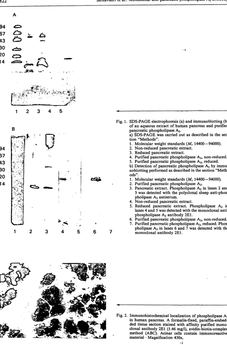
Monoclonal antibody 2E1 against human pancreatic phospholipase A
2One of the hybridomas, 2E1, produced an antibody that specifically reacted with human pancreatic phos- pholipase A
2. The purity of the monoclonal antibody 2E1 after the affmity purification with MAPS II Kit was verified by SDS-PAGE, where it gave homoge- neous protein bands with relative molecular masses of approximately 62000 (heavy chain) and 25000 (light chain). The subclass of the monoclonal antibody 2E1 was identified s IgGl.
The specificities of monoclonal and polyclonal anti- bodies were determined by immunohistochemistry and immunoblotting. An aqueous extract of human pancreas and purified pancreatic phospholipase A
2were resolved by SDS-PAGE, blotted to a nitrocel- lulose membrane and stained with monoclonal and polyclonal antibodies. The monoclonal antibody 2E1 reacted with one protein band only in both cases. The antibody 2E1 recognized the non-reduced native form but not the reduced form of the human pancreatic phospholipase A
2enzyme (fig. 1). The polyclonal sheep anti-phospholipase A
2antiserum gave one pro- tein band with purified human pancreatic phospho- lipase A
2(fig. 1). It recognized, however, one major band of apparent M
Tabout 15000 and another weak band of higher M
rin the aqueous extract of human pancreas (fig. 1). Phospholipase A
2was found in the cytoplasm of pancreatic acinar cells by immunohis- tochemistry using the monoclonal antibody 2E1 (fig.
2). No re ction was seen in pancreatic duct cells or
islets of Langerhans or in other human tissues studied
(liver, kidney, spieen, muscle, lymph node and thy-
roid). The specificity of the monoclonal antibody 2E1
was also investigated by TR-FIA (fig. 3). The anti-
body reacted with human, rat and porcine pancreatic
phospholipase A
2but not with bee venom phospho-
lipase A
2or phospholipase A
2purified from human
ascitic fluid.
E^- ·*
B
94 67 43 30 20 14
r'-r>'i
Fig. 1. SDS-PAGE electrophoresis (a) and immunoblotting (b) pf an aqueous extraet of human pancreas and purified pancreatic phospholipase A2.
a) SDS-PAGE was carried put äs described in the sec- tion "Methods".
1. Molecular weight Standards (Mr 14400-94000).
2. Non-reduced pancreatic extraet.
3. Reduced pancreatie extraet.
4. Purified pancreatic phospholipase A2, non-reduced.
5. Purified pancreatic phospholipase A2, reduced.
b) Detection of pancreatic phospholipase A2 by immu- noblotting performed äs described in the section "Meth^
ods".
1. Molecular weight Standards (Mr 14400-94000).
2. Purified pancreatic phospholipase A2.
3. Pancreatic extraet. Phospholipase A2 in lanes 2 and 3 was detected with the polyclonal sheep anti-phos- phoiipase A2 antiserum.
4. Non-reduced pancreatic extraet.
5. Reduced pancreatic extraet. Phospholipase A2 in lanes 4 and 5 was detected with the monoclonal anti^
phospholipase A2 antibody 2E1.
6. Purified pancreatic phospholipase A2, non-reduced.
7. Purified pancreatic phospholipase A2, reduced. Phos- pholipase A2 in lanes 6 and 7 was detected with the monoclonal antibody 2E1.
Fig. 2. Immunohistochemical localization of phosphoiipase A2 in human pancreas. A formaliti-fixed, paraffin-embed- ded tissue section stained with affinity pxirified mono^
clonal antibody 2E1 (i.46 mg/1), avidin-biotin-cömplex method (ABC). Acinar cells contain immunoreactive material - Magnification 430x.
Eur. J. Clin. Chem. Clin. Biochem. / Vol. 29,1991 / No. 12
Santavuori et al.: Monoclonal anti-pancreatic phospholipase A2 antibody 823 107Ί
40000- iω
g
φΰ l
20000l0 25 50 75 100 125 Monoclonal anti-phospholipase A2
antibody 2E1 [pg/l]
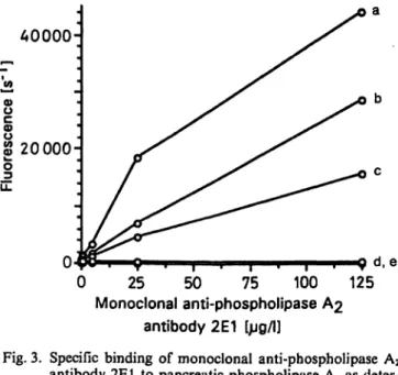
Fig. 3. Specific binding of monoclonal anti-phospholipase A2
antibody 2E1 to pancreatic phospholipase A2 s deter- mined by TR-FIA.
Microtitre plates were coated with l ng/well of phos- pholipase A2 purified from
a) human pancreas b) ral pancreas c) porcine pancreas d) bee venom and e) human ascitic fluid.
After Saturation with a Saturation solution and incu- bation with the monoclonal antibody 2E1, the immu- noreaction was detected with an Eu-labelled rabbit anti- mouse IgG second antibody. There is considerable cross-reactivity between human, rat and porcine pan- creatic phospholipase A2s, but none between human pancreatic and ascitic fluid or bee venom phospholipase A2s.
TR-FIA for human pancreatic phospholipase
A
2Standard curve, precision profile, detection limit and assay r nge
The Standard curve for TR-FIA of human phospho- lipase A2 and the precision profile calculated from 12 replicates (one strip) for each phospholipase A2 con- centration are shown in figure 4. The coefficient of intraassay Variation ranged from 2.8 to 9.9%. The sensitiyity of the assay defined s equivalent to the concentration cprresponding t the mean fluorescence of zero Standard (12 replicates) plus twice the Standard deviation was determined to be 0.28 μg/l. The r nge of values that can be measured with this method is 0.28-324 μg/l.
Analytical recovery
Two different concentrations (20,150 μg/l) of human phospholipase A2 Standards were added to five serum samples with'starting phospholipase A2 values varying from 4.11 to 28.49 μg/l. The percentage recovery of phospholipase A2 was calculated s (amount found
~ 106
10 100 Phospholipase A2 [fig/0
10
β r
ι
£
ο*ι
Φ<5
1000
Fig. 4. Dose-response curve (o) and precision profile (o) for the TR-FIA of human pancrealic phospholipase A2.
of added phospholipase A2/amount phospholipase A2 added ) χ 100. The analytical recoveries were 96.2%
and 123.1%, respectively, with an overall mean 109.6%.
Linearity
Four serum samples containing different concentra- tions of phospholipase A2 (92.81, 18.07, 15.21 and 4.33 μg/l) were diluted serially with assay buffer and analysed by TR-FIA. Excellent correlations (r = 0.99, 1.00, 1.00 and 0.99, respectively) were found in all four cases.
Reference interval
The mean concentration of phospholipase A2 in the serum of 57 apparently healthy individuals (23 women, 34 men) was 5.8 μg/l (SD 1.4). Given the parametric distribution of this analyte, the 95% ref- erence interval was 3.1—9.0 μg/l.
Ihter-niethod comparison
For comparison with a TR-FIA method using poly- clonal sheep anti-phospholipase A2 antibodies, the concentration of phospholipase A2 in 67 sera (28 normal, 39 pancreatitis) was measured by both the TR-FIA methods. The new assay was found to cor- relate well with the comparison method. The results were: y = 0.803x + 3.24; r = 0.95, n = 67 (fig. 6).
15
10
00
300-
Π Π ΠΠ
0 1 2 3 4 5 6 7 Θ 9 1 0 Immunoreactive phospholipase A2
Fig. 5. Distribution of values of pancreatic phospholipase A2
in sera of 23 healthy women and 34 men.
The mean age was 41 years (r nge 20 — 64). The mean concentration of phospholipase A2 is 5.8 ±1.4
Discussion
Phospholipase A2 exists either s an extracellular se- cretory enzyme or s an intracellular enzyme. The bee and snake venom and pancreatic secretory phospho- lipases A2 are typical extracellular enzymes. Intracell- ular phospholipases A2 have been described in many cell types, and they exist either in soluble form in the cytosol, or they are membrane-bound in various sub- cellular compartments (12 — 15). Membrane-associ- ated and soluble phospholipases A2 differ in their Substrate specificity (14) and hydrophobic properties (15), and they may also be functionally distinct. Mem- brane-bound phospholipases A2 are believed to par- ticipate in the regulation of phospholipid metabolism in biomembranes including the biosynthesis of eicos- anoids and the acylation-deacylation cycle of phos- pholipids (12).
Antibodies have been used to investigate immuno- chemical and structural relationships between phos- pholipases A2 from different sources. There are only limited and somewhat contradictory data on the an- tigenic relationship between intracellular and extra- cellular phospholipases A2. A polyclonal antiserum against human pancreatic phospholipase A2 did not recognize snake venom phospholipases A2 (16) or seminal fluid phospholipase A2 (17). kamoto et al.
(18) found immunological similarity between rat pan- creatic phospholipase A2 and rat splenic microsomal phospholipase A2. A monoclonal antibody against rat liver mitochondrial phospholipase A2 reacted with rat
11
<M 200
•ο
o
100-
o
100 200 300
Phospholipase A2 (polyclonal antibody) [pg/l]400
Fig. 6. Correlation between two TR-FIA methods using either sheep polyclonal or mouse monoclonal 2E1 anti-phos- pholipase A2 antibody s the catching antibody.
Pancreatic phospholipase A2 concentrations were as- sayed in serum samples of healthy individuals (n = 28) and patients suffering from acute pancreatitis (n = 39).
The linear regression equation is y = 0.803x + 3.24;
r = 0.95, n = 67.
liver cytosol and rat platelet phospholipase A2 but did not recognize rat or pig pancreatic phospholipase A2 or snake venom phospholipase A2 (19). Mollier et al.
(20) prepared a monoclonal antibody against mono- meric phospholipase A2 from snake venoms, and found that the antibody recognized phospholipase A2 from snake venoms but not from bee venom, porcine pancreas, rat lymphocytes pr guinea pig alveolar mac- rophages. Masliah et al. (21) demonstrated antigenic similarity between an extracellular phospholipase A2 (Naja naja venom) and membrane-bound phospholi- pase A2 from two different mammalian species and cell types (guinea pig alveolar macrophages and rat lymphocytes). In immunohistochemistry, the present monoclonal antibody 2E1 was found to be specific to pancreatic phospholipase A2. Iinmunoreactivity was found in human pancreatic acinar eells but not in other human tissues. We also investigated by TR-FIA the cross-reactivity of the monoclonal antibody 2E1 with non-pancreatic phospholipases A2 (bee Venom and human ascitic fluid phospholipase A2) and found no cross-reactivity. However, we found definite cross- reactivity with pancratic phospholipases A2 from other mammalian species. The antibody 2E1 reacted with rat and porcine pancreatic phospholipase A2, but it showed the highest affinity for human pancratic phospholipase A2. This finding confinns earlier ob- servations that phospholipases A2 from human, rat aiid porcine pancreas are closely related antigenically (2,22).
Eur. J. Clin. Chera. Clin. Biochem. / Vol. 29,1991 / No. 12
Santavuori et al.: Monoclonal anti-pancreatic phospholipase A2 antibody 825 In the present study we developed a TR-FIA for the
measurement of human pancreatic phospholipase A2
based on the new monoclonal antibody 2E1, the latter being used äs the catching antibody in a solid-phase assay. The increased concentration of immunoreactive pancreatic phospholipase A2 in serum is a specific and sensitive marker of acinar cell injury that occurs e. g.
in acute pancreatitis. In our earlier study, we found increased serum phospholipase A2 concentrations in- variably at the early stages of both mild and severe forms of acute pancratitis (5). The phospholipase A2
values were found to return to the reference interval at a somewhat slower rate than corresponding amy- lase activities in acute pancreatitis. The immunoreac- tive phospholipase A2 may include the active enzyme, the enzymatically inactive proenzyme and/or de- graded fragments of the active enzyme that may be catalytically active or inactive. Therefore immunoas- says do not give any Information äs to the catalytic activity of the phospholipase A2 enzyme molecule being measured. Since the catalytic activity of phos- pholipase A2 correlates with the severity of the disease in acute pancratitis (6), it is advisible to measure both the catalytic activity of phospholipase A2 and the concentration of immunoreactive pancreatic phos- pholipase A2 in the same serum samples in order to specifically detect pancreatic acinar cell injury (by the latter method) and to assess the severity of the disease process (by the former method). There is an ample selection of methods available for the determination of phospholipase A2 catalytic activity (23).
The previously reported radioimmunoassays and the TR-FIA for measuring the concentration of human immunoreactive pancreatic phospholipase A2 employ
polyclonal antibodies raised in rabbits (1—3, 5). The use of a monoclonal instead of a polyclonal antibody, however, would offer the advantage of unlimited sup- ply, constant and predictable quality, and high spec- ificity. Recently another RIA-method for the deter- mination of pancreatic phospholipase A2 employing a monoclonal antibody was described (4).
The mean (± SD) phospholipase A2 value, 5.8 ±1.4 g/l, measured in sera of healthy individuals by the present TR-FIA agrees well with the phospholipase A2 concentrations found by different immunoassays (RIA, TR-FIA): 6.5 ± 2.0 ^ (serum, 1), 5.1 + 1.7
§/1 (serum, 2), 4.3 ± 1.3 §/1 (plasma, 3), 2.4 ± 0.69 g/l (serum, 4) and 5.5 ±1.9 g/l (serum, 5). A good correlation between the present method and a TR- FIA described earlier by us (1) was found (data not shown). Likewise, measurements by the present method correlated very well (r = 0.95) with those obtained from the same normal and pathological serum samples by a method using a polyclonal sheep antibody both äs the catching and detecting antibody in a solid-phase TR-FIA (present study).
In summary, we describe a new monoclonal antibody 2E1 to human pancreatic secretory phospholipase A2
showing no cross-reactivity with other human tissues or ascitic phospholipase A2. A specific TR-FIA method was developed to measure pancreatic phos- pholipase A2 in serum samples.
Acknowledgement
The authors thank Ms Sinikka Kollanus and Ms Tinda Manninen for technical assistance, Mr Jaakko Lüppo for photography, Dr Juha Grönroos for serum samples and Dr Vesa Kleimola for fruitful discussions.
References
1. Eskola, J. U., Nevalainen, T. J. & Lövgren, T. N.-E. (1983) Time-resolved fluoroimmunoassay of human pancreatic phospholipase A2. Clin. Chem. 29, 1777-1780.
2. Nishijima, J., Okanaoto, M., Ogawa, M., Kasaki, G. &
Yamano, T. (1983) Purification and characterization of human pancreatic phospholipase A2 and development of a radioimmunoassäy. J. Biochem, 94, 137—147.
3. Sternby, B. & Äerström, B. (1984) Immunoreactive pan- creatic colipase, lipase and phospholipase A2 in human plasma and urine from healthy individuals. Biochim. Bio- phys. Acta 789, 164-169.
4. Oka, Y., Ogawa, M., Matsuda, Y, Murata, A., Nishijima, J., Miyauchi, K., Udä, K., Yasuda, T. & Mori, T. (1990) Serum immunoreactive pancreatic phospholipase A2 in pa- tients with variöus m^lignant tumours. Enzyme 43, 80—88.
5. Nevalainen, T. J., Eskola, J. U, Aho, A. J., Havia, V. T., Lövgren, T. N.-E. & Näntö, V. '(1985) Immunoreactive phospholipase A2 in serum in acute pancreatitis and pan- creatic cancer. Clin. Chem. 31, 1116-1120«.
6. Büchler, M., Malfertheiner, R, Schädlich, H., Nevalainen, T., Mavromatis, T. & Beger, H. G. (1989) Prognoslic value of serum phospholipase A2 in acute pancreatitis. Klin.
Wochenschr. 67, 186-189:
7. Kortesuo, P. T. & Nevalainen, T. J. (1991) Phospholipase A2 in human ascitic fluid: purification, characterization and immunochemical detection. Biochem. J. 278, 263 — 267.
8. Eskola, J. U, Nevalainen, T. J. & Aho, H. J. (1983) Puri- fication and characterization of human pancreatic phos- pholipase A2. Clin. Chem. 29, 1772-1776.
9. Galfre, G. & Milstein, C. (1981) Preparation of monoclonal antibodies: strategies and procedures. Methods Enzymol.
73, 3-46.
10. Hemmilä, L, Dakubu, S., Mukkala, V-M., Siitari, H. &
Lövgren, T. (1984) Europium äs a label in time-resolved immunofluorometric assays. Anal. Biochem. 137, 335—
11. International Federation of Clinical Chemistry (1987) Ap-343.
proved recommendation (1987) on the theory of reference values. Part 5r Statistical treatment of collected reference values. Determinations of reference limits. J. Clin. Chem.
Clin. Biochem. 25, 645—656.
12. van den Bosch, H. (1980) Intracellular phospholipases A.
Biochim. Biophys. Acta 604, 191-246.
13. van den Bosch, H. (1982) Phospholipases. In: New Com- prehensive Biochemistry, vol. 4 (Havvthorne, J. N. & Ansell,
G. B., eds), pp. 313 — 357 Eisevier, Amsterdam.
14. Gonzaiez-Buritica, H., Smith, D. M. & Turner, R. A. (1989) Characterization of soluble and cell associated phospholi- pase A2 from rheumatoid synovial fluid. Ann. Rheum. Dis.
48, 557-564.
15. Ono, T., Tojo, H., Kuramitsu, S., Kagaraiyama, H. Oka- moto, M. (1988) Purification and characterization of a membrane-associated phospholipase A2 from rat spieen. J.
Biol. Chem. 263, 5732-5738.
16. Meijer, H., Meddens, M. J. M., Dijkman, R., Slotboom, A. J. & de Haas, G. H. (1978) Immunological studies on pancreatic phospholipase A2. J- Biol. Chem. 2S3, 8564—
8569.
17. Wurl, M. & Kunze, H. (1985) Purification and properties of phospholipase A2 from human seminal plasma. Biochim.
Biophys. Acta 834, 411-418.
18. Okamoto, M., Onto, T., Tojo, H. & Yamano, T. (1985) Immunochemical relatedness between secretory phospho- lipase A2 and intracellular phospholipase A2. Biochim. Bio- phys. Res. Commun. 128, 788-794.
19. de Jong, J. G. N., Arnesz, H., Aarsman, A. J., Lenting, H.
B. M. & van den Bosch, H. (1987) Monoclonal ä^tibodies against an intracellular phospholipase A2 from rat liver and their cross-reactivity with other phospholipase A2. Eur. J.
Biochem. 164, 129-135.
20. Mollier, R, Chwetzoff, S. & Menez, A. A. (1990) A möno- clonal antibody recognizing conserved epitope in a groüp of phospholipase A2. Mol. Immunol. 27, 7—15.
21. Masliah, J., Kadir, C., Pepin, D.vilybkine, T., Etienne, J., Chambaz, J. & Bereziat, G. (1987) Antigenic relatedness between phospholipase A2 from Naja naja venom and ff öm mammalian cells. FEBS Lett. 222, 11 — 16.
22. Ono, T., Tojo, H., Inoue, K., Kagamiyama, H., Yamano9
T. & Okamoto, M. (1984) JRat pancreatic phospholipase A2: purification, characterization, and N-terminal amino acid sequence. J. Biochem. 96, 785 — 792.
23. Nevalainen, T. J. (1988) Phospholipase A2 in acute pan- creatitis. Scand. J. Gastroenterol. 23, 897—904.
Dr. Timo Nevalainen Department of Pathology Kiinamyliynkatu 10 SF-20520 Turku