Eur. J. Clin. Chem. Clin. Biochem.
Vol. 32, 1994, pp. 837-842
© 1994 Walter de Gruyter & Co.
Berlin · New York
A Comparative Stiidy of the Concentrations of Hypoxanthine, Xanthine, Uric Acid and Allantoin in the Peripheral Blood of Normals and Patients with Acute Myocardial Infarction and Other Ischaemic Diseases
By R. Kock\ ß. Delvowc1, M. Sigmund2 and H. Greiling1
1 Institute for Clinical Chemistry and Pathobiochemistry, Medical Faculty, University of Technology Aachen, Aachen, Germany
2 Medical Clinic I, Medical Faculty, University of Technology Aachen, Aachen, Germany
(Received February 14/August 4, 1994)
Summary: The aim of this study was the elucidation of the role of the xanthine oxidoreductase in the purine metabolism in ischaemic diseases of man. The serum concentrations of hypoxanthine, xanthine, uric acid and allantoin were determined in peripheral blood samples from patients with angina pectoris, cerebral insult and myocardial infarction with thrombolytic therapy and were compared with the concentrations obtained for healthy males and females. No significant differences were observed for the serum hypoxanthine concentrations, xanthine concentrations, the sum (hypoxanthine + xanthine) and the ratio (xanthine/hypoxanthine) between the healthy males, healthy females, the patients suffering from angina pectoris and the patients suffering from cerebral insult.
An increase of the serum xanthine concentration in patients with myocardial infarction indicates a significant metabolic involvement of xanthine oxidoreductase in this disease and therefore a possible role in the development of tissue damage in the postischaemic phase due to oxygen radicals generated by the oxidase activity of this enzyme.
The serum concentrations of uric acid and allantoin showed no differences between any of the studied groups.
Study of the non-enzymatic oxidation of uric acid to allantoin by oxygen radicals, a relevant radical-scavenging mechanism in other diseases, provided no indication of an increased concentration of oxygen radicals due to the xanthine oxidoreductase reaction or other radical-producing mechanisms.
Introduction xanthine, xanthine, uric acid and allantoin in samples from n = 115 patients with acute myocardial infarction Xanthine oxidoreductase (EC 1.2.3.2) is the terminal en- were compared with those in samples from n = 180
zyme of purine metabolism. Physiologically this enzyme hea,thy ma,eS5 n = M5 hea]thy females> n = 45 patients
used NAD+ äs electron acceptor, but in ischaemia it with angina pectoris and n = 19 patients suffering from may be converted from the dehydrogehase form to an cerebra] infarction. Furthermore the results of the serial oxidase form, which uses molecular oxygen äs an determinations of the serum concentrations of hypoxan- electron acceptor (1). Several studies have been per- t^n^ xanthine, the sum (hypoxanthine + xanthine) and formed on the relevance of xanthine oxidase in tissue the ratio (xanthine/hypoxanthine) for n = 53 patients destruction by oxygen-derived radicals. De Scheerder wjth clinically diagnosed acute myocardial infarction concluded from the parallel coronary release of malon- were compared with classical clinical chemical markers dialdehyde and uric acid, that there is an enhanced un- for the diagnosis and follow-up of this disease. The specific oxidation of membrane phospholipids by oxy- method used for the simultaneous determination of hy- gen-derived radicals produced by xanthine oxidase (2). poxanthine, xanthine, uric acid and allantoin from serum In the present study the serum concentrations of hypo- was presented in a previous publication (3).
Eur. J. Clin. Chem. Clin. Biochem. / Vol. 32, 1994 / No. 11
838 Kock et al.: Oxypurines and allantoin in blood of patients with ischaemic diseases
Patientswith myocardial infafction Patients with myocardial infarction
Hypoxanthine concentration [umol/l]
10 15 20 25 Hypoxanthine concentration [pmol/l]
30 £
Healthy male adults
n
Healthy female adultsPatients with angina pectoris
Patients with cerebral insult
Patientswith myocardial infarction
5 10 Xanthine concentration. [μηηοΙ/Ι]
15
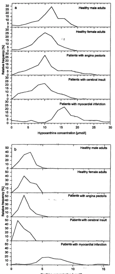
Fig. l a-b Distribution of serum hypoxanthine concentrations and serum xanthine concentrations for the reference groups (heaK thy males and healthy females), patients suffering from angina pec-
50- 40-30- 20 - 10 - 50-0 40- 30- 20-
50-
=30 -^0-
;20 -
I
050 -40 - 30 - 20- 10-0 50 -40- 30 - 20 - 10 -0
Healthy male adults
Healthy female adults
Patents with angina pectoris
Patientswith cerebral insult
Patientswith myocardial infarction
5 10 15 Xanthine concentration [μπηοΙ/Ι]
toris and patients suffering from cerebral insult versus patients with acute myocardial infarction.
Materials and Methods
Chemicals
Munichi Germany) were used. rie acid was Standard Reference Material with a.purity of 99.7% (SRM 913, National Bureau of Standards, Washington D. C. 20234). The reagents required f r the For the preparation of Standard Solutions of allantoin, hyppxanthine" determinatioii of the enzyme acti vities of creatine kinase and cre- and xanthine, materials of the highest available purity (SIGMA, atine kinase isoenzyme MB wejrp from Boehringer Mannheim.
Eur. J. Clin> Chem. Clin. Biochem. / Vol. 32,1994 / No. 11
All other Chemicals were purchased from Merck (Darmstadt, Germany) and were of analytical grade; methanol was of HPLC-grade.
Samples
For the drawing blood the Sarstedt-system with separation-gel was used and the time between blood withdrawal and centrifugation never exceeded 30 min.
The samples in this study were from 180 healthy adult males, 145 healthy adult females, 155 adult patients with acute myocardial infarction and successful thrombolytic therapy according to clinical observations, 45 patients suffering from angina pectoris, and 19 patients with a cerebral infarction. The patient samples were drawn within 3-7 h after hospitalization.
All the samples taken from 53 patients with acute myocardial in- farction for diagnostic purposes and follow-up were analysed.
These patients showed early reperfusion of the occluded coronary artery spontaneously or s a result of therapeutic lysis.
High performance liquid chromatography
HPLC was performed on a Waters chromatography System with a gradient pump Model 600, a Satellite WISP Model 700 and a tune- able wavelength detector Model 481. System control, data acquisi- tion, Integration and calibration were performed with the Waters Maxima 825 Software. Chromatographie separations were per- formed s described in 1. c. (3). For the determination of hypoxan- thine, xanthine and uric acid, UV-detection was performed at 254 nm with a r nge of 0.005 absorbance units f ll scale and a time constant of l .0 s. For the determination of allantoin s described
in 1. c. (3), UV-detection was performed at 360 nm with a r nge of 0.100 absorbance units f ll scale and a time constant of 1.0 s.
The method used in this study had a detection limit for hypoxan- thine of l pmol and for xanthine of l .5 pmol; since 50 μΐ of five- fold diluted serum were injected, the lower concentration limit for hypoxanthine and xanthine was 0.1 μηηοΐ/ΐ and 0.15 μπιοΐ/ΐ, respec- tively. For serum hypoxanthine concentrations of 5-50 μιτιοΐ/ΐ the coefficient of Variation did not exceed 5%; for xanthine concentra- tions of 2-20 μιτιοΐ/ΐ the imprecision was less than 7%.
Determination of enzyme activities
The determination of the enzyme activities of creatine kinase and creatine kinase-MB were performed on a Hitachi 747 at 25 °C with the reagent kits obtained from Boehringer Mannheim.
Statistical procedures and c a l c u l a t i o n s
For data management, Microsoft Excel 3.0a (Microsoft GmbH, Munich, Germany) was used. Statistical treatments, including the calculation of the Bayesian parameters for the sample classifica- tion, were performed with the Statistical package Sigmastat V 1.02 (Jandel Scientific GmbH, Erkrath, Germany).
Results
Hypoxanthine and xanthine from single serum samples
In figure l a—b the distribution of the hypoxanthine and xanthine concentrations in sera from patients with myo-
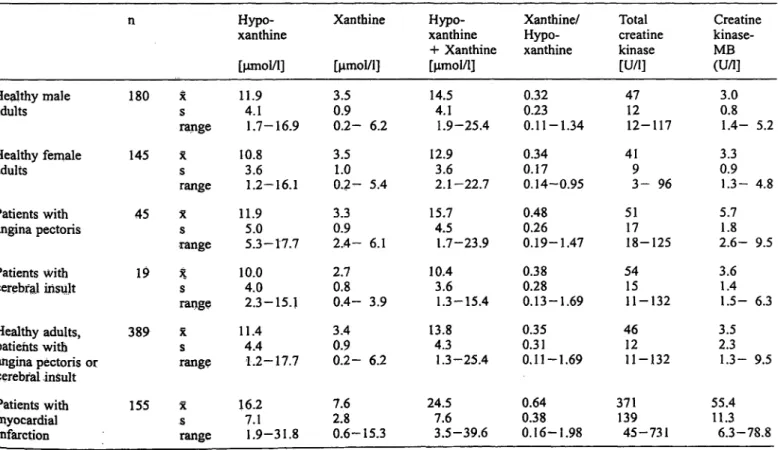
Tab. l Results for the determination of hypoxanthine, xanthine,
the sum (hypoxanthine + xanthine) and the ratio (xanthine/hypo- xanthine). The samples from the patients were drawn at the inten- sive care unit within 3—7 h after hospitalization.
n
Healthy male 180 adults
Healthy female 145 adults
Patients with 45 angina pectoris
Patients with 19 cerebfal iris jt
Healthy adults, 389 patients with
angina pectoris or cerebral insult
Patients with 155 myocardial
infarction X — mean
s = Standard deviation
*s r nge
X
sr nge
Xs r nge
*s r nge
Xs r nge
X
sr nge
Hypo- xanthine [μτηοΐ/ΐ]
11.94.1 1.7-16.9 10.83.6
1.2-16.1 11.95.0
5.3-17.7 10.04.0
2.3-15.1 11.44.4
1.2-17.7 16.27.1
1.9-31.8
Xanthine [μηιοΐ/l]
3.50.9 0.2- 6.2 3.51.0 0.2- 5.4 3.30.9 2.4- 6.1 2.70.8 0.4- 3.9 3.40.9 0.2- 6.2 7.62.8 0.6-15.3
Hypo- xanthine + Xanthine [μιτιοΙ/1]
14.54.1 1.9-25.4 12.93.6
2.1-22.7 15.74.5
1.7-23.9 10.43.6
1.3-15.4 13.84.3
1.3-25.4 24.57.6
3.5-39.6
Xanthine/
Hypo- xanthine 0.320.23 0.11-1.34 0.340.17 0.14-0.95 0.480.26 0.19-1.47 0.380.28 0.13-1.69 0.350.31 0,11-1.69 0.640.38 0.16-1.98
Total creatine kinase [U/l]
4712 12-117 419
3- 96 5117 18-125 5415
11-132 4612 11-132 371139
45-731
Creatine kinase- MB(U/l]
3.00.8 1.4- 5.2 3.30.9 1.3- 4.8 5.71.8 2.6- 9.5 3.61.4 1.5- 6.3 3.52.3 1.3- 9.5 55.411.3
6.3-78.8
Eur. J. Clin. Chem. Clin. Biochem, / Vol. 32,1994 / No. 11
840 Kock et al.: Oxypurines and allantoin in blood of patients with ischaemic diseases
cardial infarction are shown versus those obtained for healthy males, healthy females, patients with angina pectoris and patients with cerebral insult. Table l shows the statistical quantities of the distributions, and the sta- tistical quantities of the distribution of the sum (hypo- xanthine + xanthine), the ratio (xanthine/hypoxanthine), total creatine kinase activity and creatine kinase-MB ac- tivity. 7he data of the n = 45 patients suffering from angina pectoris and of the n = 19 patients suffering from cerebral insult cannot be discriminated from the data ob- tained for healthy males and healthy females. Sensitivity and specificity of the studied analytes were calculated from the results obtained for the samples from n = 155 patients suffering from myocardial infarction and the re- sults for the n = 389 samples combined from the four groups of healthy males, healthy females, angina pecto- ris and cerebral insult. Hypoxanthine showed a sensitiv- ity of 0.59 for a specificity of 0.90 at a discriminator value of 14.7 μπιοΐ/ΐ, xanthine a sensitivity of 0.83 for a specificity of 0.90 at a discriminator value of 4.5 μηηο1/1, the sum (hypoxanthine + xanthine) a sensitivity of 0.77 for a specificity of 0.90 at a discriminator value of 19.7 μιτιοΐ/ΐ, and the ratio (xanthine/hypoxanthine) a sensitivity of 0.35 for a specificity of 0.90 at a discrimi- nator value of 0.78. The ROC-curves are shown in fig- ure 2.
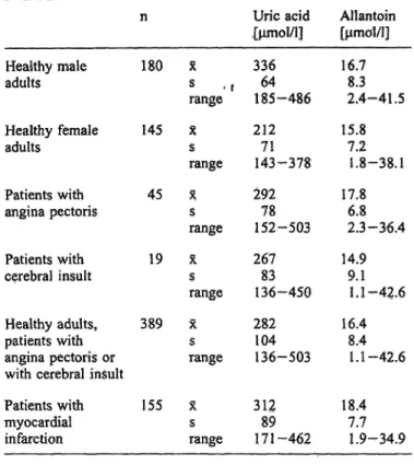
Tab. 2 Results for the determination of uric acid and allantoin.
The samples are the same s in table 1.
1.0
0.8
0.6
0.4
0.2
0.0
1.0 0.8 0.6 0.4
Specificity
0.2 0.0
Fig. 2 ROC-curves for serum hypoxanthine concentrations, se^·
rum xanthine concentrations, sum (hypoxanthine 4- xarithihe) and ratio (xanthine/hypoxanthine) for the discrimination between a ref- erence collective consisting of the normals, the patients with angina pectoris and the patients with cerebral insult, and the patients with myocardial infarction. The data used for the calculation are those shown in figure l a—d.
Hypoxanthine Xanthine
Hypoxanthine + xanthine Ratio xanthine/hypoxanthine
n
Healthy male 180 adults
Healthy female 145 adults
Patients with 45 angina pectoris
Patients with 19 cerebral insult
Healthy adults, 389 patients with
angina pectoris or with cerebral insult Patients with 155 myocardial
infarction
χ
s « f r nge Xs r nge
X
sr nge
X
sfange
X
r nges
X
sr nge
Uric acid ,[μτηο1/1]
33664 185-486 21271 143-378 29278 152-503 26783 136-450 282104
136-503
31289 171-462
Allantoin [μιτιοΐ/ΐ]
16.78.3 2.4-41.5 15.87.2
1.8-38.1 17.86.8
2.3-36.4 14.99.1
1.1-42.6 16.48.4
1.1-42.6
18.47.7 1.9-34.9 X = mean
s — Standard deviation
Uric acid and allantoin from single serum samples
Table 2 summarizes the data for the determination of uric acid and allantoin from the samples used for the data in figure l a—b. The distribution of the serum con- centrations of uric acid and the distribution of serum concentrations of allantoin in the samples from patients with myocardial infarction was not significantly dif- ferent from the distribution in the samples of the refer- ence c llectives.
Release profiles of purines and total creatine kinase/kreatine kinase-MB derived from sera of patients with myocardial infarction
Figure 3a—b shows the data for hypoxanthine, xanthine, total creatine kinase and creatine kinase-MB from serial serum samples, obtained from n = 53 patients with myocardial infarction, hospitalized at the cardiological intensive care unit between 5 h and 150 h after pain on- set. For the samples drawn initially (= 5 ± 2 h after pain onset), only the median for the serum xanthine coneen- tration exceeded the discriminator value for a specificity of 0.90. For hypoxanthine in median of the meas fed serum concentration feil below the discriminator value for a speeificity of 0.9 after 80-100 h. For xanthine, the median of the measured serum concentration< feil below
Eur. J. Clin, Chem. Ciin. Biochem. / Vol. 32, 1994 /No. 11
50
140 30
10
14.7
4.5
0 20 40 60 80 100 120 140 160 180 Time after onset of pain [h]
Fig. 3a—b Serum hypoxanthine concentrations, serum xanthine concentrations, total creatine kinase activity and creatine kinase- MB activity determined at different times after pain onset for n = 53 patients with myocardial infarction. The data are given äs
L1200 iooo
600
400
200
80 70 60 50
1 3
40°
J 20 Ö 10 m
100
20 40 60 80 100 120 140 160 180 Time after onset of pain [h]
25th-percentile (Iower broken line), 50th-percentile (medium solid line) and 75th-percentile (upper broken line). The discriminator val- ues for a specificity of 0.90 are added (right-hand axis, continuous straight line).
the discriminator value for a specificity of 0.9 after 100-l25 h. After 125h the median of the total cre- atine kinase serum activity feil below the upper limit of the reference ränge used for this method in our lab- oratory.
Cpnclusions
This clinical study of purine rrietabolism, following myocardial ischaemia fesulting frprn myocardial infarc- tion, was perfbrmed in order to elucidate the possible role of xanthine oxidoreductase iri tissüe alterations in the post-ischaemic phäse. During ischaemia there is a calcium-dependent enzymatic degradation of xanthine oxidoreductase frorn the NAD+-consurning dehydroge- nase form to an oxidase form, which directly utilizes moleculär oxygen äs an electron acceptor in the oxida- tion of hypoxanthine via xanthine to uric acid (1,4^5).
This enzyme could yield very reactive, oxygen-derived radical ions, which could, if not scavenged by appropri-
ate radical-scavengers, lead to fiirther tissüe destruction (6).
To assess the detectability of changes of purine metabo- lism in myocardial tissüe, using peripheral serum sam- ples, the results for serum hypoxanthine, xanthine, the sum (hypoxanthine -K xanthine) and the ratio (xanthine/
hypoxanthine) were determined in blood samples of n = 155 patients suffering from myocardial infarction versus appropriate reference collectives. The data shown in figure l a—b and summarized in table l indicate an increased efflux of hypoxanthine and xanthine from the injured tissüe similar to the efflux of creatine kinase.
The sensitivity and specificity data calculated from the distributions of purine serum concentrations show that the elevation of the serum xanthine concentration is most specific for the patients with myocardial infarction.
The determination of hypoxanthine is much more unspe- cific; this analyte is subject to preanalytical errors (7), but cell death and damage might also be responsible for an increase of the hypoxanthine serum concentration.
Eur. J. Clin. Chem. Clin. Biochem. / Vol. 32,1994 / No. 11
842 Kock et al: Oxypurines and allantoin in blood of patients with ischaemic diseases
The derived quantities, the sum (hypoxanthine + xan- thine) and the ratio (xanthine/hypoxanthine) are less in- formative about disease-specific metabolic changes, but the unequivocal increase of serum xanthine must be due to xanthine oxidoreductase activity directly connected with the metabolic changes of myocardial ischaemia.
Consequently, the values for xanthine in the samples drawn between approximately 5 hours and 6 days after pain onset (from the patients kept ander surveillance af- ter initial hospitalization in the cardiological intensive care unit) show an elevation above a discriminator value required for a specificity of 0.90 for the longest time after pain onset.
The main result of this study is that the xanthine concen*
tration measured in serum derived from peripheral blood is significantly increased in samples of patients suffering from myocardial infarction, compared with sera of an appropriate reference collective. The metabolic activity of xanthine oxidoreductase in myocardial infarction has therefore been proven in this study, and the fact that the release of xanthine persists for four to five days after pain onset on a level above the normal ränge, Supports the role of xanthine oxidoreductase, which has been pre- viously and controversially suggested (9—14).
On the other hand, the existence of oxygen-derived radi- cals was not supported by examination of a radical-scav-
enging mechanism, the spontaneous oxidation of uric <<
acid to allantoin. Grootveld (8) has shown that allantoin '.;
is the best marker for the detection of oxygen-radical ;·
induced spontaneous oxidation of uric acid and therefore the best index for the activity of uric acid äs a radical- scavenger. Table 2 shows that there is no difference be- tween the groups (see tab. 1) with respect to uric acid or allantoin. But the absence of a response by allantoin only hidicates that the oxygen^radicals, if generated, are preferably subject to other radical-scavenging mecha- nismSj in contrast to the diseases described by Groot- veld (8).
Therefore, the clinical relevance of the findings in this · study depends on the validity of those resülts that indi- '"' cate that the oxygen-consuming O-form of the xanthine oxidoreductase is the predöminant form in the later phase of myocardial infarction (15).
For elimination of the remaining ambiguities of the pre^
sent resülts, additional studies must be performed to clarify qf the origin, the kinetics and the fate of the xan- thine oxidoreductase involved in the metabolic changes following myocardial infarction. Additionally, the origin of the purine metabolites that lead tö the long-term ele- vation of the xanthine effusion from the heart must be elücidated.
References
1. MC Cord, J. M. (1987) Oxygen-derived radicals: A link be- tween reperfusion injury and inflammation. Fed. Proc. 46, 2402-2406.
2. De Scheerder, I. K., van de Kraay, A. M. M., Lamers, J. M.
J., Koster, J. F, de Jong, J. W. & Serruys, P. W. (1991) Myocar- dial malondialdehyde and uric acid release after short-lasting coronary occlusions during coronary angioplasty: Potential mechanisms for free radical generation. Am. J. Cardiol. 68, 392-395.
3. Kock, R., Delvoux, B. & Greiling, H. (1993) A high-perfor- mance liquid Chromatographie method for the determination of hypoxanthine, xanthine, uric acid and allantoin in serum.
Eur. J. Clin. Chem. Clin. Biochem. 31, 303-310.
4. Iriyama, K. (1987) Uric acid in ischemic tissue. Jikeikai Med.
J. 34, 145-168.
5. MC Cord, J. M. & Fridovich, L (1968) The reduction of cyto- chrome c by milk xanthine oxidase. J. Biol. Chem. 243, 5753-5760.
6. MC Cord, J. M. (1985) Oxygen-derived free radicals in post- ischemic tissue injury. N. Engl. J. Med. 312, 159-163.
7. Boulieu, R., Bory, C., Baltassat, P. & Gönnet, C. (1983) Hypo- xanthine and xanthine levels determined by high-performance liquid chromatography in plasma, erythrocyte, and urine sam- ples from healthy subjects: The problem of hypoxanthine level evolution äs a function of time. Anal. Biochem. 729,398-404.
8. Grootveld, M. & Halliwell, B. (1987) Measurement of allan- toin and uric acid in human body fluids. Biochem. J. 243, 803-808.
9. Betz, A. L., Randall, J. & Martz, J. (1991) Xanthine oxidase is not a major source of free radicals in focal cerebral ischemia. Am. J. Physiol. 260 (Heart Circ. Physiol. 29«), H563-H568.
10. Werns, S. W., Shea, J. J., Mitsos, S. E., Dysko, R. C., Fantpne, J. C., Schork, M. A., Abrams, G. D., Pitt, B. & Lucchesi, B.
R. (1986) Reduction of the size of infarction by allopurinol in the ischemicrieperrused canine heart. Cifculation 73, 518—
524.
11. Chambers, D. E., Parks, D. A., Paterson, G., Roy, R. S., MC Cord, J. M., Yoshida, S., Parmleyj L. & Downey, J. M. (1985) Xanthine oxidase äs a source of free radical in myoeardial ischemia. J. Moll. Cell. Cardiol. 17, 145-152.
12. Hammond, B., Kontos, H. A. & Hess, M. L. (1985) Oxygen radicals in the adult respiratory distress syndrome, in myocar- dial ischemia and reperfusion injury, and in cerebral vascular damage. Can. J. Physiol. Pharmaeol. 63, 173-187.
13. Peterson, D. A., Asinger, R. W., Elsperger, K. J., Homans^ D.
C. & Eaton, J. W. (1985) Reactive oxygen species may cause myocardial reperfusion injury. Biochem. Biophys. Res. Comm.
727, 87-93.
14. Stoltenberg, L., Rootvelt, T., 0yasaeter, S., Rognum; T. O. &
Saugstad, O. A. (1993) Hypoxanthine, xanthine, and uric acid concentrations in plasma, cerebrospinal fluid, vitreous humor, and urine in piglets subjected to intermittent versus cöntinuoüs hypoxemia. Pediatr. Res. 34, 767-77-1.
15. Becker, B. J. (1993) Towards the physiologieal function of uric acid. Free Radical Biol. Med. 14, 615-631.
Dr. R. Kock
Institut für Klinische Chemie und Pathpbiochemie RWTH Aachen Pauwelssträße 30
D-520741 Aachen :
Germany
Eür. J. Clin. Chem. Clin. Bioehem. / Vol. 32,1994/No. 11 '!