Digital pressure ulcer after pulse oximetry
Digitales Druckulkus nach Pulsoxymetrie
Abstract
In emergency medical service, in intensive care unit and anaesthesia oxygenation is monitored with pulse oximetry apparatus. Pulse oximetry
Philip H. Zeplin
1probe is usually attached to the finger, toe or earlobe. To the best of
1 Universitätsklinikum Leipzig, Department für Operative our knowledge this is the first case report describing the occurrence of
a pressure ulcer after finger pulse oximetry measurement.
Medizin, Abteilung für Keywords:pressure ulcer, finger pulse oximetry Plastische, Ästhetische und
spezielle Handchirurgie, Leipzig, Germany
Zusammenfassung
Sowohl in der Notfall- und Intensivmedizin als auch in der Anästhesie wird die Sauerstoffsättigung des Blutes mit Pulsoxymetern ermittelt.
Diese Pulsoxymeter werden üblicherweise an den Fingern, den Zehen oder dem Ohrläppchen angebracht. Wir beschreiben einen Fall, bei dem es nach Anlage eines Fingerclip-Pulsoxymeters zur Ausbildung eines operationsbedürftigen Druckulkus kam.
Schlüsselwörter:Druckulkus, Fingerclip-Pulsoxymeter
Case report
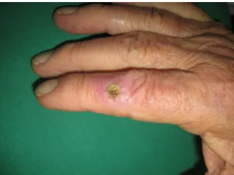
Figure 1: Grade IV pressure ulcer of the right index finger
A 76-year-old man with insulin dependent diabetes mel- litus type II, renal insufficiency caused by diabetic neph- ropathy had an arteriovenous shunt for dialysis on the right arm. Because of an intercommisural mitral valve insufficiency he underwent mitral valve clipping. The mitraclip therapy procedure was done under general an- aesthesia and takes approximately 3.5 hours. A pulse oximeter probe was placed on the right index finger to allow continuous monitoring throughout the procedure.
After removal of the pulse oximeter clip an unspecific flush occurred in the radiodorsal middle phalanx area.
Over the next four weeks a sharply demarcated skin lesion developed and continued to worsen into result shown in
Figure 1. The patient stated that he had never felt any pain. The physical and radiological examination revealed full-thickness skin damage extended into the subcu- taneous tissue layer and the extensor tendon. The lesion was diagnosed as a grade IV pressure ulcer. After surgical debridement the remaining defect was covered by a re- versed cross-finger flap.
Diabetes is strongly associated with both micro- and macrovascular complications that encourage the devel- opment of peripheral vascular disease and neuropathy.
By impairing the blood flow and loss of sensation the risk for developing pressure ulcers – particularly in the feet – increases [1]. In emergency medical service, in intensive care unit and anaesthesia oxygenation is monitored with pulse oximetry apparatus. Pulse oximetry probe is usually attached to the finger, toe or earlobe. One case of grade II pressure ulcer on a patient’s ear as a result of pulse oximetry probe has been described [2]. To the best of our knowledge this is the first case report describing the oc- currence of a pressure ulcer after finger pulse oximetry measurement. Pressure is assumed to be the most im- portant single factor in the development of pressure ul- cers. In healthy individuals capillary filling pressure of the finger arteries at heart level lies between 12–50 mmHg.
Values of >30 mmHg can only be measured in warm and vasodilated state [3]. Using a pressure measuring instru- ment we calculated the pressure of a correct attached pulse oximeter finger clip with 18 mmHg. Through longer period of time incorrect attached pulse oximeter finger clips can induce pressure ulcer. This indicates a correct use of finger pulse oximeter especially in patients with
1/2 GMS German Plastic, Reconstructive and Aesthetic Surgery 2013, Vol. 3, ISSN 2193-7052
Case Report
OPEN ACCESS
assumed capillary pressure. This case shows that pres- sure ulcers not only develop on predilection sites and continuous checks of all instruments attached to the patient are suggested.
Notes
Competing interests
The author declares that he has no competing interests.
References
1. Jeffcoate WJ, Harding KG. Diabetic foot ulcers. Lancet. 2003 May 3;361(9368):1545-51. DOI: 10.1016/S0140-
6736(03)13169-8
2. Iranmanesh S, Rafiei H, Esmaeili Abdar M. A case of pressure ulcer development on a patient's ear as a result of pulse oximetry probe. Int Wound J. 2012 Dec;9(6):701-2. DOI: 10.1111/j.1742- 481X.2011.00906.x
3. Levick JR, Michel CC. Capillary pressures in the fingers and toes [proceedings]. J Physiol. 1976 Sep;260(2):57P.
Corresponding author:
Priv.-Doz. Dr. med. habil. Philip H. Zeplin
Universitätsklinikum Leipzig, Department für Operative Medizin, Abteilung für Plastische, Ästhetische und spezielle Handchirurgie, Liebigstr. 20, 04155 Leipzig, Germany, Phone: +49-(0)341-9717188, Fax:
+49-(0)341-9717139
philip.zeplin@medizin.uni-leipzig.de
Please cite as
Zeplin PH. Digital pressure ulcer after pulse oximetry. GMS Ger Plast Reconstr Aesthet Surg. 2013;3:Doc02.
DOI: 10.3205/gpras000012, URN: urn:nbn:de:0183-gpras0000127
This article is freely available from
http://www.egms.de/en/journals/gpras/2013-3/gpras000012.shtml Published:2013-06-05
Copyright
©2013 Zeplin. This is an Open Access article distributed under the terms of the Creative Commons Attribution License
(http://creativecommons.org/licenses/by-nc-nd/3.0/deed.en). You are free: to Share — to copy, distribute and transmit the work, provided the original author and source are credited.
2/2 GMS German Plastic, Reconstructive and Aesthetic Surgery 2013, Vol. 3, ISSN 2193-7052
Zeplin: Digital pressure ulcer after pulse oximetry