The role of the pro-inflammatory cytokine Interleukin-18, its processing enzyme Caspase-1, and potential alternative IL-18-
activating pathways in atherosclerosis
Inaugural-Dissertation zur
Erlangung des Doktorgrades
der Mathematisch-Naturwissenschaftlichen Fakultät der Universität zu Köln
vorgelegt von Norbert Gerdes
aus Altenberge
Köln 2005
This work was performed between September 2001 and May 2005 under supervision of Professor Dr. Uwe Schönbeck in the group of Professor Dr. Peter Libby at the Division of Cardiovascular Medicine, Department of Medicine, Brigham & Women’s Hospital and Harvard Medical School in Boston, Massachusetts, United States of America. The work was mentored by Professor Dr. Helmut W. Klein, Institute for Biochemistry, University of Cologne, Germany.
Referees (Berichterstatter): Prof. Dr. Helmut W. Klein, University of Cologne Prof. Dr. Jens Brüning, University of Cologne
Prof. Dr. Uwe Schönbeck, Harvard Medical School
Day of disputation: 5. July 2005
For Christin And My family
Table of contents
Table of Abbreviations ….……….……….……….... 5
Zusammenfassung (German summary) ……….. 7
Summary………. 8
1. Introduction ………...………..…………... 9
1.1. Cardiovascular disease and atherosclerosis………....………. 9
1.2. Atherosclerosis: An inflammatory disease ………....…….… 10
1.3. Matrix metalloproteinases in atherosclerosis……….. 14
1.4. Cytokines in atherosclerosis ………... 15
1.5. The pro-inflammatory cytokine IL-18….………....………... 16
1.6. Caspase-1………... 17
1.7. IL-18 and atherosclerosis……….………....….. 18
1.8. Clinical association of IL-18 with cardiovascular disease and its risk factors... 20
1.9. Aim of this thesis ………... 21
2. Materials and Methods ……….………...………..…………... 22
2.1. Materials ……….………... 22
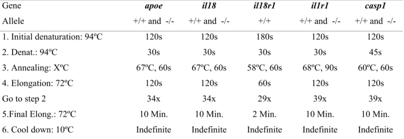
2.2. Expression of IFNγ in atheroma-associated cell types ………....……….. 22
2.3. Murine atherosclerosis model………. 30
2.4. Western blot analysis of mouse tissue lysates……… 40
2.5. MMP-mediated processing of proIL-18………...… 42
3. Results ……….. 45
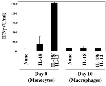
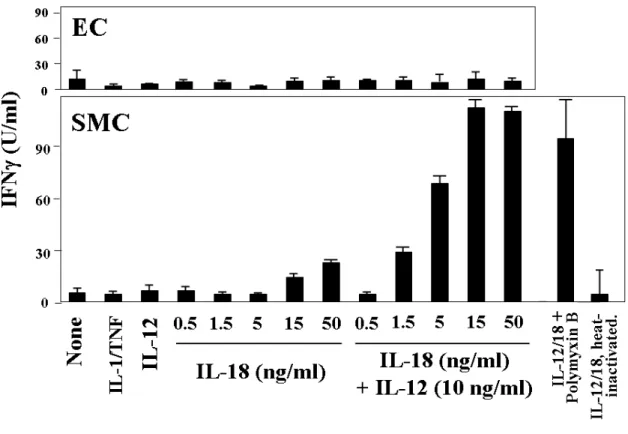
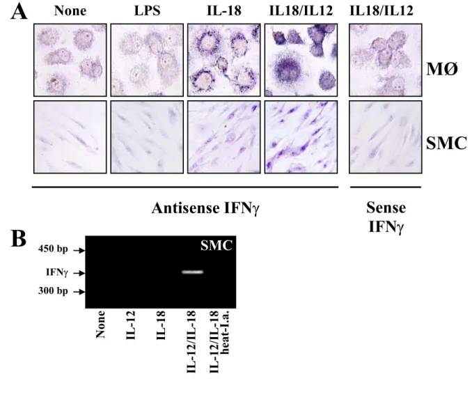
3.1. IL-18 induces IFNγ in macrophages and smooth muscle cells..………… 45
3.2. Deficiency of IL-18 decreases development of atherosclerosis in mice.... 51
3.3. The role of IL-18R in atherogenesis………... 65
3.4. Caspase-1 deficiency does not limit atherogenesis in hyperlipidemic mice.... 71
3.5. Caspase-1 independent processing of proIL-18………. 78
4. Discussion ……….…… 90
5. References ……….….…... 103
Acknowledgments ...………...……….……….. 118
Statement of Research ….………..…... 120
Curriculum vitae...………...……….……….. 122
Table of Abbreviations
APMA p-aminophenylmercuric acetate
ApoE Apolipoprotein E
apoe-/- Apolipoprotein E-deficient (mouse)
bp Base pair
BCA Bicinchoninic Acid
BMI Body mass index
BMT Bone marrow transplantation
BSA Bovine serum albumin
CAD Coronary artery disease
Caspase Cysteine aspartate protease casp1-/- Caspase-1-deficient (mouse)
CD Cluster of differentiation
CRP C-Reactive protein
CO2 Carbon dioxide
CVD Cardiovascular disease
DMEM Dulbecco’s modified eagle medium
DNA Deoxyribonucleic acid
dNTP Deoxynucleotide triphosphate
DTT Dithiothreitol
EC Endothelial cells
ECGF Endothelial cell growth factor
ECM Extracellular matrix
EDTA Ethylenediaminetetraacetic acid ELISA Enzyme-linked immunosorbent assay FACS Fluorescence activated cell sorting
FBS Fetal bovine serum
FITC Fluorescein isothiocyanate
GAPDH Glyceraldehyde 3-phosphate dehydrogenase GM-CSF Granulocyte/macrophage-colony stimulating factor
Gy Gray (SI unit for radiation dose; 1 Gy = 1 J / kg = 100 Rad
HBSS Hanks’ balanced salt solution
HCD High cholesterol diet
HRP Horse radish peroxidase
ICAM-1 Intercellular adhesion molecule 1
ICE Interleukin-1β-converting enzyme (= Caspase-1) IGIF Interferon gamma-inducing factor
IFNγ Interferon gamma
IgG Immunoglobulin class G
IHC Immunohistochemistry
IL Interleukin
IL-1β Interleukin-1 beta
IL-18BP Interleukin-18 binding protein
IL-1RacPL Interleukin-1 receptor accessory protein-like IL-1Rrp Interleukin-1 receptor related protein
IL-18R Interleukin-18 receptor
il1r1-/- Interleukin-1 receptor type 1-deficient (mouse) il18r1-/- Interleukin-18 receptor alpha-deficient (mouse)
ISH In situ hybridization
JNK c-Jun N-terminal kinase
IRAK Interleukin receptor associated kinase LC-MS/MS Liquid chromatography mass
spectrometry/mass spectrometry
LPS Lipopolysaccharide (= endotoxin)
kb Kilo base
kDa Kilo dalton
LDL Low density lipoprotein
LDLR LDL receptor
MAPK Mitogen-activated protein kinase MCP-1 Monocyte chemoattractant protein 1 MHC I Major histocompatibility complex class I MIP-1 Macrophage inflammatory protein 1
MMP Matrix metalloproteinase
MØ Mononuclear phagocytes, Macrophage
M-CSF Macrophage colony-stimulating factor
MI Myocardial infarction
MyD88 Myeloid differentiation primary response gene 88
NFκB Nuclear factor kappa B
NK cells Natural killer cells
PBS Phosphate-buffered-saline
PDGF Platelet-derived growth factor
PE Phycoerythrin
PMSF Phenylmethylsulfonyl fluoride
pNA p-Nitroaniline
PR-3 Proteinase-3
proIL-1/proIL-18 IL-1/ IL-18 precursor
(m)RNA (messenger) Ribonucleic acid RPMI Rockwell park memorial institute
RT-PCR Reverse transcriptase-polymerase chain reaction
SCID Severe combined immuno deficiency
SD Standard deviation
SDS-PAGE Sodium-dodecylsulfate-polyacrylamide gelelectrophoresis
SEM Standard error of the mean
SMC Smooth muscle cells
STAT5 Signal transducer and activator of transcription factor 5
TAE Tris-acetate-EDTA
TF Tissue factor
TGFβ Transforming growth factor beta
TH T helper (lymphocytes)
TMB Tetramethylbenzidine
TNFα Tumor necrosis factor alpha
TNFR TNF receptor
TIMP Tissue inhibitor of matrix metalloproteinases TFPI-2 Tissue factor pathway inhibitor-2
TRAF TNF Receptor Associated Factor
UV Ultraviolet
VCAM-1 Vascular cell adhesion molecule 1
Zusammenfassung
Die Arteriosklerose mit ihren klinischen Komplikationen ist eine der häufigsten Todesursachen in den westlichen Industrienationen. Abstandnehmend vom klassischen Verständnis als Erkrankung des Fettstoffwechsels mit konsekutiver Einlagerung von Lipiden in der Gefäßwand und hierdurch provozierten mechanischen Komplikationen, geht man heute vielmehr von einem komplexen dynamischen Krankheitsbild aus. Dabei sind insbesondere inflammatorische und immunologische Prozesse von entscheidender pathophysiologischer Bedeutung. In der Tat beinhalten arteriosklerotische Läsionen eine Vielzahl immunologisch aktiver Zellen. Diese produzieren verschiedenste proinflammatorische Mediatoren, welche ihrerseits die Atherogenese unterhalten und letztlich zum Auftreten klinischer Symtome führen, wie etwa der instabilen Angina, des Mykardinfarktes oder des Schlaganfalls. Unlängst konnten wir das proinflammatorische Zytokin Interleukin-18 (IL-18) als neuen, potentiellen Mediator der Arteriosklerose identifizieren.
Auf diesen Ergebnissen aufbauend besteht das Ziel der vorliegenden Arbeit darin, die in vivo Rolle von IL-18 und seiner aktivierenden Protease Caspase-1 in der Arteriosklerose am Mausmodell zu evaluieren. Interressanterweise führte die IL-18-Defizienz zu einer signifikanten Reduktion früher arteriosklerotischer Läsionen, während spätere Stadien der Erkrankung unbeeinflusst blieben. Diese Studien verdeutlichen, dass IL-18 über seine klassiche Funktion, der Induktion von Interferon-γ (INF-γ), hinausgehend von pathophysiologischer Bedeutung für die Progression der Arteriosklerose ist. Weiterführende Experimente an durch Knochenmarktransplantation erhaltenen chimären Mäusen mit IL- 18Rα defizienten hämatopoetischen bzw. vaskuläre Zellen ergaben, dass die proatherogenen Effekte von IL-18 nicht über den IL-18Rα vermittelt werden. Ferner vermochte die Caspase- 1 Defizienz überraschender Weise nicht die Atherogenese zu beeinflussen, ein Ergebnis, welches alternative Mechanismen der IL-18 Aktivierung nahelegte. Im Folgenden durchgeführte Experimente untersuchten, ob Matrixmetalloproteinasen (MMPs), welche in arteriosklerotischen Plaques überexprimiert werden, einen solchen alternativen Aktivierunsgweg darstellen könnten. In der Tat bewirkten mehrere rekombinante MMPs die proteolytische Spaltung von pro-IL-18. Insbesondere MMP-2 und MMP-8 prozessiertes proIL-18 zeigte biologische Aktivität. Sequenzanalysen des prozessierten proIL-18 identifizierten Schnittstellen, die von der klassischen Schnittstelle von Caspase-1 differierten.
Abschließend gelang der Nachweis von prozessiertem IL-18 in Casape-1 defizienten Mäusen. Dieser Befund unterstreicht die biologische in vivo Relevanz eines solchen alternativen MMP-vermittelten Aktivierungsweges.
Zusammenfassend zeigt die vorliegende Arbeit die proatherogene Rolle von IL-18 in vivo auf und weist deren Unabhängigkeit von IL-18Rα und Caspase-1 nach. Der letztere Befund ist überraschend und trägt wesentlich zum besseren Verständnis der biologischen Funktion von IL-18 bei. Die dargestellten Ergebnisse haben potentiell tiefgreifende Konsequenzen für gegenwärtige Strategien zur therapeutischen Intervention dieser biologischen Kaskaden.
Summary
Atherosclerosis is the predominant underlying pathology of cardiovascular disease, the most common cause of premature death in the industrialized world. Recent research attributed inflammation a crucial role in atherosclerosis. Indeed, atherosclerotic lesions are characterized by abundance of immune cells and their effector molecules, accelerating atherogenesis and eventually leading to clinical symptoms such as unstable angina, myocardial infarction, or stroke. We have recently implicated the pro-inflammatory cytokine interleukin (IL)-18 as a novel mediator in this disease.
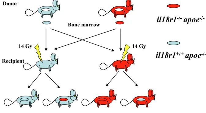
The present work aims to evaluate the in vivo role of this cytokine and its activating protease Caspase-1 in atherosclerosis. Interestingly, IL-18-deficiency limited early lesion development in hyperlipidemic mice but did not affect atherogenesis after prolonged hyperlipidemia. These studies suggest a direct role for IL-18 in disease progression extending beyond the classical function of this cytokine, the induction of interferon gamma. Additional experiments employing chimeric mice, that lacked the IL-18Rα on either the hematopoietic or the vascular cells, generated by bone-marrow transplantation, revealed that IL-18Rα does not participate in the pro-atherogenic effects of IL-18
Surprisingly, deficiency of Caspase-1 did not diminish atherogenesis, thus suggesting alternative mechanisms of IL-18 activation during atherosclerosis.
Subsequent experiments tested whether matrix metalloproteinases (MMPs), enzymes prominently expressed in atherosclerotic lesions mediate the maturation of the IL-18 precursor (proIL-18). Indeed, several recombinant MMPs proteolytically cleaved the precursor in vitro, and MMP-2- and MMP-8 processed proIL-18 exhibited IL-18 activity.
Sequence analysis of processed proIL-18 demonstrated that cleavage sites of MMP-2 and MMP-8 differ from that of Caspase-1. Finally, the presence of mature/processed IL-18 in atherosclerotic tissue of Caspase-1-deficient mice highlighted the potential in vivo relevance of such an alternative, MMP-mediated IL-18 activation mechanism for this pro-inflammatory disease.
In sum, this work directly demonstrates the pro-atherogenic role of IL-18 independent of IL-18Rα or Caspase-1. These surprising observations provide a novel understanding of IL-18 biology and may foster re-thinking of current approaches for the therapeutic intervention of this pathway in prevalent inflammatory diseases.
1. Introduction
1.1. Cardiovascular disease and atherosclerosis
Cardiovascular disease (CVD) continues to lead as a principal cause of death in developed countries,1,2 accounting for approximately 38 % and 42 % of deaths in the United States and the European Union, respectively.3,4 The total economic burden of CVD exceeds
$390 billion and €165 billion annually for these regions, respectively.3,4 Due to containment of many infectious diseases and increasing adoption of western lifestyles, this trend is projected to extend worldwide by 2020.2,5 The vast majority of CVD-related deaths is attributed to a disease of arterial blood vessels, known as ‘atherosclerosis’.3
Derived from the Greek words athera (gruel) and scleros (hard), atherosclerosis has been viewed for decades as a mere deposition of lipids along arterial walls. In the progression of the disease, these accumulations were thought to gradually narrow the lumen and finally lead to occlusion of the vessel, thus interrupting blood flow and oxygen supply to vital organs such as the heart and brain.6 This dogma was commonly accepted until studies in the late 1980s revealed that 60-70% of acute myocardial infarctions result from non-occlusive atherosclerotic lesions.7-9 Further research identified plaque disruption and subsequent thrombus formation rather than gradually developing stenosis as the final pathologic steps that cause acute clinical events such as myocardial infarction or stroke.10,11 Despite its considerable impact, the pathological mechanisms underlying this vascular dysfunction remain incompletely understood.
Research during the past two decades has focused on the cellular and molecular mechanisms responsible for the development and destabilization of atherosclerotic plaques.
Indeed, numerous studies demonstrated that most plaques did not consist of mere acellular lipid depositions, but rather harbored active inflammation characterized by accumulation of immune-competent cells.12-17 The current view of atherosclerosis hypothesizes that complex processes, that include molecular and cellular components of the immune system, successively decrease plaque stability and provoke its rupture, thus exposing highly pro-
coagulant mediators to the blood stream and resulting in thrombosis and its subsequent clinical symptoms.18-20 Hence, immune mechanisms that regulate integrity and stability as well as formation of atherosclerotic plaques have gained considerable attention among vascular biologists.14-17
1.2. Atherosclerosis: An inflammatory disease
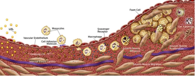
The development of human atherosclerosis usually extends over decades.13,21,22 Two independent studies found fatty streaks, the earliest visible atherosclerotic lesions, ubiquitous among teenagers.23,24 These studies revealed that atherosclerosis starts rather early in life although clinical symptoms precipitate most commonly in people of advanced age. The initial triggers of the atherogenic process still remain undetermined. Among other factors, i.e., response to endothelial injury or microbial infection, elevated levels of plasma lipoproteins such as low-density lipoproteins (LDL) are considered prerequisite for athero- genesis. Hyperlipidemia, commonly caused by environmental and/or genetic factors,25- 27 can lead to the accumulation of LDL within the vessel wall, where it can undergo modifications such as glycation or oxidation within the intima (Figure 1).28,29 Subsequently, such modified LDL can activate endothelial cells (EC), preferably at sites of hemodynamic strain,30,31 promoting recruitment of circulating T lymphocytes and monocytes from the blood via the expression of adhesion molecules (e.g., P-selectin, vascular cell adhesion molecule-1 (VCAM-1), and intercellular adhesion molecule-1 (ICAM-1)).14-17 Upon adhesion, monocytes and T lymphocytes, attracted by EC-derived chemokines (e.g., monocyte chemoattractant protein-1 (MCP-1) and macrophage inflammatory protein-1α (MIP-1α)), transmigrate into the intima.16,17 Resident monocytes, termed macrophages, within the inflamed vessel wall can incorporate native or modified LDL via phagocytic or receptor- mediated mechanisms.14,17 Following uptake, cholesterol can not be mobilized sufficiently and might instead accumulate as cytosolic droplets of cholesterol esters. These lipid-laden macrophages, also termed ‘foam cells’ due to their microscopic appearance, characterize early atherosclerotic lesions, also known as fatty streaks (Figure 1).16,17 The
accumulation of lipids, particularly their oxidatively modified derivatives furnish a strong trigger of pro-inflammatory responses.32,33 Sustained lipid uptake and accumulation causes production of pro-inflammatory cytokines, such as interleukin- (IL-)1β and tumor necrosis factor (TNF)α.14,16,17 Notably, these mediators might activate in an autocrine and paracrine fashion not only other macrophages, but also EC, smooth muscle cells (SMC), and T- lymphocytes within the lesion, thereby accelerating the inflammatory process in a positive feedback loop.14,16,17,34 Additionally, overexpressed growth factors, such as macrophage colony-stimulating factor (M-CSF), in the inflamed arterial wall function as survival and mitogenic stimulus for the inflammatory cells.35
Figure 1: Cellular mechanisms during atherosclerotic plaque evolution.
The earliest changes preceding the formation of atherosclerotic lesions take place in the endothelium. Increased endothelial permeability to lipoproteins leads to accumulation of low density lipoprotein (LDL) in the intima. Modification, particularly oxidation, of these particles induces local cytokine elaboration, which enhances in turn the expression of adhesion molecules and chemokines, e.g., monocyte chemoattractant protein 1 (MCP-1). Attracted by chemokines peripheral blood monocytes enter the lesion and express scavenger receptors in response to cytokine stimulation. These receptors mediate the uptake of modified lipoprotein particles by macrophages promoting the development of foam cells. Macrophage-derived foam cells are a source of further cytokines, smooth muscle cells (SMC) proliferative factors, and matrix metalloproteinases. SMC proliferate and can migrate from the media into the intima. Proliferation and synthesis of extracellular matrix assist establishing the fibrous cap, which separates the lesion from the blood stream. Continued influx and activation of macrophages, which release metalloproteinases, can cause degradation of extracellular matrix, eventually leading to rupture of the fibrous cap, thrombus formation, and occlusion of the artery (not shown).
Modified from: Libby. P. The vascular Biology of Atherosclerosis. Heart Disease: A Textbook of Cardiovascular Medicine. Braunwald, Zipes, and Libby Eds., 2001.
During lesion progression, foam cells in these early lesions can undergo apoptosis.16,36 This process, thought mediated in part by the Fas/FasL pathway,37,38 contributes to the development of the lipid-rich, acellular core of the plaque (Figure 1). The lipid core contains large amounts of pro-coagulants, particularly tissue factor (TF) synthesized by foam cells and potentially released during their death.20 Furthermore, SMC migrate into the lesion in response to signals provided by platelet derived growth factor (PDGF) and other chemoattractant mediators from the media, establishing the plaque’s fibrous cap, which segregates the pro-thrombotic lipid core from the lumen. SMC proliferate and contribute to formation of the fibrous cap by synthesis of extracellular matrix proteins, particularly interstitial collagen.39 The thickness and collagen content of the fibrous cap positively correlates with the plaque’s biomechanical stability.40,41 Certain pro-inflammatory cytokines such as interferon gamma (IFNγ) inhibit proliferation and collagen synthesis of SMC and can trigger apoptosis of these cells, eventually contributing to the thinning of the fibrous cap and rendering the plaque prone to rupture.16,38,39,42
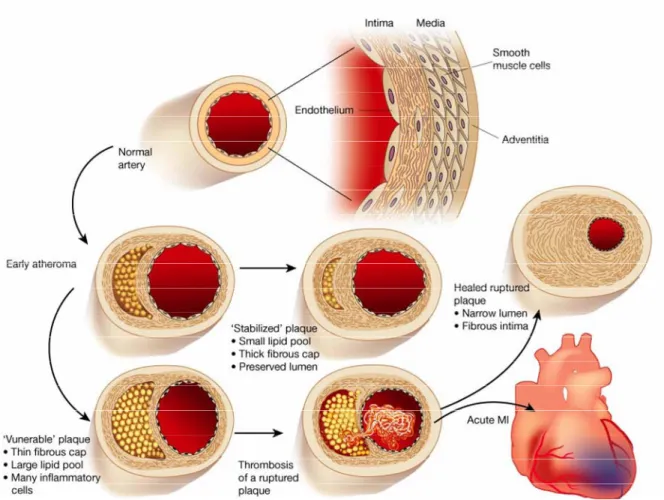
Rupture of the atherosclerotic plaque, often occurring in the macrophage-enriched shoulder region, the junction between the atheroma and the normal vessel wall, exposes the pro-coagulant content of the lesion to coagulation factors of the blood,16 resulting in thrombus formation (Figure 2)16,18,43 In subclinical plaque rupture, the thrombus does not completely occlude the lumen of the vessel and may eventually resorb. Growth factors released during the subsequent wound healing process can lead to SMC proliferation and thickening of the fibrous cap, yielding a constriction of the lumen and restriction of blood flow. Under increased cardiac demand this can lead to ischemia, provoking symptoms such as angina pectoris.16 However, if plaque rupture causes an occlusive thrombus formation it may lead to acute clinical complications, such as myocardial infarction (MI) and stroke.16,44 Therefore, the strength of the fibrous cap is considered a key determinant of atherosclerotic plaque stability.40,41 Accordingly, histological studies classify atherosclerotic lesions into those with features of ‘stable’ and ‘unstable’ (vulnerable) plaques (Figure 2). An unstable atheromatous plaque typically displays a large lipid core containing high amounts of pro- thrombotic tissue factor overlaid by a thin fibrous cap and accompanied by an accumulation of inflammatory cells, particularly in the shoulder region.16,18,21,44 In contrast, a stable fibrous
Figure 2: Schematic of the life history of an atheroma.
The normal human artery has a trilaminar structure. The endothelial cells separate the bloodstream from the intima, which contains a smattering of smooth muscle cells scattered with extracellular matrix. The media consists of much more tightly packed smooth muscle cells embedded in matrix rich in elastin as well as collagen. In early atherogenesis recruitment of inflammatory cells and accumulation of lipids lead to the formation of the lipid-rich core, as the artery enlarges to accommodate the expansion of the intima. Persistent inflammatory conditions cause further growth of the lipid core, degradation of extracellular matrix by proteases secreted by activated leukocytes, and reduced de-novo synthesis of collagen. These changes can thin the fibrous cap and leave it susceptible to rupture. Upon rupture, blood coming in contact with the thrombogenic content in the plaque coagulates and triggers thrombus formation. If the thrombus occludes the vessel persistently, an acute myocardial infarction (MI) can result. The thrombus may eventually resorb as a result of endogenous or therapeutic thrombolysis. However, growth factors released during the subsequent wound healing process can lead to smooth muscle cell proliferation and thickening of the fibrous cap, which can cause an inward directed growth of the intima, yielding a constriction of the lumen and restriction of blood flow. Increasing cardiac demand can lead to ischemia, provoking symptoms such as angina pectoris. Lipid lowering may reduce lipid content and calm the intimal inflammatory response, yielding a more “stable” plaque with a thick fibrous cap and, hence, less prone to rupture.
Modified from: Libby. P., Inflammation in atherosclerosis. Nature 2002;420:868-74
lesion features a well-developed fibrous cap and moderate lipid/leukocyte content (Figure 2).16,18,21,44
Nonetheless, the progression from fatty streak to advanced, complex lesions likely does not occur inevitably and continuously over time.16 Clinical observations rather suggest that many human lesions develop discontinuously, featuring “bursts” of growth and progression of atheroma.16,45 Therefore, comprehensive understanding of the mechanisms that cause plaque progression and destabilization is crucial for the future treatment of the disease and prevention of acute clinical events.
1.3. Matrix metalloproteinases in atherosclerosis
Matrix metalloproteinases have gained particular interest as potential mediators of plaque destabilization. These enzymes constitute a family of zinc-endopeptidases that share variations on a basic five-domain structure and are generally classified after their particular substrate (e.g., collagenases, gelatinases, elastases).46,47 Notably, atheromatous plaques overexpress MMP-1, -2, -3, -7, -8, -9, -10, 12, and -13.46-55
Within the fibrous cap, SMC express a mixture of structural proteins, including collagen, gelatin, laminin, and elastin, that comprise the extracellular matrix (ECM).46,47 The amount of collagen, the most abundant ECM protein in the fibrous cap contributing to it’s strength, is modulated by two mechanisms: de novo synthesis and degradation.47,56 Pro- inflammatory conditions can abrogate synthesis of collagen by SMC and simultaneously enhance the expression of MMPs, increasing collagen catabolism and, in turn, decreasing plaque stability.18 During lesion progression macrophages and T-cells continue to enter the lesion and aggregate particularly in the shoulder region.16,18,43 The continued activation of macrophages and T-cells as well as EC and SMC by inflammatory cytokines enhances expression and activity of MMPs.18,47 Members of two subfamilies of MMPs are considered particularly relevant to atherosclerosis and plaque stability. Interstitial collagenases (MMP-1, -8, and -13) mediate the initial breakdown of collagen fibrils, while gelatinases (MMP-2 and -9) can facilitate further degradation.47,48,51,55,57 Notably, endogenous inhibitors such as
members of the TIMP family (tissue inhibitor of matrix metalloproteinases)46,47,58 and TFPI-2 (tissue factor pathway inhibitor-2),59 regulated MMP activity. However, chronic inflammation may cause an imbalance of MMPs and their endogenous inhibitors, thereby promoting elevated MMP activity, a characteristic of atherosclerosis and other inflammatory diseases.18,46,47 Indeed, expression of these proteases localizes with sites of collagenolysis in advanced atherosclerotic lesions,53 supporting the hypothesis that imbalanced MMP regulation contributes to lesion destabilization by decreasing fibrous cap strength, thus rendering the plaque prone to rupture.
1.4. Cytokines in atherosclerosis
Substantial evidence suggests that the cytokine network orchestrates the complex cellular and inflammatory interactions underlying atherogenesis. The net pro- or anti- atherogenic function of cytokines relates to their ability to augment or antagonize inflammation, although this classification certainly oversimplifies the complex pathologic scenario of atherosclerosis. IL-1β and TNFα are classical pro-inflammatory cytokines and mediate pro-atherogenic functions,16,17 whereas IL-4 and IL-10 are considered anti- inflammatory cytokines and accordingly promote mostly anti-atherogenic pathways.16,17,60,61
Macrophages and T-cells, the dominant lymphocyte subpopulation within the atherosclerotic plaque, may partially modulate the antagonism between pro- and anti-atherogenic cytokines within the lesion and their polarization into a type 1 or type 2 helper T-cell (TH1) or (TH2) phenotypes. Lacking distinct surface markers, TH1 lymphocytes are characterized by secretion of IFNγ and TNFα,62,63 whereas TH2 polarization favors secretion of IL-3, IL-4, IL- 5, IL-10, and IL-13, which promote B-cell differentiation and immunoglobulin production,62,63 processes not commonly associated with atherosclerosis. In accord with the observed pro- or anti-inflammatory functions, expression of TH1 cytokines indeed dominates over TH2 cytokines in human atherosclerotic lesions in situ.60,64 Moreover, lack of TH1 cytokines generally diminishes the extent of atheroma in animal models,65-68 whereas deficiency of TH2 cytokines promotes atherogenesis.61,68
IFNγ crucially mediates the TH1-dominated pathways that lead to macrophage activation and the induction of a plethora of pro-inflammatory factors such as chemokines, cytokines, and growth factors from vascular as well as immune cells.16,17,63,69 In addition, IFNγ might contribute to plaque destabilization by inhibiting SMC-proliferation and collagen synthesis, as discussed above.70 Accordingly, IFNγ- or IFNγ receptor-deficiency leads to reduced severity of atherosclerosis in hyperlipidemic mice.65-67,71,72 Nonetheless, the underlying mechanisms that elicit IFNγ expression in atherosclerosis remained largely unknown until recently.
1.5. The pro-inflammatory cytokine IL-18
IL-18 is a member of the IL-1 cytokine family. Originally designated as Interferon gamma-inducing factor (IGIF) IL-18 was initially discovered as a potent inducer of IFNγ in TH1-cells and Natural Killer (NK) cells.73-75 The human IL-18 gene consists of six exons, spanning approximately 19.5 kb.76 In both humans and mice, IL-18 expression in macrophages is enhanced either directly by bacterial and viral antigens or in an auto- and paracrine manner by stimulation with IFNα, -β, or -γ.77,78 Furthermore, pro-inflammatory cytokines such as IL-1β or TNFα enhance expression of IL-18.78,79 Initially identified in Kupffer cells and activated macrophages,75 a variety of other cell types express IL-18, including osteoblasts,80 chondrocytes,81 epidermal keratinocytes,82 dendritic cells,83 as well as intestinal and airway epithelial cells.84,85
IL-18 expression and function has been associated with a variety of chronic inflammatory disorders, including cancer, rheumatoid arthritis, Crohn’s disease, psoriasis, and pulmonary sarcoidosis.79,86-89 Recently, anti-viral and anti-tumor properties were ascribed to IL-18 in addition to its role in innate immunity against bacterial infection.77,90
In contrast to most cytokines, the IL-18 gene contains only one RNA-destabilizing element, conferring unusual stability on IL-18 mRNA that highlights the importance of post- transcriptional regulation of IL-18 activity.78 Translation results in the synthesis of an 193 amino acid precursor (proIL-18) of 24 kDa, revealing 65% sequence identity with the 192
amino acid murine proIL-18.74 Similar to its analogue proIL-1β, proIL-18 lacks a conventional signal sequence and requires processing by Caspase-1, also termed IL-1β- converting enzyme (ICE), into the active, 18 kDa mature form.91-93 Cleavage via Caspase-1 occurs between Asp36 and Tyr37 for human proIL-18 and between Asp35 and Asn36 for the murine homologue, respectively.78,93 In addition, Caspase-3 has been reported to cleave proIL-18 at different sites than Caspase-1, resulting in inactive forms of the cytokine and implicating Caspase-3 in negative regulation of IL-18 activity.94 Since the IL-18 mRNA is unusually stable, as discussed above, and thus appears to evade stringent transcriptional regulation, the post-translational regulation of IL-18 activity by proteases such as Caspase-1 and -3 is considered particularly relevant to determination of the activity of this cytokine.78
1.6. Caspase-1
Caspase-1 represents the founding member of a family of cysteine proteases termed Cysteine requiring ASPartate proteASE (Caspase), sharing the active site cysteine and aspartate binding clefts.95 Originally isolated and cloned from cells of the monocytic lineage96,97 and traditionally considered the enzyme responsible for maturation of IL-1β precursor,96 Caspase-1 was identified in 1997 as the protease that mediates the activation of proIL-18.91,92 As the prototypical activator of these two prominent cytokines, Caspase-1 may participate in a number of inflammatory diseases and has received considerable attention as a potential target for therapeutic intervention.98,99
A variety of cell types express Caspase-1 including EC, SMC, fibroblasts, epithelial and epidermal cells.100-103 Although caspases are widely recognized as a family of enzymes primarily involved in apoptosis, the role of Caspase-1 in programmed cell death appears rather limited, hence, is considered a member of the subfamily of inflammatory caspases.93,104-106
Typically, Caspase-1 is synthesized as an inactive 45 kDa precursor that is autocatalytically processed to an active tetrameric complex consisting of the 10 kDa and 20 kDa subunits ((p20/p10)2).107 However, detection of mature immunoreactive Caspase-1
protein does not always correspond to its biological activity.100,108 Indeed, regulation of Caspase-1 activity remains incompletely understood. Interestingly, intracellular processing of the precursors by Caspase-1 and the secretion of IL-1β and IL-18 seem at least in part independently regulated.109 Besides passive release from dead cells, proIL-1β and proIL-18 can be actively secreted from live cells or,93,109,110 highlighting the relevance of additional regulatory control through extracellular proteases.
1.7. IL-18 and atherosclerosis
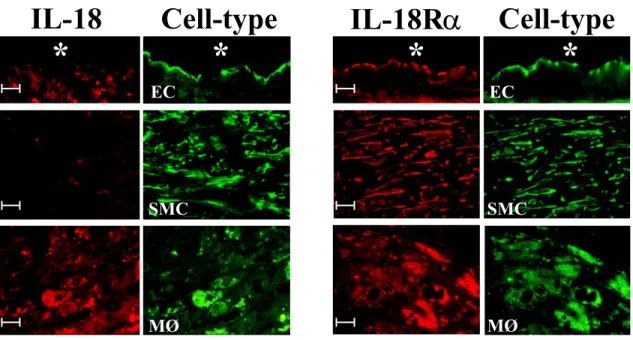
Work performed during my diploma thesis implicated the macrophage-derived cytokine IL-18 as a potent inducer of IFNγ expression and other pro-inflammatory mechanisms in atherogenesis.111,112 Besides initially demonstrating the presence of IL-18 in human atherosclerotic lesions, I also provided evidence for the expression of both subunits of the IL- 18R in this tissue as well as in atheroma-associated cell types in vitro. Notably, IL-18 localized only with macrophages of atherosclerotic lesions, whereas all three atheroma- associated cell types expressed IL-18Rα, namely EC, SMC, and macrophages (Figure 3).112 After demonstrating the expression of both receptor subunits on EC, SMC, and macrophages, additional experiments revealed several IFNγ-independent pro-atherogenic functions of IL- 18 on these cells, including enhanced expression of ICAM-1, of the cytokines IL-6 and IL-8, and of MMPs.112
Although IL-18 is a sufficient stimulus synergism with IL-12 enhances induction of its classical downstream mediator IFNγ.73,74 Reciprocal induction of the respective receptor113,114 as well as modulation of different signaling pathways leading to IFNγ promotor activation and IFNγ release may cause co-stimulatory induction of IFNγ by IL-18 and IL-12.78,115,116
Cell-type
IL-18 IL-18Rα Cell-type
MØ MØ
SMC
EC EC
SMC
*
* * *
Apart from this prototypical function, IL-18 induces the expression of additional inflammatory mediators including the cytokines IL-1β, IL-6, and TNFα, the chemokines IL-8, MCP-1, and MIP-1α, as well as adhesion molecules, such as VCAM-1 and ICAM- 1.78,81,117,118 Furthermore, IL-18 stimulation augments Fas/FasL-mediated cytotoxicity, as well as expression of perforin and granulocyte macrophage colony stimulating factor (GM- CSF), all of which contribute to cell death and local inflammation.79,81,119-121
IL-18 exerts its functions by binding to the heterodimeric IL-18 receptor, comprised of the IL-1 receptor-related protein (IL-1Rrp), termed IL-18Rα, and the IL-1 receptor accessory protein-like (IL-1RacPL), termed IL-18Rβ.122-124 Both receptor subunits belong to the IL-1R family. While IL-18Rα represents the ligand binding chain, IL-18Rβ appears to assist the formation of a high affinity complex and mediates intracellular signals.78,124 Binding of IL-18
Figure 3: Differential expression of IL-18 and IL-18Rα in human atherosclerotic lesions.
Double-immunofluorescence staining co-localized IL-18 (red, left panels) or IL-18Rα (red, right panels) with endothelial cells (EC, anti-CD31), smooth muscle cells (SMC; anti-α-actin), or mononuclear phagocytes (MØ, anti-CD68) within human carotid atherosclerotic plaques. Analysis of three atheroma from different donors showed similar results. The asterisk indicates the lumen of the vessel. The bar corresponds to 10 µm.
Modified from: Gerdes N. et al.:Expression of interleukin (IL)-18 and functional IL-18 receptor on human vascular endothelial cells, smooth muscle cells, and macrophages: implications for atherogenesis. J Exp Med 2002;195:245-57.
to the receptor complex results in recruitment of the adaptor protein MyD88, which subsequently recruits IL-1R-associated kinase (IRAK)-1 and IRAK-4 to the receptor complex.125 After activation,IRAK-1 translocates to the TNFR-associated factor 6 (TRAF- 6)-containing protein complex,126 inducing downstream signals including nuclear factor kappa b (NF-κB) and various mitogen activated phosphatase kinase (MAPK) pathways (e.g., c-Jun N-terminal kinase (JNK)).78,124,127 Indeed, IL-18signal transduction requires this common cascade, as demonstratedby gene-targeting studies showing that MyD88, IRAK-1, and IRAK-4play critical roles in IL-18 responses.125,128,129
IL-18Rα is expressed in a variety of tissues and cell types including myeloid, monocytic, and lymphocytic cell lines.78,130 Interestingly, the TH1 lymphocyte subpopulation, a hallmark cell type in atherosclerotic lesions,17 but not TH2 cells, mediators of which were considered anti-atherogenic, express the IL-18 receptor.131 Of note, inflammatory cytokines overexpressed in human atheroma, such as TNFα and IL-12,16,17,60 enhance the expression of both IL-18R chains on lymphocytes, thereby increasing the responsiveness to IL-18.78,113,130
1.8. Clinical association of IL-18 with cardiovascular disease and its risk factors
Following our initial identification of IL-18 as a novel pro-atherogenic mediator,112 several clinical studies demonstrated the correlation of elevated plasma IL-18 levels with cardiovascular risk. In a cohort of 10,600 European men followed for 5 years, IL-18 plasma concentrations in subjects who experienced MI, death, or angina were significantly higher than those in age-matched controls, independent of other inflammatory markers, such as C-reactive protein (CRP), IL-6 or fibrinogen.132 Moreover, IL-18 levels are elevated in patients with stable and unstable angina,133 and are higher in patients with MI compared to those with unstable angina.134 IL-18 plasma levels have also been associated with mortality in patients with coronary artery disease (CAD), independently of CRP, IL-6, and fibrinogen.135 Furthermore, elevated plasma IL-18 concentrations reportedly accompany acute coronary syndromes,136 a finding corroborated in an experimental model of MI in mice.137 Finally, IL-18 correlates with parameters of myocardial dysfunction,136 dilated and ischemic cardiomyopathy,138 and severity of congestive heart failure.139,140
The predictive value of IL-18 plasma levels extends beyond CVD to related dysfunctions such as metabolic syndrome. IL-18 plasma concentrations correlate with body mass index (BMI) and waist to hip ratio, and show an inverse correlation to weight loss.141-143 Furthermore, IL-18 plasma concentrations correlate with hyperglycemia and associate inversely with insulin sensitivity.143-145 In accord, several groups have reported susceptibility and occurrence of type 2 diabetes mellitus associated with elevated plasma IL-18.146- 149 In sum, these clinical data demonstrate a strong correlation between elevated levels of IL-18 and several manifestations of CVD and risk factors, highlighting the potential of IL-18 as a clinical marker as well as a target for therapeutic intervention.
1.9. Aim of this thesis
Increased expression of IL-18 and it’s receptor in atherosclerotic tissue together with its pro-inflammatory function on atheroma-associated cells in vitro suggest a prominent atherogenic role for this molecule. To verify the role of IL-18 in vivo in atherosclerosis this thesis used an experimental model of atherosclerosis in mice. Quantification of atherosclerosis in mice lacking IL-18 in comparison to control animals will allow for assessment of IL-18’s contribution to the disease.
Furthermore, it remains uncertain whether the lack of Caspase-1 limits development of atherosclerosis even more efficiently. Considering that Caspase-1 is the activating enzyme for both, IL-1β and IL-18, Caspase-1 deficiency in hyperlipidemic mice might lead to an even greater reduction of lesion development than could be attributed to lack of either substrate alone.
In sum, this thesis will evaluate the contribution of IL-18 and IL-1 signaling as well as Caspase-1 to experimental atherosclerosis and assess their potential as targets for therapeutic intervention. Furthermore, identification of cell types that participate in IL-18 signaling may provide valuable information to guide future research on IL-18-mediated mechanisms of atherogenesis and other inflammatory diseases.
2. Materials and methods
2.1. Materials
The commercial sources for recombinant proteins and other reagents are listed in Table 1.
Table 1: Recombinant proteins
Reagent Company Cat.-#
recombinant human IL-18 MB International, Woburn, MA B-003-5 recombinant human IL-12 R&D Systems, Minneapolis, MN 219-IL recombinant human IL-1β Pierce-Endogen, Cambridge, MA R-IL1B-25
recombinant human TNFα Pierce-Endogen R-TNF-50
recombinant human IFNγ Pierce-Endogen R-IFN-100
recombinant human TNFα Pierce-Endogen R-TNF-50
Polymyxin B Sigma , St. Louis, MO P4932
2.2. Expression of IFNγ in atheroma-associated cell types
2.2.1. Cell isolation and culture
All human cells were obtained according to protocols approved by the Human Investigation Review Committee at Brigham & Women's Hospital.
2.2.1.1. Endothelial Cells
Human vascular EC were isolated from saphenous veins by collagenase treatment, as originally described by Jaffe et al.150 Saphenous veins were rinsed three times using syringe and cannula with Hanks’ buffered salt solution (HBSS). Veins were clamped on one end with a hemostat, filled with 0.1 % (w/v) collagenase (Worthington Biochemicals, Freehold, NJ) solution in phosphate buffered saline (PBS), and were clamped on the other end. After incubation (30 min, 25˚C), the EC-containing collagenase solution was flushed out by three washes with HBSS. Collagenase treatment was repeated twice. All fractions were collected and EC precipitated (5 min, 300 x g, 25˚C). Subsequently, cells were resuspended in EC-
culture medium (see below) and grown (37˚C, 5% CO2) in 60 cm2 cell culture dishes (Becton Dickinson, Franklin Lakes, NJ). Medium was changed for the first time after 24 h and then routinely every 3 days. Cells were later cultured in 150 cm2 cell culture flasks (Corning, Corning, NY) and used through passages 3-4. For cell passage, confluent layers of EC were washed twice with HBSS and subsequently treated (37˚C) with trypsin-EDTA-solution (500 µg/ml trypsin and 200 µg/ml EDTA; Cambrex, cat.-#:17-161E) until cell detachment.
Trypsin was inactivated by addition of 10 % volume of fetal bovine serum (FBS; Hyclone, Logan, Utah) and cells were precipitated (5 min, 300 x g, 25˚C). Finally, 5000 cells/cm2 were sub-cultured in fresh medium in the desired culture vessels. For the experiments, EC were cultured in 60 cm2 cell culture dishes and medium was changed 24 h before experiments to serum-free starvation medium (see below). All culture dishes and flasks for EC were coated with gelatin. For this purpose, culture surface area was covered for 1 min with 0.1 % (w/v) gelatin (Becton Dickinson, Sparks, MD) solution in PBS and, after removal of the solution, dried for 30 min at 37˚C.
EC were characterized by immunohistochemical staining with mouse-anti-CD31 (Dako, Carpinteria, CA, cat.-#: M 0823). Contamination with SMC, macrophages, or T lymphocytes was excluded by employing mouse-anti-muscle actin (Enzo Diagnostics, Farmingdale, NY, cat.-#: C34931), mouse-anti-CD68 (Dako, cat.-#: M 0823), and mouse-anti-CD3 (Dako, cat.-#: T 0629) antibody, respectively, yielding no specific staining.
2.2.1.2. Smooth Muscle Cells
Human vascular SMC were isolated from saphenous veins by the explant-outgrowth method.151 Veins were cut longitudinally and adventitia and intima were removed with a scalpel. The remaining media was cut in 2 x 2 mm pieces, transferred in 60 cm2 cell culture dishes, and incubated for 30 min (25˚C) without medium. Subsequently, SMC culture medium (see below) was carefully added to cover the tissue. After the first outgrowing SMC were observed microscopically, medium was changed every 3 days and the tissue pieces were removed after a confluent SMC culture was established (approximately 3 weeks after isolation). Cells were sub-cultured, as described above for EC, with 3000 cells/cm2. SMC were used through passages 3-4. For the experiments cells were cultured in 60 cm2 cell culture dishes and medium was changed 24 h before stimulation to serum-free medium (see
below). SMC were characterized by immunohistochemical staining and contamination with EC, macrophages, or T-lymphocytes was excluded as described above for EC, yielding no specific staining.
2.2.1.3. Monocytes and macrophages
Monocytes were isolated by density gradient centrifugation from freshly prepared leukocyte concentrates (Leukopac) from healthy donors obtained from the Brigham &
Women's Hospital/Dana Faber Cancer Institute blood donor center. One volume (10 ml) of Lymphocyte separation medium (ICN Biomedicals, Aurora, OH) was gently overlaid with 4 volumes of leukocyte concentrate. Following 45 min centrifugation (450 x g, 25˚C, without brake), the upper phase was aspirated and the leukocyte containing interphase was transferred into fresh tubes, while avoiding interference with the granulocyte- and erythrocyte-containing bottom phase.
Leukocytes were washed three times with HBSS. Cells were precipitated by centrifugation for 10 min (300 x g, 25˚C) between each washing step. After the final washing step, the number of viable cells was determined by staining with trypan blue (0.4 % (w/v);
Sigma), and subsequent counting in a hematocytometer (Neubauer chamber; Reichert, Buffalo, NY). Trypan blue penetrates membranes of non-viable cells and thus yields blue intracellular staining, whereas viable cells do not stain.
For purification purposes, 1 x 10⁶ cells/cm2 in monocyte medium were (see below) plated in 60 cm2 cell culture dishes. After one hour of incubation (37˚C, 5% CO2), plates were vigorously washed three times with HBSS to remove non-adherent cells. Macrophages were cultured (37˚C, 5% CO2) in macrophage culture medium supplemented with 2 % (v/v) human serum (see below) for 10 days. Medium was changed every 3 days, and differentiated macrophages (day 10) were cultured 24 h before stimulation in serum-free medium. The purity of macrophages was ≥ 96%, as determined routinely by FACS analysis (PE- conjugated mouse-anti-human CD68; 20 µl/0.5 x 106 cells; PharMingen; San Diego, CA).
2.2.1.4. Limulus amoebocyte assay
All culture media, FBS, and human serum contained less than 40 pg endotoxin/ml, as determined by the chromogenic Limulus amoebocyte assay following the instructions of the
manufacturer (QCL-1000® Chromogenic LAL Kit; Cambrex, cat.-# 50-647U). Endotoxin derived from gram-negative bacteria catalyses the activation of a pro-enzyme in the Limulus Amoebocyte Lysate. In the second reaction step, the activated enzyme catalyses the cleavage of p-nitroaniline (pNA) from a colorless substrate. The pNA released can be measured photometrically at 405 nm after the reaction is stopped with stop reagent (25% (v/v) glacial acetic acid). The concentration of endotoxin in a sample was calculated from the absorbance values of a serial dilution of a known concentration of endotoxin standard.
2.2.1.5. Cell culture media
Culture medium for EC Medium M199 (Cambrex) Supplemented with:
5 % (v/v) Fetal Bovine Serum (FBS; Hyclone)
250 µg/ml Endothelial Cell Growth Factor (ECGF; kindly provided by Dr. M Muszynski, Brigham & Women's Hospital)
100 U / 100 µg/ml Penicillin/streptomycin (Cambrex)
1.25 µg/ml Amphotericin (Fungizone; Apothecon, Princeton, NJ) 100 µg/ml Heparin (Sigma)
Starvation medium for EC
Same as Culture medium for EC but without FBS and ECGF
Culture medium for SMC
Dulbecco’s Modified Eagle Medium (DMEM; Cambrex) Supplemented with:
10 % (v/v) FBS
100 U / 100 µg/ml Penicillin/streptomycin 1.25 µg/ml Amphotericin
200 µM L-glutamine (Cambrex)
Starvation medium for SMC
Same as Culture medium for SMC but without FBS
Culture medium for macrophages
Rockwell Park Memorial Institute medium 1640 (RPMI 1640; Cambrex) Supplemented with:
100 U / 100 µg/ml Penicillin/streptomycin 1.25 µg/ml Amphotericin
2 % (v/v) Human serum (ICN Biomedicals, heat- inactivated (30 min/56˚C) before use)
Culture medium for monocytes (also used for starvation of differentiated macrophages) Same as Culture medium for macrophages but without human serum
2.2.2. Enzyme-Linked Immunosorbent Assay (ELISA)
Release of IFNγ from EC, SMC, monocytes/macrophages, and KG-1 cells was measured by ELISA. Cell supernatants (50 µl) were added for 2 h to 96-well Maxisorb plates (Nunc, Rochester, NY) pre-coated with a monoclonal IFNγ capturing antibody (Pierce- Endogen; 1.5 µg/ml in PBS, 4°C, overnight). The plates were subsequently washed three times with PBS 0.1% (v/v) Tween-20 in an automatic plate washer, and the respective biotin- labeled anti-human IFNγ antibody (Pierce-Endogen; 0.5 µg/ml diluted in 2% (w/v) bovine serum albumin (BSA) in PBS) was added for 1 h (25°C, shaking). Following incubation for 30 min at room temperature with Horse-radish peroxidase (HRP)-conjugated Streptavidin (Pierce-Endogen; 0.125 µg/ml in PBS/2%BSA) and three washing steps, antibody binding was detected by the addition of tetramethylbenzidine (TMB) solution (Pierce-Endogen) and measuring absorbance at 650 nm in a plate reader (Molecular Devices, Spectra Max plus 384). The concentration of IFNγ was calculated from a standard curve prepared from recombinant IFNγ (Pierce-Endogen). Samples were assayed in duplicates.
2.2.3. In situ hybridization (ISH)
In situ hybridization was performed according to the instructions of the manufacturer (Innogenex, San Ramos, CA). Briefly, frozen tissue sections as well as cultures of SMC and macrophages on 4-chamber slides were fixed in cold acetone, air-dried, and incubated (10 min, 65°C; subsequently 2 h, 37°C) with a mixture of FITC-labeled IFNγ (5’- CATCGTTTCCGAGAGAATTAAGCCAAAGAAGTTGAAATCA-3’; 5’-AAGAGAA CCCAAAACGATGCAGAGCTGAAAAGCCAAGATA-3’; 5’-TTTTCTGTCACTCTCCT CTTTCCAATTCTTCAAAATGCCT-3’) antisense oligomers, or the respective sense (control) oligomers (all 4 µg/ml, final concentration) in hybridization-buffer. Finally, slides were washed 3 times, incubated with biotinylated mouse-anti-FITC antibody (1 h), followed by alkaline phosphatase-conjugated streptavidin (30 min) and NBT/BCIP chromogen solution (1 h). Adjacent sections were analyzed for SMC, macrophage, and T-cell content by immunohistochemistry as described below.
2.2.4. Immunohistochemistry (IHC)
Serial cryostat sections (6 µm) of surgical specimens of human carotid atheroma (n=3), normal carotids from autopsies, and non-diseased aorta from cardiac transplantation donors (n=3) were cut, air dried onto microscope slides, fixed in acetone (-20oC, 5 min), and pre- incubated with PBS containing 0.3% hydrogen peroxide. Subsequently, sections were incubated (30 min) with primary mouse-anti-human muscle actin mAb for SMC (1:200;
Enzo Diagnostics), mouse-anti-human CD31 mAb for EC (1: 35; Dako, Carpinteria, CA), mouse-anti human CD68 for macrophages (1:500; Dako), mouse-anti-human CD3 mAb for T cells (1:500; Dako), or control antibody (mouse myeloma protein MOPC-21; 0.25 µg/ml;
Sigma), diluted in PBS supplemented with 5% appropriate serum, and processed according to the recommendations provided by the supplier (Universal Dako LSAB Kit, Dako).