• Keine Ergebnisse gefunden

WATCHING THE HEALING BRAIN: MULTIMODAL AND NON-INVASIVE IMAGING OF REGENERATIVE PROCESSES AFTER EXPERIMENTAL CEREBRAL ISCHEMIA

N/A
N/A
Protected

Academic year: 2021

Aktie "WATCHING THE HEALING BRAIN: MULTIMODAL AND NON-INVASIVE IMAGING OF REGENERATIVE PROCESSES AFTER EXPERIMENTAL CEREBRAL ISCHEMIA"

Copied!
141
0
0

Wird geladen.... (Jetzt Volltext ansehen)

Volltext

(1)

REGENERATIVE PROCESSES

AFTER EXPERIMENTAL CEREBRAL ISCHEMIA

Inaugural-Dissertation

zur

Erlangung des Doktorgrades

der Mathematisch-Naturwissenschaftlichen Fakultät vorgelegt von

Joanna Magdalena Adamczak

aus Skwierzyna

Universität zu Köln Max Planck Institut

für neurologische Forschung

(2)

Berichterstatter: PD. Dr. Heike Endepols Prof. Dr. Wolfgang Walkowiak Prof. Dr. Mathias Hoehn

Tag der mündlichen Püfung: 11.4.2013

(3)

Abstract ... 5

Zusammenfassung ... 7

Introduction ... 9

1. Stroke ... 9

1.1. Pathophysiology ... 9

1.2. Rodent model of transient focal cerebral ischemia ... 11

1.3. Established therapies and new approaches ... 12

2. Endogenous regenerative processes after stroke ... 13

2.1. Angiogenesis ... 14

2.1.1. Stroke-induced angiogenesis ... 14

2.1.2. Temporal and spatial profile of post-stroke angiogenesis ... 16

2.2. Adult neurogenesis ... 16

2.2.1. Stroke-induced neurogenesis ... 17

2.2.2. Temporal and spatial profile of post-stroke neurogenesis ... 18

3. Non-invasive imaging strategies ... 18

3.1. Bioluminescence imaging ... 19

3.1.1. The principles of bioluminescence imaging ... 19

3.1.2. In-vivo bioluminescence imaging in pre-clinical research ... 20

3.2. Magnetic resonance imaging ... 21

3.2.1. The principle of nuclear magnetic resonance ... 21

3.2.2. In vivo magnetic resonance imaging in pre-clinical research ... 23

3.2.3. Imaging brain vasculature with magnetic resonance ... 23

3.2.4. Imaging brain function with magnetic resonance ... 25

4. Aims ... 29

Publications ... 31

Discussion ... 105

1. Angiogenesis after stroke ... 106

1.1. Imaging angiogenesis on the molecular level ... 106

1.2. Imaging angiogenesis on the structural level ... 108

1.3. New insights into vascular remodeling after stroke ... 109

2. Adult neurogenesis after stroke ... 110

2.1. Brain-specific bioluminescence imaging ... 111

(4)

3. Functional brain activity after stroke ... 115

3.1. Establishment of mouse functional magnetic resonance imaging ... 115

3.2. Imaging functional activity after stroke ... 117

4. Summary ... 119

5. Outlook ... 120

References ... 121

Appendix ... 131


(5)

Stroke is a severe disease of the brain, which leads to cell death and loss of function.

Neuroprotective therapy to prevent neuronal loss has not been effective in human stroke patients. Therefore, new therapeutic strategies are needed. Spontaneous recovery can be observed in some patients. However, the basis of this phenomenon is not completely understood yet. Several endogenous regenerative processes have been observed following cerebral ischemia, which may be the reason for functional recovery and can be used as a basis for new therapeutic strategies. Shortly after the insult, endothelial cells start to proliferate and eventually lead to revascularization of ischemic brain tissue (angiogenesis).

Furthermore, resident neural progenitor cells increase their proliferative activity, migrate towards the ischemic tissue and even differentiate into new neurons (neurogenesis). Detailed knowledge about the molecular mechanisms and interactions between angiogenesis and neurogenesis in response to stroke is needed in order to reveal new therapeutic targets. This PhD thesis established novel non-invasive imaging strategies to followed post-stroke angiogenesis and neurogenesis with particular regard to their dynamic temporal profiles.

Bioluminescence imaging and magnetic resonance imaging were chosen for this purpose.

The vascular endothelial growth factor receptor 2 was used as a molecular marker for angiogenesis, and for the first time the molecular basis of post-stroke vascular remodelling was observed non-invasively with bioluminescence imaging in an angiogenesis-specific reporter mouse. Structural changes of the vascular system were monitored with a magnetic resonance imaging strategy. Initial pronounced decrease of vessel density in ischemic tissue was followed by vessel density normalization. Non-invasive observation of endogenous neurogenesis is limited by the small number of neural progenitor cells within the adult brain.

This work established the first bioluminescence protocol optimized for highly sensitive

bioluminescence imaging of neurogenesis in a neurogenesis-specific reporter mouse. For

the first time, increased proliferation of neural progenitor cells after stroke was observed with

bioluminescence imaging. As post-stroke angiogenesis and neurogenesis may lead to

regeneration of brain function, this PhD thesis established the first functional magnetic

resonance imaging protocol for the specific application in mice. First investigations of brain

function after stroke were performed and future studies will have the opportunity to follow

functional recovery in transgenic mouse models. All methods used in this thesis bear the

exceptional potential to be combined into a multimodal approach. Screening for new

therapeutic targets within the brain endogenous regenerative capacity will be possible non-

invasively. Furthermore, the effect of new therapies on angiogenesis, neurogenesis or

functional recovery can be quickly tested.

(6)


(7)

Der Schlaganfall ist eine schwere Erkrankung des Gehirns, welche zu Zelltod und Funktionsverlust führt. Da neuroprotektive Therapien bei Schlaganfallpatienten bisher unwirksam waren, werden neue therapeutische Ansätze benötigt. Einige Patienten zeigen nach dem Schlaganfall eine spontane Erholung, deren Grundlage noch nicht vollkommen entschlüsselt ist. Endogene, d.h. körpereigene Prozesse, können die Ursache der spontanen Erholung sein und können des weiteren als Grundlage für neue Therapien genutzt werden.

Neue Blutgefäße entstehen in der Nähe das ischämische Hirngewebes (Angiogenese) und endogene neurale Vorläuferzellen wandern in das ischämische Gewebe, wo sie sich zu neuen Neuronen weiterentwickeln (Neurogenese). Detaillierte Kenntnisse über die endogenen Prozesse der Angiogenese und Neurogenese nach Schlaganfall werden nun benötigt, um auf ihrer Grundlage neue Therapien zu entwickeln. In dieser Arbeit werden erstmals nicht-invasive bildgebende Strategien verwendet um die endogenen Prozesse der Angiogenese und Neurogenese nach Schlaganfall zu beobachten und insbesondere ihren dynamischen Zeitverlauf zu dokumentieren. Für die Angiognese wurde der vaskuläre endotheliale Wachstumsfaktor Rezeptor 2 als molekularen Marker verwendet und erstmals mittels Biolumineszenzbildgebung die molekulare Regulation der Gefäßsystemver- änderungen nach Schlaganfall nicht-invasive beobachtet. Strukturelle Veränderungen des Gefäßsystems wurden mittels einer kernspintomographischen Methode mit hoher räumlicher Auflösung observiert. Auf eine anfängliche Verringerung der Gefäßdichte in ischämischen Bereichen folgte einer Normalisierung. Die nicht-invasive Beobachtung der endogenen Neurogenese wird durch die geringe Anzahl von neuronalen Vorläuferzellen im adulten Gehirn erschwert. Diese Arbeit erstellte ein Protokoll für hochempfindliche Biolumineszenzbildgebung der Neurogenese in einer Neurogenese-spezifischen Reportermaus. Damit ließ sich erstmals die endogene Neurogenese nach Schlaganfall nicht- invasiv untersuchen. Da die untersuchten spontanen regenerativen Prozesse die Grundlage funktioneller Erholung sein können, wurde in dieser Arbeit erstmals ein nicht-invasives funktionelles Bildgebungsverfahren für die spezifische Anwendung an Mäusen etabliert.

Erste Untersuchungen der Hirnaktivität nach Schlaganfall wurden in dieser Arbeit

durchgeführt und künftige Studien haben die Möglichkeit transgene Mäuse für

Untersuchungen der funktionelle Erholung nach Schlaganfall zu verwenden. Die in dieser

Arbeit etablierten Verfahren stellen neue, nicht-invasiven Methoden zur Untersuchung von

endogenen regenerativen Prozessen nach Schlaganfall dar. Insbesondere können sie nun

zu innovativen multimodalen Ansätzen kombiniert werden. Neue Erkenntnisse über die

Wechselwirkung von Angiogenese und Neurogenese können erzielt werden und Therapien

schneller und einfacher auf ihre Wirksamkeit getestet werden.

(8)
(9)

1. Stroke

Stroke remains the third-leading cause of death and the main cause of disability worldwide, leaving the majority of survivors dependent on institutional care (Donnan et al., 2008). Stroke describes the pathological situation when sudden oxygen and glucose deprivation results in severe brain damage. Depending on the cause, stroke can be either hemorrhagic or ischemic. Hemorrhagic stroke results from intra-cerebral bleeding caused by the rupture of a blood vessel in the brain parenchyma or in the subarachnoid space, which causes damage to the brain due to the elevation of intra-cranial pressure but also due to the lack of oxygen and glucose supply. Ischemic stroke can be further subdivided into thrombotic and embolic. Thrombotic stroke is the blockage of a cerebral artery by a gradual formation of a clot within this artery, while embolic stroke results from a traveling blood clot (embolus) formed somewhere else in the body. With 85-90% of all incidences, ischemic stroke represents the most common kind of stroke. Stroke symptoms are versatile, including sensory and motor dysfunction, paralysis, aphasia, nausea, and headache (Fatahzadeh and Glick, 2006) and are dependent on the size and area of the brain affected. Although stroke represents a severe and frequent disease, only few therapeutic interventions with limited applicability exist. Therefore, stroke continues to cause personal and familial tragedies.

Furthermore, stroke and stroke-related rehabilitation places a heavy economical burden upon society (Meairs et al., 2006).

1.1. Pathophysiology

The blockage of a cerebral artery leads to oxygen and glucose deprivation within the area supplied by the artery. Very quickly the production of adenosine-tri-phosphate (ATP) by oxidative phosphorylation ceases and the high energy demand of the brain cannot be fulfilled (energy failure) (Dirnagl et al., 1999). This energy deficit is most severe in areas with the lowest residual blood flow (ischemic core), resulting in rapid anoxic depolarizations (Mitsios et al., 2006). Energy dependent ion pumps fail to restore ionic gradients, leading to increased intracellular ion concentration with water passively following the concentration gradient (cytotoxic edema) (Dirnagl et al., 1999). In the ischemic core, cells die quickly by necrosis.

Areas with collateral blood supply experience less severe ischemia (ischemic penumbra) but

incomplete restoration of ion gradients generates waves of depolarizations (peri-infarct

(10)

impaired glial re-uptake of neurotransmitters, extracellular concentrations of glutamate become toxic through excessive activation of glutamate receptors (excitotoxicity) causing an accumulation of intracellular calcium (Iadecola and Anrather, 2011b). Excessive intracellular calcium triggers several deleterious events including activation of lytic enzymes, mitochondrial dysfunction and oxidative stress (Moskowitz et al., 2010). As a result, free radicals are produced in detrimental amounts, which are potent destroyers of cell membranes, DNA and any other cellular component. Damaged endothelium causes a break- down of the protective blood-brain barrier, which leads to an increased permeability to plasma proteins and consequently to increased fluid content within the extracellular space (vasogenic edema) (Durukan and Tatlisumak, 2007). Starting already a few hours after stroke, upregulation of pro-inflammatory cytokines calls peripheral immune cells to infiltrate the injured brain parenchyma, which form together with activated astrocytes and brain resident microglia a glial scar (Iadecola and Anrather, 2011a). The above described events after ischemia activate programmed cell death (apoptosis) which expands tissue damage in the ischemic penumbra (Mitsios et al., 2006, Ginsberg, 2003, Durukan and Tatlisumak, 2007). The temporal aspect of the events within the ischemic cascade is illustrated in Figure 1. Details of the underlying separate events are reviewed in (Chavez et al., 2009, Dirnagl et al., 1999, Durukan and Tatlisumak, 2007, Iadecola and Anrather, 2011b, Mitsios et al., 2006, Moskowitz et al., 2010)

Figure 1: Time line of major events of the ischemic cascade  

Detrimental effects of energy failure, excitotoxicity and oxidative stress predominantly occur during the

acute phase. The inflammatory reaction starts during the first few hours after onset of ischemia, while

regenerative processes are effective during the chronic phase.

(11)

Figure 2: Middle cerebral artery occlusion in rodents  

A Architecture of major cerebral arteries. A filament is inserted through the common carotid artery and blocks the blood flow to the middle cerebral artery. B Magnetic resonance angiography shows blood flow through the major cerebral arteries before (left panel) and after (right panel) insertion of the filament. C Top: MR T2 map at 3 days after MCAO in a mouse (30 min occlusion time). Bottom:

Corresponding brain section taken from the mouse brain atlas (Paxinos and Franklin, 2001). Cerebral lesions after MCAO form in the caudate putamen, thalamus, and the cortex, including the somatosensory and piriform cortex.

1.2. Rodent model of transient focal cerebral ischemia

In order to study molecular mechanisms or to investigate the benefit of therapeutic interventions, ischemic stroke is modeled in experimental animals. The majority of experiments are carried out on small animals such as rats and mice, which have a similar cerebrovascular system as humans. Several animal models have been developed to study ischemic stroke. The most frequently used model in experimental stroke research uses the intraluminal filament technique, which was first developed in the rat model by Koizumi et al.

(1986) (Koizumi et al., 1986). A filament is introduced into the lumen of an extra-cranial artery and advanced through the internal carotid artery until it blocks the blood flow to the middle cerebral artery (Figure 2A+B). Therefore, this technique is commonly referred to as middle cerebral artery occlusion (MCAO). With this model the duration of ischemia can be controlled. Leaving the filament in the artery results in a permanent occlusion model.

Retraction of the filament models successful reperfusion (transient ischemia). Infarcts

develop in the vascular territory of the middle cerebral artery (MCA), including the lateral

striatum, the thalamus and the fronto-parietal cortex (Figure 2C). A very good reproducibility

of infarct size was achieved by coating the filament with poly-L-lysine, which increases

adhesion to the vascular wall (Belayev et al., 1999). Early studies used predominantly rats

(12)

species (Belayev et al., 1999, Hata et al., 2000), allowing investigation of molecular mechanisms of stroke. The advantage of this method is the relative low invasiveness with no need for craniectomy and the high reproducibility with control over the ischemic duration.

Intraluminal MCAO was used to model transient cerebral ischemia in this thesis. While rat MCAO was already established in our laboratory, modifications of the surgical procedure for mouse MCAO were evaluated during the course of this thesis and an optimal mouse MCAO protocol established for the use in our laboratory. Detailed description of the procedures can be found in the Materials and Methods section of the publications.

1.3. Established therapies and new approaches

Current treatments for stroke are very limited, focusing on removal of the clot in the acute phase for fast restoration of blood supply. Pharmacological lysis of the clot with intravenous administration of recombinant tissue plasminogen activator (rt-PA) within the first 4.5 h after the insult is an effective therapy, approved by the US Food and Drug Administration (Albers et al., 2002, Hacke et al., 2008, Hacke et al., 1998, Wahlgren et al., 2008). However, only around 15% of stroke patients arrive at the hospital within this time window and only 10% of the ischemic stroke patients receive this therapy (Minnerup et al., 2011) due to an increased risk of hemorrhage (Hacke et al., 2008). Mechanical endovascular recanalization with a wire system has proven similar effectiveness and can be applied within an extended therapeutic time window for up to 8h post infarction (Clark et al., 2009, Kulcsar et al., 2010, Smith et al., 2008, Smith et al., 2005).

Besides reperfusion therapy for the acute phase, several agents have been reported to

rescue damaged brain tissue by modulating the biochemical events following cerebral

ischemia. Summarized under the term of neuroprotection these agents target the detrimental

effects of excitotoxicity, oxidative stress, inflammation and apoptosis within the ischemic

cascade. Over 1000 neuroprotective agents have been tested preclinically (O'Collins et al.,

2006) and around 200 have entered clinical trials – but none was able to reproduce

effectiveness in the clinics so far (Minnerup et al., 2012). Currently under clinical

investigation are growth factors (erythropoietin (Ehrenreich et al., 2002, Ehrenreich et al.,

2009, Minnerup et al., 2009), granulocyte colony-stimulating factor (Schabitz et al., 2010)),

free radical scavengers (Edaravone (Tohgi et al., 2003); Ebselene (Ogawa et al., 1999),

NXY-059 (Lees et al., 2006, Macleod et al., 2008, Shuaib et al., 2007)), antibiotics

(13)

stroke onset (Tohgi et al., 2003, Naritomi et al., 2010).

Very few patients are eligible to receive thrombolytic therapy and hundreds of neuroprotective compounds were unsuccessful in clinical trials, therefore new therapeutic strategies begin to focus onto the chronic phase. The main objective of the new strategies is to restore lost function. Potential routes to achieve this goal are being identified, including stem cell transplantation and brain endogenous regenerative processes. Transplantation of various types of cells directly into the ischemic brain or intravenous infusion improved functional outcome in animal models of stroke and first transplantations were already performed on human stroke patients (Bang et al., 2005, Kondziolka et al., 2005, Nelson et al., 2002). Possible mechanisms of action include cell replacement (Oki et al., 2012), trophic support and modulation of the inflammatory processes after ischemia (Lee et al., 2008). It was recently recognized that even the adult brain has some capacity to react to cerebral injury. The endogenous regenerative processes initiated after ischemia include the formation of new blood vessels (angiogenesis) and the endogenous production of new neurons (neurogenesis). These processes may represent promising targets for the development of new therapies.

2. Endogenous regenerative processes after stroke

Early in the ischemic cascade hypoxia-induced up-regulation of growth factors and

cytokines initiates the endogenous regenerative processes of angiogenesis and

neurogenesis. Angiogenesis is the formation of new blood vessels from pre-existing ones. In

the context of cerebral ischemia the angiogenic response describes the whole process of

vascular remodeling which possibly translates into an increase of cerebral micro vessel

density. Stroke patients with increased vessel density survived longer and showed a better

neurological performance (Krupinski et al., 1994, Szpak et al., 1999). Angiogenesis is closely

linked to endogenous neurogenesis (Sun et al., 2003). Endogenous neurogenesis describes

the formation of new neurons from neural stem cells, which reside in the adult brain. The

phenomenon of adult neurogenesis and its responsiveness to injury are well documented in

animal models. However, endogenous neurogenesis does not seem potent enough to

restore lost neuronal functions. As both processes share signal molecules, e.g. vascular

endothelial growth factor (VEGF) (Carmeliet and Tessier-Lavigne, 2005), and manipulation

with pro-angiogenic growth factors results in an enhanced neurogenic response to stroke

(14)

processes in conjunction. The evaluation of their temporal profiles will allow determination of key events within each process, which may represent new therapeutic targets.

2.1. Angiogenesis

Angiogenesis is the formation of new blood vessels. In general, blood vessels can form via two distinct processes: vasculogenesis and angiogenesis. While vasculogenesis denotes de novo formation of vessels from proliferating and differentiating mesoderm-derived endothelial progenitor cells during embryonic development, angiogenesis represents new vessel formation from pre-existing blood vessels. Almost no changes of the cerebral vasculature appear under healthy conditions. However, ischemic injury will trigger molecular and structural changes.

2.1.1. Stroke-induced angiogenesis

The occlusion of a cerebral artery leads to a lack of oxygen (hypoxia) and glucose (hypoglycemia) in the affected brain region. Hypoxia inducible factor 1α (HIF1α) stabilizes under hypoxic conditions and forms a dimer with HIF1β [65] which functions as a transcription factor and binds to the hypoxia-response element (HRE) promoter. Thus, hypoxia triggers the elevation or de novo expression of several growth factors and cytokines, including vascular endothelial growth factor (VEGF), fibroblast growth factor (FGF), transforming growth factor (TGF), hepatocyte growth factor (HGF), platelet-derived growth factor (PDGF), insulin-like growth factor (IGF), epidermal growth factor (EGF) and angiopoietins (Ang1 and Ang2) (Hayashi et al., 2003, Lin et al., 2000).

VEGF is the major effector of angiogenesis (Adams and Alitalo, 2007, Ferrara, 2004).

Under healthy conditions low levels function as endothelial survival factor (Ferrara, 1999, Lee et al., 2007). Following stroke, VEGF expression increases starting already 1 h after onset of ischemia (Hayashi et al., 2003). VEGF activates vascular endothelial growth factor receptor 2 (VEGFR2) on endothelial cells and results in endothelial cell proliferation, differentiation and migration. Therefore, an increased expression of VEGFR2 is characteristic for the process of active vascular remodeling after stroke.

Structural changes of the vascular system are initiated in the presence of above listed

pro-angiogenic growth factors. Angiogenesis starts with vasodilation and an increase in

vascular permeability. Secretion of matrix metalloproteinases (MMPs) results in degradation

(15)

to the site of angiogenesis and integrate into the new sprout. Release of PDGF by the tip cells promotes the recruitment of pericytes to stabilize the new sprout. Eventually, the tip cells encounter another sprout to fuse and establish a continuous lumen. The lumen is most likely formed by intercellular fusion of large vacuoles. Reestablished blood flow improves oxygen delivery and thereby reduces the pro-angiogenic signals. Subsequently, Ang1 signaling leads to maturation and stabilization of the new capillary. A distinct process of so called intussuceptive angiogenesis can also lead to an increase in vessel density without the need of endothelial cell proliferation. The lumen of an existing vessel is divided into two tubes by the deposition of extracellular matrix.

Figure 3: Major steps of post-stroke angiogenesis

A Brain capillaries are formed by endothelial cells, pericytes and basement membrane. B Hypoxia

changes the expression of pro-angiogenic growth factors, like VEGF and Ang2, which induce

destabilization of the endothelial wall. Matrixmetalloproteinases (MMPs) degrade the basement

membrane. C Proliferating endothelial cells follow guidance molecules and invade the parenchyma to

form an endothelial sprout. D Fusion of sprouts establish a new caplillary. E Re-established blood flow

enhances anti-angiogenic signals, including PDGF and Ang1 expression, which results in pericyte

recruitment and vessel stabilization. Modified from (Clapp et al., 2009).

(16)

2.1.2. Temporal and spatial profile of post-stroke angiogenesis

Initially, ischemic neuronal cell death is accompanied by pronounced vascular regression and endothelial apoptosis within the ischemic core (Bosomtwi et al., 2008, Lin et al., 2008a, Hayashi et al., 2003). Endothelial cell proliferation was detected as early as 3 days after stroke in rodent models of MCAO (Hayashi et al., 2003, Beck et al., 2000). A similar time profile was observed in human stroke patients (Krupinski et al., 1994). Increases in vascular density were discovered in peri-lesional areas starting 3-4 days after stroke (Hayashi et al., 2003, Beck and Plate, 2009). In particular, intensive vascular sprouting was found in the caudo-ventral area next to the subventricular zone in a mouse model of MCAO (Ohab et al., 2006, Thored et al., 2007) and in the pial network of the brain surface in rats with cortical stroke (Lin et al., 2002, Lin et al., 2008a). In some cases, the increase in vessel density was only transient (Thored et al., 2007). Although no increase in vascular density was detected in an embolic model of stroke, changes in cerebral blood flow and volume, as well as increased blood-brain barrier leakage indicate active vascular remodeling in peri- infarct regions (Lin et al., 2008a, Li et al., 2007). As literature gives only few and model- specific pieces of information about the location and the extent of vascular changes, much more knowledge is needed to fully elucidate its spatio-temporal profile.

2.2. Adult neurogenesis

Neurogenesis describes the formation of mature neurons from neuronal stem or

progenitor cells. Long time it has been believed that neuronal proliferation in the central

nervous system ceases after birth and that the adult mammalian brain does not have the

ability to regenerate after injury. However, in 1962 Joseph Altman made the initial discovery

that new neurons were continuously added to the adult rat brain. To date, the concept of

adult neurogenesis is well established and signs of neurogenesis were confirmed in the adult

human brain (Eriksson et al., 1998, Curtis et al., 2007). The adult brain has two established

neurogenic regions, the subventricular zone (SVZ) and the subgranular zone (SGZ), where

neural stem cells are present and proliferate throughout life (Alvarez-Buylla et al., 2002). The

SVZ is located along the lateral ventricles and under physiological conditions gives rise to

new interneurons in the olfactory bulb. The architecture of SVZ is subject of intensive

research. Slowly dividing, primary neural stem cells located in the SVZ give rise to highly

proliferative cells, therefore named transient amplifying cells. From these cells migratory

neuroblasts arise, which follow the rostral migratory stream towards the olfactory bulb (Figure

(17)

Figure 3: Adult neurogenic niches in the rodent brain  

Neural stem/progenitor cells lie within the subventricular zone (SVZ) along the lateral ventricle (LV) and within the subgranular zone (SGZ) within the hippocampus. The former stem/progenitor cells give rise to neuroblasts, which migrate along the rostral migratory stream (RMS) to the olfactory bulb to form olfactory bulb specific interneurons. The stem/progenitor cells of the SGZ give rise to new granule cells of the granule cell layer (see insert). Adapted form (Ekdahl et al., 2009)

2.2.1. Stroke-induced neurogenesis

Cerebral ischemia induces changes in adult neurogenesis. Molecular cues involved in angiogenesis, including FGF, EGF, brain-derived neurotrophic factor, Ang and VEGF signaling, induce increased proliferation of neural stem cells within the SVZ and SGZ (Christie and Turnley, 2013). Neuroblasts of the ipsilateral SVZ, normally migrating to the olfactory bulb, change their direction and follow chemo-attractive signals of VEGF and stromal cell-derived factor 1α towards the site of injury (Zachary, 2005). During this journey they use newly built vessels as travelling scaffold (Ohab et al., 2006). A subset of the arriving neuroblasts differentiates into mature neurons of the appropriate neuronal type and even electrophysiological activity was observed in some studies.


Figure 4: Neurogenic response to ischemia Subventricular zone (SVZ) neural stem/

progenitor cells increase their proliferation.

Neuroblasts migrate into the ischemic striatum

and differentiate into the appropriate striatal

neuron.

(18)

2.2.2. Temporal and spatial profile of post-stroke neurogenesis

Increased proliferation of SVZ neural progenitor cells was observed already 2 days after the onset of ischemia (Jin et al., 2001). In some studies it was found bilaterally but most studies report significantly higher proliferation in the ipsilateral SVZ. Proliferation peaked at 1-2 weeks and returned to normal levels by 3-4 weeks (Thored et al., 2007, Jin et al., 2001, Zhang et al., 2001), although continuous proliferation was observed several months after stroke (Thored et al., 2007). New neurons can be found in the ischemic striatum at 2 weeks after ischemia (Arvidsson et al., 2002, Parent et al., 2002). Some studies also report post- ischemic neurogenesis in the cerebral cortex (Gould et al., 1999, Palmer et al., 2000), but the origin of those cells is being debated. Proliferating cells from the SVZ were seen to migrate into the corpus callosum and the penumbral cortex (Jin et al., 2003) but almost no mature neurons differentiated from these cells were observed at later time points (Arvidsson et al., 2002, Parent et al., 2002).

3. Non-invasive imaging strategies

The angiogenic and neurogenic responses after cerebral ischemia are highly dynamic

processes. Traditional experimental methods are invasive and require sacrificing high

numbers of animals at many different time points during the experiment to resolve critical

steps within those processes. Non-invasive imaging overcomes the limitation of one time

point results by allowing the investigation of the whole dynamic process within the

physiological or pathological context of the living organism. Thus, non-invasive imaging

offers exclusive advantage for the field of regenerative stroke research. The non-invasive

observation of biological processes holds the advantage of only mildly affecting the

physiological homeostasis an organism. Various techniques exist for small animal imaging

based either on optical probes (e.g Bioluminescence Imaging - BLI), magnetic properties

(e.g. Magnetic Resonance Imaging - MRI), or radio nucleotides (e.g. Positron Emission

Tomography - PET). These methods differ in spatial and temporal resolution, sensitivity,

signal specificity and tissue penetration. BLI offers high sensitivity but is limited in spatial

resolution. Radiotracer methods are also highly sensitive but offer only slightly higher spatial

resolution. Finally, MRI is superior for spatial resolution but is limited in sensitivity. This thesis

employs the methods of BLI and MRI for the investigation of regenerative processes after

cerebral ischemia.

(19)

Bioluminescence imaging (BLI) belongs to the group of optical imaging methods, which are based on the sensitive detection of light to visualize cellular and molecular processes. In the special case of BLI the source of light is a photon-producing enzymatic reaction between a specific enzyme (luciferase) and its specific substrate (luciferin). The phenomenon of bioluminescence has been observed in several species, including jellyfish (Aequorea), sea pansy (Renilla), corals (Tenilla), click beetle (Pyrophorus plagiophthalamus), and several bacterial species (Vibrio fischeri) (Hastings, 1996). The most commonly used bioluminescence reporter for research purposes is the luciferase from the North American firefly (Photinus pyralis) (Figure 5A).

BLI has quickly developed into a powerful tool for non-invasive imaging of cells or molecular processes within a living organism. Although spatial resolution is quite poor due to tissue induced light scattering, BLI offers very high sensitivity. Mammalian cells do not express the enzyme luciferase, so that light emission of the reporter can be unambiguously assigned to the process under investigation, generating images with high signal to noise ratio.

Figure 5: Principles of bioluminescence imaging

A Firefly (Phontinus pyralis) showing the endogenous production of light by a biochemical reaction (www.firefly.org). B Oxidation of luciferin by luciferase results in the emission of light (530-640 nm) (Inouye, 2010).

3.1.1. The principles of bioluminescence imaging

The light emitting reaction of luciferase-luciferin interaction varies between the

luminous organisms. All reactions rely on an oxidative process with molecular oxygen and

the conversion of chemical energy into light (Inouye, 2010). Firefly luciferase produces

photons in a two-step reaction that requires ATP, magnesium, and a benzothiazoyl–thiazole

luciferin (Wilson and Hastings, 1998). Initially, the firefly luciferase catalyzes the formation of

(20)

releasing an inorganic pyrophosphate (PPi) and adenylating the D-luciferin (Figure 5B).

During the second step the adenylated D-luciferin is oxidized to produce an excited state of oxyluciferin. This reaction releases AMP and CO 2 . When the excited oxyluciferin relaxes to its ground state, light of a broad spectrum (530–640 nm) is emitted (Figure 6A). This emission spectrum is pH and temperature dependent and further in vivo related factors change the photon emission, which has to be considered for in vivo application.

3.1.2. In-vivo bioluminescence imaging in pre-clinical research

The utilization of BLI for in vivo application includes stable over-expression of the luciferase enzyme as a molecular reporter in mammalian cells. The expression of luciferase allows subsequent sensitive imaging of molecular processes like promotor activity of a gene of interest (Figure 6B+C) or cell tracking of luciferase-expressing cells in the living organisms (Figure 6D) (Contag et al., 1997). Photon emission of the luciferase labeled cells is initiated upon injection of the substrate D-luciferin. Another benefit of BLI is that the light reaction of firefly luciferase is energy dependent, therefore only viable cells contribute to the BLI signal.

Different injection routes (subcutaneous sc, intra-peritoneal ip, intra-venous iv) result in distinct light emission kinetics. BLI is simple to execute and allows monitoring of processes over a long period of time, with multiple measurements in the same animal, reducing the number of animals needed and minimizing the effects of biological variation.

Light emission from living tissue is detected by a specialized charged coupled device (CCD) camera, which converts the photon signal into an electrical signal. CCD cameras spatially encode the intensity of incident photons into electrical charge patterns to generate an image. Such cameras are installed in dark chambers and active cooling of the CCD camera reduces the noise of the system.

Figure 6: In-vivo application of bioluminescence imaging in this thesis  

A Emission spectrum of firefly (Phontinus pyralis) luciferase under in-vivo condition (temperature

37°C, pH=7.4). B VEGFR2-luc knock-in mouse, expressing luciferase under the control of the

vascular endothelial growth factor VEGFR2 promotor. C DCX-luc mouse, expressing luciferase under

(21)

through several centimeters of tissue, allowing the detection of light emitted from deeper structures inside an experimental animal (Sadikot and Blackwell, 2005). Nevertheless, images are surface weighted, meaning that light sources closer to the surface of the animal appear brighter compared to deeper sources, which has to be considered for quantitative analysis (Sutton et al., 2008). The interaction of light and tissue results in signal scattering, which decreases spatial resolution to mm scale.

Stable and reproducible imaging protocols are needed for achieving comparability between measurements at different time points and between different studies. For example, the luciferase reaction is temperature-dependent, indicating the need for close physiological monitoring of the animal's body temperature in order to gain comparable results. A variety of anesthesia and injection routes (sc, ip, iv) of the substrate D-luciferin have been used in literature so far, resulting in different photon emission intensities and kinetics, although the underlying luciferase expression is identical (Virostko et al., 2004, Keyaerts et al., 2008, Inoue et al., 2009, Keyaerts et al., 2011). Furthermore, in the specific case of BLI of the central nervous system substrate availability is aggravated by the presence of the blood- brain barrier (BBB), although luciferin is a small molecular weight molecule und freely diffusible through the BBB. In conclusion, BLI is a powerful tool for longitudinal non-invasive in vivo imaging of small animals, the potential of which can be exploited if an optimal protocol is evaluated for every new application with regard to its aim.

3.2. Magnetic resonance imaging

Magnetic resonance imaging (MRI) is one of the most powerful tools for non-invasive imaging approaches and is commonly used in clinical diagnosis. Compared to other imaging methods, MRI combines high resolution with excellent soft tissue contrast. The underlying physical principle is termed nuclear magnetic resonance (NMR) and was discovered by Purcell and Bloch in 1952. Further work by Lauterbur and Mansfield around 20 years later enabled the use of NMR for imaging (Lauterbur, 1989).

3.2.1. The principle of nuclear magnetic resonance

The basis of NMR lies in the magnetic properties of certain atomic nuclei, which posses

a non-zero spin. The spin describes the charge-related rotation of the nucleus around its own

(22)

clinical research, because of its abundance in molecules and organisms. The orientation of the proton spins is random under normal conditions. When introduced into an external magnetic field (B 0 ), the spins will align paralelle or anti-parallel with the external field, resulting in a net sum magnetization along the magnetic field axis M z (Figure 7A). The proton spins start to precess around the axis M z with a special frequency, which is dependent on the external magnetic field strength and termed Larmor frequency. The actual MR signal is generated by applying a radiofrequency pulse with Larmor frequency. It induces a flip of the net magnetization into the transverse plane of B 0 . The new transverse magnetization (M T ) continues to precess around the M z axis and induces a current in a conductive coil, which represents the MR signal (Figure 7B). Once the radiofrequency pulse is removed, the M T magnetization will gradually disappear, by a process called relaxation, until the proton spins reach their equilibrium in realigning with the external magnetic field M z .

Figure 7: Principles of nuclear magnetic resonance  

A Proton spins behave like small bar magnets and can orientate along an external magnetic field.

Their magnetization sums up to a net magnetization M z in the direction of the external field B 0 . B Proton spins can be excited by a radio frequency wave, so that the net magnetization M z flips into the xy-plane. Continuous precession of M z around B 0 results in an electrical signal in a receiver. The signal decreases with tissue specific time curves, the so-called free induction decay (FID). Adapted from (http://wikidoc.org/index.php/Basic_MRI_Physics).

The relaxation can be divided in two distinct components: The transverse relaxation and the longitudinal relaxation. Transverse relaxation describes the decay of the transverse component of the magnetization, which is caused by an exchange of energy between the protons as well as by the impact of constant external inhomogeneities of the magnetic field.

This decay is called free induction decay (FID) and follows a specific time constant termed

T2* relaxation time (Figure 7B). Utilization of a second radiofrequency pulse, a so-called

refocusing pulse, can reverse the impact of external magnetic field inhomogeneities on the

signal by generating an echo for signal detection. The amplitude of the echos decays with a

tissue specific time constant termed T2 relaxation. The longitudinal component of the net

magnetization recovers with a tissue specific time constant T1 until the equilibrium of spin

alignment with the external magnetic field is reached. The above described relaxation

(23)

applications.

3.2.2. In vivo magnetic resonance imaging in pre-clinical research

Performing MR imaging in small animals requires the position of an anesthetized animal into the center of an external magnetic field created by superconductive coils within an MR system. Several different sequences can be acquired on the same animal during one same imaging session, which illustrates the exceptional potential of this method to assess different aspects of biological structures and processes.

For the application of small animal MRI, field strengths up to 16 tesla are available and highly specialized equipment enables imaging with resolutions down to 50 µm. This allows visualizing fine neuroanatomical structures in small animal brains. Besides imaging of cerebral anatomical structures, MRI can be employed to image cerebral blood flow (CBF), cerebral blood volume (CBV), and oxygenation status of the blood. The integration of these pieces of information can be used to gain knowledge even about brain activity. The use of additional contrast agents can further extend the application range to investigate the functional integrity of the blood-brain barrier and the microvascular architecture.

As in most in-vivo imaging approaches, tight control of the physiological state of the animal is necessary to avoid confounding influence on the measurement. For example, temperature and partial pressure of CO 2 in the blood have effects on the cerebral blood flow and can alter any measurement based on this parameter. Dedicated equipment is needed for physiological monitoring, which allows controlling temperature, respiration rate, heart rate, and oxygen saturation. In conclusion, MRI qualifies for pre-clinical small animal imaging, due to its non-invasive, longitudinal, and especially multimodal characteristics.

3.2.3. Imaging brain vasculature with magnetic resonance

MRI can be employed to image the architecture of large to medium size arteries in the

brain (angiography). However, changes following stroke pathology appear predominantly in

the microvasculature system. In order to gain information about changes of the

microvasculature, measurements of CBF and CBV have been used as correlates (Lin et al.,

2002, Jiang et al., 2005, Li et al., 2007, Ding et al., 2008, Hayward et al., 2011). But CBF and

CBV based measurements provide relative low specificity for angiogenesis-related vascular

changes, since CBF and CBV increases may also arise in response to autoregulatory

(24)

state contrast enhanced MRI, which provides direct information about the characteristics of the microvasculature, namely mean vessel density and mean vessel size.

Steady-state contrast-enhanced magnetic resonance imaging

In steady-state contrast-enhanced MRI (SSCE-MRI), transverse relaxation rates R2 and R2* (R2=1/T2, R2*=1/T2*) are measured before and after the administration of an intravascular contrast agent. The difference in relaxivity ΔR2 and ΔR2* within a given voxel can be used to quantify mean vessel density and mean vessel size of that voxel on the basis of mathematical modeling. Monte Carlo simulations showed that ΔR2 is predominantly sensitive to small vessels (<10µm), representing blood volume within the microvasculature, while ΔR2* is sensitive to vessels of all sizes, representing a measure of regional total cerebral blood volume.


Figure 8: Steady state contrast enhanced MRI

Intravascular injection of superparamagnetic contrast agent results in changes in tissue relaxivity ΔR2 and ΔR2*. ΔR2 represents CBV in microvessels, while ΔR2* represents regional total CBV.

Mathematical modeling allows determination of microvessel density and size on a voxel-wise basis.

(25)

Imaging brain function with MRI can employ CBV or CBF changes as a correlate for functional activity but is most commonly performed with blood oxygenation level-dependent (BOLD) contrast. Functional MRI (fMRI) approaches rely on neurovascular coupling, a phenomenon, which describes the vascular response to neuronal activity.

Neurovascular coupling

Neural activity is energy demanding, in particular, the re-uptake of neurotransmitters and the re-establishment of the ion gradients (Attwell and Laughlin, 2001). The exact mechanism by which the brain achieves the tight control of CBF to locally changing energy demands is not yet completely understood and several concepts exist. Local cerebral blood flow is controlled on the arteriole level. Arterioles are surrounded by smooth muscle cells, which can constrict or dilate vessel diameter. Blood flow in capillaries which do not possess smooth muscle cells can be regulated by pericytes serving as capillary sphincters. Originally, decreased oxygen and glucose concentration and increased carbon dioxide concentration were thought to trigger metabolic signals for increased blood flow. However, more recent concepts established that neurotransmitters and lactate, both released by active neurons, are metabolic regulators of blood flow (Attwell et al., 2010). Glutamate activates neuronal NMDA receptors, resulting in neuronal nitric oxide synthase activation and nitric oxide production, which dilates vessels. As part of the neurovascular unit, astrocytic end feet surrounding brain vessels are key players of neurovascular coupling. Upon neural activity, the extracellular presence of neurotransmitters, in particular glutamate, provokes astrocytic release of vasoactive substances, including adenosine, potassium, arachidonic acid and its derivates like prostaglandins, which lead to vasodilation and regionally increased CBF (Iadecola and Nedergaard, 2007, Koehler et al., 2009).

BOLD contrast

BOLD contrast was discovered in the early 90s (Ogawa et al., 1990) and uses the

differential magnetic properties of hemoglobin. Hemoglobin consists of 4 subunits, each

containing an iron complex. Binding of oxygen changes the molecular conformation and

further results in pairing of previously single electrons of the iron atoms. Oxygenated

hemoglobin (oxyHb) is diamagnetic and has no effect on the local magnetic field, thus not

changing tissue related relaxivity. However, deoxygenated hemoglobin (deoxyHb) is

paramagnetic and thereby changes the local magnetic field resulting in small field

inhomogeneities which shorten the T2* relaxivity. A blood vessel containing excessive

amounts of deoxygenated hemoglobin will induce enhanced signal drop in the tissue

(26)


Figure 9: Principle of functional magnetic resonance imaging

A Neural activity increases the energy demand and thereby the supply of oxygenated hemoglobin. In the presence of deoxygenated hemoglobin (deoxyHb) the signal void is stronger than in the presence of oxygenated hemoglobin (oxyHb) (compare inserts of MR images of glass capillaries filled with deoxyHb (insert a) and oxyHb (insert b); inserts from Ogawa et al. 1990). B Repetitive fast imaging during the resting phase and the activation phase allows to statistically determine areas of activation.

increases the amount of deoxyHb and thus enhances signal loss in the area of activation

(decreased BOLD signal). However, neural activity also triggers a hemodynamic response as

described above, which results in increased CBV and CBF due to vasodilation. The increase

in CBF lowers the amount of deoxyHb due to a washout and dilution with fresh, oxygen rich

arterial blood. The CBF increase is the dominating process that eventually leads to a net

increase in the level of oxyHb at the site of activation, resulting in the BOLD signal. Thus, the

BOLD contrast represents an indirect measure of neural activity, with an inherent temporal

(27)

forepaw stimulation. Subsequent statistical comparison of both phases reveals areas with BOLD changes, which are only in the order of a few percent and are visualized in parametric maps.


(28)


(29)

Stroke is a severe disease with extensive impact on personal life quality. The failure of adequate oxygen supply to the brain quickly leads to widespread cell death and loss of function. Stroke therapy has to prevent cell death or restore cellular circuits in order to retain and restore functional integrity. As neuroprotective therapies could not show beneficial effects for human stroke patients, new hope is laid on restoration therapies. Especially the discovery of the endogenous regenerative capacity of the brain led to several strategies, which aim to enhance the endogenous processes. One strategy is based on the spontaneous revascularization of ischemic tissue, starting early after the insult by the induction of endothelial cell proliferation. Another strategy considers endogenous neuronal stem cells of the established adult neurogenic niches, which respond to stroke by enhanced proliferation and directed migration towards the injured brain area. However, it is not clear if these endogenous mechanisms can be utilized to have an effect on functional recovery.

Thorough knowledge about post-stroke angiogenesis and endogenous neurogenesis will reveal potential new therapeutic targets.

The aim of this PhD thesis was to monitor the endogenous regenerative processes of angiogenesis and neurogenesis following cerebral ischemia with new non-invasive imaging methods based on bioluminescence imaging and magnetic resonance imaging. Rat and mouse models of cerebral ischemia were employed and specific transgenic mice were used.

Since regeneration is supposed to lead to functional recovery, this thesis also aimed to monitor functional changes of specific brain regions following stroke. As no such method existed for specific application in mice, this thesis established a mouse specific protocol.


(30)


(31)

I. Monitoring angiogenesis after stroke using a VEGFR2-luc mouse model.

Joanna M. Adamczak, Gabi Schneider, Melanie Nelles, Ivo Que, Ernst Suidgeest, Louise v.d. Weerd, Clemens Löwik, Mathias Hoehn

Angiogenesis (2012), (under review)

II. Vascular changes after stroke in the rat – a longitudinal study using optimized magnetic resonance imaging.

Philipp Boehm-Sturm*, Tracy D. Farr*, Joanna Adamczak, Jan Jikeli, Luam Mengler, Dirk Wiedermann, Therése Kallur, Valerij Kiselev, and Mathias Hoehn

Contrast Media and Molecular Imaging (2013) (accepted) (*: authors contributed equally)

III. Optimizing bioluminescence sensitivity for noninvasive imaging of neural stem cell grafts in the mouse brain.

Markus Aswendt*, Joanna Adamczak*, Sebastian Couillard-Depres, Clemens Löwik, Mathias Hoehn

PLoS ONE (2013) 8(2): e55662 (*: authors contributed equally)

IV. High field BOLD response to forepaw stimulation in the mouse.

Joanna M. Adamczak, Tracy D. Farr, Jörg Seehafer, Daniel Kalthoff and Mathias Hoehn NeuroImage (2010). 51(2): 704-712


(32)
(33)

I.

Monitoring angiogenesis after stroke using a VEGFR2-luc mouse model.

Joanna M. Adamczak, Gabi Schneider, Melanie Nelles, Ivo Que, Ernst Suidgeest, Louise v.d. Weerd, Clemens Löwik, Mathias Hoehn

Angiogenesis (2012), (under review)

(34)
(35)

$QJLRJHQHVLV

,QYLYRELROXPLQHVFHQFHLPDJLQJRIYDVFXODUUHPRGHOLQJDIWHUVWURNH 0DQXVFULSW'UDIW

0DQXVFULSW1XPEHU

)XOO7LWOH ,QYLYRELROXPLQHVFHQFHLPDJLQJRIYDVFXODUUHPRGHOLQJDIWHUVWURNH

$UWLFOH7\SH 2ULJLQDO$UWLFOH

.H\ZRUGV 9(*)5ION&HUHEUDOLVFKHPLD$QJLRJHQHVLV9HVVHOGHQVLW\LQYLYR ELROXPLQHVFHQFHLPDJLQJ

&RUUHVSRQGLQJ$XWKRU 0DWKLDV+RHKQ

0D[3ODQFN,QVWLWXWHIRU1HXURORJLFDO5HVHDUFK

&RORJQH*(50$1<

&RUUHVSRQGLQJ$XWKRU6HFRQGDU\

,QIRUPDWLRQ

&RUUHVSRQGLQJ$XWKRUV,QVWLWXWLRQ 0D[3ODQFN,QVWLWXWHIRU1HXURORJLFDO5HVHDUFK

&RUUHVSRQGLQJ$XWKRUV6HFRQGDU\

,QVWLWXWLRQ

)LUVW$XWKRU -RDQQD$GDPF]DN

)LUVW$XWKRU6HFRQGDU\,QIRUPDWLRQ

2UGHURI$XWKRUV -RDQQD$GDPF]DN

*DEULHOH6FKQHLGHU 0HODQLH1HOOHV ,YR4XH (UQVW6XLGJHHVW

/RXLVHYDQGHU:HHUG'U

&OHPHQV/¸ZLN 0DWKLDV+RHKQ 2UGHURI$XWKRUV6HFRQGDU\,QIRUPDWLRQ

$EVWUDFW 7KURPERO\VLVUHPDLQVWKHRQO\EHQHILFLDOWKHUDS\IRULVFKHPLFVWURNHEXWLVUHVWULFWHG WRDVKRUWWKHUDSHXWLFZLQGRZGXULQJWKHDFXWHSKDVHIROORZLQJWKHLQIDUFW&XUUHQWO\

PXFKUHVHDUFKLVIRFXVLQJRQVSRQWDQHRXVUHJHQHUDWLRQSURFHVVHVGXULQJWKHVXE DFXWHDQGFKURQLFSKDVH$QJLRJHQHVLVZDVGHVFULEHGLQWKHERUGHU]RQHVRIWKH LQIDUFWEXWIXUWKHULQVLJKWLQWRWKHWHPSRUDOSURILOHDQGWKHPROHFXODUPHFKDQLVPVLV QHHGHGWRIXOO\DSSUHKHQGLWVWKHUDSHXWLFSRWHQWLDO$QJLRJHQHVLVLVDPXOWLVWHS SURFHVVLQYROYLQJH[WUDFHOOXODUPDWUL[GHJUDGDWLRQLQFUHDVHGEORRGEUDLQEDUULHU SHUPHDELOLW\HQGRWKHOLDOFHOOSUROLIHUDWLRQPLJUDWLRQDQGILQDOO\QHZYHVVHOIRUPDWLRQ DQGPDWXUDWLRQ,QWHUDFWLRQEHWZHHQYDVFXODUHQGRWKHOLDOJURZWKIDFWRU9(*)DQGLWV UHFHSWRU9(*)5SOD\VDFHQWUDOUROHLQDQJLRJHQLFVLJQDOLQJFDVFDGHVDQG 9(*)5WUDQVFULSWLRQLVXSUHJXODWHGIROORZLQJVWURNH,QWKHSUHVHQWVWXG\ZH LQYHVWLJDWHGWKH9(*)5H[SUHVVLRQDVDPROHFXODUPDUNHUIRURQJRLQJSRVWVWURNH DQJLRJHQHVLV:HXVHGDWUDQVJHQLFPRXVHPRGHOH[SUHVVLQJILUHIO\OXFLIHUDVHXQGHU WKHFRQWURORIWKH9(*)5SURPRWRUWRQRQLQYDVLYHO\HOXFLGDWHWKHWHPSRUDOSURILOHRI 9(*)5H[SUHVVLRQDIWHUVWURNH:HIRXQGLQFUHDVHG9(*)5H[SUHVVLRQXSWRG DIWHUWKHLQVXOWZKLFKZDVSDUDOOHOHGE\LQFUHDVHGSURWHLQOHYHOVRI9(*)5LQ :HVWHUQEORWVRIWKHLVFKHPLFFRUWH[DQGVWULDWXP)XUWKHUZHREVHUYHGLQFUHDVHG YDVFXODUYROXPHLQWKHSHULLQIDUFWUHJLRQDQGDOVRLQWKHVWULDWDOFRUHRQKLVWRORJLFDO VHFWLRQV7KLVPRXVHPRGHOHQDEOHVQRQLQYDVLYHJOREDOWUDFNLQJRIWKHYDVFXODU UHPRGHOLQJSURFHVVDIWHULVFKHPLFVWURNHZKLFKODVWVDWOHDVWXSWRZHHNVDIWHUWKH LQVXOW

Powered by Editorial Manager® and Preprint Manager® from Aries Systems Corporation

(36)

J.M. Adamczak et al. Non-invasive VEGFR2 imaging after stroke !

I n vivo bioluminescence imaging of vascular remodeling after stroke

Joanna M. Adamczak 1 , Gabriele Schneider 1 , Melanie Nelles 1 , Ivo Que 2 , Ernst Suidgeest 3 , Louise van der Weerd 3,4 , Clemens Löwik 2,3 , Mathias Hoehn 1

1 In-vivo-NMR Laboratory, Max-Planck-Institute for Neurological Research, Cologne, Germany

2 Department of Endocrinology, Leiden University Medical Center, Leiden, The Netherlands

3 Department of Radiology, Leiden University Medical Center, Leiden, The Netherlands

4 Department of Human Genetics, Leiden University Medical Center, Leiden, The Netherlands

Running title: Non-invasive VEGFR2 imaging after stroke

Key words: VEGFR2, flk-1, Cerebral ischemia, Angiogenesis, Vessel density

Address correspondence to:

Prof. Dr. Mathias Hoehn In-vivo-NMR Laboratory

Max Planck Institute for Neurological Research Gleuelerstrasse 50

D-50931 Köln, Germany Phone: +49-221-4726 315 Fax: +49-221-4726 337 Email: mathias@nf.mpg.de

0DQXVFULSW

&OLFNKHUHWRGRZQORDG0DQXVFULSW$GDPF]DNHWDOB0DQXVFULSWBGRF[

&OLFNKHUHWRYLHZOLQNHG5HIHUHQFHV

(37)

J.M. Adamczak et al. Non-invasive VEGFR2 imaging after stroke !

! "!

Abstract

Thrombolysis remains the only beneficial therapy for ischemic stroke, but is restricted to a short

therapeutic window during the acute phase following the infarct. Currently much research is focusing

on spontaneous regeneration processes during the sub-acute and chronic phase. Angiogenesis was

described in the border zones of the infarct, but further insight into the temporal profile and the

molecular mechanisms is needed to fully apprehend its therapeutic potential. Angiogenesis is a

multistep process, involving extracellular matrix degradation, increased blood brain-barrier

permeability, endothelial cell proliferation/migration, and, finally, new vessel formation and

maturation. Interaction between vascular endothelial growth factor (VEGF) and its receptor 2

(VEGFR2) plays a central role in angiogenic signaling cascades, and VEGFR2 transcription is

upregulated following stroke. In the present study we investigated the VEGFR2 expression as a

molecular marker for ongoing poststroke angiogenesis. We used a transgenic mouse model expressing

firefly luciferase under the control of the VEGFR2 promotor to non-invasively elucidate the temporal

profile of VEGFR2 expression after stroke. We found increased VEGFR2 expression up to 14d after

the insult, which was paralleled by increased protein levels of VEGFR2 in Western blots of the

ischemic cortex and striatum. Further, we observed increased vascular volume in the peri-infarct

region and also in the striatal core on histological sections. This mouse model enables non-invasive

global tracking of the vascular remodeling process after ischemic stroke, which lasts at least up to 2

weeks after the insult.

(38)

J.M. Adamczak et al. Non-invasive VEGFR2 imaging after stroke ! I ntroduction

Angiogenesis, the formation of new blood vessels from pre-existing ones, is recognized as a potential new therapeutic target in ischemic stroke [1-3]. Increased vascularization in areas surrounding the infarct has been observed in human [4,5] as well as in animal brain tissue [6-10] and is associated with improved functionality [11-13,4]. These results support the hypothesis, that angiogenesis after stroke is therapeutically advantageous.

The absence of adequate blood supply caused by the blockage of a cerebral artery leads to tissue hypoxia, which triggers the angiogenic response [8,14] +\SR[LD LQGXFLEOH IDFWRU Į LV VWDELOL]HG XQGHUK\SR[LFFRQGLWLRQVDQGGLPHUL]HVZLWK+,)ȕWRIRUPDWUDQVFULSWLRQIDFWRUZKLFKELQGVWRWKH hypoxia responsible element in the promotor region of several hypoxia inducible cytokines and growth factors [15,16]. The most potent angiogenic factor is the vascular endothelial growth factor (VEGF), which exerts its effect through its main receptor, i.e. vascular endothelial growth factor receptor two (VEGFR2) [15,17]. Activation of the VEGFR2 results in endothelial cell proliferation, migration and differentiation and thus plays a key role in adult angiogenesis. VEGFR2 is upregulated as early as 1h poststroke in mouse models of middle cerebral artery occlusion (MCAO), continues to increase for up to one week and decreases thereafter [18,6,8]. This time profile of VEGFR2 expression after stroke is mainly based upon invasive methods like immunohistochemistry, Western blot and mRNA analysis.

Non-invasive tracking of VEGFR2 expression as a correlate for angiogenesis after stroke was performed with PET in combination with a VEGFR tracer [18]. Increased VEGFR2 expression was observed in the ischemic hemisphere of rats until 16 days after stroke, and subsequently decreased to almost normal levels at 23 days post-stroke, correlating with immunohistological VEGFR2 quantification. However, a correlation to actual vessel density in the post-stroke tissue is missing in this study.

In the following study we aim to investigate the usefulness of VEGFR2 as a molecular marker for

longitudinal observation of post-stroke vascular remodeling. We performed longitudinal and non-

(39)

J.M. Adamczak et al. Non-invasive VEGFR2 imaging after stroke !

! "!

invasive bioluminescence imaging of the VEGFR2 expression following middle cerebral artery occlusion in VEGFR2-luc mice, which express firefly luciferase under the control of the VEGFR2 promoter. After validation of elevated VEGFR2 levels in the ischemic hemisphere, we further investigated the changes in vessel density in immunohistological sections.

M ethods

Animal model

All animal experiments were conducted according to the guidelines laid out in the German Animal Welfare Act, in accordance with the European Council Directive 2010/63/EU, and were approved by the Landesamt für Natur, Umwelt und Verbraucherschutz North Rhine-Westphalia, reference number 84-02.04.2011.A123, as well as by the bioethics committee from Leiden University Medical Center, Leiden, The Netherlands, reference number 10215. A transgenic knock-in mouse model [19] which expresses firefly luciferase under the control of the VEGFR2 promotor was used for all experiments.

The animals were kept under ad libitum supply of food and water in a 12h/12h day and night cycle.

All measurements and surgical interventions were performed under isoflurane anesthesia.

Experimental groups

In total 30 male VEGFR2-luc knock-in mice (7-13 weeks old) were randomly assigned into groups of

different survival times and different post mortem tissue processing. Six healthy control animals were

measured at three different days for the assessment of inter- and intra-animal stability of

bioluminescence kinetics from the brain. Subsequently, these mice were sacrificed for Western

blotting (n=3) and immunohistochemistry (n=3). Eighteen mice received a 30 min occlusion of the

right middle cerebral artery. Of this group, 2 animals were excluded and prematurely sacrificed due to

(40)

J.M. Adamczak et al. Non-invasive VEGFR2 imaging after stroke !

lack of stroke (n=1) and strong weight loss (n=1). Of the remaining 16 animals, 4 were sacrificed at 7 days and brain tissue was collected for Western blotting (n=2) and immunohistochemistry (n=2). 12 animals were let to survive to 14 days post MCAO and brain tissue was collected for Western blotting (n=6) and immunohistochemistry (n=6). 6 animals received a sham surgery of which 1 had to be excluded due to spontaneous lesion formation detected as hyperintense brain areas on T2-weighted MR images. The remaining 5 sham animals were sacrificed at 14 days post sham surgery. Tissue was collected for Western blotting (n=3) and immunohistochemistry (n=2). MCAO and sham animals were imaged 3 to 7 days before surgery (baseline acquisition) and 3, 7 and 14 days post surgery. Each bioluminescence imaging session was directly followed by a MRI acquisition of T2 maps. Between day 3 and day 7 post surgery, sham and stroke animals received injections of 5-bromo-2'-deoxyuridine (BrdU, Sigma Aldrich, Taufkirchen, Germany) twice daily (50mg/kg). An overview of the study design is presented in Fig. 1a.

Middle cerebral artery occlusion

The ischemic lesion was induced by transient occlusion of the right middle cerebral artery (MCAO),

using the intraluminal filament model adapted from rat. The specific surgical method used in this

study equals previously described MCAO in mice [20]. Mice were anesthetized with 1-2% isoflurane

in a 30/70 oxygen/air mixture and received a subcutaneous injection of 4 mg/kg buprenorphin

(Temgesic, Merck, Darmstadt, Germany) for analgesia. A neck incision exposed the common carotid

artery and a silicon rubber-coated filament ZLWK D WLS GLDPHWHU RI ȝP 3.5H 'RFFRO

Corporation, Sharon, MA USA) was introduced into its lumen. The filament was advanced through

the internal carotid artery until it blocked the blood flow to the middle cerebral artery. Animals were

allowed to recover under a red light lamp during the occlusion. After 30 min of occlusion, animals

were re-anesthetized and reperfusion was initiated by filament removal. The common carotid artery

(CCA) was permanently ligated. Sham surgery involved the partial introduction of a filament into the

common carotid artery without blocking the blood flow to the MCAO. Animals were also recovered

for 30 min and re-anesthetized for filament removal. The CCA was also ligated permanently.

Abbildung

Figure 1: Time line of major events of the ischemic cascade     
Figure 2: Middle cerebral artery occlusion in rodents      
Figure 3: Major steps of post-stroke angiogenesis
Figure 3: Adult neurogenic niches in the rodent brain      
+7

Referenzen

ÄHNLICHE DOKUMENTE

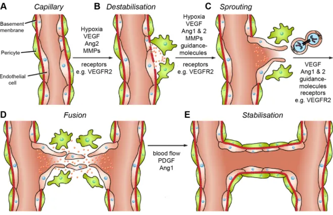
Fruttiger konnte mit Hilfe des Endothelzellmarkers VEGFR1 (vascular endothelial growth factor receptor 1) und des Endothelzell- und Angioblastenmarkers VEGFR2 nachweisen,

Earlier studies conducted in our group showed TrkB was upregulated in spinal cord of oligodendrocyte specific Fgfr1 knockout mice (Rajendran, 2014).. However, in

Im Vergleich mit den verschiedenen Child-Pugh-Stadien zeigten sich nur für VEGF bei Patienten mit einer Zirrhose Child B oder C höhere Mediane als bei gesunden Probanden

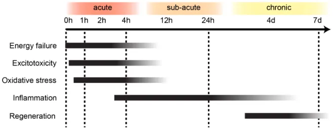
When tissue suffers impaired oxygen supply eventually resulting in cell death, and after focal cerebral ischemia in most cases through necrosis, there is activation of

Evans blue (EB, 50 mg/kg in saline, Sigma–Aldrich, Hamburg, Germany) was injected intravenously in MCAo mice either immediately after or 4, 8, 12 or 16 hours after reperfusion (n =

We have previously shown that total knockout of fibroblast growth factor-2 (FGF-2) results in prolonged survival and improved motor performance in superoxide dismutase 1 (SOD1 G93A

Neben VEGF gilt auch FGF (fibroblast growth factor) als einer der Haupt-Wachstumsfaktoren der Angiogenese in der Plazenta, denn diese beiden Proteinfamilien sind möglicherweise

Diese Annahme wird unterstützt durch Experimente, in denen beispielsweise die Steigerung der Expression von VEGF auf m-RNA-Ebene unter erniedrigtem