Edited by:
Debra Ann Fadool, Florida State University, United States
Reviewed by:
Adam Puche, University of Maryland, United States Bela Volgyi, University of Pécs, Hungary
*Correspondence:
Veronica Egger veronica.egger@ur.de
Specialty section
This article was submitted to Cellular Neurophysiology, a section of the journal Frontiers in Cellular Neuroscience
Received:
30 August 2020
Accepted:05 October 2020
Published:05 November 2020
Citation:
Egger V and Diamond JS (2020) A17 Amacrine Cells and Olfactory Granule Cells: Parallel Processors of Early Sensory Information.
Front. Cell. Neurosci. 14:600537.
doi: 10.3389/fncel.2020.600537
A17 Amacrine Cells and Olfactory Granule Cells: Parallel Processors of Early Sensory Information
Veronica Egger
1* and Jeffrey S. Diamond
21
Department of Neurophysiology, Institute of Zoology, Universität Regensburg, Regensburg, Germany,
2Synaptic Physiology Section, National Institute of Neurological Disorders and Stroke, National Institutes of Health, Bethesda, MD, United States
Neurons typically receive synaptic input in their dendritic arbor, integrate inputs in their soma, and send output action potentials through their axon, following Cajal’s law of dynamic polarization. Two notable exceptions are retinal amacrine cells and olfactory granule cells (GCs), which flout Cajal’s edict by providing synaptic output from the same dendrites that collect synaptic input. Amacrine cells, a diverse cell class comprising
>60 subtypes, employ various dendritic input/output strategies, but A17 amacrine cells (A17s) in particular share further interesting functional characteristics with GCs: both receive excitatory synaptic input from neurons in the primary glutamatergic pathway and return immediate, reciprocal feedback via GABAergic inhibitory synapses to the same synaptic terminals that provided input. Both neurons thereby process signals locally within their dendrites, shaping many parallels, signaling pathways independently.
The similarities between A17s and GCs cast into relief striking differences that may indicate distinct processing roles within their respective circuits: First, they employ partially dissimilar molecular mechanisms to transform excitatory input into inhibitory output; second, GCs fire action potentials, whereas A17s do not. Third, GC signals may be influenced by cortical feedback, whereas the mammalian retina receives no such retrograde input. Finally, A17s constitute just one subtype within a diverse class that is specialized in a particular task, whereas the more homogeneous GCs may play more diverse signaling roles via multiple processing modes. Here, we review these analogies and distinctions between A17 amacrine cells and granule cells, hoping to gain further insight into the operating principles of these two sensory circuits.
Keywords: retina, olfactory bulb, reciprocal synapse, inhibition, parallel processing, local feedback, sensory processing
CIRCUITRY
Both the retina and olfactory bulb are strictly layered early sensory processing areas
with myriad interneuron types that provide local and lateral interactions between
sensory input channels (Figure 1A). In the retina, these channels correspond to the
local receptive fields of photoreceptors which transduce incident light from the visual
world into a neural signal that is passed through glutamatergic synapses to bipolar cells
and then onto the retinal projection neurons, the ganglion cells (RGCs; Figure 1B).
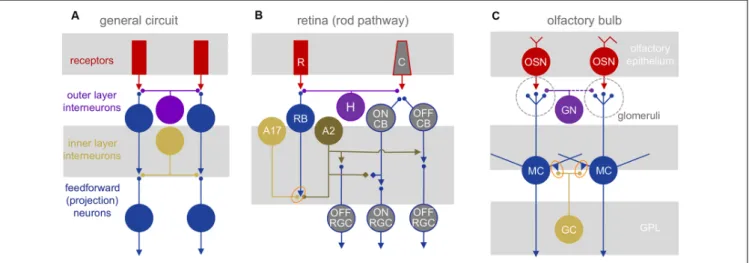
FIGURE 1 |
Neuronal circuit architectures.
(A)General network elements common to the retina and olfactory bulb: two layers of inhibitory interactions are mediated by segregated subsets of local interneurons.
(B)Mammalian retinal circuitry, with the rod pathway highlighted. Rod photoreceptors (R) contact rod bipolar cells (RB), which in turn contact A17 and A2 amacrine cells. The A2 relays the ON signal from rod bipolar cells to the cone pathway. C, cone photoreceptor; H, horizontal cell;
CB, cone bipolar cell; RGC, retinal ganglion cell. Orange oval highlights reciprocal synapse between RB and A17.
(C)Olfactory bulb circuitry. Olfactory sensory neurons (OSN) project into glomeruli, contacting glomerular neurons (GN) and the apical dendritic tufts of mitral (and tufted) cells (MC). The MC lateral dendrites form reciprocal synapses (orange oval) with granule cell (GC) and other interneuron dendrites. GN, glomerular neurons.
In the olfactory bulb, the channels correspond to the glomerular modules that are innervated exclusively by one of several 100s-2,000 olfactory receptor neuron types—each expressing a distinct olfactory receptor—in the nose. These receptor neurons are excited by volatile odorants and pass a glutamatergic signal to mitral and tufted cells (MTCs), the bulbar projection neurons (Figure 1C).
While the bulb contains no direct neuronal analog to retinal bipolar cells, we propose that the highly excitable dendritic tufts of MTCs, which can produce regenerative signals on their own (Chen et al., 1997; Yuan and Knöpfel, 2006) may represent their counterparts. In both systems, neighboring excitatory projection neurons (RGCs and MTCs) typically are not directly interconnected via chemical or electrical synapses, although MTC tufts within the glomeruli may interact via glutamate spillover between synapses or electrical coupling (Schoppa and Westbrook, 2001, 2002).
Signals in both primary sensory pathways are sculpted in two stages by distinct, laterally structured inhibitory networks: in the outer retina, horizontal cells feed back onto photoreceptors to craft center-surround receptive fields (Baylor et al., 1971). In the outer layer of the bulb, a diverse set of glomerular neurons (GN) mediates intra- and interglomerular interactions between the sensory axons and the dendritic tufts of MTCs (reviewed in Wachowiak and Shipley, 2006; Burton, 2017). In the inner retina, amacrine cells (ACs) provide feedback and feedforward inhibition to both bipolar cells and/or RGCs, and in the inner bulb, MTCs interact with local interneurons that consist mostly of granule cells, although other interneuron subtypes contribute substantially to odor processing (Toida et al., 1994; Lepousez et al., 2010; Huang et al., 2013; Kato et al., 2013; Miyamichi et al., 2013). Both GCs and some ACs make GABAergic feedback inhibitory synapses onto the same synaptic terminals that provide them excitatory input (Rall et al., 1966; Kolb and
Famiglietti, 1974). Both GCs and many ACs are connected by gap junctions (Reyher et al., 1991; Vaney, 1994; Menger and Wässle, 2000). GCs also receive powerful glutamatergic, centrifugal inputs (Price and Powell, 1970c; Balu et al., 2007;
Pressler and Strowbridge, 2017), whereas ACs do not.
ACs are molecularly and morphologically diverse:
63 molecularly defined subtypes also differ concerning dendritic arbor size, branching patterns, projection depth in the inner plexiform layer (IPL), and synaptic partners (Diamond, 2017; Yan et al., 2020). GC subtypes are less well characterized; the current count of six morphological subtypes likely underestimates their molecular diversity, especially considering differences between GCs born neonatally and during adult neurogenesis (Breton-Provencher and Saghatelyan, 2012; Nagayama et al., 2014; Takahashi et al., 2018); there is no adult neurogenesis of ACs. Greater interneuron diversity in the retina may be required to support more parallel output channels:
the number of distinct RGC types (currently Baden et al., 2016;
Rheaume et al., 2018; Laboissonniere et al., 2019; Tran et al., 2019) may exceed that of MTC projection neurons by an order of magnitude (Imamura et al., 2020).
GCs make all of their synaptic outputs from apical dendritic spines that receive excitatory inputs primarily from the lateral dendrites of MTCs (Price and Powell, 1970b; Naritsuka et al., 2009). Because this prominent feature equips GCs for parallel processing, we compare them here with A17 cells, the AC subtype that is most similar concerning synaptic interactions: A17 cells also perform local signal processing within reciprocal synapses that are contained in dendritic varicosities from which they provide reciprocal feedback onto rod bipolar cells (RBCs; Chávez et al., 2006; Grimes et al., 2010). In the rod pathway which mediates night vision, A2 ACs relay RBC signals to the cone pathway (Famiglietti and Kolb, 1975; Pourcho and Goebel, 1985;
Strettoi et al., 1992), whereas A17s interact exclusively with RBC
terminals to modulate signal transfer to the A2s. Glutamatergic inputs from RBCs to A2s and A17s occur at ‘‘dyad’’ synapses in which each RBC active zone is apposed to two postsynaptic elements, usually one A2 and one A17. Individual GCs and A17s contain similar numbers of reciprocal synapses (150–200; Price and Powell, 1970a; Grimes et al., 2010; Geramita et al., 2016).
BIOPHYSICAL CHARACTERISTICS
A17s and GCs exhibit distinctive morphological and membrane properties that enable them to provide reciprocal feedback inhibition in parallel through a large number of dendrodendritic synapses that can operate largely independently of one another within the same cell. They achieve these analogous goals using markedly different strategies. The similar outcomes highlight interesting parallels between the two systems, and the differences may provide insights into distinct computational requirements of different sensory circuits.
Morphological Specializations Isolate Feedback Synapses
The clearest morphological similarity between GCs and most ACs—the absence of an axon—was first pointed out more than a century ago by Ramón y Cajal (1911) and posed a counterpoint to his Law of Dynamic Polarization (Ramón y Cajal, 1891).
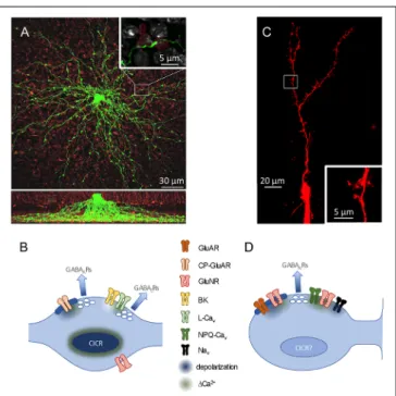
Cajal typically identified distinct, segregated input and output regions to infer the direction of information flow through a neuron, but these clues are absent in GCs and most ACs. In A17s, for example, dozens of very thin (∼130 nm diameter;
Grimes et al., 2010) dendrites radiate, unbranched, from the soma like spokes on a wheel, extending deep into the inner plexiform layer and studded with varicosities ( ∼ 1 µm diameter) at ∼ 20 µm intervals (Zhang et al., 2002; Grimes et al., 2010;
Figure 2A). Synaptic outputs are confined to the varicosities, which also receive synaptic inputs from RBCs (Nelson and Kolb, 1985). Models of this distinct morphology predicted that synaptic potentials would attenuate rapidly along the thin dendrites, possibly isolating neighboring varicosities from each other (Ellias and Stevens, 1980). Accordingly, imaging experiments showed that synaptic activation of single varicosities typically elicits only comparatively small Ca
2+signals in neighboring varicosities (Grimes et al., 2010).
GCs morphologically isolate their synapses differently. Each GC soma extends a large primary dendrite up to a branched arbor in the external plexiform layer (EPL) and only a few smaller dendrites down in the GC layer (Figure 2C). The dendrites are studded with prominent spines that receive synaptic input;
in the EPL, a subset of particularly large spines (‘‘gemmules;’’
Rall et al., 1966), deliver reciprocal synaptic outputs to MCs.
GC apical dendrites are quite thick ( ∼ 350–1,100 nm diameter;
Rall and Shepherd, 1968) and, together with active conductances detailed below, enable membrane depolarizations to traverse the GC dendritic arbor (Egger et al., 2003). GC spine necks are long (nearly 2 µm) and thin ( ∼ 230 nm diameter) and often contain mitochondria (Woolf et al., 1991) that likely increase their axial resistance. These spine necks reduce the extent to which signals in one spine influences neighboring spines, while also creating an
FIGURE 2 |