dendritic cells to microbial stimuli in patients with inflammatory bowel disease
Dissertation
zur Erlangung des akademischen Grades doctor rerum naturalium
(Dr. rer. nat.) im Fach Biologie eingereicht an der
Mathematisch-Naturwissenschaftlichen Fakultät I der Humboldt-Universität zu Berlin
von
Dipl. Biochem. Saskia Thomas
Präsident der Humboldt-Universität zu Berlin Prof. Dr. Dr. h.c. Christoph Markschies
Dekan der Mathematisch-Naturwissenschaftlichen Fakultät I Prof. Dr. rer. nat. Andreas Herrmann
Gutachter: 1. Prof. Dr. Andreas Radbruch
2. Prof. Dr. med. Daniel C. Baumgart 3. Prof. Dr. med. Hans-Dieter Volk
A ship in port is safe,
but that is not what ships are for.
Sail out to sea and do new things.
-- Admiral Grace Hopper
Für meine Eltern.
Die Ätiologie der chronisch entzündlichen Darmerkrankungen Morbus Crohn und Colitis ulcerosa ist bis heute ungeklärt. In zahlreichen Studien konnte jedoch an Mausmodellen gezeigt werden, dass dendritische Zellen eine wichtige Rolle im Rahmen der mukosalen Immunabwehr spielen. Eine unkontrollierte Aktivierung von immunologischen Effektorzellen durch antigenpräsentierende Zellen ist die Folge, welche die Antigene der luminalen Flora folglich falsch erkennen und damit letztendlich zu einer Schädigung des Gewebes führen. In der vorliegenden Arbeit wurden humane CD1c+CD11c+CD14-CD19- myeloide dendritische Zellen (mDCs) aus dem peripheren Blut und der intestinalen Mukosa von CED Patienten sowie von gesunden Probanden phänotypisch und funktionell näher charakterisiert.
mDCs von Patienten reagieren auf mikrobielle Modellstimuli wie LPS im Gegensatz zu dendritischen Zellen von Gesunden mit der Ausbildung eines aktivierten Phänotyps und der Sekretion pro-inflammatorischer Zytokine. Die Daten lassen vermuten, dass ihre tolerogene Rolle gestört ist und die Zellen so möglicherweise aktiv zum Entzündungsgeschehen durch eine Fehlreaktion auf die kommensale Flora beitragen. Die TLR4 Expression von mDCs war bei CED Patienten in Remission höher und stieg während eines akuten Schubes weiter signifikant an. Es konnte außerdem gezeigt werden, dass zirkulierende mDCs von Erkrankten mehr LPS aufnehmen. Des Weiteren ist die Häufigkeit von mukosalen und aktivierten mDCs bei CED Patienten im Vergleich zu Gesunden signifikant erhöht. Die vermehrte Häufigkeit von aktivierten mDCs in der entzündeten Mukosa ist ein Hinweis auf intestinales „homing“, also ein Wiedereinwandern der gereiften Lymphozyten in die Darmwand. Die Daten zeigen außerdem eine abweichende LPS Antwort von mDCs bei CED Patienten, die folglich mit einem entzündeten Phänotyp einhergeht.
Es ist bekannt, dass die probiotische Hefe Saccharomyces boulardii (Sb) eine Wirksamkeit bei entzündlichen sowie infektiösen Erkrankungen des Gastrointestinaltraktes hat. Daher wurde untersucht, ob Sb die Funktion von dendritischen Zellen ebenfalls anpasst. Kulturexperimente von humanen mDCs mit Zellkulturüberständen von Sb (SbS) und LPS zeigten eine deutliche Reduzierung in der Expression der co-stimulatorischen Moleküle CD40 und CD80 sowie des Reifemarkers CD197 (CCR7) sowohl bei gesunden Probanden als auch bei CED Patienten.
SbS reduzierte außerdem die Sekretion zweier wichtiger pro-inflammatorischer Zytokine, TNF- und IL-6. Während es die Sekretion von IL-10 bei gesunden Probanden erhöhte, konnte bei CED Patienten eine leichte Abnahme verzeichnet werden. Zusammenfassend
stimulieren mDCs effektiv naïve TCs und führen so zu einer Differenzierung in TH1- oder TH2- Zellen. Mittels Membran-Verteilungschromatographie konnte außerdem gezeigt werden, dass die aktive Komponente der probiotischen Hefe ein molekulares Gewicht von weniger als 3 kDa haben muss.
Persistence of inflammatory bowel disease is associated with a breakdown of tolerance against the commensal microflora. Various animal studies have provided insights that mucosal dendritic cells play a key role in this process. However, the specific function of certain dendritic cells in IBD is still unknown. Thus, primary CD1c+CD11c+CD14-CD19- myeloid blood (mDCs) and mucosal dendritic cells from IBD patients and healthy controls were compared phenotypically and functionally in this study.
More mDCs from IBD patients exhibited an activated phenotype shown by expression of co- stimulatory molecules as control mDCs. Further, mDCs from IBD patients secrete higher levels of pro- and anti-inflammatory. TLR4 expression by mDCs was higher in remission and significantly increased in flaring UC and CD patients compared with remission and controls.
Circulating mDCs from IBD patients take up more LPS and the uptake begins earlier compared with controls. The frequency of mucosal mDCs and the number of activated, i.e.
CD40 and CD80 expressing mucosal mDCs, is significantly greater in UC and CD compared with non-IBD controls. The increased frequency of activated mDCs in the inflamed mucosa suggests intestinal homing of mDCs in acute stages of IBD. Further, the data suggests an aberrant LPS response of mDCs in patients suffering from IBD which results in an inflammatory phenotype. The most widely accepted hypothesis for the cause of IBD is a disturbed interaction of the host immune system with commensal microflora and other luminal antigens. The well controlled balance of the intestinal immune system is disturbed and luminal antigens like LPS gain access to the underlying mucosal tissue via the leaky barrier.
Furthermore, it was investigated whether the probiotic yeast preparation Saccharomyces boulardii (Sb) modulates dendritic cell function which has shown efficacy in inflammatory and infectious disorders of the gastrointestinal tract. Culture experiments of mDCs from healthy volunteers and IBD patients in the presence of Sb culture supernatant (SbS) significantly reduced the expression of the co-stimulatory molecules CD40 and CD80 as well as the DC maturation marker CD197 (CCR7) induced by the prototypical microbial antigen LPS. Moreover, SbS reduced secretion of two key pro-inflammatory cytokines TNF- and IL- 6, while the secretion of anti-inflammatory IL-10 increased. However, IBD patients showed also a reduction in their secretion level of IL-10. Finally, SbS inhibited proliferation of naïve T cells (TCs) in a mixed lymphocyte reaction with healthy mDCs. Without the probiotic yeast
kDa, as evaluated by membrane partition chromatography.
dendritische Zellen, antigenpräsentierende Zelle, Entzündung, chronisch entzündliche Darmerkrankungen, Morbus Crohn, Colitis ulcerosa, Saccharomyces boulardii, T-Zellen
Keywords:
dendritic cells, antigen-presenting cell, inflammation, Inflammatory Bowel Disease, Crohn’s Disease, Ulcerative colitis, Saccharomyces boulardii, T cells
Zusammenfassung
Abstract
Abbreviations
1 Introduction ... 1
1.1 The Immune System... 1
1.2 Dendritic Cells... 3
1.2.1 Subsets of human dendritic cells... 3
1.2.2 Dendritic cell maturation and migration ... 5
1.2.3 Dendritic cell mediated activation of naïve T cells... 9
1.3 The gastrointestinal immune system ... 11
1.3.1 Barrier and unspecific defense mechanisms ... 12
1.3.2 The intestinal immune system in healthy state... 12
1.3.3 Cytokine regulation of the mucosal immune response via mucosal T cells ... 15
1.3.4 Oral tolerance ... 15
1.4 Inflammatory Bowel Disease ... 16
1.4.1 Crohn’s Disease... 16
1.4.2 Ulcerative Colitis... 16
1.4.3 Epidemiology ... 17
1.4.4 Etiology and pathophysiology... 17
1.4.5 Genetic factors... 17
1.5 Malfunction of the immune system in Inflammatory Bowel Disease... 18
1.6 Probiotics and Inflammatory Bowel Disease ... 20
1.6.1 Saccharomyces boulardii in the treatment of Inflammatory Bowel Disease ... 21
1.7 Aim... 22
2 Material ... 23
2.3 Magnetic cell separation reagents ... 27
2.4 Antibodies ... 28
2.5 Primer ... 29
2.6 Commercial kits ... 29
2.7 Consumables ... 30
2.8 Material for cell separation ... 32
2.9 Equipment ... 32
2.10 Software ... 33
3 Methods ... 35
3.1 Overview ... 35
3.2 Sample collection and preparation ... 36
3.2.1 Human blood and tissue sampling ... 36
3.2.2 Scoring of disease activity... 37
3.2.3 Peripheral blood mononuclear cells ... 37
3.2.4 Mucosal mononuclear cells... 38
3.3 Isolation of human blood and mucosal cells ... 38
3.3.1 Principle of the magnetic cell sort technology ... 38
3.3.2 Purification of CD45+ mucosal cells... 39
3.3.3 Purification of myeloid dendritic cells ... 39
3.3.4 Isolation of naïve CD4+ T cells ... 40
3.3.5 Characterization of mucosal myeloid dendritic cells ... 40
3.4 Cell culture ... 40
3.4.1 Preparation of Saccharomyces boulardii culture supernatant ... 40
3.4.2 Culture and stimulation of dendritic cells ... 41
3.4.3 Quantification of intracellular antigen uptake by myeloid dendritic cells ... 42
3.4.4 Mixed lymphocyte reaction of myeloid dendritic cells and naïve T cells... 42
3.5 FACS ... 43
3.5.1 Principles of flow cytometry ... 43
3.5.4 CFSE staining of T cells... 47
3.5.5 Intracellular cytokine staining... 48
3.5.6 Cytometric bead array analysis of cytokine secretion by dendritic cells ... 48
3.6 RNA Isolation and cDNA synthesis ... 49
3.7 Quantitative real-time RT-PCR ... 50
3.8 Statistical Analysis ... 51
4 Results ... 52
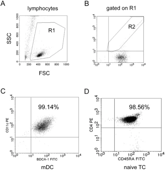
4.1 High purity of isolated human myeloid dendritic cells from peripheral blood... 52
4.2 Phenotype and development of human dendritic cells in the periphery ... 53
4.2.1 More myeloid dendritic cells from IBD patients display an activated phenotype than controls ... 53
4.2.2 Myeloid dendritic cells from IBD patients secrete more inflammatory cytokines upon LPS stimulation than controls ... 55
4.2.3 Myeloid dendritic cells effectively stimulate naïve T cells in an alloreaction and lead to a differentiation into effector T cells ... 59
4.2.4 Increased LPS uptake by myeloid dendritic cells in IBD patients... 61
4.2.5 Increased TLR2 and TLR4 expression by myeloid dendritic cells from IBD patients... 64
4.3 Effects of the probiotic yeast Saccharomyces boulardii on human myeloid dendritic cells... 66
4.3.1 Saccharomyces boulardii culture supernatant decreases the number of CD40, CD80 and CCR7 positive myeloid dendritic cells after incubation with LPS in healthy controls ... 67
4.3.2 Saccharomyces boulardii culture supernatant reduces secretion of TNF-α and IL-6 and increases secretion of IL-10 by myeloid dendritic cells ... 69
4.3.3 Saccharomyces boulardii culture supernatant inhibits T cell proliferation in an allogenic mixed lymphocyte reaction... 70
4.3.4 Saccharomyces boulardii culture supernatant diluted 1:2 induced substantial cell death ... 72 4.3.5 The active component in Saccharomyces boulardii culture supernatant has a
of CD40, CD80 and CCR7 positive myeloid dendritic cells ... 73
4.3.5.2 Saccharomyces boulardii culture supernatant permeates have an effect on cytokine secretion... 75
4.4 Effects of Saccharomyces boulardii on human myeloid dendritic cells from IBD patients ... 76
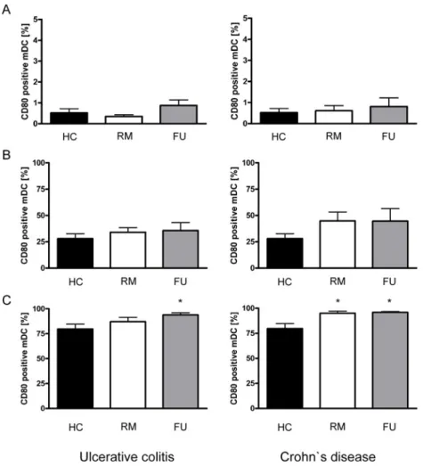
4.4.1 Saccharomyces boulardii culture supernatant reduces the number of CD40, CD80 and CCR7 positive myeloid dendritic cells in patients with Ulcerative Colitis and Crohn’s Disease following LPS stimulation ... 77
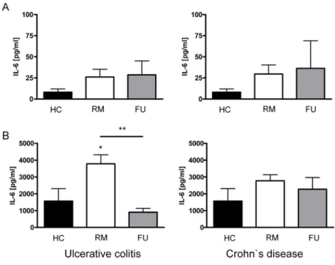
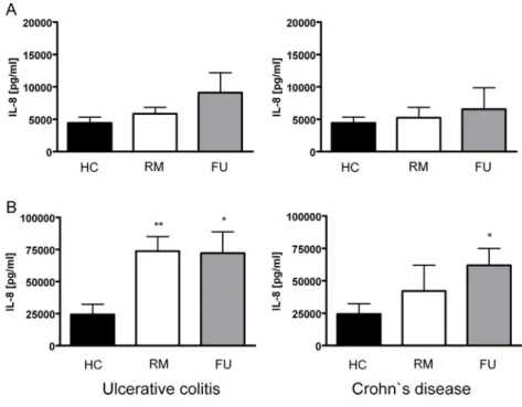
4.4.2 Saccharomyces boulardii culture supernatant reduces secretion of important pro- and anti-inflammatory cytokines by myeloid dendritic cells in IBD patients... 79
4.5 Phenotype and development of human dendritic cells in the mucosa... 81
4.5.1 Increased frequency of activated myeloid dendritic cells among lamina propria mononuclear cells in IBD patients ... 81
5 Discussion... 84
5.1 Human dendritic cells in the periphery and in secondary lymphatic organs... 84
5.1.1 Increased frequency of dendritic cells in the inflamed mucosa ... 85
5.1.2 Dendritic cells from IBD patients display a different phenotype... 87
5.1.3 Disturbed cytokine balance in IBD ... 88
5.1.3.1 Myeloid dendritic cells from IBD patients secrete higher levels of pro- inflammatory cytokines... 89
5.1.3.2 Myeloid dendritic cells from IBD patients secrete higher levels of anti- inflammatory IL-10 ... 91
5.1.4 Inflammatory response by myeloid dendritic cells of patients with IBD ... 92
5.2 Saccharomyces boulardii exhibits anti-inflammatory potential through modulation of dendritic cells ... 93
5.2.1 Administration of Saccharomyces boulardii culture supernatant results in changes of dendritic cell function ... 94
5.2.2 Administration of Saccharomyces boulardii culture supernatant results in changes of dendritic cell immune responses ... 95
5.2.3 Saccharomyces boulardii culture supernatant induces phenotypic and functional changes in myeloid dendritic cells from IBD and IC patients ... 96
Saccharomyces boulardii culture supernatant... 97
5.2.5 The active component of Saccharomyces boulardii culture supernatant ... 98
5.3 Perspective ... 100
References ... 102
Table of figures ... 124
Table directory... 126
Appendix ... 127
Danksagung... 134
List of publications ... 136
Presented poster ... 136
Eidesstattliche Erklärung ... 138
Abbreviations
ab Antibody Ag Antigen
APC Antigen-presenting cell
B-cell Bursa-dependent; bone-marrow-derived (lymphocytes) BCR B cell antigen receptor
BM Bone marrow
BSA Bovine serum albumin
CBA Cytometric Bead Array
CD Crohn’s Disease
cDC Conventional dendritic cell
cDNA Copy deoxyribonucleic acid CDP Common dendritic cell precursor
DC Dendritic cell
DNA Deoxyribonucleic acid
DMEM Dulbecco’s Modified Eagle Medium DTE Dithienylethene
EDTA Ethylenediaminetetraacetic acid ELISA Enzyme-linked immunosorbent assay FACS Fluorescence-activated cell sorting
Fc Fragment crystallisable
FCS Fetal calf serum Fig. Figure
FITC Fluorescein isothiocyanate FU Flare-up
g Gravitational acceleration
GM-CSF Granulocyte macrophage-colony stimulating factor HBSI Harvey Bradshaw Severity Index
HBSS Hanks' Buffered Salt Solution
HC Healthy control
HeBSS HEPES buffered saline solution HEV High endothelial venules hr(s) Hour(s)
HS Human serum
IBD Inflammatory Bowel Disease
IC Infectious Colitis
IFN- Interferon-
IFN- Interferon-
Ig Immunoglobulin IL Interleukin
kDa Kilo Dalton
LC Langerhans cells
LN Lymph node
LPMC Lamina propria mononuclear cells LPS Lipopolysaccharide MACS Magnetic activated cell sorting MALT Mucosal-associated lymphoid tissue mDC Myeloid dendritic cell
MDP Macrophage and dendritic cell precursor MFI Mean fluorescence activity
MHC Major histocompatibility complex min Minute(s)
MLN Mesenteric lymph node MLR Mixed lymphocyte reaction
MTWSI Modified Truelove Witts-Severity Index NK cell Natural killer cell
ODN Oligodeoxynucleotides
PAMP Pathogen-associated molecular pattern
PB Peripheral blood
PBMC Peripheral blood mononuclear cell PBS Phosphate buffered saline
PCR Polymerase chain reaction pDC Plasmacytoid dendritic cell PE Phycoerythrin PerCP Peridinin chlorophyll protein
PI Propidium Iodide
PMA Phorbol myristate acetate pre-DC Precursor dendritic cell
PRR Pattern recognition receptor RM Remission
RPMI Rosewell Park Memorial Institute
RT Room temperature
Sb Saccharomyces boulardii
SbS Saccharomyces boulardii culture supernatant Sc Saccharomyces cerevisiae
SDS Sodium dodecylsulphate
SEB Staphylococcal enterotoxin B sec Second(s)
SLE Systemic lupus erythematosus
TC T cell
TLR Toll like receptor
TNF- Tumor necrosis factor-
UC Ulcerative Colitis
vs Versus v/v Volume per volume
1 Introduction
1.1 The Immune System
Historically, immunity meant protection from disease and more specifically, infectious disease. The cells and molecules responsible for immunity constitute the immune system, and their collective and coordinated response to the introduction of foreign substances is called the immune response. The mammalian immune system can be divided into two different but cooperating compartments – the innate (non-adaptive) and the adaptive (acquired) immune response (Table 1) 1.
Table 1: Major properties of the Innate and Adaptive Immune System.
Property Innate Adaptive
antigen non-specific antigen specific rapid response (minutes) slow response (days) Characteristics
no memory memory
natural barriers (e.g. skin) Lymphocytes (B and T cells) Phagocytes antigen-recognition molecules
(B cell and T cell receptor) soluble mediators (e.g. complement) secreted molecules (e.g. antibody) Immune components
pattern-recognition molecules
Every immune response against a pathogen has different requirements and involves both, appropriate recognition of foreign molecular structures and mounting of an adequate reaction.
Thus, both immune responses pursue diverse but complementary defensive strategies and therefore contribute in their own way to successfully resolve an infection.
The innate immune system reacts immediately to invading pathogens 2. The defence mechanisms of innate immunity range from external physical and biochemical barriers (epithelial cells, mucosal surfaces) to an internal defence (phagocytes, dendritic cells (DCs), natural killer (NK) cells) and also soluble factors such as plasma proteins (i.e. complement cascade, C-reactive protein). Innate immunity is based on a set of germ-line encoded receptors (pattern recognition receptors, PPRs). These receptors are not specific to particular pathogens
but they recognize conserved molecular patterns associated with pathogens (pathogen associated molecular patterns, PAMPs) 3.
In contrast to the innate immune system, there are two types of adaptive immune responses called humoral and cell-mediated immunity, which are accomplished by different components of the immune system. Cell-mediated immunity is arranged by T lymphocytes (T cells, TCs).
TCs can efficiently recognize and eliminate infected cells. An important issue of the adaptive TC response is the large diversity of the TC repertoire, which is generated by random rearrangement of gene sequences coding for a functional TC receptor (TCR) during TC development in the thymus. Humoral immunity in the peripheral blood and mucosal secretion is mediated by antibodies that are produced by B lymphocytes (B cells) which mature in the bone marrow (BM). B cells are initially activated to secrete antibodies after the binding of antigens to specific membrane immunoglobulin (Ig) molecules (B cell receptors, BCRs) which are expressed by these cells 4-6. Once engaged, the B cell receives signals to start the production of the secreted form of this Ig, a process that initiates the antibodies response whose purpose is to eliminate the antigen from the host. B and T cells differ in many functional aspects but share one of the important properties of the immune response – they exhibit specificity toward an antigen. Thus the major recognition and reaction functions of the immune response are contained within the lymphocytes.
Indeed, the high degree of diversity might lead to a reaction against endogenous structures of the organism. Therefore, sophisticated mechanisms are required to ensure tolerance within the immune system. Thus, the prime task of the immune system is to sustain the subtle balance between immunity and tolerance. A failure of mechanisms that control central and peripheral tolerance can lead to autoimmune diseases like type 1 diabetes, lupus or rheumatoid arthritis (RA) 7. The defining characteristics for adaptive immunity are exquisite specificity for distinct molecules and the ability to remember and respond more quickly and efficiently to repeated infections (immune memory) 8-10.
1.2 Dendritic Cells
DCs are professional antigen-presenting cells (APCs) that play a central role in the immune system. They were first described in the human skin by Paul Langerhans in 1868, who assumed that they were cells of the nervous system based on their morphology and finally called them Langerhans cells (LCs) 11. However, the discovery of a small population of “large stellate” cells in peripheral lymphoid organs of mice in 1973 by Steinman and Cohn reinitiated the modern research of DCs 12;13. It could be established that LCs as well as DCs in the skin and the thymus are not nerve cells but originate from hematopoetic progenitors of the BM 14-16. They are the most important cell type responsible for antigen presentation. As APCs they control the initiation and the maintenance of adaptive immune responses, as well as the induction of peripheral tolerance 17-20.
1.2.1 Subsets of human dendritic cells
DCs make a rare cell population within the human body. They constitute only 1-3% of all skin cells and 0.2% of peripheral blood mononuclear cells (PBMCs) 21. Due to their low frequency and the absence of specific antibodies it was long very difficult to identify, isolate and enrich these cells. In 1994 Romani et al. were the first who successfully isolated DCs from human blood 22.
DCs present a heterogenous population of specialized BM derived leukocytes. They are derived from multiple lineages and have distinct stages of cell development, activation and maturation. Further, DCs feature the potential to induce both immunity and tolerance 13;23. Under steady-state conditions DCs exist as either conventional (cDCs) or precursor DCs (pre- DCs). Different subsets of DCs are described in human peripheral blood. However, the majority of DC subsets are of myeloid origin and they are generated in large numbers in the BM every day. These cells are also termed CD11c+ DCs, DC1 or myeloid DCs (mDCs) and express on the one hand high levels of the granulocyte macrophage-colony stimulating factor (GM-CSF) and on the other hand low levels of the IL-3 receptor (CD123). These progenitor cells initially transform into immature DCs and are characterized by a high endocytic activity and with a low potential for TC activation. The second major subset of DCs in human population express high levels of CD123 but little GM-CSF receptor and need to be activated
plasmacytoid DCs (pDCs). Subsets of DC precursors circulate in the blood stream and migrate into tissues, where they reside as sentinels in an immature state 24.
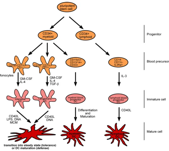
Figure 1: Subsets of human dendritic cells. Myeloid progenitors differentiate into monocytes which are CD14+CD11c+CD1- DC precursors that yield to immature DCs in response to granulocyte/macrophage colony- stimulating factor (GM-CSF) and IL-4. On the other site myeloid progenitors can also differentiate into CD14- CD11c+CD1+ precursors which yield to LCs in response to GM-CSF, IL-4, and transforming growth factor-
(TGF-). A second lymphoid-related DC differentiation pathway can be described, that give rise to plasmacytoid DCs (pDCs) where CD14-CD11c-IL3R+ DC precursors can originate from the lymphoid CD34+ progenitor 25;26. The CD14-CD11c-IL3R+ precursor cells differentiate into immature DCs in response to IL-3. All immature cells finally differentiate into mature cells in response to cytokines or pathogen products. (Adapted from Banchereau et al, 2000 26)
Recent work unveilednew aspects of the developmental and lineage relationships amongDC populations 27. However, this unique work by Liu and colleagues is limited on in vivo
experimental approaches in the mouse. The authors were able to show that DC development progresses from the macrophage and DC precursor (MDP) that is indentified by its surface phenotype (Lin- cKithi CD115+ CX3CR1+ Flt3+) to common DC precursors (CDPs). CDPs no longer rise to monocytes but they can produce pDCs and classical (conventional) spleen DCs (cDCs). Finally, they rise to pre-cDCs and commit to cDC development. Furthermore, intravital imaging studies indicated that pre-cDCs emerge from the BM, travel through blood and finally reach the lymph node (LN) through high endothelial venules (HEVs). In the first instance pre-cDCs migrate along HEVs and later disperse throughout the LN TC area to finally integrate into the DC network. The origin and differentiation cues for many tissue macrophages, monocytes, and DCsubsets in mice, and the corresponding cell populations in humans,remain to be elucidated.
1.2.2 Dendritic cell maturation and migration
DCs are leukocytes, distributed throughout lymphoid and non-lymphoid tissues, in peripheral blood and afferent lymph vessels. The main function of immature DCs is the uptake of exogenous antigens in the periphery. They constantly sample their environment for antigens at potential sites of pathogen entry. This is done through receptors that recognize specific chemical signatures which are found on subsets of pathogens. DCs express numerous receptors like toll-like receptors (TLRs) that recognize conserved structures, known as PAMPs that are unique to the microbial world and found on entire classes of pathogens (i.e.
bacterial LPS or double-stranded viral RNA) 28. Several C-type lectin and lectin-like receptors have been characterized that are expressed abundantly on the surface of these professional APCs 29. Furthermore, DCs express Fc-receptors which bind to antibody-opsonising particles or cells as well as receptors of the complement system 30.
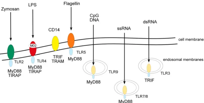
Figure 2: Toll-like receptors and their ligands. The family of TLRs consists of 13 members. Each individual receptor recognizes viral and/ or bacterial components and initiates the activation of immune cells. TLR2, 4, and 5 expressed on the cell surface recognize either zymosan, bacterial LPS and flagellin, respectively. Nucleic acids mainly originating from viral patghogens like CpG DNA, dsRNA and ssRNA are recognized by TLR3, 7/8 and 9. They are localized intracellular and detect nucleic acids in compartments that are normally not accessible to the nucleic acids derived from the host.
Upon encountering an antigen and the exposure to so-called ‘danger’ signals like pathogens, tissue damage, and local inflammation, immature DCs migrate to lymphoid organs (i.e. LN).
During this process, which is regulated by an altered expression of chemokine receptors, DCs undergo maturation. Immature DCs phagocytose pathogens and degrade its proteins into small pieces and upon maturation present those fragments at their cell surface using MHC molecules. When TLR ligands bind their receptors, an intracellular signalling cascade induces DC activation and maturation as defined by an up-regulation of MHC class I and II, an increased expression of co-stimulatory molecules and the secretion of specific cytokines 31. It could be shown that human DC subsets express distinct patterns of TLRs and may subsequently be suited to encounter different pathogens and finally regulate immune responses 32;33. That process is characterized by a down-regulation of the capacity to capture antigen and an up-regulation of antigen processing and presentation, as well as an up- regulation of co-stimulatory molecules which act as co-receptors in TC activation greatly enhancing their ability to activate TCs.
Figure 3: Life cycle of a dendritic cell. DCs originate from bone marrow-derived CD34+ progenitors and finally migrate into tissues all over the body. DC progenitors reside in tissues in an immature state. Invading pathogens are captured by immature DCs followed by antigen processing. Consequently, DCs mature and migrate to secondary lymphoid organs where they present the antigen to TCs and initiate an immune response.
Activated TCs finally migrate to the site of inflammation whereas B cells become activated after the contact with TCs and DCs migrate into various areas in the body. Here, B cells mature into plasma cells which are secreting antibodies that can neutralize the invading pathogen. B – B cell, DC – dendritic cell, T – T cell.
pDCs are sparsely distributed and found in blood and lymphoid tissues (i.e. thymus, tonsils, spleen) 25;34-37. mDCs which are widely distributed are also found in peripheral blood. Mature DCs are a final stage of differentiation, and they cannot be converted into either macrophages or lymphocytes. All DC populations show a characteristic process of maturation which leads to phenotypic and functional change.
Figure 4: Phenotypic and functional properties of immature and mature dendritic cells. Features like phenotype and function change during DC maturation. Immature DCs take up antigen i.e. via phagocytosis, macropinocytosis or adsorptive pinocytosis. DC – dendritic cell, IL – interleukin, mDC – myeloid DC, pDC – plasmacytoid DC. (Adapted from Banchereau et al, 1998 13)
Functionally, DCs exert various effects on other immune cells, particularly in secondary lymphoid organs. Here, they present non-self peptide-MHC complexes to naïve and memory T lymphocytes to mobilize specific immunity. The capacity of DCs to induce stimulatory TCs is dependent on the state of DC maturation. By contrast, in order to induce TC tolerance in the thymus, DCs present self peptide-MHC complexes to thymocytes. Tolerance is induced in the absence of inflammatory stimuli when immature, resting DCs are very poor stimulators for the proliferation of resting TCs. During that process DCs are not activated and they are not able to express any co-stimulatory molecule. The capacity of DCs to initiate primary immune responses is due to their ability to deliver specific co-stimulatory signals which are essential for TC activation from the resting or naïve state into distinct classes of effector cells. These immunogen-specific immune responses are critical for tumor resistance, prevention of metastasis, and blocking infections.
In addition, DCs play an important role in innate immunity by secreting cytokines, i.e. IL-12
activate NK cells which rapidly eradicate selected targets. Therefore, additional pro- inflammatory signals are necessary for the activation of DCs. Immature DCs express different receptors on their surface that are specific for pro-inflammatory cytokines (i.e. TNF-, IL-1, IL-18) 38. A lack of these signals lead to a release of anti-inflammatory cytokines such as IL- 10 or TGF- by immature DCs. Consequently, TCs are being inactivated and regulatory TCs are differentiated which are able to protract or even avoid an immune response 39.
A third known mechanism for the activation of DCs is mediated by direct cell-cell contact and the interaction of the surface receptor CD40 on DCs with the CD40 ligand (CD40L) on activated TCs 40. For an ideal initiation of DC maturation the presence of several signals is necessary.
1.2.3 Dendritic cell mediated activation of naïve T cells
DCs are cells of the innate immune system and are acutely activated when a pathogen invades the body, but they also play a decisive and instructive role in the adaptive immune response that arises 41. Given that the induction of productive TC responses depends upon activation of DCs, it follows that the DC serves as a pivotal interface bridging the innate and adaptive immune system. After delivery of their pathogen-related information, DCs are no longer necessary for the acute response to antigen, presumably they undergo apoptotic cell death and are eliminated.
In the secondary lymphoid organs, mature DCs present antigens captured in the periphery to resting or naïve TCs, inducing an adaptive immune response. Therefore, the TCR binds the presented antigen and the two cells form an immunological synapse from DCs with the according ligands of the TC. The activation and clonal expansion of naïve TCs depends on three independent signals. Without these signals the TC becomes anergic and shifts into a non-proliferating state. The first signal is provided by binding of the TCR receptor to a short peptide presented by MHC on the DC. This ensures that only TCs with a TCR specific to that peptide are activated. The second signal comes from co-stimulation, in which surface receptors on the DC are induced by a relatively small number of stimuli, usually products of pathogens, but sometimes breakdown products of
signal is intensified by an additional interaction of CD40 and CD40L 40. The combination of these two signals induces the expression of IL-2 and his receptors in the TC. The autocrine linkage of IL-2 leads to an expansion of the antigen-specific TC 43. Other receptors are expressed upon activation of the TC, such as TCs. Therefore, DCs and other immune cells secrete specific cytokines 44. Important for the differentiation of CD4+ TCs to TH1 cells are IL-12, IFN- and IFN- whereas the attendance of IL-4 promote a differentiation in TH2 cells 45;46. The proliferation as well as the cytotoxic activity of CD8+ TCs is stimulated by signalling of IL-15 and other interferons 47;48.
Figure 5: Signals for T cell activation. For the activation of TCs specific signals are necessary. Signal 1 is the antigen-induced signal which is delivered to the TC by the TCR-peptide-MHC interaction. CD4+ TCs recognize the antigen in association with MHC II molecules. Signal 2 is conducted through the co-stimulatory CD28 on the TC which binds to CD80 (B7.1) and/ or a binding to CD86 (B7.2) on the APC. The CD40L-CD40 interaction displays an additional signal for CD4+ TCs. Cytokines as well as other exogenous factors like LPS provide the third signal for the activation of naïve TCs. APC – antigen-presenting cell, TCR – T cell receptor, TC – T cell.
DCs are the most important cell type responsible for antigen presentation, and they control both the initiation and maintenance of adaptive immune responses, as well as the induction of peripheral tolerance 17-20.
1.3 The gastrointestinal immune system
The intestinal mucosa implies a large number of mononuclear cells, including macrophages and DCs. They are all together believed to play a central role in regulating mucosal adaptive and innate immune responses 49. The gut contains the largest proportion of the mucosa- associated lymphoid tissue (MALT) in the organism. Here one can find more lymphocytes than in any other lymphatic organ. The lymphatic tissue of the gut covers TCs, B cells, granulocytes, mastocytes, macrophages and DCs 50;51.
The intestinal tract encounters an enormous load of antigens while coevally maintaining a normal homeostatic environment. Most antigens are beneficial to the host, such as dietary antigens and symbiotic bacteria, and some are harmless like commensals. All together the intestine harbours the largest and most diverse microbiota which consists of more than 500 species of bacteria 52;53. On the other hand, pathogens need to be recognized and eliminated before damage occurs. Whereas the systemic immune system elicits an aggressive immune response to exposure of any non-self antigen, the intestinal immune system needs to be more flexible. Antigens need to be sampled, processed, and presented in a way that allows the elimination of pathogens and tolerance to nonpathogens. The challenge of facing billions of bacteria, limitless dietary antigens, and the largest pool of lymphocytes in the body necessitated the development of unique cells as well as mediator and regulatory processes
52;53. Cells of the gut-associated lymphoid tissue (GALT) include conventional cells of the innate and adaptive immune system (i.e. B and T lymphocytes, macrophages, DCs), non- classical APCs, such as intestinal epithelial cells (IECs), and finally lymphocytes specific for the GALT, called lamina propria lymphocytes (LPLs) and intestinal epithelial lymphocytes (IELs).
1.3.1 Barrier and unspecific defense mechanisms
Mucosal surfaces are physical interfaces between the immune system and the massive antigenic load represented by the commensal and potentially pathogenic enteric bacteria 54. A variety of mechanisms contribute to the ability of the gut to either react or remain tolerant to antigen present in the intestinal lumen. The epithelial cell layer forms a barrier against exposure to mucosal microflora and other mucosal antigens. Thus it plays a key role in the regulation of mucosal immune responses. Crucial for an efficient barrier function are specialized adaptations of the intestine, including mucus secretion, tight junctions between epithelial cells, defensins and IgA. Mucus is produced as a thick layer along the intestinal membrane and it offers numerous functions like trapping bacteria and viruses, preventing them from gaining initial access to the host and serving as a microenvironment for the accumulation of bacteriocidal and bacteriostatic chemical enzymes. Furthermore, IECs form intercellular tight junctions that effectively restrict transepithelial movement of particulate and even hydrophilic molecules. Therefore, they prevent an uncontrolled uptake of bacteria and many of their metabolites 55. However, the defense function of the intestinal epithelium is not limited to providing a barrier. Instead, the intestinal epithelium actively interacts with microbes and immune cells by secretion of cellular immune mediators.
1.3.2 The intestinal immune system in healthy state
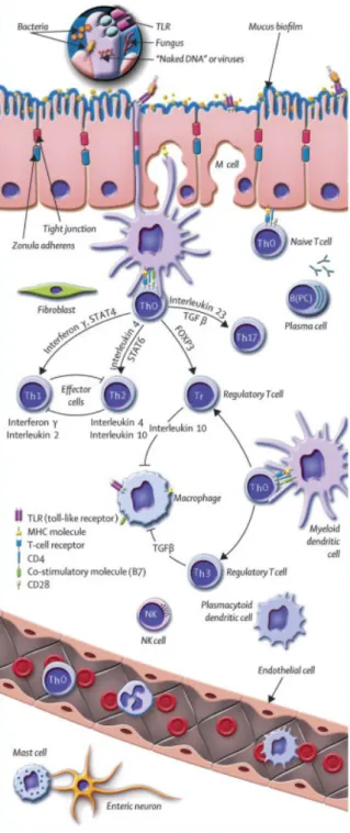
The intestinal immune system represents a complex network of lymphoid and non-lymphoid cells as well as humoral factors. In contrast to DCs from the systemic immune system, mucosal DCs seem to preferentially induce regulatory TCs 56-59. These properties of the mucosal immune system might be due to the high concentration of anti-inflammatory cytokines, such as TGF-β, IL-4 and IL-10. Luminal antigens are sampled by DCs. Uptake and presentation of antigens to naïve T and B cells to induce an adequate immune response is the primary function of the M cells in the mucosa. Following ingestion, antigens and microorganisms are transported from the gut lumen to the dome region through these specialized M cells. In place they encounter APCs leading to typical interactions between DCs and naïve TCs (TH0) through MHC II receptors. In the healthy intestinal immunes system, immature DCs control not only the adaptive immune response, like the balanced differentiation of naïve TCs into effector TCs (T 1, T 2, T 17) that are necessary to
antagonize pathogens and induce regulatory TCs (Treg, TH3), but also the innate immune response that provides activation of NK cells. TH3 cells, a population of CD4+ TCs that produce transforming growth factor-β (TGF-β) can be generated by repeated restimulation of mesenteric lymph nodes (MLNs) or splenic lymphocytes from mice that have been fed low dose of antigen for oral tolerance induction. In the presence of commensals and the absence of inflammation in this area, a balance between effector and regulatory immune cells is maintained by a complex and controlled cytokine network. In the presence of cytokines like IL-12 and IFN-α TCs can differentiate into TH1 cells, whereas IL-4 can induce the differentiation into TH2 cells. TH1 cells express the IL-12 receptor β2 chain and the IL-18 receptor, whereas TH2 cells express an IL-1-like molecule that appears to regulate TH2 effector functions both in the peripheral and mucosal immune system 60. After induction into the GALT, mature lymphocytes leave the inductive sides and finally migrate to the effector sides such as the lamina propria. Here they can induce pro-inflammatory as well as suppressive immune responses. Among the pro-inflammatory signals mucosal TH1 and TH2 effector cells also produce cytokines that have a central regulatory role in the immunhomoeostasis.
Figure 6: The intestinal immune system in healthy state. Antigen can pass the epithelium via M cells and after transfer to local DCs it might directly be presented to naïve TH0 cells. TH0 cells direct further differentiation by the production of different cytokines. Under healthy conditions, a balance between the generation of pro-inflammatory TH1/ TH2 and anti-inflammatory Tr/ TH3 cells is established. After the activation the cells spread widely via the lymphatic system and finally reach the lamina propria and the epithelium of the intestine. M cell – microfold cell; TC – T cell; TH – T helper cell; TH0 cells – naïve T cell; TH, TH1, TH2, TH17 cells – effector TC; Tr, TH3 – regulatory T cells; B – B cell; B(PC) – plasma cell; NK – natural killer T cell.
(Adapted from Baumgart et al, 2007 61).
1.3.3 Cytokine regulation of the mucosal immune response via mucosal T cells
Antigens can pass the mucosal epithelium through M cells and finally encounter with local DCs which direct the differentiation of naïve CD4+ TH0 cells to one of the several states of polarized cytokine production under the influence of cytokines and their associated signaling pathways 62.
TH1 cells secrete pro-inflammatory cytokines like IFN-γ, IL-2, and TNF-α whereas TH2 cells secrete IL-4, IL-5, IL-6, IL-9, IL-10 and IL-13, and promote expression of IgA and other Ig isotypes. Moreover, TH3 cells are also known to secrete TGF-β whereas regulatory TCs predominantly produce IL-10. Many of the TC functions in the gastrointestinal immune system are mediated by secreted cytokines. Previous studies have shown that lamina propria TCs produce higher levels of IL-10 compared to peripheral blood lymphocytes 63;64. This indicates an inhibition of TH1 activation by IL-10 secreting regulatory TCs. Furthermore, the production of IL-10 in the intestine acts on macrophages to prevent their activation and the induction of pro-inflammatory cytokines, thereby inhibiting the recruitment of TCs to the intestine. Other studies demonstrated that regulatory TCs, induced by oral antigen uptake, have characteristics of TH2 or TH3 cells 65;66. On the other hand, work with TCR transgenic mice showed that continuous feeding of low dose antigen induces a TH1 response 67. However, there is also data indicating that TH1 cytokine production may not only have a pro- inflammatory effect as it also can be protective in the immune regulation of certain infectious or autoimmune diseases 68.
1.3.4 Oral tolerance
The ability of the mucosal immune system to distinguish between harmful and harmless antigens is essential to establish highly protective immune responses and further prevent the induction of mucosal pathology. One mechanism that inhibits reactive immune responses is the induction of oral tolerance. Oral tolerance is defined as the induction of a state of systemic immune unresponsiveness to orally administrated antigen upon subsequent antigen challenge such as the absence of antigen-specific TCs. This mechanism presumably prevents the development of an immune reaction or allergy against intestinal intraluminal antigens.
tolerance to the microbiota and food-derived antigens are not yet defined. They involve a complicated interplay of anatomical, cellular as well as humoral factors that prevent immunity against antigens that approach from the lumen. Otherwise the intestinal lumen would trigger an inflammatory response, when presented to the immune system by a non-oral way 69. The mucosal immune system matures during microbial colonisation meanwhile immune or oral tolerance is established.
1.4 Inflammatory Bowel Disease
Inflammatory Bowel Disease (IBD) is a chronic relapsing, immunologically mediated disorder of the gastrointestinal tract. There are two main forms of IBD - ulcerative colitis (UC) and Crohn’s disease (CD) - which have many similarities and also several clinical and pathological differences. IBD leads to long-term and in some cases to an irreversible impairment of the gastrointestinal function and structure 70;71.
1.4.1 Crohn’s Disease
CD was first recognized by the German surgeon Wilhelm Fabry in 1623 72. Later, in 1932 it was described and named after the physician Burril B. Crohn 73. It primarily causes abdominal pain, diarrhea, vomiting, and weight loss. It may affect any part of the gastrointestinal tract, but most commonly the disease starts at the terminal ileum 74.
1.4.2 Ulcerative Colitis
UC was first described by the British physician Sir Samuel Wilks in 1859 75. The main symptoms in active UC are diarrhea mixed with blood and mucus, of gradual onset 76. In 50- 70% the disease is limited to the rectum and sigmoid colon 77. Sometimes it is limited to the rectum as ulcerative proctitis.
1.4.3 Epidemiology
Both diseases have a prevalence range of 10-200 cases per 100,000 individuals in Europe and North America and are becoming more common in the rest of the world as different countries adopt a western lifestyle 78. These observations indicate that there are strong environmental influences on IBD. This was already confirmed in twin studies by the relatively low concordance rate in identical twins, ~50% for CD and ~10% for UC 79.
1.4.4 Etiology and pathophysiology
Although the exact etiology remains uncertain, it is hypothesized that IBD results from inappropriate and ongoing activation of the mucosal immune system which is driven by the presence of the normal luminal flora. This aberrant response is most likely facilitated by genetic prepositions and defects the barrier function of the intestinal epithelium as well as the mucosal immune system.
1.4.5 Genetic factors
It has been previously shown in mouse models that genetic defects can lead to the development of spontaneous mucosal inflammation. Genetic studies highlight the importance of host-microbe interactions in the pathogenesis of IBD 70;80-83. Entirely different genetic abnormalities can lead to similar features of the inflammation in the intestine. IBD is a complex polygenic disease with differential concordance rates in twins. In CD, monozygotic twins have a concordance rate of 58%, while dizygotic twins have rates similar to siblings. On the other hand, in patients with UC the concordance rates are lower than in its counterpart (monozygotic twins, 6-17%; dizygotic twins, 0-5%) 84-87. Furthermore, it could be demonstrated that 20% of patients suffering from CD carry mutations in the nod2/card15 gene, which is involved in the regulation of host response to bacteria 88. CARD15 is an innate immunity receptor which has been strongly associated with the pathogenesis of CD.
Homozygotes carrying two risk mutations and have a 44 times greater probability of contracting CD than noncarriers of the mutations 89. Another important genetic factor is chromosome 6 that has been implicated in the etiology of IBD. Work from several groups
(TNF-) alleles 90-94. Alltogether, these facts illustrate that genetic changes can cause colonic inflammation.
1.5 Malfunction of the immune system in Inflammatory Bowel Disease
The well controlled balance of the intestinal immune system is disturbed at all levels in IBD.
The immunological nature of the disease arises from the observation that IBD is characterized by massive cellular infiltrates and the disease is associated with abnormalities in the immune system including inappropriate production of antibodies and TC dysfunction. A leaky barrier gains access for luminal antigens in IBD. Innate and adaptive immune cells express a different profile of PRRs. On the other hand microbial antigens from commensals trigger and maintain an inflammatory response. Consequently, mDCs falsely recognize commensals as pathogens, enter a maturation program consisting of an increased expression of PRRs, MHC, and co- stimulatory molecules and finally stop migrating. This process promotes the differentiation of naïve TCs into effector TCs. As a result epithelial cells now also express co-stimulatory molecules which enable them to function as APCs and further contribute to an effector TC response. Several studies provide evidence that the two major forms of IBD in humans are a consequence of dysregulated or excessive TH1 or TH2 responses 95-97. There is considerable evidence that IBD patients have an inappropriate TC response to their own intestinal flora, due to a dysfunction in the primary or secondary mechanisms that normally drive and regulate such responses or based on dysfunction in the intestinal epithelial cell barrier that leads to an inappropriate penetration of microbial agents 98-100.
Figure 7: The intestinal immune system in Inflammatory Bowel Disease. In patients suffering from IBD the well controlled balance of the intestinal immune system is destroyed. Antigens gain easy access due to the leaky mucosal tissue. TC – T cell; TH – T helper cell; TH0 cells – naïve T cell; TH, TH1, TH2, TH17 cells – effector TC;
Tr, TH3 – regulatory T cells; B – B cell; B (PC) – plasma cell; NK – natural killer T cell; MLN – mesenteric lymph node. (Adapted from Baumgart et al, 2007 61).
Taken together, IBD patients have a failure in maintaining oral tolerance including down- regulation of responses to harmless luminal antigens like commensals or food, while allowing effector cell responses to mucosal pathogens. In active IBD effector TCs predominate over regulatory TCs. Especially in CD, naïve TCs preferably differentiate into IFN- and IL-12 producing TH1 cells whereas in UC these cells differentiate into aberrant IL-5 producing TH2 cells.
1.6 Probiotics and Inflammatory Bowel Disease
Probiotics are living microorganisms that have a beneficial effect on the host by positively affecting the microbial environment. Typical probiotic candidates are lactobacilli or bifidobacter, but E.coli and other species like yeasts have also been used to study probiotic effects. Probiotic bacteria for humans are preferably of human origin. They have to be safe for the host, genetically stable, and capable of surviving passage through the gastrointestinal tract
101. Among the effects claimed for probiotics are beneficial immunomodulation, reduction of serum cholesterol, improved lactose digestion and protection against colon cancer 101;102. It is widely accepted that the intestinal bacterial flora contributes to the pathogenesis of IBD. This could be supported by several experimental and clinical observations in human. The parts of the gut with highest bacterial counts are the sites which are most affected by IBD and antibiotic treatment has lessened disease activity in both patient groups 103. Commensal bacteria-host interactions are essential in healthy organisms and for immune homeostasis and thus it is not too surprising that disruption of the physiologic bacteria–immune balance may lead to gut inflammation 104;105.
Previously it could be shown that probiotic treatment seems to be effective in patients suffering from IBD 106-108. The probiotic mixture VSL#3 is effective in both maintenance and prophylactic treatment of pouchitis and also helps to induce remission in patients with UC 109-
111. Further studies could show that the nonpathogenic Escherichia coli Nissle 1917 is as effective as mesalazine in preventing relapse of UC and Saccharomyces boulardii appears useful as maintenance treatment for CD 112. However, there are also negative sides, as L rhamnosus GG and Lactobacillus johnsonii LA1 are ineffective in preventing postoperative recurrence of CD 113;114. Additional support for a favorable action of probiotics in gut
sodium sulfate- (DSS) and hapten-induced colitis, and HLA-B27 transgenic rats 115-118. Although clinical results in IBD patients are also encouraging, the data are limited and few studies are placebo-controlled. If probiotics do prove to have beneficial effects in IBD, investigation of the mechanisms may well lead to further advances in treatment.
1.6.1 Saccharomyces boulardii in the treatment of Inflammatory Bowel Disease
The probiotic yeast Saccharomyces boulardii (Sb) in a lyophilized form has demonstrated efficacy in inflammatory and infectious disorders of the gastrointestinal tract in controlled clinical trials 119-124. The yeast belongs to the group of simple eukaryotic cells, like fungi and algae, and therefore differs from other bacterial probiotics that are prokaryotes. Recent oberservations classified Sb genetically within the species of Saccharomyces cerevisiae 125-127. However, it is known that Sb differs metabolically as well as physiologically from S.
cerevisiae 128. Previous studies evaluated the effect of Sb in patients with IBD. It could be demonstrated that the addition of Sb to conventional treatment significantly reduced the stool frequency in patients suffering from CD 129. Furthermore, a beneficial effect in the maintenance of remission in CD has been reported 130. In patients with left-sided UC, the addition of Sb together with mesalamine significantly improved the clinical activity index of these patients 131. Certainly, little is known about how the probiotic yeast Sb unfolds its anti- inflammatory properties in human, especially during IBD.
1.7 Aim
Persistence of IBD is associated with a breakdown of tolerance against the commensal microflora. Previous animal studies have provided insights in the role of mucosal DCs which play a key role in this process. However, the specific function of certain DCs in IBD is still unknown. Thus, primary CD1c+CD11c+CD14-CD19- mDCs or mucosal DCs from IBD patients and healthy controls were compared in this study. Since our group has previously shown that IBD patients with an acute flare-up experience a significant drop in their peripheral mDCs and pDCs populations as well it was important to further characterize the cells from these patients. In the present study experiments were carried out to further specify the localisation and phenotype of human mDCs to gain further insights in possible functional roles of these cells, especially in IBD. Therefore, peripheral blood DCs as well as mucosal DCs were quantified and further analyzed by measuring surface and maturation marker as well as co-stimulatory molecules. Moreover, cytokine secretion of key pro-inflammatory and anti-inflammatory cytokines was investigated to allow a better understanding of the functional role of DCs. Since probiotics become very important as dietary supplements and an effect has been previously shown after probiotic treatment in patients suffering from IBD an additional aim of the current study was to investigate how the probiotic yeast Saccharomyces boulardii unfolds its anti-inflammatory properties in human and exceptionally in IBD patients.
2 Material
2.1 Chemicals and supplements
Table 2: Chemicals in alphabetical order.
Name Company
Ammonium chloride (NH4Cl) Sigma
Steinheim, Germany
BactoTM Agar Difco Laboratories
Becton Dickinson Sparks, U.S.A.
Bovine serum albumin (BSA) Sigma
Steinheim, Germany Carboxyfluorescein-diacetate-succinimidylester (CFSE) Invitrogen
Karlsruhe, Germany Collagenase NB 8 Broad Range
from Clostridium histolyticum
Serva Electrophoresis Heidelberg, Germany DNase I (deoxyribonuclease I)
from bovine pancreas Lyophilisate 100 mg
Roche Diagnostics GmbH Mannheim, Germany
DMSO ICN Biomedicals Inc.
Aurora, U.S.A.
Dithienylethene (DTE) Sigma,
Steinheim, Germany
Ethanol Aaper Alcohol and Chemical
Shelbyville. U.S.A.
Fetal calf serum (FCS) Invitrogen
Karlsruhe, Germany
Ficoll-Paque Plus Amersham Pharmacia Biotech
Freiburg, Germany
Fluoromount-GTM Southern Biotech
Eching, Germany
Fungizone Invitrogen Karlsruhe, Germany
Gentamycin GIBCOTM
New York, U.S.A.
Herculase® Enhanced DNA Polymerase Stratagene La Jolla, U.S.A.
Human serum type AB Cambrex
Charles City, U.S.A.
L-glutamine GIBCOTM
New York, U.S.A.
Lipopolysaccharide, E. coli O55:B5 Calbiochem®
Darmstadt, Germany Lipopolysaccharide,
from E. coli serotype O55:B5 Alexa Fluor 488 conjugate
Molecular Probes Göttingen, Germany
O.C.T.™ Compound Tissue-Tek®
Zoeterwoude, Netherlands
Penicillin-Streptomycin Biochrom AG
Berlin, Germany
Potassium hydrogen carbonate (KHCO3) Sigma
Steinheim, Germany
Propidium Iodide, 50 mg Calbiochem®
Darmstadt, Germany
Pyruvate GIBCOTM
New York, U.S.A.
Recombinant human IL-3 R&D Systems Inc.
Minneapolis, U.S.A.
RNase Zap® Sigma
Steinheim, Germany
Sodium azide (NaN3) Sigma
Steinheim, Germany
Staphylococcal enterotoxin B (SEB) from Staphylococcus aureus
Sigma
Steinheim, Germany
Trypan blue (0.5%) Sigma
Steinheim, Germany
YPD-Broth Sigma Steinheim, Germany
2.2 Buffers and solutions
The following buffers and solutions were purchased from commercial suppliers.
Table 3: Buffers purchased from commercial suppliers.
Name Company
0.5 M EDTA, pH 8.0 Sigma
Steinheim, Germany
Cytofix/Cytoperm BD Biosciences
San Diego, U.S.A.
Dulbecco`s Modified Eagle Medium (DMEM) GIBCOTM
Grand Island, U.S.A.
FACS Clean Solution BD Biosciences
San Diego, U.S.A.
FACS Flow Solution BD Biosciences
San Diego, U.S.A.
FACS Rinse Solution BD Biosciences
San Diego, U.S.A.
Hanks' Buffered Salt Solution (HBSS) GIBCOTM
New York, U.S.A.
Perm/Wash Buffer BD Biosciences
San Diego, U.S.A.
Phosphate Buffered Saline (PBS) PAA Laboratories Pasching, Austria
RLT-Buffer (Lysis Buffer) Qiagen
Maryland, U.S.A.
RPMI Medium 1640 GIBCOTM
Grand Island, U.S.A.
The following buffers and solutions were prepared freshly before use.
Table 4: Freshly prepared buffers and solutions.
Name Composition
Blocking buffer PBS
10% FCS
Co-Culture medium RPMI
0.2% L-glutamine 10% HS type AB
1% Penicillin-Streptomycin
DC culture medium RPMI
0.2% L-glutamine
10% human serum (HS) type AB 1% Penicillin-Streptomycin 1% Pyruvate
Digest-Mix Tissue wash buffer
15 mg collagenase 0.2% DNase
Erythrocyte lysis buffer Aqua dest.
0.1% KHCO3
0.8% NH4Cl
0.1 M EDTA at pH 7.5
MACS buffer PBS
0.4% EDTA 0.5% HS type AB
FACS buffer PBS
0.05% BSA 0.1% NaN3
HBSS-DTE HBSS 5 mM DTE
Tissue wash buffer HBSS
1% Penicillin-Streptomycin 0.2% Gentamycin
0.4% Fungizone
Washing Buffer PBS
1% FCS
2.3 Magnetic cell separation reagents
All products for magnetic-activated cell sorting (MACS) were purchased from Miltenyi Biotech, Bergisch Gladbach, Germany.
Table 5: Kits purchased for cell isolation using MACS technology.
Name Contents
CD1c (BDCA-1)+ Dendritic Cell Isolation Kit
2 ml FcR Blocking Reagent, human: human IgG
2 ml CD19 MicroBeads, human: conjugated to a monoclonal antibody (isotype: mouse IgG1)
2 ml CD1c(BDCA-1)-Biotin antibody, human:
monoclonal CD1c(BDCA-1) antibody conjugated to biotin (clone: AD5-8E7; isotype: mouse IgG2a)
2 ml Anti-Biotin MicroBeads: conjugated to monoclonal anti-biotin antibody (isotype: mouse IgG1)
CD4 MultiSort Kit CD4-Multisort Beads MS Release Reagent MS Stop Reagent
CD45 MicroBeads 2 ml CD45 MicroBeads, human: conjugated to monoclonal anti-human CD45 antibodies (isotype:
mouse IgG2a)
CD45RA MicroBeads 2 ml CD45RA MicroBeads, human: conjugated to monoclonal anti-human CD45RA antibodies (isotype:
mouse IgG1)
2.4 Antibodies
Table 6: Antibodies, isotype controls and secondary antibodies used for FACS and immunofluorescence staining.
Specificity Label Species Clone Dilution Supplier
human CCR7 Alexa 647 rat IgG2a, κ 3D12 1:20 BD
human CD11c PE mouse IgG2b, sB-ly6 1:10 BD
human CD14 APC
FITC PerCp
mouse IgG2a, κ
mouse IgG2b, κ
mouse IgG2a, κ
TüK4 MΦP9
1:10 1:10 1:10
BD Caltag BD
human CD19 Cy5 mouse IgG1, κ HIB19 1:10 BD
human CD3 Bio
PE
mouse IgG2a
mouse IgG2a
1:12 1:10
Caltag Caltag
human CD4 FITC
PE mouse IgG2a
S2.5 SK3
1:20 1:10
Caltag BD
human CD40 PE mouse IgG1, κ 5C3 1:10 BD
human CD45 APC mouse IgG1, κ HI30 1:20 BD
human CD45RA FITC mouse IgG2b, κ HI100 1:10 BD
human CD80 PE mouse IgG1, κ L307.4 1:10 BD
human CD83 APC
FITC PE
mouse IgG1, κ
mouse IgG1, κ
mouse IgG1, κ
HB15e
1:10 1:10 1:10
BD BD BD
human CD86 FITC
PE
mouse IgG1, κ
mouse IgG1, κ
2331 (FUN-1) 1:10 1:10
BD BD human anti-CD1c (BDCA-1) APC
FITC
mouse IgG2a
mouse IgG2a
AD5-8E7 AD5-8E7
1:10 1:10
Miltenyi Miltenyi
IFN- PE mouse IgG1 1:100 BD
IL-4 PE mouse IgG1 1:133 BD