Grases, Lesch, Stein, Heissmeyer and Reutter: Aggravating and Protecting Effects in Galactosaminc Induced Hepatitis 539
Z. Klin. Chem. Klin. Biochem.
10. Jg. 1972, S. 539—542
Aggravating and Protecting Effects of Triton and Prednisolone in Galactosamine Induced Hepatitis
1)
• By P. J. GRASES
1), R. LESCH, U. STEIN, H. HEISSMEYER and W. REUTTER
Pathologisches Institut, Medizinische Universitätsklinik und Biochemisches Institut\
Universität Freiburg^ FreiburgJ Germany
(Eingegangen am 20. April/3. August 1972)
The biochemical and morphological modifications that Triton and prednisolone produce on galactosamine hepatitis in rats were studied. — Determinations of acid phosphatase, N-acetyl-ß-glucosaminidase, cathepsin D, and acid carboxypeptidase in tissue fractions indicate that they can be liberated from lysosomes when galactosamine and Triton are given either alone or simultaneously; in the latter case the alter- ations are more severe. — Prednisolone not only inhibits the changes produced by galactosamine, but it is also capable of inhibiting marked elevation of the serum levels of aspartate and alanine transaminase and of sorbitol dehydrogenase, and of morphologic signs of liver damage.
In der vorliegenden Arbeit werden bei der Galaktosamin-Hepatitis biochemische und morphologische Veränderungen beschrieben, die durch die Verabreichung von Triton WR-1339 beziehungsweise Prednisolon hervorgerufen werden. — Durch Bestimmung der sauren Phosphatase, N-Acetyl-ß-glucosaminidase, Kathepsin D und sauren Carboxypeptidase in Lysosomenfraktionen konnte sowohl durch Triton WR-1339 als auch durch Galaktosamin eine Labilisierung der Lysosomenmembran nachgewiesen werden. Gleichzeitige Gabe von Galaktosamin und Triton WR-1339 führt zu einer verstärkten Labilisierung lysosomaler Membranen. — Nach vorheriger Prednisolon- Gabe reagiert die Leber nach Galaktosamin-Gabe nur mit einer gering ausgeprägten Entzündung: die Aktivitäten der Aspartat- und Alanintransaminase sowie Sorbitdehydrogenase im Plasma steigen nur wenig an; lichtmikroskopisch zeigt sich eine geringe entzündliche Infiltration ohne Nekrosen. Wird Prednisolon nach der Galaktosamin-Applikation gegeben, tritt die Hepatitis abgeschwächt auf.
As in other experimental hepatocellular damages (I, 2) galactosamine hepatitis is characterized by the presence of structural alterations in more than one organelle (2, 3, 4). The difficult task of establishing the so called
"primary target organelle" has so far been accomplished with only a few toxic agents (5, 6). An increase of catabolic lysosomal enzyme activities in the rat liver can be observed after administration of galactosamine (7), and for this reason, it was of interest to study the possible role of lysosomes in this type of liver injury by the application of compounds which can produce labilization and injury (8, 9, 10) or stabilization (11) of lysosomes.
The present study deals with morphological and bio- chemical findings, obtained by application of Triton WR-1339 and prednisolone to rats with acute galactos- amine hepatitis. Triton was used because of its capacity for increasing lysosomal fragility (12), and prednisolone, because of its well known lysosomal stabilizing effect (11) and its ability to arrest the development of ex- perimental hepatic disease (13, 14).
Materials and Methods
Animals
Male Wistar rats (Ivanovas, Kisslegg, Germany) were fed a standard diet (Altromin R; Altromin GmbH, Lage, Germany) and water ad libitum. The animals were divided into two ex- perimental groups:
*) Institute Anatomopatologico, U. C. V., Caracas, Venezuela.
Recipient of an Alexander von Humboldt-Stiftung Postdoctoral Research Fellowship.
Group I
In this group the body weights ranged between 150 and 160 g, and the animals were divided into the following subgroups2):
A) GalN (4 rats) — a single dose of an aqueous solution (0.45 mol/1) of galactosamine (purchased as D-galactosamine · HC1 by Carl Roth OHG, Karlsruhe, Germany) was injected intraperitoneally at a level of 375 mg/kg;
B) Triton (4 rats) — 1 ml of a Triton WR-1339 (Serva, Heidelberg, Germany) solution diluted in saline to a concentration of 200 mg/
ml, was injected into the tail vein;
C) GalN plus Triton (9 rats) — galactosamine and Triton were given simultaneously in the previously mentioned amounts;
D) Prednisolone (4 rats) — 0.15 ml of an aqueous solution of prednisolone (Deltacortril-Pfizer GmbH, Karlsruhe, Germany) containing 10 mg/ml were given 4 times intravenously at 3 h intervals;
E) GalN plus prednisolone (4 rats) — the first dose of prednisolone was given immediately after galactosamine and the amounts, routes of administration and subsequent treatment were the same as stated above;
F) Controls (11 rats) — no treatment.
Group II ·
In this second group, the animals weighed between 180 and 200 g and were also divided into 6 equivalent subgroups. Each subgroup contained 6 rats with the exception of the GalN plus Triton subgroup which contained 7. The experiment was designed in the same way as in Group I, but prednisolone was given sub- cutaneously at slightly higher dose (up to 0.2 ml). In the GalN plus prednisolone subgroup prednisolone was also administered for one day before galactosamine injection (0.2 ml X 4 at 3-hour intervals).
2) The number of rats indicated in each subgroup corresponds to those in which total activities of acid phosphatase and N-acetyl-/?- glucosaminidase were determined. The numbers in which other enzymes were studied are given in Table 2.
2. Klin. Chem. Klin. Biochem. / 10. Jahrg. 1972 / Heft 12
540 Grases, Losch, Stein, Hcissmeycr and Reutter: Aggravating and Protecting Effects in Galactosamine Induced Hepatitis
An additional subgroup of 7 rats treated with galactosaminc and 10 rats \vith combined GalN plus prednisolone treatment as pre- viously described, was included for determination of serum enzyme levels.
After 24 h without food but with water, the rats were killed by exsanguination under light ether anaesthesia. The livers were removed and divided into several portions for light microscopic examination and biochemical enzyme determinations.
Light microscopy
Tissue fragments were fixed in 4% neutral buffered formalin and paraffin sections were stained conventionally with hematoxylin and eosin, PAS, and PAS after diastase treatment.
Biochemical enzyme determinations
Total activities were determined in liver homogenates prepared, by homogenizing the samples (0.6—1.5 g) with 9 volumes of distilled water in an Ultraturrax homogenizer (Jahnke und Kunkel, S tauf en, Germany).
Tissue fractionation studies were done according to the principles given by DE Du VE et al. (15) modified by PLATT and KATZEN- MEIER (16). Before biochemical study samples were frozen and thawed 3 times.
Acid phospbatase (EC 3.1.3.2) was determined according to the method described by GIANETTO and DE DUVE (17) but \vith addition of Triton X-100 as proposed by WATTIAUX and DE DUVE (18). The liberated inorganic phosphate was measured following the principle described by FISKE and SUBBAROW (19).
N-Acetjl-ß-glueosaM/nidase (EC 3.2.1.30) was estimated by the method of VAES and JAQUES modified by VAN HOOF and HERS (20).
Catbepsin D (EC 3.4.2) was measured by the method of ANSON (21).
Acid carboxypepfidase (EC 3.4.2.1) was determined as described by BUDDECKE et al. (22) using the technique proposed by MATHESON and TATTRIE (23).
One unit of hydrolytic activity represents the amount of enzyme needed to degrade 1 micromole of substrate per minute at 37°C (20). Protein was measured by means of the Biuret technique (24).
Standard procedures were used for determination of serum levels of aspartate awinotransferase (EC 2.6.1.1) (25), alanine aminotrans- j-erase (EC 2.6.1.2) (26) and sorbitol dthydrogmase (EC 1.1.1.14) (27).
Histological examination was performed in all animals. Deter- minations of cathepsin D and acid carboxypeptidase were only done in rats from experimental Group I. Total activities of acid phosphatase and N-acetyl-/?-glucosaminidase were also determined in this group and repeated when fractionation studies of these two enzymes were done in experimental Group II.
Results
Light microscopic study
The histological findings were similar in both groups.
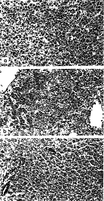
In the galactosamine treated subgroups the usual findings were noted: mainly disseminated single cell and spotty necroses; activation of mesenchymal cells, and inflammatory infiltrates not only in the parenchyma, but also in the portal tracts (Fig. la).
The hepatocytes of Triton-treated animals showed no alterations. The KUPFFER cells were moderately in- creased in number but no evidence of erythrophago- cytosis was found.
The animals given galactosamine plus Triton displayed the most severe hepatic lesions. Although the lobular architecture was preserved, the areas of parenchymal necroses were extensive and inflammatory cells were more numerous than with galactosamine alone (Fig.
Fig. la
Liver of rat 24 h after administration of galactosamine. Hematoxylin and eosin, x 125
Fig. I b
Liver of a galactosamine plus Triton-treated rat. Extensive paren- chymal necroses and numerous inflammatory cells are evident Hema-
toxylin and eosin, x 125 Fig. Ic
Liver of a galactosamine plus prednisolone-treated rat. Single cell eosin, x 125 necroses and KUPFFER cell hyperplasia are only discrete. Note the
absence of inflammatory cells. Hematoxylin and
1 b). Conspicuous edema of the portal tracts as well as focal parenchymal hemorrhages appeared as additional features.
When prednisolone was given in addition to galactos-
amine, the lesions were similar to those present in the
galactosamine-treated. animals, but much less marked
(Fig. Ic). Entire lobules free of necroses could be
identified. Variations in hepatocytic size and shape, as
well as variations in the staining quality of the cyto-
plasm, were less conspicuous. No differences could be
detected between the animals pretreated with predni-
2. Klin. Chem. KHn. Biochem. / 10. Jahrg. 1972 / Heft 12Grases, Lesch, Stein, Heissmeyer and Reütter: Aggravating and Protecting Effects in Galactosamine Induced Hepatitis 541
solone (Group II) and those without previous admin- istration of the corticosteroid (Group I).
The prednisolone-treated subgroups showed fine vacu- olated fatty degeneration, limited mostly to the lobular periphery.
Biochemical enzyme determinations
Serum levels of aspartate, and alanine transaminase, and of sorbitol dehydrogenase of those animals treated with grvlactosamine plus prednisolone were much lower than those present in the animals treated with galactos- amine alone (Tab. 1). Mean values for the three enzymes were almost normal.
Total activities of cathepsin D and acid carboxypeptid- ase were decreased in the animals treated with galactos- amine. Both catabolic enzymes were increased in the supernates from the rats in which galactosamine plus Triton were given. Elevations of enzyme activities were also present in the supernates from the galactosamine- treated rats. When Triton was injected alone, only cathepsin D was increased (Tab. 2).
In experimental Group total activities of acid phos- phatase and N-acetyl-/?-glucosaminidase, whether rel-
Tab. I
Serum enzyme activities [U/l]a
Treatment Aspartate Alanine- Sorbitol
transaminase transaminase dehydro- genase GalNGalN + Predni-
solone
7 10
1258 ±528 85.3 ±21. 9
667 ±365 23.5 ±6
353 ±168 8.0 ±6.6 x ± s
ated to protein content or wet weight, showed similar values with exception of an increase of acid phos- phatase in the galactosamine-treated rats. Both enzymes were elevated in the supernates from the GalN plus Triton-treated subgroup, but only N-acetyl-ß-glucos- aminidase showed increased acitivity in the supernates from the animals treated with galactosamine alone or Triton alone (Tab. 3).
In both groups, enzyme activities of the combined galactosamine plus prednisolone treatement were com- parable to those found in the control animals.
Discussion
The present study indicates that Triton WR-1339 is capable of aggravating the lesions produced by galactos- amine in the liver of rats. It also suggests that a protec- tive effect can be obtained by administering predni- solone either simultaneously or before giving galactos- amine.
Total activities of the lysosomal enzymes acid phos- phatase, N-acetyl-ß-glucosaminidase, cathepsin D, and acid carboxypeptidase in the liver homogenates were determined on the premise that lysosomal damage induced by galactosamine or galactosamine plus Triton would liberate lysosomal enzymes. Only cathepsin D and acid carboxypeptidase were found to be decreased in the galactosamine treated animals, but acid phos- phatase was increased in the same subgroup.
The use of fractionation techniques demonstrates that the significant increase of several enzymes in the majority of supernates from those animals treated with galactos-
Tab. 2 Liver enzyme activities
Subgroup Cathepsin D
Total Supernate [mU/g] [mU/mg Protein]
x±s x±s
Acid carboxypeptidase Total Supernate [mU/g] [mU/mg Protein]
x±s x±s Control
GalNTriton GalN .+ Triton Prednisolone GalN + Prednisolone
44 4 3 4 4
246 ±15.3 155±19.6b
246 ±42.5 242 ±45 21 2 ±30.4 21 8 ±43.7
0.66 ±0.04 1.14±0.23a
1.26±0.17&
1.48 ±0.09*
0.50 ±0.11 0.59 ±0.09
649 ±84 410 ±75.4*
696 ±105 557 ±93 721 ±145 630 ±82.5
0.66 ±0.08 1.64±0.14c 0.58 ±0.06
1.93±0.13C
0.58 ±0.06 0.76 ±0.11 p of paired samples (vs controls) a < 0.01; b < 0.0025; c < 0.0005
Tab. 3 Liver enzyme activities Acid Phosphatase Subgroup
Control GalNTriton GalN -f Triton Prednisolone GalN + Prednisolone
h
6 6 6 7 6 6
Total [U/g]
x±s 2.67 ±0.23 3.35±0.1815
2.68 ±0.26 2.42 ±0.45 2.59 ±0.35 2.79 ±0.29
Supernate [mU/mg Protein]
x±s 1.17 ±0.25 1.39 ±0.33 1.33 ±0.72 2.87 ±1.03*
1.14 ±0.40 0.89 ±0.23
N-acetyl-jS Total [U/g]
5c±s 1.55 ±0.23 1.73 ±0.22 1.75 ±0.1 4 1.48 ±0.29 1. 68 ±0.10 1 .43 ±0.26
t-glucosaminidase Supernate [mU/mg Protein]
x±s 0.66 ±0.11 1.81 ±0.54 b 1.80±0.2Qc 4.86±1.82c 0.88 ±0.1 7 0.62 ±0.15 p of paired samples (vs controls) a < 0.005; b < 0.0025; c < 0.0005
Z. Klin. Chem. Klin. Biochem. / 10. Jahrg. 1972 / Heft 12 74
542 Grases, Lcsch, Stein, Heissmeyer and Reutter: Aggravating and Protecting Effects in Galactosamine Induced Hepatitis
amine and/or Triton reflect the release of enzymes from lysosomes. This confirms previous findings in which cathepsin D and acid carboxypeptidase showed similar changes 25 h after the administration of galactosamine (7). An increase in lysosomal enzyme activities in the liver is not due to an enhanced activation of mesenchymal cells, because normal enzyme values could be measured in liver diseases with mesenchymal activation, and also in isolated leucocytes stimulated by inflammation (28, 29).
Whether the liberation of these enzymes is the result of primary damage already present in the early stages of liver injury, or is only an expression of cell necroses (8), is difficult to establish in this experiment. But in any case, the increase of 3 of the 4 assayed enzymes in the supernates from those animals treated with Triton alone cannot be attributed to focal necroses or to the con- currence of an extrahepatic cell population, since neither necroses nor inflammatory infiltrates were observed.
The absence of enzyme variations in the animals treated with galactosamine and prednisolone indicates that corticosteroids have a protective role. Furthermore, the impressive inhibition of an increase of serum levels of aspartate and alanine transaminase and of sorbitol dehydrogenase observed when prednisolone is given together with galactosamine constitutes additional evidence for the protective role of prednisolone. To what degree this protection is achieved with parti- cipation of other organelles, including the cell mem- brane, is not known with certainty (11, 30). The stabilizing effect of corticosteroids in connection with
lysosomes has been demonstrated in the liver but without any relation at all to toxic agents (31), and in in vitro experiments (30). Confirmatory evidence based on the antagonistic effects of vitamin A and cortisone indicate that this can also be accomplished in vivo (32).
The increase in total activities of acid phosphatase after administration of galactosamine alone may suggest new synthesis as the result of an early regenerative response to the diffuse damage (28). Increased synthesis of N- acetyl-/8-glucosaminidase, suggested to be the conse- quence of parenchymal cell division, has been found in rats 24 h after partial hepatectomy (33).
If the aggravating effect of Triton on the hepatic necroses induced by galactosamine is due at least partially to lysosomal injury, it would seem to support the assumption that alterations of this organelle play a role in the conversion of spotty viral hepatitis to massive hepatic necroses (10). On the other hand further studies of the protective role of corticosteroids using galactos- amine induced hepatitis as an experimental model might be useful in order to ckrify the inconsistency in the response of human viral hepatitis to corticosteroid therapy.
Acknowledgments
The authors wish to express their gratitude to Miss BARBARA HASSELS and Miss CLAUDIA GENTNER for their skillfull technical assistance.
This investigation was supported by Deutsche Forschungsge- meinschaft, Bonn-Bad Godesberg and Wissenschaftliche Ge- sellschaft, Freiburg.
References
l, SCHAFFNER, F. (1966), Medicine 45, 547—552. — 2. MEDLINE, A., SCHAFFNER, F. & POPPER, H. (1970), Exp. Molec. Pathol. 12, 201—211. τ- 3. REUTTER, W., LESCH, R., KEPPLER, D. & DECKER, K. (1968), Naturwiss. 55, 497. — 4. KEPPLER, D., LESCH, R., REUTTER, W. & DECKER, K. (1968), Exp. Molec. Pathol. 9, 279—290. — 5. NOVIKOFF, A. B., ROHEIM, P. S. & QUINTANA, N.
(1966), in Biochemical Pathology (Farber, E. & Magee, D. F., eds.) p. 27—49, Williams & Wilkins, Baltimore, Maryland. — 6. RECKNAGEL, R. O. (1967), Pharmacol. Rev. 19, 145—208. — 7. STEIN, U., HEISSMEYER, H., WANGEMANN, G., LESCH, R., REUTTER, W. & KEPPLER, D. (1971), Klin. Wschr. 49, 550—554^
8. SLATER, T. F. (1969), in Lysosomes in Biology and Pathology (Dingle, J. T. & Fell, H. B., eds.) Vol. I, 469—492, North- Holland Publishing Co., Amsterdam & London. — 9. ROELS, O. A. (1969), in 1. c. (8) Vol. I, 254—275. — 10. ITURRIAGA, H.,' POSALAKI, I. & RUBIN, E. (1969), Exp. Molec. Pathol. 10, 231—
239. — 11. WEISSMANN, G. (1969), in 1. c. (8) Vol. I, 276—295. — 12. WATTIAUX, R., WIBO, M. & BAUDHUIN, P. (1963), in Lysoso- mes (de Reuck, A. V. S. & Cameron, M. P. eds.) 176—196, Ciba Foundation Symposium, Little Brown, Boston. — 13. HUTTERER, E., RUBIN, E., GALL, E. & POPPER, H. (1962), Exp. Molec!
Pathol. /, 85—95. —14. ANTHONY, D. D., SCHAFFNER, F., POPPER, H. & HUTTERER, R. (1962), Exp. Molec. Pathol. /, 113—12Ϊ. — 15. DE DUVE, C., PRESSMAN, B. C., GIANETTO, R., WATTIAUX, R. & APPELMANS, F. (1955), Biochem. J. 60, 604—617. — 16.
PLATT, D. & KATZENMEIER, U. (1970), Arzneim. Forsch. (Drug
Res.) 20, 258--261. — 17. GIANETTO, R. & DE DUVE, C. (1955), Biochem. J. 59, 433—438. — 18. WATTIAUX, R. & DE DUVE, C.
(1956), Biochem. J. 63, 606—608. — 19. FISKE, C. H. & SUB- BAROW, Y. (1925), J. Biol. Chem. 66, 375—400. — 20. VAN HOOF, F. & HERS, H. G. (1968), European J. Biochem. 7, 34—44. — 21. ANSON, M. L. (1938), J. Gen. Physiol. 22, 79—89. — 22.
BUDDECKE, E., REICH, G. & STEIN, U. (1966), Hoppe-Seyler's Z. Physiol. Chem. 347, 192—206. — 23. MATHESON, A" T. &
TATTRIE, B. L. (1964), Canad. J. Biochem. 42, 95—103. — 24.
BEISENHERZ, G., BOLTZE, H. J., B CHER, ΤΗ., CZOK, R., GAR- BADE, K. H., MEYER-ARENDT, E. & PFLEIDERER, G. (1953), Z.
Naturforsch. 8b, 555—577. — 25. BERGMEYER, H. U. & BERNT, E.
(1962a) in Methoden der enzyrriatischen Analyse (Bergmeyer, H. U. ed.) 837—842, Verlag Chemie, Weinheim. — 26. BERG- MEYER, H. U. & BERNT, E. (1962b) in 1. c. (25) 846—851. — 27. GERLACH, U. (1962), in 1. c. (25) 761—764. — 28. STEIN, U., HEISSMEYER, H., ZIMMERMANN, W. & LESCH, R. (1971), Klin.
Wschr. 49, 1271—1274. — 29. HEISSMEYER, H., STEIN, U. &
STRASSER, K. (1971), Blut 23, 228—232. — 30. DE PUVE, C, WATTIAUX, R. & WIBO, M. (1962), Biochem. Pharmacol. 9, 97—116. — 31. BEA FAY, H., CAMPENHOUT, E. & DE DUVE, C.
(1959), Biochem. J. 73, 617—623. — 32. WEISSMANN, G. &
PINGLE, J. T. (1961), Exptl. Cell. Res. 25, 207—210. — 33.
SUEMATSU, T., IWABURI, N. & KOIZUMI, T. (1970), Bioohem.
Biophys. Acta 201, 378—380.
Doz. Dr. Rainer Lesch D 78 Freiburg i. Br.
Albertstr. 16
Z. Klin. Chem. Klin. Biodiem,'/ 10. Jahrg. 1972 / Heft 12
![Tab. 3 Liver enzyme activities Acid Phosphatase Subgroup Control GalN Triton GalN -f Triton Prednisolone GalN + Prednisolone h 666766 Total[U/g]x±s 2.67 ±0.233.35±0.18 152.68 ±0.262.42 ±0.452.59 ±0.352.79 ±0.29 Supernate [mU/mg Protein]x±s1.17 ±0.251.39 ±0](https://thumb-eu.123doks.com/thumbv2/1library_info/5584717.1690485/3.894.59.816.988.1143/activities-phosphatase-subgroup-control-prednisolone-prednisolone-supernate-protein.webp)
