Janecki and Fijalkowska: Estimation of the subtractions of human serum small molecular diameter lipoproteins 789 J. Gin. Chem. Clin. Biochem.
Vol. 17,1979, pp. 789-794
A Simple Method of Quantitative Estimation of the Subfractions of Human Serum Small Molecular Diameter Lipoproteins
By /. Janecki and Anna Fijalkowska
Institute of Pediatrics, Medical Academy, Warszawa, Poland (Received September 22, 1978/June 8,1979)
Summary: The method presented allows the separation and fractionation of up to 12 subfractions of human serum small molecular diameter lipoproteins (SMDL) by polyacrylamide gel electrophoresis. The method was checked and confirmed by more than 2000 separations. The frequency of the appearence of the particular subfractions was in- vestigated in 300 persons - children and adults. The method of quantitative estimation of each subfraction was evaluated. The precision of this method was 3 % to 11 % (mean 6 %). A good reproducibility of separations was found within 2 to 4 days after blood collection (with refrigeration). The stability of the separation patterns was satisfactory for up to 9 days in five healthy persons. Examples of differences in the SMDL-subfraction patterns, depending on sex, age and pathology, are given.
Eine einfache Methode zur quantitativen Bestimmung der Subfraktionen von Lipoproteinen geringen Molekuldurch- messers (SMDL) im Serum vom Menschen
Zusammenfassung: Die vorgestellte Methode erlaubt die Trennung von Lipoproteinen geringen Moleküldurchmessers im Serum vom Menschen in bis zu zwölf Subfraktionen von unterschiedlicher elektrophoretischer Beweglichkeit in Polyacrylamidgel. Die Methode wurde an mehr als 2000 Trennungen geprüft und bestätigt. Die Häufigkeit des
Auftretens einzelner Subfraktionen wurde bei 300 Probanden — Erwachsenen und Kindern - untersucht. Die Methode der quantitativen Bestimmung jeder Subfraktion wurde geprüft. Die Präzision der Methode betrug 3—11 % (x = 6 %).
Eine gute Reproduzierbarkeit der Trennungen wurde innerhalb 2—4 Tagen nach Probennahme bei Probenverwahrung im Kühlschrank gefunden. Zufriedenstellende Stabilität der Trennbilder innerhalb neun Tagen wurde durch je fünf Untersuchungen von Gesunden bestätigt. Einige Beispiele für SMDL-Subfraktionen-BUder bei verschiedenem Alter, Geschlecht.und Erkrankungen werden gegeben.
Introduction were children following small accidents, such as foreign body in the ear or respiratory tract, children with epilepsy or psychopathy Separation of HDL, a-lipoproteins and small molecular and healthy young adult persons (students of Technical High diameter lipoproteins (SMDL) has been described by Sch°o1 in Warsaw)·
many authors (1-8) Our study was aimed at finding We collected sera from various patients in order to observe the a simple but quantitative, precise and reproducible JJJJ«J^JJ6 of *' SMDL subfractions from a wide
method of differentiation of lipoproteins, which pene- ~ ' '
A ie / -j i/r.»*¥^T\ - xt_ j Blood samples were taken after overnight fasting; sera were used träte 75.0 g/l polyacrylamide gel (SMDL), i.e. a method immediately or kept in a refrigerator for not more than 3 days.
which would allow the observation of subfraction changes
depending on sex, age or various pathologic stages. Electrophoresis
Sera were prestained with Sudan Black B by the method of Ressler et al. (9) and separated in polyacrylamidc gel according to the method of Wollenweber ct al. (10) modified by the authors.
The gel columns were made from Cyanogum 41 (Fischer USA) as follows:
Materials and Methods . Spacer gel _ 27.5 g/i Cyanogum in tris-HCl buffer (60 ml 1 mol/1 HC1+ 7.4 g tris per liter = pH 6.7).
More than 2000 sera were investigated. Blood was collected
from the umbilical cord and from children of different ages, Separation gel 1-34.0 g/1 Cyanogum and separation gel II- hospitalised in the Institute of Pediatrics at the Medical Academy 75.0 g/1 Cyanogum in tris-HCl buffer (60 ml l mol/1 HC1 + in Warsaw, as well as from adults. The group of "healthy persons" 33.0 g tris per liter = pH 8.9).
0340-076X/79/0017-0789S2.00
©toy Walter de Gruyter & Co. · Berlin · New York
1 μΐ tetramethylenediamine (BDH) and 2 μΐ of 100 g/1 solution of ammonium persulphate (POCH) were added per 1 ml of Cyanogum solution immediately before use. The Chromatographie columns were prepared in glass tubes of 7 cm length and 5 mm internal diameter. They were fixed vertically in special rubber stoppers. The electrophoresis was carried out not earlier than 24 hours after the gel had been made but not later than 4 days of storage at + 4 °C.
40 μΐ of a mixture containing serum, dye solution and 200 g/1 sucrose solution (volumes, 2 + 1 + 1) was put on top of the column. A current of 2 mA per column was used. Electro- phoresis was carried out with buffer pH 8.6 (28.8 g glycine + 0.6 g tris per liter) for about 2 hours until the albumin front was 5-7 mm from the lower end of the column.
Documentation
Immediately the electrophoresis was finished all results were registered by photography using both black-and-white negative film and colour positive film and by densitometry using the Modular Vitatron Photometer (0.5 mm slit, 616 nm wave length).
Results
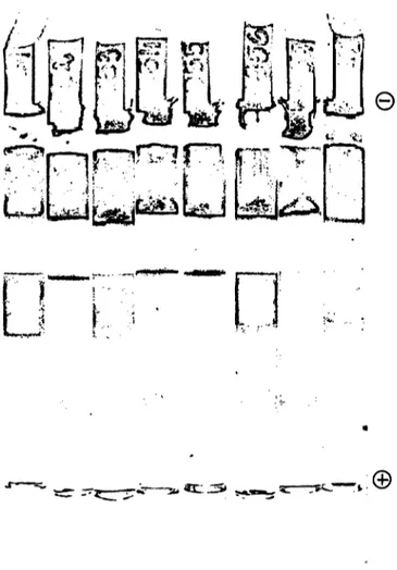
The electrophoretic run resulted in 2 coloured lipo- protein fractions: the first penetrated separation gel II and the second one stopped in separation gel I (fig. 1).
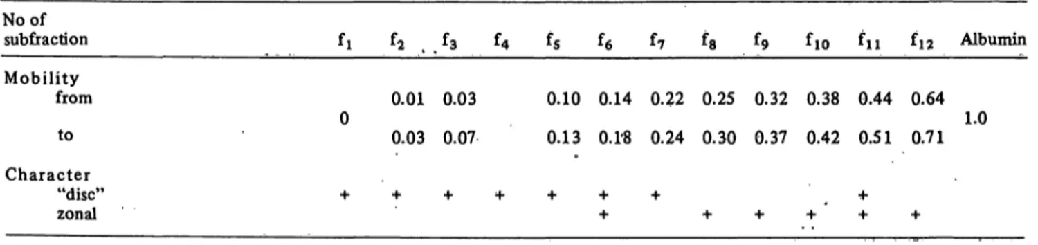
In the gel region between both groups no lipoproteins bin- ding Sudan Black B were observed. A brown-gray frac- tion, which moved in front of the separation, was identified immunologically as albumin. The main SMDL group in separation gel II ran with a relative mobility of 0 to 0.5 towards the albumin. Between this group and albumin we always found a more or less intense sub- fraction with a relative mobility about 0.7.
SMDL subfractions of lower mobility had a classical
"disc" character, and those of higher mobility were more zonal (tab. 1).
-r;
i»
W m
^^ ^VrQj
Wf*'**. „ -4
• V''··'··· I . **-<"
Fig. 1. A batch of columns showing SMDL-differeiitiation in serum from healthy people. Note the differences in in- tensity of the staining in different columns.
The disc subfractions were generally easier to identify, especially subfractions f3 and f7. In a large number of separations of different sera it was possible to find some,
in which the zonal subfractions were also distinct. Com- Not all subfractions were seen on every column. Some of paring the columns with densitograms just after separation, them appeared very often, others rather rarely. Table 2 it was not difficult to identify zonal subfractions, when shows the frequency of the SMDL subfractions as found they were not very distinct (fig. 2). in the sera of 300 healthy persons.
Tab. 1. The electrophoretic mobility of SMDL subfractions toward albumin in separation gel II.
No of sub fraction Mobility
from to Character
"disc"
zonal
fi f2 £3 £» £5 *6 f? *8 f9 fio ^ii ^12 Albumin
0.01 0.03 0.10 0.14 0.22 0.25 0.32 0.38 0.44 0.64 0 1.0
0.03 0.07 0.13 Ο.Γ8 0.24 0.30 0.37 0.42 0.51 0.71
+ + + * + ; + + + + - : + '
J. Clin. Chem. Clin. Biochem. / Vol. 17,1979 / No. 12
Janecki and Fijalkowska: Estimation of the subfractions of human serum small molecular diameter lipoproteins 791
Fig. 2. Comparision of the column (separation gel II) with the corresponding densitogram. Note the diffeiences of
"disc" (f i, f2, f3, fs, f7) and zonal-fractions (f6, f8, ^ 0, fii,fi2,Alb,).
To evaluate the amount of each subtraction, the peaks of the densitometric curve were measured. A repro- ducible length of separation of SMDL was difficult to obtain. A correction of densitometrical readings was therefore sometimes used: galvanometer settings were multiplied by 100, then divided by the length of the pathway of albumin in separation gel II (mostly 100 mm in the standard densitogram). This mathematical treat- ment gave a better comparison of results.
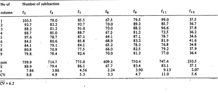
The precision of the quantitative estimation of the par- ticular SMDL subfractions in the same serum was investigated in 9 columns (fig. 3). The variation co- efficient varied from 3.3% to 11 %, mean 6.2% (tab. 3).
To prove the reproducibility of the quantitative esti- mation of the subfractions, which reflects the stability of the material and methodical parameters, the electrqphpresis of some sera, kept in the refrigerator at + 4 °C, was carried out after 2 to 3 days, in separately prepared columns. The recorded values were 95 % to 108 %, mean 101% of the initial values
•^^
?.
Fig. 3. Separation of the same serum in 9 parallel columns. Usu- ally the differences in the length are smaller than in this batch (compare fig. 1 and 4).
(tab. 4). We did not therefore find any systematical loss or increase of the SMDL subfraction due to storage in the refrigerator for this short period.
The stability of the patterns of SMDL subfractions were proved in 4 healthy persons (1 man and 3 women). The blood was collected every second day for 9 days, the way of life and nutrition were normal. The mean devia- tion of particular subfractions was not higher than 15% of the mean value.
In the above described method the sera investigated were taken from healthy persons of different sex and age, especially children. In table 5 it is evident that some subfractions are mostly higher than others (e.g. sub- fractions f6, f7, f8, f9, f j o). It was also observed that some differences depend on sex and age, e.g. sub- fraction f2 diminished generally with age especially in females, while subfraction f7 increased rapidly in young women when compared with girls.
Changes of SMDL subfractions in different diseases were qualitative as well as quantitative. Generally, a decrease of all SMDL subfractions was observed in patients with a decreased HDL level in various diseases, e.g. severe neoplasma, nephropathy and liver failure. In other diseases we observed some changes in particular subfractions, e.g. f6 and f7 by some gynecologi- cal, endocrinological dysfunctions. The most impressive changes were found in cases of acute liver failure, e.g. acute viral hepatitis (fig. 4). Often during this disease all sub-
Tab. 2. The frequency (%) of the appearence of SMDL subfractions in sera of healthy persons.
No of subfraction
%
f? f3 f4
83 96 8
f. fe f, f. f, 95 80 96 76 65
fio fn 87 95
fn
100
J. Clin. Chem. Clin, Biochem. / Vol. 17,1979 / No. 12
Tab. 3. Precision of subfractions (calculated as height of peaks on densitogram corrected for the length of separation) No of
column 21 43 56 78 9 χsum CVs
Number of subtraction
f3 f5
103.5 92.788.2 88.797.6 84.584.1 79.8.80.8 799.9 88.97.86
8.8
78.082.2 81.285.0 78.780.6 79.170.9 78.4 714.7 . 79.4
4.93.86
f?
85.592.7 91.088.7 87.185.8 84.177.5 82.6 775.0
86.14.56 5.3
fe 67.570.0 70.067.5 64.168.9 65.266.0 70.0 609.1 67.72.24
3.3
*9
79.589.2 88.281.2 87.183.2 78.382.5 81.2 750.4
83.43.90 4.7
f l l
99.085.7 96.672.5 78.781.9 79.276.8 77.0 747.4 83.19.13 11.0
f l 2
37.536.7 37.836.2 41.634.6 34.837.9 36.4 333.5 37.12.07
5.6 CV = 6.2
Tab. 4. Reproducibility of subfractions investigated after 2-3 days (calculated as percent of first measurement).
No of serum 06730693 07310745 08290881 11021163 11651167 12371314 sum
X
s
Number of subfraction fa fs fe
10398 12397 10788 9688 8369 10497 115396.1
13.5
11499 10397 11678 104104 103132 8781 1218101.5
15.1
10897 101109 100111 108103 114102 8392 1228102.3
8.7
f?
102102 10294 108103 123106 109100 10190 1240108.0
8.2
fe 10898 10499 100110 136113 109106 11796 1296102.2
10.9
f9
10494 100100 10697 10193 102119 11298 122694.8
7.4
fio 10489 105101 9883 8186 10086
• 95110 113894.8
9.6
f l l
134104 9591 12977 9984 7081 120101 118598.8
20.3
f!2
13060 9381 10387 100131 109111 12989 1223101.9
21.7 χ = 100.99
Tab. 5. Patterns of SMDL subfractions in groups of different sex and age.
Age Sex Number of subfraction
M O
Babies 3- 10 years old 11-16 years old * Young adults
96 90
96
9 a
31.825.8 24.424.5 27.318.9 12.5++
18.7
44.535.8 32.237.1 . 30.P,28.8
22.9 22.3
60.043.9 46.928.7+
57.340.9 44.7 39.6
52.542.0 49.257.1 53.340.0 64.4
I I
39.0
55?5 46.0 55.761.3+
54.746.7 80.1++
++
50.8
58.648.7 55.366.4+
59.245.0+
57.6 56.1
58.654.4 55.762.3 55.740.0+
53.5 62.3+
55.550.0 56.951.2 50.035.0+
53.6 62.5++
41.732.6 35.838.8 50.028.3+
38.2 35.8 + statistical significance of p 0.05
++ statistical significance of p 0.005
asterisk behind the number: significant difference to the younger group asterisk between numbers: significant difference between sexes
J. Glin. Chem. Clin. Bipchem. / Vol. 17,1979 / No. 12
Janecki and Fijalkowska: Estimation of the subfractions of human serum small molecular diameter lipoproteins 793
Fig. 4. A batch of columns with sera of patients on different days of acute hepatitis. Compare with fig. 1 and 3. Note the absence of the most of the SMDL in columns Nos.
1954, 1955, 1957.
fractions disappeared temporary, except f 12 and the largest subfractions, fl 9 f2 and f3, which increased sig- nificantly. Our experiments on animals confirmed the constancy and high significance of these phenomena.
Discussion
The study described in this paper is a continuation of our earlier investigations on the dye-labeling of serum pro- teins (11,12). In these studies, we showed the dis- appearance of an ICG-labeled protein (possibly an alpha- lipoprotein) in some liver diseases. We required an alter- native, but a simple method, for the measurement of the amount of this protein in the serum of patients suffering from liver diseases. We did not want to investigate all biochemical and physical properties of HDL, SMDL or alpha-lipoproteins. A diagnostic method was required with satisfactory precision and reproduc- ibility, easy to perform and standardise. We therefore chose the simplest and most standardisable method using the prestauiing of lipoproteins (9). Poststaining probably carries a smaller risk of artefacts, but it is much more complicated and difficult to standardise;
our experience with poststained paper and cellulose- acetate electrophoresis of proteins (13) shows that there are many problems connected with the standardisa- tion of this method.
The inhomogeneity of the lipoproteins from α-region (or HDL), when separated by polyacrylamide gel elec- trophoresis, was observed byNarayan et al. (3) in 1965. These authors used the HDL fraction obtained by ultracentrifugation. Dangerfield & Pratt (4) fractio- nated whole serum and observed a fast migrating group of lipoproteins that penetrated 6.5 % polyacrylamide gel and separated into several subfractions. They defined the lipoproteins of this group as "small molecular diameter lipoproteins" (SMDL) in contrast to the slow migrating ones, which did not penetrate more concentrated gel and which were defined as "large molecular diameter lipo- proteins" (LMDL).
We used a different concentration of gel from that of Dangerfield & Pratt (75 g/1 instead of 65 g/1), but we have retained the nomenclature SMDL. The difference was not significant; after leaving the spacer gel, the lipoproteins differentiated themselves very early into 2 groups: a fast moving one penetrating separation gel II and the other, slower group, remaining in separation gel I. There were no lipoproteins, between these 2 groups, so it seemed that the name SMDL is equally appropriate for lipoproteins penetrating 6.5 % or 7.5 % gels.
The inhomogeneity of the fast migrating lipoproteins was also observed by other authors (5, 7, 8). Anderson et al. (8) found, by ultracentrifugation, 3 major components of HDL; Utermann (5) identified, by gel electrophoresis, 4 subfractions in this group, but the author obtained satisfactory separations only after the addition of laurinic acid to the serum.
The subfractions of SMDL appeared in the form of
"discs" or "zonal" fractions. This is probably indicative of smaller or greater inhomogeneity of the lipoprotein molecules in the particular subfractions.
Particular subfractions appeared in investigated sera with different frequency. This has also been observed by other authors (4, 5). Possible causes are genetic factors, physiological oscillation of the amount of some proteins etc.
For clinical purposes it is necessary to have quanti- tative results. Some authors (2, 8) have reported quantitative data based on centrifugal analysis. This method could not be used for clinical purposes be- cause of its complexity. Dangerfield & Pratt ob- served changes in the amount of particular sub- fractions of SMDL, but the results were not strictly quantified. Utermann also found qualitatively different patterns of SMDL in various persons; he also as- certained quantitative differences, but did not give any values for the disc-electrophoretic subfractions.
The main aim of our experiments was to obtain a really
j. Clin. Chem. Clin. Biochem. / Vol. 17,1979 / No. 12
quantitative method for the objective statement of relative small differences of SMDL subfraction levels for clinical purposes. We thus developed the above described method, which enables us to state numeral differences between particular persons. On the other hand, the pattern of SMDL subfractions in any one individual is relatively stable over a period of a few days (this was not reported by other authors).
It was also possible to show differences depending on sex, age and pathology.
In contrast to individual differences between particular persons these differences were sometimes very charac-
teristic and distinct e.g. women in comparison with girls, or in the umbilical cord blood when compared with blood of small children. This confirms the practical utility of our method. The differences connected with pathology were sometimes even more distinct, e.g.
by some gynecological disturbances or in some liver diseases, which indicates some of the fields of clinical medicine where this method could be used in the diagnosis and/or understanding of the physiopathological mechanism in some diseases.
References
1. Burstein, M. (1961) Bull. Schweiz. Akad. Med. Wiss. 17, 92-110.
2. Barclay, M., Trebus-Kekish, O., Skipski, V. P. & Barclay, R. K. (1965) Clin. Chim. Acta 11, 389-394.
3. Narayan, Κ. Α., Narayan, S. & Kumerow, F: A. (1965) Nature 205, 246-248.
4. Dangerfield, W. G. & Pratt, J. J. (1970) Clin. Chim. Acta 50,273-278.
5. Utermann, G. (1972) Clin. Chim. Acta 36,521-529.
6. Feliste, R., Dousset, N. & Douste-Blazy, L. (1973) Clin.
Chim. Acta 47, 329-333.
7. MacKenzie, S. L, Sundaram, G. S. & Shodi, H. S. (1973) Clin. Chim. Acta 47, 329-333.
8. Anderson, D. W., Nichols, A. V., Forte, T. M. & Lindgren, F. T. (1977) Biochim, Biophys. Acta4P5,55-68.
9. Ressler, N., Springgate, R, & Kaufman, J. (1961) J.
Chromatog. 6,409-415.
10. Wollenweber, J. & Kahlke, W. (1970) Clin. (him. Acta 29, 411-420.
11. Janecki, J. & Krawczynski, J. (1970) Clin. Chim. Acta 16, 1008-1011.
12. Janecki, J. (1973) Zbl. Pharm. 772, 357-366.
13. Hertel, Z. & Janecki, J. (1976) Diagn. Lab. XII, 81-87.
Doc. Dr. Jerzy Janecki Leiter der Abteilung f r Labor-Diagnostik
Institute of Pediatrics Medical Academy ul. Litewska 14/16 PL-00-576Warszawa
J. Clin. Chem. Clin. Bipchem. / Vol. 17,1979 /No. 12